1 Novel human adenosine receptor antagonists based on the 7-amino-thiazolo[5,4-d]pyrimidine scaffold. Structural investigations at the 2-, 5- and 7- positions to enhance affinity and tune selectivity.
Flavia Varano a,*, Daniela Catarzi a, Matteo Falsini a, Diego Dal Ben b, Michela Buccioni b, Gabriella Marucci b, Rosaria Volpini b, Vittoria Colotta a
a Dipartimento di Neuroscienze, Psicologia, Area del Farmaco e Salute del Bambino, Sezione di
Farmaceutica e Nutraceutica, Università degli Studi di Firenze, via Ugo Schiff, 6, 50019, Sesto Fiorentino, Italy. b Scuola di Scienze del Farmaco e dei Prodotti della Salute, Università degli Studi di Camerino, Via S. Agostino 1, 62032 Camerino, MC, Italy
Corrisponding author:
*Tel: +39 055 4573732. Fax: +39 055 4573780. E-mail: [email protected].
*Manuscript
2 Abstract
This paper describes the synthesis of novel 7-amino-thiazolo[5,4-d]pyrimidines bearing different substituents at positions 2 , 5 and 7 of the thiazolopyrimidine scaffold. The synthesized compounds 2-27 were evaluated in radioligand binding (A1, A2A and A3) and adenylyl cyclase activity (A2B and
A2A) assays, in order to evaluate their affinity and potency at human adenosine receptor subtypes.
The current study allowed us to support that affinity and selectivity of 7-amino-thiazolo[5,4-d]pyrimidine derivatives towards the adenosine receptor subtypes can be modulated by the nature of the groups attached at positions 2, 5 and 7 of the bicyclic scaffold. To rationalize the hypothetical binding mode of the newly synthesized compounds, we also performed docking calculations in human A2A, A1 and A3 structures.
KEYWORDS. G protein coupled receptors; adenosine receptors; adenosine receptor antagonists; thiazolopyrimidine derivatives; bicyclic heteroaromatic system.
3 Adenosine receptors (ARs) are members of the G protein-coupled receptors superfamily and comprise four subtypes designated A1, A2A, A2B, and A3 [1]. A2A and A2B ARs are mainly coupled
to Gs proteins while A1 and A3 ARs are generally coupled to Gi proteins, thus leading to an
enhancement or decrease of intracellular cAMP levels, respectively. Additionally, the various AR subtypes may interact with other classes of G proteins leading to activation of various second messenger pathways, such as phospholipase C, mitogen activated protein kinase, potassium and calcium channels, arachidonic acid pathways, and phospholipase D [1].
It is well known that ARs, due to their different organ and tissue distribution in the human body, are implicated in numerous and important physiopathological processes [2, 3]. In particular, the blockade of ARs was studied for the treatment of heart and renal failure [4-6], cognition disorders [7] (A1 AR), neurodegenerative diseases [8-9], dermal fibrosis [10-12], retinal dysfunctions [13],
cancer [14-17], and pain [18] (A2A AR), inflammatory lung diseases such as asthma [19] (A2B AR),
airway contraction, glaucoma and cancer [20-22] (A3 AR).
In continuation of our studies directed towards the identification of new AR antagonists [23-28], in a recent paper we disclosed the 5-methyl-2-phenylthiazolo[5,4-d]pyrimidin-7-one 1 [29] which proved to be a potent and selective human (h) A3 AR antagonist, being inactive at the other AR
subtypes (Fig. 1). To further explore the structure affinity relationships (SARs) in this class of AR antagonists, we here describe the synthesis of the 7-amino-5-methyl-2-phenylthiazolo[5,4-d]pyrimidine 2 which is the 7-amino analogue of 1 (Fig. 1). The replacement of the 7-oxo function with a 7-amino group is a structural modification that in other classes of bicyclic AR antagonists significantly modified AR affinity and selectivity [30, 31]. Moreover, we recently disclosed the thiazolo[5,4-d]pyrimidine-5,7-diamino series which shows an extraordinary high affinity for the hA2A AR subtype [18, 32-33]. Anticipating the results, it emerged that the 7-amino derivative 2
showed good affinity for the hA2A and for the hA1 AR and a low binding activity at the hA3
4 by combining a phenyl or a furan-2-yl ring at position 2 with an aryl or heteroaryl group at position 5 (compounds 3-20, Fig. 1). Furthermore, we also investigated the effect on AR affinity and selectivity of introduction of different acyl residues on the 7-amino moiety (compounds 21-27, Fig. 1). Acyl residues on the 7-amino group were chosen since they increased hA3 AR affinity and
selectivity in many classes of AR antagonists of similar size and shape [20, 22, 28]. All the newly synthesized compounds (2-27) were tested in binding assays to evaluate their affinity at cloned hA1,
hA2A and hA3 ARs, stably expressed in Chinese Hamster Ovary (CHO) cells. Compounds were also
tested at hA2A and hA2B ARs by measuring their inhibitory effects on NECA-stimulated cAMP
levels in CHO cells.
5 Along with the pharmacological evaluation, docking studies at hA1, hA2A and hA3 AR structures
were performed to rationalize the observed affinity data.
Compounds 2-27 were synthesized according to the procedures depicted in Schemes 1-3. The 7-amino-2-phenyl-5-methyl derivative 2 was obtained (95% yield) starting from its 7-oxo analog 1 synthesized as previously reported by us (Scheme 1) [29]. The 7-oxo compound 1 was treated with POCl3 to yield the 7-chloro derivative 28 [29], which upon reaction with aqueous ammonia solution
furnished the desired 7-amino derivative 2.
Scheme 1
Reagents and conditions: (a) POCl3, dimethylaniline, reflux, 3h; (b) NH3(g), EtOH, sealed tube, 130
°C, overnight, 95% yield.
Compounds 2-phenyl- 3-11 and 2-(furan-2-yl)- substituted 12-20 were synthesized reacting the corresponding 5-chloro derivatives 29 [33] and 30 [18] and the suitable boronic acids under Suzuki conditions (Scheme 2).
Finally, the 7-amino derivatives 2, 3 and 12 by reaction with the proper acyl chlorides yielded the desired 7-acylamino substituted 21-27 (Scheme 3).
N N H N S O C H3 N N N S C H3 Cl N N N S C H3 NH2 a b 1 28 2
6 Schema 2.
Reagents and conditions: (a) R5B(OH)2, tetrakis, Na2CO3, DME/H2O, microwave irradiation, 160
°C 30 min (3-4, 6-7, 12-13, 17-18), 35-68% yield, or reflux 4h (5, 8-11, 14-16, 19-20), 30-72% yield. R5 3, 12 C6H5 4, 13 C6H4-3-CH2OH 5, 14 C6H4-4-CH2OH 6, 15 C6H4-4-OCH3 7, 16 C6H4-3-OCH3 8, 17 C6H4-3-CN 9, 18 C6H4-3-OH 10, 19 furan-2-yl 11, 20 5-methylfuran-2-yl N N N S NH2 Cl R2 N N N S NH2 R5 R2 29 R2 = phenyl 30 R2 = furan-2yl 3-11 R2 = phenyl 12-20 R2 = furan-2yl a
7 Scheme 3
Reagents and conditions: (a) RCOCl, Pyridine dry, CH2Cl2, reflux, 3-4 days, 20-55% yield.
R 21, 24, 26 C6H5 22 C6H4-4Cl 23 C6H4-4OCH3 25, 27 furan-2-yl N N N S R5 NH2 R2 N N N S R5 N H R2 O R 2 R2 = C6H5 R5 = CH3 3 R2 = C6H5 R5 = C6H5 12 R2 = furan-2yl R5 = C6H5 21-23 R2 = C6H5 R5 = CH3 24-25 R2 = C6H5 R5 = C6H5 26-27 R2 = furan-2yl R5 = C6H5 a
8 The new thiazolo[5,4-d]pyrimidine derivatives 2-20 and 21-27 were evaluated for their affinity to hA1, hA2A and hA3 ARs, stably transfected in CHO cells, and were also tested at the hA2B AR
subtype by measuring their inhibitory effects on 5’-(N-ethyl-carboxamido)adenosine (NECA)-stimulated cAMP levels in hA2B CHO cells. The selected derivatives 9, 10, 18 and 19, showing high
affinity for the hA2A AR, were investigated to determine their antagonistic potency by measuring
their effects on cAMP production in CHO cells, stably expressing hA2A ARs.
The results of binding experiments carried out on the new thiazolo[5,4-d]pyrimidine derivatives 2-20 and 21-27 are displayed in Tables 1 and 3, respectively. Concerning the hA2B AR, compounds
2-20 and 21-27 were inactive in inhibiting the NECA-stimulated cAMP levels in hA2B CHO cells
(IC50> 30000 nM, data not shown).
In general, results of some interest have been obtained for the 7-amino derivatives 2-20 (Table 1). In fact, all the reported compounds bind the hA1, hA2A and hA3 ARs with good affinities. More
specifically, compounds 9, 12, 17, 18, 19, 20 showed nanomolar affinity toward the hA2A AR
subtype and a good degree of selectivity for this receptor. Worthy of note is the presence of a furan-2-yl at both 2 and 5 positions producing the best combination of hA2A AR affinity and selectivity
(compound 19). Compounds 10, 13, 14, 15, 16, were able to bind both the hA2A and hA3 ARs
showing comparable Ki values falling in the nanomolar range. Finally, two derivatives (compounds
4 and 5) having nanomolar affinity and good selectivity for the hA3 AR subtype were identified.
Analyzing the binding data, we observe that the replacement of the 5-methyl group of 2 by a phenyl residue (compound 3) led to a noteworthy increase in hA2A and hA3 AR affinity while the hA1 AR
9 Table 1. Binding affinity of compounds 2-20 at hA1, hA2A and hA3 ARs.a
aData (n = 3−5) are expressed as means ± standard errors. bDisplacement of specific [3H]-CCPA binding at hA
1 AR expressed in CHO cells. cDisplacement of specific [3H]-NECA binding at hA2A AR expressed in CHO cells. dDisplacement of specific [3 H]-HEMADO binding at hA3 AR expressed in CHO cells.
R2 R5 binding experiments Ki (nM) A2A AR selectivity hA1b hA2Ac hA3d A1/A2A A3/A2A 2 CH3 34 ± 1 248 ± 33 2656 ± 235 0.14 10.7 3 C6H5 148 ± 16 19 ± 6.2 84 ± 13 7.8 4.4 4 C6H4-3-CH2OH 32 ± 9 53 ± 13 3.7 ± 1.2 0.6 0.07 5 C6H4-4-CH2OH 82 ± 17 82 ± 19 8.2 ± 0.5 1 0.1 6 C6H4-4-OCH3 224.6 ± 61.3 115.3 ± 5.1 46.7 ± 12 1.9 0.4 7 C6H4-3-OCH3 175.9 ± 34.7 109.8 ± 0.35 35.6 ± 9.2 1.6 0.3 8 C6H4-3-CN 594 ± 124 33.6 ± 8.9 247 ± 74 17.7 7.3 9 C6H4-3-OH 58.5 ± 6.1 5 ± 1.5 190 ± 16.7 11.7 38 10 furan-2-yl 67 ± 6.8 1.7 ± 0.2 2.8 ± 0.4 39.4 1.6 11 5-CH3-furan-2-yl 28.9 ± 6.6 22.4 ± 4.03 24.9 ± 3.4 1.3 1.1 12 C6H5 33 ± 2 3 ± 0.04 15 ± 2.9 11 5 13 C6H4-3-CH2OH 28 ± 5.5 2.1 ± 0.3 0.9 ± 0.1 13 0.4 14 C6H4-4-CH2OH 77.7 ± 14.5 7.7 ± 0.45 3.58± 0.41 10.1 0.5 15 C6H4-4-OCH3 47.7 ± 3.4 11.2 ± 0.83 4.66 ± 1.2 4.2 0.4 16 C6H4-3-OCH3 62.3 ± 5.1 1.9 ± 0.33 4.90 ± 0.19 32.8 2.6 17 C6H4-3-CN 102.7 ± 6.8 7.7 ± 1.4 61.5 ±14.1 13.3 8.0 18 C6H4-3-OH 51.0 ± 1.6 1.64 ± 0.35 15.73 ± 3.5 31.1 9.6 19 furan-2-yl 69 ± 15 3.4 ± 0.9 99 ± 15 20.3 29.1 20 5-CH3-furan-2-yl 45.9 ± 4.4 2.36 ± 0.4 13.76 ± 1.7 19.4 5.8 N N N S R2 NH2 R5 2-20 O
10 Introduction of substituents on the appended 5-phenyl ring (compounds 4-9) results in different effects depending on the receptor subtype. In fact, only the presence of a meta hydroxy substituent (compound 9) increases the affinity leading to a potent and selective hA2A AR antagonist (Ki = 5
nM). For hA3 AR anchoring the best substituent was the hydroxymethyl one both in the meta or
para positions (compounds 4 and 5). Finally, binding at the hA1 AR was only slightly enhanced by a
hydroxy substituent both directly attached (compound 9) or spaced by a methylene group (compound 4-5).
Replacing the 5-phenyl moiety of derivative 3 with a furan-2-yl ring resulted in compound 10 which showed enhanced affinity towards all three receptors, with the greatest increase observed for the hA2A and hA3 AR subtypes, (Ki = 1.7 nM and Ki = 2.8 nM, respectively).
Subsequently, we synthesized compounds 12-20 which are the 2-(furan-2-yl) analogues of 3-11, respectively. The furan-2-yl group seemed the ideal substituent at this position to obtain good affinity at all three receptors. In fact, hA1, hA2A, and hA3 ARs binding affinities of 12-20 were
comparable to or higher than those of the corresponding 2-phenyl derivatives 3-11 with the only exception being the hA3 AR affinity of 19 which is 35-fold lower than that of compound 10. This
trend was expected since the furan-2-yl ring at this position is a profitable substituent in other classes of AR antagonists and in our 5,7-diaminothiazolo[5,4-d]pyrimidine derivatives. [34, 18, 32, 33]
It has to be noted that in the 2-(furan-2-yl)-5-aryl derivatives 13-18, differently from what was observed in the corresponding 2-phenyl derivatives 4-9, the presence and nature of the substituents on the appended 5-phenyl ring seem to have no influence on the binding activity at the hA2A AR. In
fact, all the 2-(furan-2-yl)-5-aryl substituted derivatives 13-18 showed a comparable binding affinity falling in the nanomolar range. On the contrary, looking at the hA1 and hA3 AR binding
11 the best substituent for both hA1 and hA3 AR affinities is still the 3-hydroxymethyl one, derivative
13 being the most active at these two hARs (hA1 Ki = 28 nM; hA3 Ki = 0.9 nM).
It has to be noted that the 2,5-difuran-2-yl derivative 19 in addition to be highly hA2A vs hA1
selective, such as the corresponding 2-phenyl-5-(furan-2-yl) 10, it has also the best selectivity vs the hA3 subtype. A similar behavior can be observed for the 5-(methylfuran-2-yl)-2-furan-2-yl 20 with
respect to its corresponding 5-(methylfuran-2-yl)-2-phenyl 11. In fact, compound 20 is A2A
selective vs the A1 AR and showed a hA2A binding affinity about 6-fold higher compared to that at
the hA3 ARs subtype, whereas the corresponding 2-phenyl derivative 11 is a non-selective ligand.
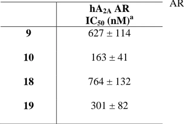
Derivatives 9, 10, 18 and 19, able to bind hA2A ARs with nanomolar affinity and a good degree of
selectivity were chosen to be tested for their antagonistic properties by evaluating their effect on cAMP production in CHO cells, stably expressing the hA2A AR. The obtained results (Table 2)
showed that the compounds behaved as antagonists as they were able to counteract NECA-stimulated cAMP accumulation.
Table 2. Potencies of compounds 9, 10, 18, and 19 at hA2A AR.
hA2A AR IC50 (nM)a 9 627 ± 114 10 163 ± 41 18 764 ± 132 19 301 ± 82 a
IC50 values obtained by inhibition of NECA-stimulated adenylyl cyclase activity in CHO cells expressing hA2A AR.
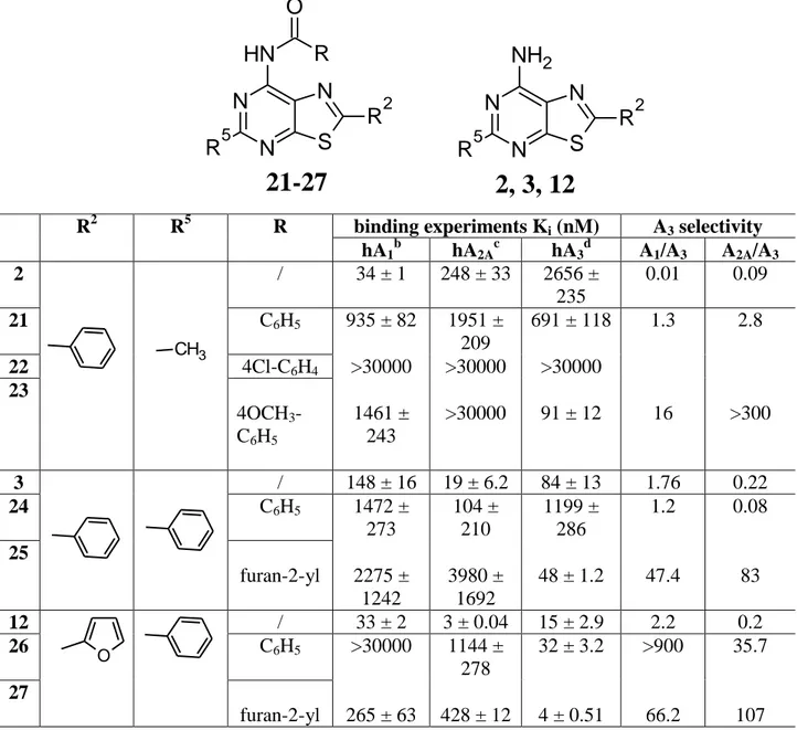
Table 3 reports the binding affinities at hA1, hA2A and hA3 ARs of derivatives 21-27 which were
obtained by introduction of different acyl groups on the 7-amino function of the parents 2, 3 and 12. This modification was performed in order to shift affinity and selectivity toward the hA3 receptor
12 N N N S R2 N H R5 O R
21-27
N N N S NH2 R2 R52, 3, 12
subtype. As expected, this structural modification, in general, afforded an increase in the hA3
binding, depending on the nature of the acyl group.
Table 3. Binding affinity of compounds 21-27 at hA1, hA2A and hA3 ARs.a
a
Data (n = 3−5) are expressed as means ± standard errors. bDisplacement of specific [3H]-CCPA binding at hA1 AR expressed in CHO cells. cDisplacement of specific [3H]-NECA binding at hA2A AR expressed in CHO cells. dDisplacement of specific [3 H]-HEMADO binding at hA3 AR expressed in CHO cells.
In fact, the best combination for hA3 affinity and selectivity is a furoylamine function at position 7
together with a furan-2-yl and a phenyl moiety at positions 2 and 5, respectively (see compound 27 R2 R5 R binding experiments Ki (nM) A3 selectivity
hA1b hA2Ac hA3d A1/A3 A2A/A3 2 / 34 ± 1 248 ± 33 2656 ± 235 0.01 0.09 21 C6H5 935 ± 82 1951 ± 209 691 ± 118 1.3 2.8 22 4Cl-C6H4 >30000 >30000 >30000 23 4OCH3 -C6H5 1461 ± 243 >30000 91 ± 12 16 >300 3 / 148 ± 16 19 ± 6.2 84 ± 13 1.76 0.22 24 C6H5 1472 ± 273 104 ± 210 1199 ± 286 1.2 0.08 25 furan-2-yl 2275 ± 1242 3980 ± 1692 48 ± 1.2 47.4 83 12 / 33 ± 2 3 ± 0.04 15 ± 2.9 2.2 0.2 26 C6H5 >30000 1144 ± 278 32 ± 3.2 >900 35.7 27 furan-2-yl 265 ± 63 428 ± 12 4 ± 0.51 66.2 107 CH3 O
13 hA3 Ki = 4 nM). Analyzing the results, we observe that introduction of an acyl group is always
detrimental for the hA1 and hA2A binding activity. This is in accordance with previously reported
data on our bicyclic analogues confirming that the free 7-amino group is important for the anchoring of the ligand at the A1 and A2A subtypes [28].
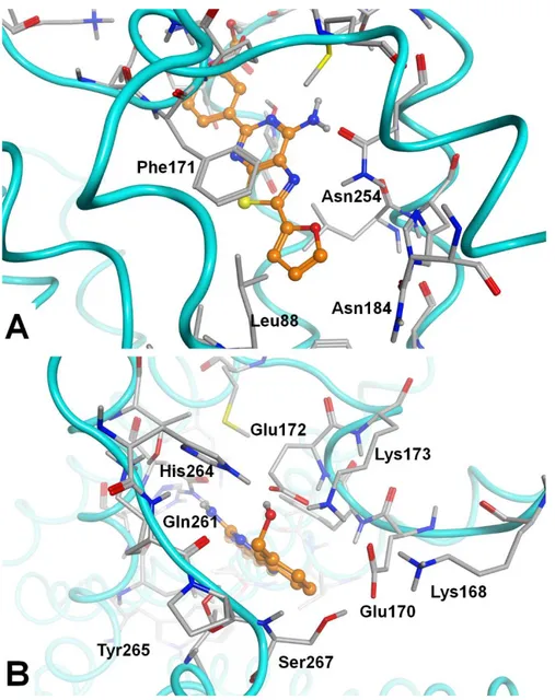
Molecular docking analyses were performed to simulate the binding mode of the synthesized compounds at the hA2A AR cavity. As molecular target, we chose the high-resolution crystal
structure of the hA2A AR in complex with the antagonist/inverse agonist
4-(2-(7-amino-2-(furan-2-yl)[1,2,4]triazolo[2,3-a][1,3,5]triazin-5-ylamino)ethyl)phenol (ZM241385) (http://www.rcsb.org; pdb code: 5NM4; 1.7-Å resolution [35]). MOE (Molecular Operating Environment 2014.09 [36]) docking tool (“induced fit” setting) and Gold [37] and Autodock software [38, 39] were employed for this task. Analogously to a previous study at the same receptor [24], we performed docking analyses with various docking tools to get a sort of average binding mode prediction of the synthesized compounds at the hA2A AR binding cavity.
According to docking results, the synthesised molecules 2-20 get inserted into the binding site of the hA2A AR with the thiazolo[5,4-d]pyrimidine scaffold located analogously to the triazolo-triazine
core of the co-crystallized derivative ZM241385. Even the ligand-target interactions appear conserved with respect to the co-crystallized ligand, as the new compounds generally engage a double H-bond interaction with the Asn2536.55 side chain (through the N1 atom and the 7-amine function) and an additional polar contact with Glu169 (EL2). Further key interactions are a π-π interaction with Phe168 (EL2) and a non-polar interaction with the Leu2496.51 side chain (Fig. 2). The 5-substituent points toward the extracellular environment and gets in proximity to Ile662.64, Ser672.65, Leu167 (EL2), Glu169 (EL2), Leu267 (EL3), and Tyr2717.36, while the 2-substituent is inserted into the depth of the binding cavity, in proximity to residues belonging to TM3, TM5 and TM6 segments (Val843.32, Leu853.33, Thr883.36, Met1775.38, Trp2466.48, Leu2496.51, His2506.52, and Asn2536.55).
14 Figure 2. The general binding mode of the synthesized compounds at the hA2A AR cavity,
according to docking-scoring results; compound 18 is represented and key receptor residues are indicated.
The 5-substituent is generally a substituted phenyl group, where the substituents on this ring modulate the affinity for the three ARs. In particular, compounds bearing meta-substituents are endowed with higher hA2A AR affinity with respect to compounds presenting the same substituents
at the para-position. Figure 3A shows a top-view of the compound binding mode with the hA2A AR
residues in proximity to the 5-substituent. Polar groups at the meta-position of the 5-phenyl ring are able to engage H-bond interactions with H-bond functions of the receptor, like the backbone NH groups of Phe168 and Glu169 (EL2) and the hydroxyl group of Tyr2717.36. Additional interactions may be given with Ile662.64, Ser672.65, Leu167 (EL2), and Leu267 (EL3) residues. For the 2,5-diphenyl derivative 3, the replacement of the 5-unsubstituted phenyl ring with a furan-2-yl moiety (10) affords a significant improvement of affinity. Docking results showed that compounds bearing at least one furan-2-yl substituent are located in the binding cleft with this group inserted into the depth of the cavity. In fact, the binding mode of 5-(furan-2-yl)-2-phenyl derivative 10 is a mirror-version of the generally observed docking conformations (see above), with the 5-substituent (i.e. furan-2-yl group) deeply inserted into the cavity and the 2-substituent (i.e. phenyl group) pointing
15 toward the external environment. Compounds bearing two furan-2-yl groups (i.e. 19) may be inserted in both arrangements, according to docking results.
Figure 3. A. Top view of the docking conformation of compound 18 at the hA2A AR; the key
residues for the interaction with 5-substituent are indicated. B. Detail of the depth of the hA2A AR
binding cavity, represented as molecular surface. In this cavity is generally located the 2-substituent. The residues indicated are the ones in the closest proximity to the 2-substituent and have been considered for the calculation of the interaction forces with the svl IF-E 6.0 tool (see in main text).
16 It is generally observed at the hA2A AR that compounds presenting a furan-2-yl substituent into the
depth of the binding cavity (in proximity of Asn2536.55) are endowed with higher affinity with respect to those presenting a phenyl ring in the same position. Docking studies of compounds 2-20 showed that the substituent inserted in this region is the one bound to the C2 atom of the bicyclic core. Moreover, binding data reported in Table 1 indicated that compounds bearing a furan-2-yl group as 2-substituent (compounds 12-20) are endowed with higher affinity with respect to the corresponding derivatives bearing a 2-phenyl substituent (compounds 3-11). To justify this trend, we performed a post-docking analysis of the interactions between the compounds and the receptor binding site by using the IF-E 6.0 [40] tool that is retrievable at the SVL exchange service (Chemical Computing Group, Inc. SVL exchange: http://svl.chemcomp.com). This tool was previously employed for other analyses at ARs [26, 29, 41]. The script calculates atomic and residue interaction forces and displays them as 3D vectors. Furthermore, it calculates the per-residue interaction energies (values in kcal/mol), where negative and positive energy values are associated to favorable and unfavourable interactions, respectively. Hence, for this task we employed three pairs of compounds (14 and 5, 16 and 7, 18 and 9) bearing the same substituent at the 5-position, and a furan-2-yl or a phenyl ring as 2-substituent. The results of this analysis are reported in Table 4.
Table 4. Interaction energies (values in kcal/mol) between 2-(furan-2-yl) derivatives (14, 16, 18) and 2-phenyl derivatives (5, 7, 9) and the binding site residues located in proximity of the 2-position of the analyzed compounds. See text for details.
14 5 16 7 18 9
Val84 0,01 -0,28 -0,24 -0,41 -0,52 -0,36
Leu85 -1,52 -1,42 -1,32 -1,25 -1,51 -1,38
Thr88 -0,73 -1,23 -0,82 -1,31 -1,04 -1,36
17 Met177 -1,52 -1,47 -1,76 -1,62 -1,89 -1,68 Trp246 -0,10 0,20 0,02 0,43 0,44 0,45 Leu249 -2,27 -1,45 -2,68 -1,40 -2,97 -0,94 His250 -2,65 -4,15 -2,43 -4,15 -2,09 -3,64 Asn253 -11,09 -8,79 -11,05 -9,12 -9,94 -8,60 tot -26,35 -25,39 -26,58 -24,70 -25,90 -24,04
From the results reported in Table 4 it may be observed that the sum of contributes of aminoacids located in proximity of the 2-substituent appears favorable for compounds bearing a furan-2-yl group in this position with respect to those bearing a phenyl ring at the same position. The polar interaction between the oxygen atom of the furan-2-yl substituent and the side chain of Asn2536.55 represents the strongest ligand-target interaction. Substitution of the furan-2-yl moiety with a phenyl ring makes this polar contact impossible, leading to a lower interaction.
Docking experiments were also performed at a hA1 AR crystal structure (pdb code: 5UEN; 3.2-Å
resolution [42]) with the same docking tools and protocols as above. Docking conformations obtained at this receptor model appear quite in agreement with the ones observed at the hA2A AR
(see Fig. 4A). Interactions between compounds 2-20 and the hA2A and hA1 ARs are generally
conserved, and this could explain the similar nanomolar affinity data at these two ARs, even if the affinity at the hA1 AR appears generally lower for all compounds respect to the one at the hA2A AR.
Comparing the hA1 AR and hA2A AR docking data, it appears that some residues at the entrance of
the hA1 AR binding cavity (Glu170, Ser267, and Tyr2717.36, Fig. 4B) and in proximity to the
5-substituents are different in some cases with respect to the corresponding ones in the hA2A AR
(Leu167, Leu267, and Tyr2717.36). In particular, the hA1 AR presents a set of charged residues in
proximity to the 5-substituent and these aminoacids make an inter-residue polar interaction network (Fig. 4B). This could be one of the factors leading to a slightly different interaction with the
18 compounds with respect to the hA2A AR subtype that presents in the same region a set of less polar
aminoacids.
Figure 4. A The general binding mode of the synthesized compounds at the hA1 AR cavity,
according to docking-scoring results; compound 13 is represented and key receptor residues are indicated. B. Top view of the docking conformation of compound 13 at the hA1 AR; the key
residues for the interaction with 5-substituent are indicated.
Docking experiments were also performed (with the same settings) at a homology model of the hA3
AR [24], obtaining analogue docking conformations with respect to the ones observed at the hA2A
AR and hA1 AR (Fig. 5A). As described above, the interactions between compounds and the
19 differences, which are located at the entrance of these receptor regions. The residues of this region of the hA3 AR are not particularly polar, with only one charged residue (the EL3 residue Glu258).
Similarly, the hA2A AR presents Glu169 (EL2) as the sole charged residue. Instead, the hA1 AR
shows several charged residues (see above), hence a significantly different chemical-physical profile at this receptor region can be noted. Moreover, the hA3 AR has two aminoacids able to
engage H-bond interactions (Gln167 in EL2 and Gln261 in EL3, Fig. 5B) with the 5-substituent, similarly to some H-bond functions present at the entrance of the hA2A AR cavity (see above).
These similarities could help interpret the comparable trend of affinities of compounds for the hA2A
20 Figure 5. A The general binding mode of the synthesized compounds at the hA3 AR cavity,
according to docking-scoring results; compound 13 is represented and key receptor residues are indicated. B. Top view of the docking conformation of compound 13 at the hA3 AR; the key
residues for the interaction with 5-substituent are indicated.
In conclusion, the simplicity of the synthetic processes and the decoration capability of the 7-aminothiazolo[5,4-d]pyrimidine core make it a privileged structure for the design of novel AR ligands with distinct selectivity profiles for the AR subtypes. The obtained results allowed us to conclude that affinity and/or selectivity of the 7-aminothiazolo[5,4-d]pyrimidines towards ARs can be modulated by the nature of the substituents attached at positions 2, 5 and 7 of the bicyclic scaffold. For instance, the presence of a furan-2-yl in both the 2 and 5 positions produces the best combination of hA2A AR affinity and selectivity (compound 19). Moreover, two derivatives
(compounds 4 and 5) bearing a phenyl and aryl moieties at positions 2 and 5, respectively, and possessing nanomolar affinity and good selectivity for the hA3 AR subtype, were identified. As
expected, substitution of the 7-amino group with acyl moieties was profitable for the anchoring at the hA3 AR subtype (i.e. compound 27). Additionally, molecular docking simulations have been
supportive to rationalize the observed affinity data at the hA2A, hA1 and hA3 AR structures.
Author Contributions
All authors have given approval to the final version of the manuscript. Notes
The authors declare no competing financial interest. Acknowledgment
The synthetic work was financially supported by the University of Florence and the Italian Ministry for University and Research (MIUR, PRIN 2010-2011, 20103W4779_004 project).
21 1. Borea PA, Gessi S, Merighi S, Vincenzi F, Varani K. Pharmacology of adenosine receptors:
the state of the art. Physiol. Rev. 2018; 98:1591-1625.
2. Borea PA, Gessi S, Merighi S, Varani K. Adenosine as a multi-signalling guardian angel in human diseases: when, where and how does it exert its protective effects? Trends
Pharmacol. Sci. 2016; 37:419-434.
3. Gessi S, Merighi S, Varani K, Borea PA. Adenosine receptors in health and disease. Adv.
Pharmacol. 2011; 61:41-75.
4. Varani K, Vincenzi F, Merighi S, Gessi S, Borea PA. Biochemical and pharmacological role of A1 adenosine receptors and their modulation as novel therapeutic strategy. Adv. Exp.
Med. Biol.- Protein Reviews 2017; 19:193-232.
5. Mitrovic V, Seferovic P, Dodic S, Krotin M, Neskovic A, Dickstein K, Voogd HDE, Bocker C, Ziegler D, Godes M, Nakov R, Essers H, Verboom C, Hocher B. Cardio-renal effects of the A1 adenosine receptor antagonist SVL320 in patients with heart failure. Circ. Hear. Fail.
2009; 2:523-531.
6. Yap SC, Lee HT. Adenosine and protection from acute kidney injury. Curr. Opin. Nephrol.
Hypertens. 2012; 21:24-32.
7. Mihara T, Mihara K, Yarimizu J, Mitani Y, Matsuda R, Yamamoto H, Aoki S, Akahane A, Iwashita A, Matsuoka N. Pharmacological characterization of a novel, potent adenosine A1
and A2A receptor dual antagonist,
5-[5-amino-3-(4-fluorophenyl)pyrazine-2-yl]-1-isopropylpyridine-2(1H)-one (ASP5854), in models of Parkinson’s disease and cognition. J.
22 8. Preti D, Baraldi PG, Moorman AR, Borea PA, Varani K. History and perspectives of A2A
adenosine receptor antagonists as potential therapeutic agents. Med. Res. Rev. 2015; 35:790-848.
9. Jenner P. An overview of adenosine A2A receptor antagonists in Parkinson's disease. Int.
Rev. Neurobiol. 2014; 119:71-86.
10. Fernández P, Trzaska S, Wilder T, Chiriboga L, Blackburn MR, Cronstein BN, Chan ES. Pharmacological blockade of A2A receptors prevents dermal fibrosis in a model of elevated
tissue adenosine. Am. J. Pathol. 2008; 172:1675-1682.
11. Perez-Aso M, Fernandez P, Mediero A, Chan ES, Cronstein BN. Adenosine A2A receptor
promotes collagen production by human fibroblasts via pathways involving cyclic AMP and AKT but independent of Smad2/3. FASEB J. 2014; 28:802-812.
12. Perez-Aso M, Mediero A, Low YC, Levine J, Cronstein BN. Adenosine A2A receptor plays
an important role in radiation-induced dermal injury. FASEB J. 2016; 30:457-465.
13. Boia R, Ambrosio AF, Santiago AR. Therapeutic opportunities for caffeine and A2A
receptor antagonists in retinal diseases. Ophthalmic Res. 2016; 55:212-218.
14. Vijayan D, Young A, Teng MWL, Smyth MJ. Targeting immunosuppressive adenosine in cancer. Nat. Rev. Cancer. 2017; 17:709-724.
15. Ohta A, Gorelik E, Prasad SJ, Ronchese F, Lukashev D, Wong MK, Huang X, Caldwell S, Liu K, Smith P, Chen JF, Jackson EK, Apasov S, Abrams S, Sitkovsky M. A2A adenosine
receptor protects tumors from antitumor T cells. Proc. Natl. Acad. Sci. U. S. A. 2006; 103:13132-13137.
23 16. Young A, Ngiow SF, Gao Y, Patch AM, Barkauskas DS, Messaoudene M, Lin G, D. Coudert JD, Stannard KA, Zitvogel L, Degli-Esposti MA, Vivier E, Waddell N, Linden J, Huntington ND, Souza-Fonseca-Guimaraes F, Smyth MJ. A2AR adenosine signaling
suppresses natural killer cell maturation in the tumor microenvironment. Cancer Res. 78 2018; 78:1003-1016.
17. Gessi S, Bencivenni S, Battistello E, Vincenzi F, Colotta V, Catarzi D, Varano F, Merighi S, Borea PA, Varani K. Inhibition of A2A adenosine receptor signaling in cancer cells
proliferation by the novel antagonist TP455. Front. Pharmacol. 2017; 8:888.
18. Varano F, Catarzi D, Vincenzi F, Betti M, Falsini M, Ravani A, Borea PA, Colotta V, Varani K. Design, synthesis and pharmacological characterization of 2-(2-furanyl)thiazolo[5,4-d]pyrimidine-5,7-diamine derivatives: new highly potent A2A adenosine
receptor inverse agonists with antinociceptive activity. J. Med. Chem. 2016; 59:10564-10576.
19. Zablocki J, Elzein E, Kalla R. A2B adenosine receptor antagonists and their potential
indications. Expert Opin. Ther. Pat. 2006; 16:1347-1357.
20. Baraldi PG, Preti D, Borea PA, Varani K. Medicinal chemistry of A3 adenosine receptor
modulators: pharmacological activities and therapeutic implications. J. Med. Chem. 2012; 55:5676-5703.
21. Borea PA, Varani K, Vincenzi F, Baraldi PG, Tabrizi MA, Merighi S, Gessi S. The A3
adenosine receptor: history and perspectives. Pharmacol. Rev. 2015; 67:74-102.
22. Jacobson KA, Merighi S, Varani K, Borea PA, Baraldi S, Tabrizi MA, Romagnoli R, Baraldi PG, Ciancetta A, Tosh DK, Gao ZG, Gessi S. A3 adenosine receptors as modulators
24 23. Poli D, Falsini M, Varano F, Betti M, Varani K, Vincenzi F, Pugliese AM, Pedata F, Dal Ben D, Thomas A, Palchetti I, Bettazzi F, Catarzi D, Colotta V. Imidazo[1,2-a]pyrazin-8-amine core for the design of new adenosine receptor antagonists: structural exploration to target the A3 and A2A subtypes. Eur. J. Med. Chem. 2017; 125:611-628.
24. Falsini M, Squarcialupi L, Catarzi D, Varano F, Betti M, Dal Ben D, Marucci G, Buccioni M, Volpini R, De Vita T, Cavalli A, Colotta V. The 1,2,4-triazolo[4,3-a]pyrazin-3-one as a versatile scaffold for the design of potent adenosine human receptor antagonists. Structural investigations to target the A2A receptor subtype. J. Med. Chem. 2017; 60:5772-5790.
25. Squarcialupi L, Catarzi D, Varano F, Betti M, Falsini M, Vincenzi F, Ravani A, Ciancetta A, Varani K, Moro S, Colotta V. Structural refinement of pyrazolo[4,3-d]pyrimidine derivatives to obtain highly potent and selective antagonists for the human A3 adenosine
receptor. Eur. J. Med. Chem. 2016; 108:117-133.
26. Squarcialupi L, Falsini M, Catarzi D, Varano F, Betti M, Varani K, Vincenzi F, Dal Ben D, Lambertucci C, Volpini R, Colotta V. Exploring the 2- and 5-positions of the pyrazolo[4,3-d]pyrimidin-7-amino scaffold to target human A1 and A2A adenosine receptors. Bioorg. Med.
Chem. 2016; 24:2794-2808.
27. Catarzi D, Varano F, Poli D, Squarcialupi L, Betti M, Trincavelli L, Martini C, Dal Ben D, Thomas A, Volpini R, Colotta V. 1,2,4-Triazolo[1,5-a]quinoxaline derivatives and their simplified analogues as adenosine A3 receptor antagonists. Synthesis, structure-affinity
relationships and molecular modeling studies. Bioorg. Med. Chem. 2015; 23:9-21.
28. Squarcialupi L, Colotta V, Catarzi D, Varano F, Filacchioni G, Varani K, Corciulo C, Vincenzi F, Borea PA, Ghelardini C, Di Cesare Mannelli L, Ciancetta A, Moro S. 2-Arylpyrazolo[4,3-d]pyrimidin-7-amino derivatives as new potent and selective human A3
25 adenosine receptor antagonists. Molecular modeling studies and pharmacological evaluation. J. Med. Chem. 2013; 56:2256-2269.
29. Varano F, Catarzi D, Squarcialupi L, Betti M, Vincenzi F, Ravani A, Varani K, Dal Ben D, Thomas A, Volpini R, Colotta V. Exploring the 7-oxo-thiazolo[5,4-d]pyrimidine core for the design of new human adenosine A3 receptor antagonists. Synthesis, molecular modeling
studies and pharmacological evaluation. Eur. J. Med. Chem. 2015; 96:105-121.
30. Colotta V, Catarzi D, Varano F, Cecchi L, Filacchioni G, Martini C, Trincavelli L, Lucacchini A. 1,2,4-Triazolo[4,3-a]quinoxalin-1-one: a versatile tool for the synthesis of potent and selective adenosine receptor antagonists. J. Med Chem. 2000; 43:1158-1164.
31. Colotta V, Catarzi D, Varano F, Calabri FR, Lenzi O, Filacchioni G, Martini C, Trincavelli L, Deflorian F, Moro S. The 1,2,4-triazolo[4,3-a]quinoxalin-1-one moiety as an attractive scaffold to develop new potent and selective human A3 adenosine receptor antagonists:
synthesis, pharmacological and ligand-receptor modeling studies. J. Med. Chem. 2004; 47:3580-3590.
32. Varano F, Catarzi D, Vincenzi F, Falsini M, Pasquini S, Borea PA, Colotta V, Varani K. Structure-activity relationship studies and pharmacological characterization of N5 -heteroarylalkyl-substituted-2-(2-furanyl)thiazolo[5,4-d]pyrimidine-5,7-diamine-based derivatives as inverse agonists at human A2A adenosine receptor. Eur. J. Med. Chem. 2018;
155:552-561.
33. Varano F, Catarzi D, Falsini M, Vincenzi F, Pasquini S, Varani K, Colotta V. Identification of novel thiazolo[5,4-d]pyrimidine derivatives as human A1 and A2A adenosine receptor
26 34. de Lera Ruiz M, Lim YH, Zheng J. Adenosine A2A receptor as a drug discovery target. J.
Med. Chem. 2014; 57:3623-3650.
35. Weinert T, Olieric N, Cheng R, Brunle S, James D, Ozerov D, Gashi D, Vera L, Marsh M, Jaeger K, Dworkowski F, Panepucci E, Basu S, Skopintsev P, Dore AS, Geng T, Cooke RM, Liang M, Prota AE, Panneels V, Nogly P, Ermler U, Schertler G, Hennig M, Steinmetz MO, Wang M, Standfuss J. Serial millisecond crystallography for routine room-temperature structure determination at synchrotrons. Nat. Commun. 2017; 8(1):542.
36. Molecular Operating Environment, C.C.G., Inc., 1255 University St., Suite 1600, Montreal, Quebec, Canada, H3B 3X3;
37. Jones G, Willett P, Glen RC, Leach AR, Taylor R. Development and validation of a genetic algorithm for flexible docking. J. Mol. Biol. 1997; 267(3):727-748.
38. Morris GM, Goodsell DS, Halliday RS, Huey R, Hart WE, Belew RK, Olson AJ. Automated docking using a Lamarckian genetic algorithm and an empirical binding free energy function. J. Comput. Chem. 1998; 19:1639-1662.
39. Morris GM, Huey R, Lindstrom W, Sanner MF, Belew RK, Goodsell DS, Olson AJ. AutoDock4 and AutoDockTools4: Automated docking with selective receptor flexibility. J.
Comput. Chem. 2009; 30:2785-2791.
40. Shadnia H, Wright JS, Anderson JM. Interaction force diagrams: new insight into ligand-receptor binding. J. Comput. Aided Mol. Des. 2009; 23:185-194.
41. Dal Ben D, Buccioni M, Lambertucci C, Marucci G, Thomas A, Volpini R, Cristalli G. Molecular modeling study on potent and selective adenosine A3 receptor agonists. Bioorg.
Med. Chem. 2010; 18:7923-7930.
42. Glukhova A, Thal DM, Nguyen AT, Vecchio EA, Jorg M, Scammells PJ, May LT, Sexton PM, Christopoulos A. Structure of the adenosine A1 receptor reveals the basis for subtype
Table 1. Binding affinity of compounds 2-20 at hA1, hA2A and hA3 ARs.a
aData (n = 3−5) are expressed as means ± standard errors. bDisplacement of specific [3H]-CCPA binding at hA
1 AR expressed in CHO cells. cDisplacement of specific [3H]-NECA binding at hA2A AR expressed in CHO cells.
d
Displacement of specific [3 H]-HEMADO binding at hA3 AR expressed in CHO cells.
R2 R5 binding experiments Ki (nM) A2A AR selectivity hA1b hA2Ac hA3d A1/A2A A3/A2A 2 CH3 34 ± 1 248 ± 33 2656 ± 235 0.14 10.7 3 C6H5 148 ± 16 19 ± 6.2 84 ± 13 7.8 4.4 4 C6H4-3-CH2OH 32 ± 9 53 ± 13 3.7 ± 1.2 0.6 0.07 5 C6H4-4-CH2OH 82 ± 17 82 ± 19 8.2 ± 0.5 1 0.1 6 C6H4-4-OCH3 224.6 ± 61.3 115.3 ± 5.1 46.7 ± 12 1.9 0.4 7 C6H4-3-OCH3 175.9 ± 34.7 109.8 ± 0.35 35.6 ± 9.2 1.6 0.3 8 C6H4-3-CN 594 ± 124 33.6 ± 8.9 247 ± 74 17.7 7.3 9 C6H4-3-OH 58.5 ± 6.1 5 ± 1.5 190 ± 16.7 11.7 38 10 furan-2-yl 67 ± 6.8 1.7 ± 0.2 2.8 ± 0.4 39.4 1.6 11 5-CH3-furan-2-yl 28.9 ± 6.6 22.4 ± 4.03 24.9 ± 3.4 1.3 1.1 12 C6H5 33 ± 2 3 ± 0.04 15 ± 2.9 11 5 13 C6H4-3-CH2OH 28 ± 5.5 2.1 ± 0.3 0.9 ± 0.1 13 0.4 14 C6H4-4-CH2OH 77.7 ± 14.5 7.7 ± 0.45 3.58± 0.41 10.1 0.5 15 C6H4-4-OCH3 47.7 ± 3.4 11.2 ± 0.83 4.66 ± 1.2 4.2 0.4 16 C6H4-3-OCH3 62.3 ± 5.1 1.9 ± 0.33 4.90 ± 0.19 32.8 2.6 17 C6H4-3-CN 102.7 ± 6.8 7.7 ± 1.4 61.5 ±14.1 13.3 8.0 18 C6H4-3-OH 51.0 ± 1.6 1.64 ± 0.35 15.73 ± 3.5 31.1 9.6 19 furan-2-yl 69 ± 15 3.4 ± 0.9 99 ± 15 20.3 29.1 20 5-CH3-furan-2-yl 45.9 ± 4.4 2.36 ± 0.4 13.76 ± 1.7 19.4 5.8 N N N S R2 NH2 R5 2-20 O Table(s)
Table 2. Potencies of compounds 9, 10, 18, and 19 at hA2A
AR.
a
IC50 values obtained by inhibition of NECA-stimulated adenylyl cyclase activity in CHO cells expressing hA2A AR.
hA2A AR IC50 (nM)a 9 627 ± 114 10 163 ± 41 18 764 ± 132 19 301 ± 82 Table(s)
N N N S R2 N H R5 O R
21-27
N N N S NH2 R2 R52, 3, 12
Table 3. Binding affinity of compounds 21-27 at hA1, hA2A and hA3 ARs.a
aData (n = 3−5) are expressed as means ± standard errors. bDisplacement of specific [3H]-CCPA binding at hA
1 AR expressed in CHO cells. cDisplacement of specific [3H]-NECA binding at hA2A AR expressed in CHO cells.
d
Displacement of specific [3 H]-HEMADO binding at hA3 AR expressed in CHO cells.
R2 R5 R binding experiments Ki (nM) A3 selectivity hA1b hA2Ac hA3d A1/A3 A2A/A3 2 / 34 ± 1 248 ± 33 2656 ± 235 0.01 0.09 21 C6H5 935 ± 82 1951 ± 209 691 ± 118 1.3 2.8 22 4Cl-C6H4 >30000 >30000 >30000 23 4OCH3 -C6H5 1461 ± 243 >30000 91 ± 12 16 >300 3 / 148 ± 16 19 ± 6.2 84 ± 13 1.76 0.22 24 C6H5 1472 ± 273 104 ± 210 1199 ± 286 1.2 0.08 25 furan-2-yl 2275 ± 1242 3980 ± 1692 48 ± 1.2 47.4 83 12 / 33 ± 2 3 ± 0.04 15 ± 2.9 2.2 0.2 26 C6H5 >30000 1144 ± 278 32 ± 3.2 >900 35.7 27 furan-2-yl 265 ± 63 428 ± 12 4 ± 0.51 66.2 107 CH3 O Table(s)
Figure 1.
Scheme 1
Reagents and conditions: (a) POCl3, dimethylaniline, reflux, 3h; (b) NH3(g), EtOH, sealed tube, 130
°C, overnight, 95% yield. N N H N S O C H3 N N N S C H3 Cl N N N S C H3 NH2 a b 1 28 2 Figure(s)
Schema 2.
Reagents and conditions: (a) R5B(OH)2, tetrakis, Na2CO3, DME/H2O, microwave irradiation, 160
°C 30 min (3-4, 6-7, 12-13, 17-18), 35-68% yield, or reflux 4h (5, 8-11, 14-16, 19-20), 30-72% yield. R5 3, 12 C6H5 4, 13 C6H4-3-CH2OH 5, 14 C6H4-4-CH2OH 6, 15 C6H4-4-OCH3 7, 16 C6H4-3-OCH3 8, 17 C6H4-3-CN 9, 18 C6H4-3-OH 10, 19 furan-2yl 11, 20 5-methylfuran-2yl N N N S NH2 Cl R2 N N N S NH2 R5 R2 29 R2 = phenyl 30 R2 = furan-2yl 3-11 R2 = phenyl 12-20 R2 = furan-2yl a Figure(s)
Scheme 3
Reagents and conditions: (a) RCOCl, Pyridine dry, CH2Cl2, reflux, 3-4 days, 20-55% yield.
R 21, 24, 26 C6H5 22 C6H4-4Cl 23 C6H4-4OCH3 25, 27 furan-2yl N N N S R5 NH2 R2 N N N S R5 N H R2 O R 2 R2 = C6H5 R5 = CH3 3 R2 = C6H5 R5 = C6H5 12 R2 = furan-2yl R5 = C6H5 21-23 R2 = C6H5 R5 = CH3 24-25 R2 = C6H5 R5 = C6H5 26-27 R2 = furan-2yl R5 = C6H5 a Figure(s)