International Journal of Pharmaceutics 597 (2021) 120346
Available online 2 February 2021
0378-5173/© 2021 The Authors. Published by Elsevier B.V. This is an open access article under the CC BY-NC-ND license (http://creativecommons.org/licenses/by-nc-nd/4.0/).
LinTT1 peptide-functionalized liposomes for targeted breast cancer therapy
Nicola d’Avanzo
a,b, Giulia Torrieri
b, Patrícia Figueiredo
b, Christian Celia
c, Donatella Paolino
d,
Alexandra Correia
b, Karina Moslova
e, Tambet Teesalu
f, Massimo Fresta
a,*, H´elder
A. Santos
b,g,*aDepartment of Health Sciences, University of Catanzaro “Magna Græcia”, Campus Universitario “S. Venuta”, Viale Europa, I-88100 Catanzaro, Italy bDrug Research Program, Division of Pharmaceutical Chemistry and Technology, Faculty of Pharmacy, University of Helsinki, FI-00014 Helsinki, Finland cDepartment of Pharmacy, University of Chieti-Pescara “G. d’Annunzio”, Via dei Vestini 31, I-66100 Chieti, Italy
dDepartment of Experimental and Clinical Medicine, University of Catanzaro “Magna Græcia”, Campus Universitario “S. Venuta”, Viale Europa, I-88100 Catanzaro, Italy
eDepartment of Chemistry, University of Helsinki, FI-00014 Helsinki, Finland fLaboratory for Cancer Biology, University of Tartu, Tartu 50411, Estonia
gHelsinki Institute of Life Science (HiLIFE), University of Helsinki, FI-00014 Helsinki, Finland
A R T I C L E I N F O Keywords:
Nanomedicines Functionalized-liposomes Breast cancer
Targeted therapy: multidrug approach
A B S T R A C T
Breast cancer, with around 2 million new cases in 2019, is the second most common cancer worldwide and the second leading cause of cancer death among females. The aim of this work is to prepare a targeting nanoparticle through the conjugation of LinTT1 peptide, a specific molecule targeting p32 protein overexpressed by breast cancer and cancer associated cells, on liposomes’ surface. This approach increases the cytotoxic effects of doxorubicin (DOX) and sorafenib (SRF) co-loaded in therapeutic liposomes on both 2D and 3D breast cancer cellular models. The liposome functionalization leads to a higher interaction with 3D breast cancer spheroids than bare ones. Moreover, interaction studies between LinTT1-functionalized liposomes and M2 primary human macrophages show an internalization of 50% of the total nanovesicles that interact with these cells, while the other 50% results only associated to cell surface. This finding suggests the possibility to use the amount of associated liposomes to enrich the hypoxic tumor area, exploiting the ability of M2 macrophages to accumulate in the central core of tumor mass. These promising results highlight the potential use of DOX and SRF co-loaded LinTT1-functionalized liposomes as nanomedicines for the treatment of breast cancer, especially in triple negative cancer cells.
1. Introduction
Breast cancer is the second most commonly diagnosed cancer worldwide and the second leading cause of cancer death among women (Bray et al., 2018). The statistics indicate that about 16.2% of 850,000 cancer deaths in 2018 in European countries were associated to breast cancer (Dafni et al., 2019), and 279,100 new breast cancer cases are estimated to occur in the United States in 2020 (Siegel et al., 2020). Oncological patients who have been diagnosed with breast cancer can be subjected to different therapies, such as surgery, radiation therapy and
chemotherapy, often used in association (Akram et al., 2017). Despite the improved survival rate during the last three decades (DeSantis et al., 2019), mainly due to the early diagnosis (Hawkes, 2019; Wang, 2017) and the development of endocrine and hormone receptor targeted treatments (Masoud and Pag`es, 2017; Tremont et al., 2017), conven-tional chemotherapies still play a crucial role in adjuvant and neo-adjuvant setting (Waks and Winer, 2019), especially in triple negative breast (TNB) cancer that are not responsive to hormonal therapies (Wahba and El-Hadaad, 2015). Unfortunately, conventional chemo-therapeutic agents show several restrictions: (i) inadequate * Corresponding authors at: Drug Research Program, Division of Pharmaceutical Chemistry and Technology, Faculty of Pharmacy, University of Helsinki, FI-00014 Helsinki, Finland (H.A. Santos). Department of Health Sciences, University of Catanzaro “Magna Græcia”, Campus Universitario “S. Venuta”, Viale Europa, I-88100 Catanzaro, Italy (M. Fresta).
E-mail addresses: [email protected] (N. d’Avanzo), [email protected] (G. Torrieri), [email protected] (P. Figueiredo), [email protected] (C. Celia), [email protected] (D. Paolino), [email protected] (A. Correia), [email protected] (K. Moslova), [email protected] (T. Teesalu), [email protected] (M. Fresta), [email protected] (H.A. Santos).
Contents lists available at ScienceDirect
International Journal of Pharmaceutics
journal homepage: www.elsevier.com/locate/ijpharm
https://doi.org/10.1016/j.ijpharm.2021.120346
pharmacokinetic profiles; (ii) physicochemical instability after in vivo administration; (iii) the development of resistance mechanisms; and (iv) the poor or lack specificity towards the pathological tissues. These drawbacks lead to several side effects, which strongly compromise the patients’ health and decrease their compliance to the therapies, thus making priority the development of a “new generation” of chemother-apeutic agents (Arruebo et al., 2011; Wicki et al., 2015; Du et al., 2019).
In this scenario, the use of nanocarriers as drug delivery systems (DDSs) had a significant impact in cancer therapy, as shown by the presence of nanomedicines that are currently available on the market like Doxil®, Myocet®, Abraxane®, Onivyde® DaunoXome®, Thermo-Dox® and many others in clinical trials for the treatment of breast cancer (Anselmo and Mitragotri, 2019; Di Wu et al., 2017). The use of DDSs overcomes several limitations related to the conventional chemother-apies, by preserving payloads from degradation (Maggisano et al., 2019), providing a sustained and controlled release (Yong et al., 2019; Barone et al., 2019), and increasing the therapeutic efficacy by allowing the selective targeting of nanocarriers to the tumor tissue (Almeida et al., 2014), thus improving the pharmacokinetic profiles (Unnam et al., 2019; Celia et al., 2021) and decreasing the administration frequency of drug dosage (Bulbake et al., 2017).
In particular, liposomes have been widely studied as drug delivery systems for anticancer use due to their biocompatibility (Wolfram et al., 2014a,b), the high formulation versatility that provides a nanoplatform to the delivery of hydrophilic and lipophilic payloads (as single agents or combination) (Cosco et al., 2012) and the opportunity to modify their physicochemical characteristics, such as size, surface properties and composition (Olusanya et al., 2018). These last features can be oppor-tunely optimized in order to take advantage from pathophysiological changes that occur into the tumor microenvironment (TME), increasing the nanoparticle specificity (Olusanya et al., 2018).
Liposomes can be properly modified, by conjugating directly to phospholipids and/or polymers (Riaz et al., 2018) specific ligands capable to target selectively receptors overexpressed in TME compo-nents, thus improving their accumulation inside the tumor tissue and increasing their therapeutic effects (Paolino et al., 2014; Luo et al., 2020). To date, several molecules, i.e. antibodies, peptides and proteins, are used to modify the surface of DDSs and provide targeted breast cancer therapies (Khan et al., 2015). In particular, tumor homing pep-tides are one of the most promising strategies in this field (Singh et al., 2019; Qu et al., 2017). These molecules consist of <30 amino acids and are accumulated in the tumor tissues, providing a potential use as spe-cific targeting and diagnostic agents in cancer therapy (Laakkonen and Vuorinen, 2010). These peptides have several advantages compared to other targeting molecules, such as higher tumor penetrating properties than conventional antibodies, cheap synthetic process, higher selectivity than small targeting molecules, i.e. aptamers, and non-immunogenic properties (Vlieghe et al., 2010). In these attempts, the trans-mem-brane gC1q receptor (gC1qR), also known as p32 protein, is one of the most promising molecules to target aggressive adenocarcinoma, such as breast cancer (Saha and Datta, 2018; Peerschke and Ghebrehiwet, 2014; Rubinstein et al., 2004). The protein p32 is over-expressed on the cellular surface of cancer (Rubinstein et al., 2004; Fogal et al., 2008) and tumor associated cells, like active angiogenic endothelial cells, cancer associated fibroblast, and tumor associated macrophages (TAMs) (Agemy et al., 2013; Sharma et al., 2017). The protein p32 is further overexpressed in malignant cancers (Peerschke et al., 2019) and play a crucial role in tumor progression (Saha and Datta, 2018; Chen et al., 2009). These results demonstrated that p32 protein is a new target for breast cancer, and particularly TNB cancers, where the common re-ceptors that are currently used for targeted therapy are lacking ( Khos-ravi-Shahi et al., 2019).
Linear TT1 (LinTT1) peptide (AKRGARSTA), a specific molecule targeting p32 protein (Sim´on-Gracia et al., 2018b; Sim´on-Gracia et al., 2018a), was used in this study to modify the surface of liposomes. As a result of its lower affinity for p32 protein in comparison with parental
disulfide-bridged cyclic TT1 peptide, it showed a limited interaction with the receptor located on “binding-site barrier” in the TME, resulting in an increased extravasation and a significant accumulation in inner tumor tissues (Sharma et al., 2017). Furthermore, as previously demonstrated in vivo, urokinase-type plasminogen activator (uPA), a serine protease aberrantly expressed in malignant tumor, makes the cleavage of LinTT1 peptide, leading to the exposition of C-end moiety (AKRGAR) (Sim´on-Gracia et al., 2018b; Braun et al., 2016). This C-end Rule (CendR) motif binds the transmembrane receptor Neuropilin-1 (NRP-1) wich results over-expressed in tumor tissue, leading to an improved penetration of the peptide and conjugated-cargo into the tumor (Sim´on-Gracia et al., 2018b).
The aim of this work was the design and synthesis of therapeutic targeting liposomes through the conjugation of LinTT1 peptide onto the surface of preformed liposomes. The resulting functionalized liposomes were physicochemically characterized and the conjugation of peptide to liposomal surface was measured by elemental and fluorescent analysis, while blood safety was evaluated using human red blood cells (RBCs). Sorafenib (SRF) and doxorubicin hydrochloride (DOX) were co-loaded inside the LinTT1-functionalized liposomes (LinTT1-Lipo) to provide a synergistic effect of the two drugs, and improve the cytotoxic activity of liposomal doxorubicin (Doxil®/Caelyx® or Myocet®) that is currently on market and used in clinic. Despite SRF is currently approved for hepatocellular (Keating, 2017), iodine resistant thyroid (Pitoia and Jerkovich, 2016) and renal cell carcinoma (Wilhelm et al., 2006), several studies demonstrated its efficacy in breast cancer therapies when it was co-administered with other drugs which are commonly used in breast anticancer therapy (Lei et al., 2019; Chen et al., 2019a). The anticancer activity of therapeutic LinTT1-Lipo was evaluated on both 2D (MCF-7 and MDA-MB-231 cells) and 3-D (MDA-MB-231) cancer cell models. The interaction between LinTT1-Lipo and 3D spheroids was evaluated in vitro by flow cytometry and confocal laser scanning mi-croscopy analyses. Finally, the interaction between LinTT1-Lipo and primary human M2 macrophages was also studied. TAMs are commonly present in TME and play a crucial role in cancer progression (Chen et al., 2019b). In response to the physical stimuli in TME, such as hypoxia in the tumor core area and the high levels of chemokine, i.e. IL-4, TAMs population is off-balanced toward M2-phenothype, thus showing onco-genic properties (Chen et al., 2019b; Park et al., 2019). For these rea-sons, the targeting of TAMs is one of the most promising approaches to prepare innovative anticancer nanomedicines (Cassetta and Pollard, 2018). In this investigation, we focused our efforts on the opportunity to use these cells to increase the liposomes accumulation in tumor core through a “cellular hitchhiking” approach (Torrieri et al., 2020). We hypothesize that this approach and the intrinsic ability of these cells to accumulate in the central area of the solid tumor, could increase the drug concentration in the hypoxic tumor tissue where anticancer ther-apy with drug delivery systems that are currently used have failed so far.
2. Materials and methods
2.1. Materials
Cholesterol (CHOL), trypan blue solution and polysorbate 80 (Tw80) were obtained from Sigma-Aldrich. 1,2- dipalmitoyl-sn-glycero-3-phos-pocholine (DPPC), N-(carbonyl-methoxypolyethylene glycol-2000)-1,2- distearoylsn-glycero-3 phosphoethanolamine (DSPE-mPEG2k), 1,2-dis-tearoyl-sn-glycero-3-phosphoethanolamine-N-[maleimide (polyethylene glycol)-2000] (DSPE-PEG2000mal) and Ganglioside were purchased from Avanti Polar (Suffolk, UK). Doxorubicin hydrochloride (DOX) was provided from Tokyo Chemical Industry Co. Ltd, Japan. Hank’s balance salt solution (HBSS), trypsin, Dulbecco’s modified Eagle’s medium (DMEM), Rosewell Park Memorial Institute (RPMI) culture-medium, penicillin–streptomycin (PEST), L-glutamine, fetal bovine serum (FBS), non-essential amino acids (NEAA) and phosphate buffered saline (PBS) solution were provided from HyClone (USA). N-(fluorescein-5-
tiocarbamoyl)-1,2-dihexadecanoyl-sn-glycero-3-phosphoethanolamine triethylammonium salt (fluorescein-DHPE) and DAPI-405 were pur-chased from Thermo Fisher Scientific Co. (St. Louis, USA). Sorafenib (SRF) was obtained from LC laboratories® (USA). Paraformaldehyde (PFA) was purchased from Sigma-Aldrich,USA. Ficoll-Paque was ob-tained from GE Healthcare Bio-sciences (Piscataway, NJ). All the other reagents used in the experiments were of analytical grade.
Human breast cancer cell lines (MDA-MB-231 and MCF-7) and human Foreskin fibroblast cell lines were obtained from American Type Culture Collection (ATCC), USA. Red blood cells and M2 macrophages were collected starting from Heparin-stabilized fresh human blood provided by the Finnish Red Cross Blood Service by anonymous donors.
2.2. Synthesis of LinTT1-liposomes
Liposomes were synthesized using the thin layer evaporation (TLE) method (Paolino et al., 2014). Briefly, lipids were dissolved in round- bottomed vials using an organic solvent mixture (chloroform/meth-anol 3:1 v/v). The final lipid molar ratio was 6:3:0.6:0.4 for DPPC:CHOL: GANGLIOSIDE:DSPEmPEG2000-maleimmide, respectively. The organic solvents were removed using a rotavapor Büchi R-210 at 40 ◦C (Flawil, Switzerland) and the residual solvent was dried overnight by a Büchi T51 glass drying oven (Flawil, Switzerland) connected to a vacuum pump. The resulting lipid film was hydrated with a PBS solution (10 mM, pH 7.4) to obtain a final lipid concentration of 20 mg mL−1. Three alternate cycles (3 min each) of warming at 60 ◦C, in a thermostatic water bath, and vigorous mixing, by vortex at 750 rpm, were used during the hydration process. The resulting multilamellar liposomes were kept at 60 ◦C for 1 h to anneal the bilayer structure and then extruded by a Lipex extruder at 60 ◦C (Vancouver, Canada) through polycarbonate membrane filters with pore sizes from 800 to 100 nm (Nucleopore® Polycarbonate). After extrusion, the liposomal suspension was incubated with a PBS solution (10 mM, pH 7.4) containing LinTT1 peptide (lipid:peptide ratio at 60:1 w/w) for 3 h at room temperature under continuous magnetic stirring (≈200 rpm). The obtained LinTT1- functionalized liposomes (LinTT1-Lipo) was purified by Amicon® Ultra centrifugal filters (cut-off 100 kDa, 13000 rpm for 5 min) and washed twice with fresh PBS solution (10 mM, pH 7.4).
To obtain therapeutic LinTT1-functionalized liposomes, SRF, if necessary, was added to lipid mixture with a final concentration of 0.5 mg mL−1 of drug per final volume of liposomes, while DOX was entrapped using a pH gradient and remote loading procedure ( Bare-nholz, 2012). Briefly, for DOX loading, lipid film hydration was obtained using an ammonium sulphate solution (250 mM, pH 5.5), followed by warming and extrusion, as described above. After extrusion, liposomes were centrifuged at 90,000g for 1 h, at 4 ◦C by using a Beckman Opti-maTM Ultracentrifuge (Fullerton, Canada). The resulting pellet was re- suspended with a DOX solution to have a final drug concentration of 1 mg mL−1 (PBS 10 mM, pH 7.4). The samples were then heated for 1 h up to the transition temperature or Tm (60 ◦C), and under continuous stirring to facilitate the DOX crystallization in the aqueous core of li-posomes. The unentrapped DOX was removed by a dialysis tube (cut-off 10,000 Da, Spectrum Labs, Breda, Netherlands) under continuous slowly stirring at room temperature for 2 h. A fresh PBS solution (10 mM; pH 7.4) was used as receptor medium. Then the peptide was conjugated to therapeutic liposomes, as described above.
Unconjugated liposomes (bare-Lipo) were synthetized using the same procedures as described above by replacing DSPEmPEG 2000-mal-eimide with DSPEmPEG2000 at the same molar ratio. When required, fluorescent liposomes were obtained by adding DHPE-fluorescein (0.1% w/w) to the lipid mixture of bare-Lipo during the preparation proced-ure, while LinTT1-functionalized liposomes were per se fluorescent due to FAM group included in the backbone structure of the peptide.
2.3. Physicochemical characterization
The average hydrodynamic diameter, polydispersity index (PDI) and zeta-potential were measured with a Zetasizer Nano ZS (Malvern In-struments Ltd, Malvern, UK), set with a 4.5 mW laser diode, operating at 670 nm, and a detection angle of 173◦. Samples were properly diluted 50-folds with PBS or MilliQ-water for size and zeta-potential analyses, to avoid multi-scattering phenomena. Results were expressed as the average of three different experiments ± S.D. Moreover, the concen-tration of nanovesicles (liposomes/mL) was quantified by using Zeta-sizer Ultra (Malvern Panalytical Instruments Ltd, Malvern, UK) and this parameter was used to calculate the density of LinTT1 conjugated to liposomal surface.
FTIR analysis was obtained on freeze-dried liposomes using a FTIR instrument (Vertex 70, Bruker, USA). The resulting ATR-FTIR spectra were recorded between 3600 and 700 cm−1 with a resolution of 4 cm−1 using an OPUS 5.5 software, at room temperature.
The elemental composition of bare-Lipo and LinTT1-Lipo, for the relative percentages of carbon (C), hydrogen (H), nitrogen (N) and sulfur (S), was measured on freeze-dried samples by using a Vario MICRO cube CHNS analyzer (Elementar Analysensysteme GmbH, Langenselbold, Germany). Different nitrogen percentage amounts between the two samples were used to confirm the conjugation of LinTT1 on the surface of liposomes via PEG. The instrument was daily calibrated through sulphanilamide, that is the recommended calibration standard to calculate the systematic error for each analysis (Vergallo et al., 2020).
The successful conjugation of LinTT1 peptide was also confirmed by a Varioskan™ LUX multimode microplate reader (Thermo Fisher Sci-entific Inc., USA). Briefly, after purification the fluorescence of LinTT1 peptide was measured after its conjugation to liposomal surface and the analysis was carried out by measuring the fluorescence of 5-FAM group that is included in the backbone structure of peptide. Data was compared with fluorescent signals of bare-Lipo. A wavelength of 495 and 515 nm were used as excitation and emission wavelength, respec-tively, during the analysis.
Finally, the peptide’s conjugation efficiency on liposomes surface, was also evaluated in a fluorescent mode, using a suitable calibration curve (Figure S1).
TEM analysis was carried out, as described elsewhere (Barone et al., 2020). Briefly, liposomes were appropriately diluted (1:200, v/v) in isotonic and inert buffer, and then deposited on 200-mesh formvar- coated copper grid (TABB Laboratories Equipment, UK). The resulting samples were stained by uranyl acetate solution (2%, w/v) and then dried at 23 ◦C. The images were acquired using a JEM 2010 microscope (Jeol, MA, USA) (Figure S2 and S3).
2.4. NMR analysis
The conjugation of LinTT1 to DSPE-PEG2000mal by sulfhydryl- maleimide reaction was tested by 1H NMR, as reported elsewhere with some modifications (Wang et al, 2019). Briefly, LinTT1 was dissolved in PBS (pH = 7.4, 10 mM), while DSPE-PEG2000mal was dissolved in N,N dimethylformamide (DMF). The resulting lipid solution was then added to the LinTT1 solution under continuous magnetic stirring at room temperature. The final molar ratio between peptide and DSPE- PEG2000mal was 1.5:1. After 3 h of incubation, the excess of DMF and unconjugated peptide was removed by dialysis using a dialysis tube of 3.5 kDa (Spectra/Por 1 Standard RC Dry Dialysis Tubing, Spectrum Labs, USA.) versus deionized water. The dialysis was carried out at room temperature for 6 h. The resulting product was freeze-dried (Christ Alpha 1–4 LCG, Osterode am Harz Germany), and then analyzed by Varian Mercury 300 MHz instrument (Varian Inc., Palo Alto, CA, USA). The analysis was carried out according to manufacture instructions and software set-up of both instruments, and 1H NMR of LinTT1, DSPE- PEGmal, and DSPE-PEGmal-LinTT1 was analyzed (Figure S4).
2.5. Hemocompatibility test
Red blood cells (RBCs) were isolated starting from whole human blood and used to evaluate the hemocompatibility of LinTT1- functionalized liposomes, as reported elsewhere (Shahbazi et al., 2013; Yu et al., 2011; Bhatt et al., 2018). Briefly, RBCs were washed for five times with sterile PBS (10 mM, pH 7.4). About 2 mL of cell sus-pension was then diluted up to 40 mL with PBS (≈5% hematocrit). This diluted RBC suspension (0.1 mL) was added to LinTT1-Lipo (0.4 mL) that has been previously diluted with PBS in order to have a final lipid concentration of 25, 50, 100, and 200 µg mL−1. The resulting suspension was gently mixed and then incubated under continuous shaking at 37 ◦C, for 48 h. At fixed time points (1, 4, 8, 12, 24, and 48 h), samples were vortexed again and centrifuged for 5 min at 13,000 rpm. For each time point, 100 µL of the supernatant were transferred to a 96-well plate and the absorbance of hemoglobin was measured at 577 nm, using a refer-ence wavelength of 655 nm. The analysis was carried out by a Vari-oskan™ LUX multimode microplate reader (Thermo Fisher Scientific Inc., USA). MilliQ-water and PBS solution (0.4 mL) were used as positive and negative controls, respectively, during the analysis.
2.6. Drugs loading capability and in vitro release kinetic of liposomes
Liposomes were lyophilized and then disrupted with cooled absolute methanol and the amount of DOX and/or SRF entrapped inside lipo-somes were evaluated by VarioskanTM LUX multimode microplate reader (Thermo Fisher Scientific Inc., USA) and HPLC, respectively. Empty liposomes with the same lipid composition were used as blank during the analysis. For both drugs, the entrapment efficiency (E.E.%) and drug loading (D.L.%) were evaluated, according to Equations (1) and (2): E.E.% =Den Dtot×100 (1) D.L.% =Den Lw ×100 (2)
where, Den is the amount of encapsulated drug, Dtot is the total amount of drug added during preparation procedure and Lw is the amount of lipids used to make liposomes. The HPLC analysis for SRF quantification was performed, as reported elsewhere (Almeida et al., 2017). The analysis was carried out using a C18 column (4.6 × 100 mm, 3 μm, Gemini-Nx plus C18; Phenomenex, CA, USA) at room tempera-ture. The mobile phase consisted of 0.2% (v/v) of trifluoroacetic (TFA) acid buffer (pH 2) and acetonitrile (42:58 v/v ratio), and the flow rate was set at 1.0 mL min−1. For each sample, 20 μlL was injected and SRF was detected at the maximum wavelength of 254 nm. For DOX quanti-fication, 100 µL of disrupted liposomes were placed in a 96-well plate and drug was detected by a VarioskanTM LUX multimode microplate reader (Thermo Fisher Scientific Inc., USA) in a fluorescent mode (λex 470 nm; λem 585 nm). An appropriate external calibration curve for each drug was used to calculate the amount of DOX and SRF entrapped inside the liposomes (Figure S5). SRF amount to be included in the lipid bilayer of liposomes was studied and the one that provided the maximum drug loading efficiency percentage (Figure S6), was selected for further studies.
In vitro drug release of DOX and SRF co-loaded into LinTT1-
functionalized liposomes (LinTT1-Lipo/D + S) was evaluated by the dialysis bag method, using a cellulose acetate dialysis tube (Spectra/Por 1 Standard RC Dry Dialysis Tubing, 12–14 kDa, Spectrum Labs, USA.). Two different solutions were used as receptor medium for DOX and SRF: PBS solution supplemented with 1% (v/v) of Tw80 and PBS solution supplemented with 10% of FBS. The receptor medium was adjusted to have a final pH of 7.4, 6.5 and 5.5. Liposomes (1 mL) were placed in the dialysis bag and transferred into a beaker containing 100 mL of the re-ceptor medium. The release medium was constantly warmed (37 ±
0.5 ◦C) and gently stirred up to 72 h of incubation (GR 150 thermostat, Grant Instruments Ltd., Cambridge, UK). At fixed time points, 1 mL of receptor medium was withdrawn and replaced with the same volume of fresh fluid. Samples were then analyzed by HPLC and by VarioskanTM LUX multimode microplate reader (Thermo Fisher Scientific Inc., USA) for SRF and DOX, respectively. To remove the autofluorescence of serum proteins, and thus, prevent potential interferences during the analysis, the collected samples were pre-treated, as reported elsewhere (Charrois and Allen, 2004; Di Francesco et al., 2021). Briefly, samples were mixed with slight acid methanol (sample/methanol 1:3, v/v, ratio) and then stirred for 1 min. The resulting mixture was then centrifuged (11,500g for 10 min) and the supernatant was used for the analysis. A sample containing only the receptor medium was used as blank for different media and pH values to normalize the resulting data. Equation (3) was used to calculate the percentage of released drugs:
Drug released% = ( drugrel drugload ×d.f. ) ×100 (3)
where, drugrel is the amount of drug released at selected time point, drugload is the amount of drug entrapped inside liposomes and d.f. is the dilution factor that was used during the analysis. To optimize results, the drug release was calculated using the appropriate external calibration curves consisting of the same buffer used during release studies (composition and pH values) of DOX and SRF (Figure S7 and S8). Results are the average of three independent experiments ± S.D.
2.7. Stability studies in human plasma.
The human plasma stability of DOX/SRF-co-loaded LinTT1-func-tionalized liposomes was tested with some modifications, as previously reported elsewhere (Almeida et al., 2014). The human plasma was pu-rified starting from the whole human blood obtained from anonymous donors through the Finnish Red Cross Blood Service. The human plasma was isolated and collected as reported in the Section 2.11 and stored at − 20 ◦C until the day of experiment. Briefly, 400 μL of DOX/SRF-co-loaded LinTT1-functionalized liposomes were incubated with 2 mL of medium (saline solution NaCl 0.9%/human plasma, 50:50 (v/v)) at 37 ◦C. The resulting mixture was maintained under continuous stirring (300 rpm) up to 72 h. At fixed time points (30 min, 1 h, 2 h, 4 h, 6 h, 8 h, 24 h, 48 h, 72 h) 100 µL of samples was withdrawn and its relative average size (liposomes) was measured by DLS. The resulting data was compared to that obtained for HSPC:Chol:DSPE-PEG2000 (6.5:3:0.5 lipid molar ratio) loading DOX (2 mg/mL) (Caelyx-like Lipo) which is similar to Doxil/Caelyx commercial liposomes. The incubation of liposomes with saline solution (0.9% NaCl) was used as negative control (data not shown).
The stability of DOX/SRF-co-loaded LinTT1-functionalized lipo-somes and Caelyx-like Lipo (control) was analyzed using Turbiscan Lab® Expert (L’Union, France), which can predict the potential desta-bilization phenomena (sedimentation, coagulation or flocculation) of intact liposomes and colloidal nanocarriers in general, as reported elsewhere (Caddeo et al., 2018; Tai et al., 2018). Turbiscan Lab® Expert analysis was used to test how the human plasma can affect the stability of DOX/SRF-co-loaded LinTT1-functionalized liposomes and Caelyx-like Lipo (control). Briefly, 600 µL of liposomes were diluted up to 6 mL with medium (saline solution NaCl 0.9%/human plasma, 50:50 (v/v)) or with saline solution (NaCl 0.9%), as a negative control. The resulting mix-tures were then placed into a glass tube and analyzed for 1 h. The analysis was carried out at 37 ◦C, which is equivalent to body temper-ature, using a pulsed near infrared LED (wavelength set at 880 nm). Significant variation of Back Scattering (ΔBS) and Transmission (ΔT) signals vs sample height (Figura S9 and S10) were used to test the po-tential destabilization phenomena of liposomes (Celia et al., 2009). Moreover, the destabilization kinetic profile vs incubation time was further evaluated (Figure S11).
2.8. 3D tumor spheroids preparation
3D spheroids of MDA-MB-231 cells were obtained using the bio-printing method, as previously described (Figueiredo et al., 2019), with some modifications. Briefly, MDA-MB-231 and human Foreskin fibro-blast cell lines were seeded in 6-well plates at density of 4 × 10⁵ cells per well and left to attach overnight. Afterwards, each well was treated with 50 µL of NanoShuttle-PL (Nano3D Biosciences Inc., Germany) and incubated for 10 h. After incubation time, cells were washed with sterile PBS, detached by trypsin and mixed again to obtain a final number ratio of 8:2 for cancer cells and fibroblast, respectively. The cell mixture was then seeded in ultralow attachment 96-well plates at a density of 8 × 10³ cells per well and the resulting plate was placed on the top of 96-well spheroid magnetic drive for 15 h (Nano3D Biosciences Inc., Germany). Cells aggregated under a magnetic field and the resulting spheroids were cultured for 2 days before the treatment.
2.9. In vitro cytotoxic studies
The cytotoxic effects of DOX, with or without SRF, as free drugs and entrapped in bare or in LinTT1-functionalized liposomes, were evalu-ated using 3D and 2D cellular models.
For 2D cell models, MCF-7 and MDA-MB-231 breast cancer cell lines were used. Cells were placed in 96-well plates (8 × 10³ cells per well) and allowed to attach overnight at 37 ◦C. The culture medium was then removed, replaced with medium containing therapeutic bare and LinTT1-functionalized liposomes, with a final DOX concentration from 0.01 to 10 µM and incubated for 24, 48 and 72 h. Based on the relative drug loading percentage (%), the SRF concentration tested during the experiments, at different incubation times, was one third of the DOX. CellTiter-GloR® luminescence cell viability assay kit (Promega Corpo-ration, USA) was used to calculate the cell viability percentage (%), according to the Equation (4). Luminescence was evaluated by Vari-oskanTM LUX multimode microplate reader (Thermo Fisher Scientific Inc., USA).
Cell viability(%) =LumT LumC
×100 (4)
where LumT is the luminescence associated to treated cells and Lumc is the luminescence associated to untreated cells (negative control).
For 3D cell model, spheroids of MDA-MB-231 cells, obtained as described above, were gently transferred to 96-well plates (PerkinElmer Inc., USA) and the cell viability was evaluated by using a RealTime- Glo™ MT Cell Viability Assay. Briefly, DOX and SRF as free drugs or co- loaded into bare- and LinTT1-Lipo, were diluted in 50 µL of culture medium and mixed with 50 µL of cell culture medium containing NanoLuc luciferase and MT Cell Viability Substrate. Drugs, both in free form or co-loaded into liposomes, were tested at the same range of concentration that was used for 2D cell culture models. The analysis was carried out at different time points (6, 24, 48 and 72 h). The lumines-cence was measured using a VarioskanTM LUX multimode microplate reader (Thermo Fisher Scientific Inc., USA), at different incubation times. The cell viability percentage (%) was calculated according to Equation (4). All the experiments for 2D and 3D cellular models were performed in triplicate, and results are expressed as the average ± S.D.
2.10. 3D spheroids and liposomes interaction studies
Interactions between 3D spheroids of MDA-MB-231 cells and lipo-somes were quantitatively and qualitatively evaluated by flow cytom-etry and confocal laser scanning microscopy, respectively. 3D spheroids of MDA-MB-231 cells were washed with fresh PBS and then incubated with culture medium containing fluorescent bare-Lipo or LinTT1-Lipo with a final lipid concentration of 500 µg mL−1 for 3 and 6 h.
For flow cytometry analysis, 3D spheroids of MDA-MB-231 cells were firstly incubated with fluorescent bare- and LinTT1-Lipo, and then,
after suitable incubation times, were harvested for 5 min with trypsin- PBS-EDTA, collected by centrifugation at 1,500 RCF for 5 min and washed twice with PBS solution. The resulting cell suspension was incubated with trypan blue solution (0.005% v/v) for 4 min, then washed twice with PBS-EDTA and re-suspended with fresh PBS-EDTA. The analysis was carried out using LSR II flow cytometer (BD Bio-sciences, USA), with FACS Diva software. At least 5,000 events were collected for each sample, and data was analyzed by FlowJo VX software (Tree Star, Ashland, OR, USA). All experiments were performed in triplicate.
For confocal laser scanning microscopy analysis, 3D spheroids were treated with fluorescent bare- and LinTT1-Lipo, and then, after incu-bation times, the cells were washed twice with PBS solution. 3D spheroids of MDA-MB-231 cells were fixed for 24 h at 37 ◦C using PFA (4% v/v). 3D spheroids of MDA-MB-231 cells were then washed with PBS solution and the nuclei were stained by adding of DAPI-405 (100 µL; 2.8 µg mL−1) and then incubated again for 24 h at 37 ◦C. Afterward, 3D spheroids of MDA-MB-231 cells were further washed twice with PBS and the localization of bare and LinTT1-functionalized liposomes was observed with a Leica SP5 inverted confocal microscope (Leica Micro-systems, Germany), equipped with a 20 × objective.
2.11. Isolation of CD14+peripheral blood mononuclear cells (PBMC)
from whole human blood and polarization of M2 macrophage
Monocytes were isolated from the whole human blood and polarized in M2 macrophage, as reported elsewhere (Torrieri et al., 2020). Briefly, the whole human blood, collected from anonymous donors of the Finnish Red Cross Blood Service and used within 2 h, was diluted with an equal volume of PBS (10 mM, pH 7.4) at room temperature. About 20 mL of diluted blood was layered over 15 mL of Ficoll®-Paque solution in a falcon tube to separate blood constituents through density gradient centrifugation at 400g for 40 min at room temperature. Afterward, RBCs were stored at 4 ◦C and used for hemocompatibility test, while the PBMCs (the middle layer on the interface between plasma and Ficoll solution) were transferred in new falcon tubes and washed with fresh PBS buffer. The supernatant was discarded, and the pelleted cells were re-suspended in 5 mL of magnetic-activated cell sorting (MACS) buffer. After counting, CD14+monocytes were isolated from PBMCs by mag-netic labeling using MAb CD14 conjugated microbeads (Miltenyi, Biotech, GmBH, USA), followed by physical separation through mag-netic column, according to manufacturers’ instructions. CD14+ mono-cytes obtained were seeded in a petri capsule at a density of 2 × 10⁶ cells per 10 mL of Roswell Park Memorial Institute (RPMI) 1640 medium supplemented with 10% of FBS. To promote the differentiation in M2 macrophage, RPMI medium was further supplemented with cytokines macrophage colony-stimulating factor (M− CSF) at the final concentra-tion of 20 ng mL−1. After three days the medium was removed and replaced with a fresh one with the same composition. At day 6 cells were washed twice, detached with PBS/EDTA solution (EDTA concentration 5 mM), centrifuged at 500g for 5 min, re-suspended with RPMI medium, and seeded in 12-well plates at the density of 2.5 × 10⁵ cells per well for flow cytometry analysis. For each well the medium was supplemented with 20 ng mL−1 of interleukin 4 (IL-4). Three wells for each plate were supplemented only with 20 ng mL−1 of M− CSF and were used as negative control during the analysis (MØ macrophages). On day 8, the markers expression was evaluated in order to confirm the macrophage differentiation. The successful polarization of M2 macrophages was evaluated by flow cytometer analysis using two different fluorescent human antibodies: allophycocyanin (APC)-CD86 and fluorescein iso-thiocyanate (FITC)-CD206. The two antibodies were incubated with cells for 15 min at 4 ◦C, and then washed twice to remove the uncon-jugated antibodies. The expression of two markers was evaluated through LSR II flow cytometer (BD Biosciences, USA). Analyses were performed in triplicate and MØ macrophages were used as control. Finally, at day 9, the interaction between human M2 macrophages and
bare and LinTT1-functionalized liposomes was evaluated.
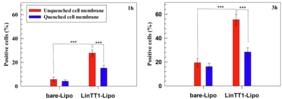
2.12. Human M2 macrophage-liposomes interaction studies
Flow cytometry analysis was used to study the interaction between primary human macrophages, and bare and LinTT1-functionalized li-posomes. Briefly, after polarization, M2 macrophages were seeded in 12- well plates at a density of 2 × 10⁵ cells per well overnight. After removing the cell culture medium, the wells were washed once with PBS solution (pH 7.4). Then, 1.5 mL of bare and LinTT1-functionalized li-posomes, with a final lipid concentration of 250 µg per well, were incubated with the cells for 1 and 3 h at 37 ◦C. Cells were washed twice with PBS solution and then harvested with PBS-EDTA solution. The resulting cell suspensions were centrifuged and washed with PBS buffer three times, and then pelleted cells were re-suspended in an appropriate volume of PBS-EDTA solution for flow cytometer analysis. The interac-tion extent between liposomes (both bare- and LinTT1-Lipo) and M2 macrophages were evaluated without and with trypan blue solution (0.005% v/v) that was used as cell membrane quencher agent during the experiments. When required, trypan blue solution was incubated with cell suspension for 4 min and then washed twice with PBS-EDTA solu-tion. The analysis was performed with an LSR II flow cytometer (BD Biosciences, USA). Data were analyzed using Flowjo VX software (Tree Star, Ashland, OR, USA).
2.13. Statistical analysis
One-way analysis of variance (ANOVA), followed by Tukey’s mul-tiple comparison test was used to analyze the significant difference among results. The analysis was carried out using SigmaPlot v.12 and Excel (Office 2010). Probabilities were set at three different significance
levels: *p < 0.05; **p < 0.01 and ***p < 0.001.
3. Results and discussion
3.1. Physicochemical characterization
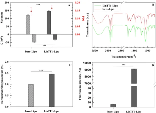
Physicochemical properties, such as size, zeta-potential and size distribution play a crucial role to design DDSs, particularly on colloidal nanoparticles suitable for a potential systemic administration (Zhao et al., 2019). In this investigation, we analyzed the physicochemical properties of LinTT1-Lipo using dynamic light scattering technique. The average diameter of bare-Lipo and LinTT1-Lipo were 121 ± 3 nm and 146 ± 4 nm, respectively. The increased hydrodynamic radius of LinTT1-Lipo depends on the presence of an additional hydrophilic molecule (LinTT1 peptide) conjugated onto the surface of liposomes (Fig. 1A) (Paolino et al., 2014). LinTT1 peptide also increased the zeta- potential value of liposomes changing from − 49.4 ± 3.1 (bare-Lipo) to –32.6 ± 2.3 mV (LinTT1-Lipo) (Fig. 1A). Differences on zeta-potential values between bare and functionalized liposomes may depend on the cationic aminoacids, i.e. arginine and lysine, present on the peptide’s structure. The presence of a guanidinium group and an additional amino group in arginine and lysine backbone, respectively, may provide a slight positive zeta-potential value to the free peptide, increasing the overall zeta-potential value of nanosystem after peptide’s conjugation to the liposomal surface. Conversely, there was no significant modification of the polydispersity index (PDI) between bare-Lipo and LinTT1-Lipo, having a narrow size distribution with a PDI<0.2 for both formula-tions (Wolfram et al., 2014a) (Fig. 1A). This is in agreement with data previously reported elsewhere, that demonstrated for other types of nonosystems, a similar PDI before and after functionalization with LinTT1 peptide (Sim´on-Gracia et al., 2018b). Moreover, the ganglioside
Fig. 1. Physicochemical properties of liposomes evaluated before and after conjugation of LinTT1. (A) Average hydrodynamic diameter, PDI, and zeta-potential value (ζ). (B) ATR-FTIR spectra of bare and LinTT1-functionalized liposomes. (C) Nitrogen amount quantification by elemental analysis of liposomes. (D) Fluo-rescent intensity of bare and LinTT1-functionalized liposomes. Results are the average of three independent experiments ± standard deviation (S.D.). Statistical significance was obtained by a *p < 0.05, **p < 0.01, and ***p < 0.001.
was used to synthesize liposomal formulations in order to reduce the potential immunogenicity of PEG (d’Avanzo et al., 2020; Mima et al., 2017). Indeed, despite this study is focused on in vitro analysis, nano-vesicles were optimized for a potential in vivo application. Based on this evidence, ganglioside was used to make liposomes and data demon-strated that the presence of this molecule in liposomal structure did not modify the stability of nanovesicles in vitro.
Attenuated total reflectance − Fourier transform infrared (ATR- FTIR) analysis demonstrated a more intense carboxylic acid-indicative band at 1745 cm−1 (carboxylic acid C = O stretching) in LinTT1-Lipo than bare-Lipo, thus suggesting the presence of a greater number of carboxylic groups on liposomes’ surface, after peptide’s conjugation. Moreover, the absence of the specific thiol-indicative band at 2550–2600 cm−1 (S-H stretching) in both formulations evidenced the lack of free thiolic groups in LinTT1-Lipo, hence confirming the reaction between thiolic group of cysteine in the peptide structure and the mal-eimide group of polyglycol ethylene (PEG) residual. Nevertheless, in agreement with data reported elsewhere (Torrieri et al., 2020) for LinTT1-functionalized dextran nanoparticles, the conjugation of LinTT1 peptide to the liposomal surface did not significantly modify the FTIR spectrum (Fig. 1B), making it necessary the use of further techniques to confirm the peptide conjugation.
For this, the conjugation of LinTT1 on the surface of liposomes was further studied using elemental composition analysis, which showed a significant increase of the nitrogen amount in LinTT1-Lipo compared to bare-Lipo (Fig. 1C). This finding demonstrated that LinTT1 was suc-cessfully conjugated onto the liposomes’ surface due to the large number of nitrogen atoms found in the peptide structure. Furthermore, in response to the presence of carboxy-fluorescein group (FAM) in the peptide structure, LinTT1-Lipo showed fluorescent properties (Fig. 1D). The resulting LinTT1-functionalized liposomes had a final peptide amount of 2.59 × 10⁴ peptides/liposomes which corresponds to a den-sity of ~ 0.66 peptide molecules/nm2. The resulting peptide density, which has been reported as number of peptide molecules/nm2 of lipo-somal surface, was very similar to data published by Sim´on-Gracia in a previous work (0.7 peptide molecules/nm2 for LinTT1-funtionalized polymersomes), thus showing that LinTT1 peptide molecules is pre-sent on liposomal surface and this amount of peptide can provide a specific targeting of resulting nanosystems versus biological models (Sim´on-Gracia et al., 2018b). These values were obtained starting from a liposomal concentration of 1.29 × 10¹² nanovesicles/mL, and the final amount of conjugated LinTT1peptide (5.7 × 10¹⁶ peptide molecules/mL of final formulation) as reported in the Supplementary materials (Figure S1).
The conjugation between LinTT1 and DSPE-PEG2000mal by sulfhydryl-maleimide reaction caused the synthesis of DSPE-PEG2000- LinTT1. The reaction between maleimide group of DSPE-PEG2000 and sulfhydryl of LinTT1 form a stable synthetic derivative. The synthesis success was confirmed by 1H NMR spectra of LinTT1, DSPE-PEGmal and DSPE-PEG2000-LinTT1. 1H NMR of LinTT1, DSPE-PEG2000mal and DSPE-PEGmal-LinTT1 showed that the characteristic maleimide sharp peak at 7 ppm was present in the spectrum of DSPE-PEG2000mal, but not in that of DSPE-PEG2000-LinTT1, confirming the happened reaction (Figure S4). These results agree with data previously reported for a similar reaction process (Wang et al, 2019).
DOX, alone or in combination with SRF, was loaded in LinTT1-Lipo and tested in vitro, as described subsequently. Physicochemical charac-terization of therapeutic liposomes demonstrated that the hydrody-namic diameter of DOX-loaded liposomes (alone or in association with SRF) increased around 10 nm in comparison with empty ones, however no significant changes were found for the PDI and zeta-potential values (Table S1). Similar values of zeta-potential of empty and drugs-loaded nanovesicles demonstrated that DOX and SRF were entrapped inside the aqueous core and in the phospholipid bilayer of liposomes, respec-tively, and they were not adsorbed on nanovesicles’ surface. The slight increase of average sizes of DOX-loaded liposomes can be ascribed to the
DOX crystallization in the aqueous core of liposomes, as previously re-ported (Pasut et al., 2015). TEM analysis showed that liposomes have spherical-like shapes and their shape is not modified by the conjugation of LinTT1 on the surface of nanocarriers (Figures S2 and S3). All drug- loaded liposomes, both functionalized and bare ones, exhibited the physicochemical characteristics suitable for their systemic use as drug delivery systems for anticancer therapy: (i) an average diameter below than 200 nm suggesting their ability to penetrate intact through fenes-trated vasculature of neo-formed tumor vessels and accumulate in tumor tissue (Maruyama, 2011; Blanco et al., 2015); (ii) a net negative charge suggesting colloidal stability of nanosuspension (Di Francesco et al., 2017a,b); and (iii) a PDI below than 0.2 showing a narrow size distri-bution of nanovesicles (Vakili-Ghartavol et al., 2020)
3.2. Hemocompatibility test
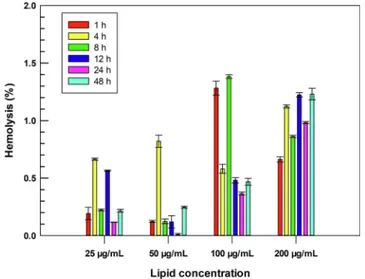
Erythrocytes are the main cells in the bloodstream and closely interact with nanomedicines after their intravenous injections (Pretini et al., 2019), suggesting that the hemocompatibility of a potential sys-temic nanoparticle needs to be investigated during the early stages of its development (de la Harpe et al., 2019). In this scenario, the hemo-compatibility of LinTT1-Lipo was investigated and their incubation with human RBCs demonstrated that the conjugation of LinTT1 peptide on the surface of liposomes via PEG does not lead to a hemolytic nano-system. LinTT1-Lipo induced a hemolysis percentage below than 2% for all tested lipid concentrations up to 48 h of incubation (Fig. 2), which is lower than the threshold of 5% fixed by ISO/TR 7405–1984 for hemo-lytic samples (Zhang et al., 2018). Results demonstrated that LinTT1- Lipo are safe and biocompatible and do not cause hemolysis of human RBCs, suggesting their suitable use for intravenous administration.
3.3. Drugs loading and in vitro release kinetic profiles of payloads
Multidrug liposomes were obtained by co-loading of SRF and DOX inside the liposomes based on their physicochemical properties and solubility (Xiao et al., 2016; Cai et al., 2014), and the relative entrap-ment efficiency (E.E.%) and drug loading (L.D.%) were evaluated using the proper calibration curve Equations (Figure S5). SRF, which is co- loaded in the bilayer of therapeutic liposomes, did not affect the high E.E. of DOX in the aqueous compartment. DOX-loaded liposomes, with
Fig. 2. Hemocompatibility of LinTT1-functionalized liposomes. Hemolysis was monitored up to 48 h of incubation with human erythrocytes at 37 ◦C. Different
lipid concentrations (25, 50, 100, and 200 µg mL−1) were tested during the
analysis. Lysed hemoglobin was quantified in the supernatant at the wavelength of 577 nm using a UV–Vis spectrophotometer. Results are the average of three independent analysis ± S.D.
or without SRF, showed an E.E. of around 90% and a L.D. of 4.5% (Table S1). High E.E. of DOX inside the therapeutic liposomes depended on the crystallized drug that precipitates inside the aqueous core of li-posomes, thus forming a gel-like structure as a consequence of trans-membrane pH gradient and remote loading procedures that were used for the liposomes’ preparation (Cheung and Al-Jamal, 2019; Fritze et al., 2006). Conversely, the E.E. and L.D. efficiencies of SRF-loaded lipo-somes were around 50% and 1.25%, respectively (Table S1). In partic-ular, the co-loading of both drugs into the LinTT1-Lipo resulted in a slight reduction of E.E. of SRF from 51.6 ± 1.1 to 49.8 ± 0.4, while no- significant changes were observed for DOX (Table S1). These results were in agreement with data previously published for liposomes con-taining gemcitabine and paclitaxel in the same colloidal nanocarrier and the very slight decrease or the absence of significant changes in the drugs loading capacity could be explained by the different compartment localization of the two bioactive compounds (Cosco et al., 2011). Moreover, the entrapment and drug loading efficiencies for both drugs were very similar between bare-Lipo and LinTT1-Lipo, suggesting that the surface architecture of liposomes did not affect these parameters (Table S1).
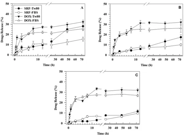
The release kinetics of DOX and SRF from LinTT1-Lipo/D + S were evaluated into two different receptor media: PBS supplemented with FBS (10% v/v) and PBS supplemented with Tween 80 (Tw80) (1% v/v). The receptor media were chosen based on media previously reported elsewhere (Tahir et al., 2020; Guo et al., 2017) for the SRF release study. The analysis was performed at physiological pH and in acid condition (pH 5.5 and pH 6.5) in order to mimic the TME (Justus et al., 2013). Around 25% of DOX was released after 72 h of incubation in the medium supplemented with FBS and ≈30% of drug in the receptor medium supplemented with Tw80. No significant difference was observed for the cumulative release of DOX in the media that have different pH, thus having similar results after 72 h of incubation (Fig. 3). In particular, only slight differences were observed in the release profile of DOX after 3 h of
incubation between physiological pH and pH = 5.5. These results demonstrated that the release kinetic of DOX from LinTT1-Lipo/D + S was independent of the pH of selected receptor media. This is in agreement with data previously reported elsewhere that demonstrated a similar kinetic release of DOX from PEGylated liposomes in PBS solution at pH 5.5 and 7.4 (Shibata et al., 2015). The lack of significant variations in the cumulative release of DOX at different tested pH-values could depend on the ammonium sulfate, and particularly the sulfate, used to generate the pH gradient during the remote loading procedures. In fact, sulfate is a base conjugate obtained after dissociation of strong acid, and the slight decrease of pH from 7.4 to only 5.5 does not allow its pro-tonation, thus resulting in a massive interaction of negative sulfate with protonated DOX at specific pH range (from 5.5 to 7.4) used during the study as previously reported (Fritze et al., 2006). This property affects similar results obtained for the release kinetic of DOX at different pH- values (Fritze et al., 2006). Conversely, 10 ± 2% of SRF was released at pH 5.5 for both receptor media, while 12 ± 2% and 28 ± 4% of drug was released at pH 7.4 for receptor medium supplemented with FBS and Tw80, respectively (Fig. 3). Instead, the cumulative release of SRF at pH 6.5 was 17 ± 3% and 10 ± 1% in the medium supplemented with Tw80 and FBS, respectively, after 72 h of incubation.
Therefore, the release of SRF in the medium with Tw80 at pH 7.4 was twice that of the drug released in medium supplemented with FBS at the same pH (28 ± 4% for Tw80 versus 12 ± 2% for FBS). This difference needs more investigations, but probably the higher release of SRF in the receptor medium containing Tw 80 at pH 7.4 in comparison with pH 5.5 may depend on the lower critical micelle concentration (CMC) of this surfactant at physiological pH than the acid one, as previously demon-strated elsewhere (Bloor et al., 1970) for Tween 40 (Tw40) that have the same hydrophilic head of Tw80 and differs only for the acyl chain. This hypothesis is further supported by release data obtained at pH 6.5. Indeed, despite the SRF released in the medium supplemented with Tw80 is higher than that obtained for the medium supplemented with
Fig. 3. Release kinetic of DOX and SRF from LinTT1-functionalized liposomes in two different media at pH 7.4 (A), pH 6.5 (B) and pH 5.5 (C), at 37 ◦C. The amount
of released drugs was calculated by using external calibration curves as reported in Figure S7 and S8 of Supplementary material. Results are the average of three independent experiments ± S.D. Error bars, if not shown, are within symbols.
FBS after 72 h (17 ± 3% vs. 10 ± 1%, respectively), this difference was significantly lower than results obtained at physiological pH, thus showing that pH-value can affect the SRF release kinetic of LinTT1- liposomes in the accepting medium supplemented with Tw80.
Moreover, Tw80 is a non-ionic surfactant that can be adsorbed on the external surface of liposomal bilayer, thus making mixed micelles with lipids of liposomes, as previously reported for other lipids (Paolino et al., 2017). This complex can favor strong interaction between Tw80 and SRF diffused across lipid bilayer resulting in the leakage of liposomes su-pramolecular structure. This may increase the solubility of SRF and its release in the medium supplemented with Tw80, especially at pH 7.4, where the lower CMC of this surfactant improves its solubilizing properties.
Release kinetic of SRF and DOX in different experimental conditions have a biphasic release kinetic with rapid and continuous release following a zero-order kinetic up to 12 h of incubation, and a plateau from 24 to 72 h of incubation (Fig. 3). Results showed that LinTT1-Lipo were able to co-deliver continuously DOX and SRF up to 72 h in phys-iological and acid media without a massive and rapid release of pay-loads. These findings showed a sustained drugs release from functionalized liposomes, suggesting the ability of this nanosystem to avoid the rapid cargo leakage, thus potentially reducing the side effects of delivered chemotherapeutic agents (Bozzuto and Molinari, 2015). In particular, DOX crystallization hampered the quick leakage of drug after
in vitro and in vivo use and significantly increased the encapsulation
efficiency of doxorubicin hydrochloride in the aqueous core of lipo-somes. This is a direct effect of drug-gel-like structure occurred in the aqueous core of liposomes using pH-gradient and remote procedure as previously reported for Doxil/Caelyx (Barenholz, 2012) and depend on different physicochemical parameters that are specific for drug candi-dates (Cern et al., 2014; Cern et al., 2017).
However, the gel-like structure resulted by crystallizing doxorubicin hydrochloride and the resulting slow and constant drug release did not affect and/or decrease the anticancer efficacy of payload and its cyto-toxicity on breast cancer cells. In fact, LinTT1-liposomes can be targeted from breast cancer cells overexpressing LinTT1 specific receptors and colloidal nanocarriers were like a depot system, which favored the accumulation of crystallized doxorubicin hydrochloride inside the can-cer cells and provided the slow dissolution of payload in the intracellular compartment, as reported elsewhere (Li et al., 2019; Wei et al., 2015). Based on these, the therapeutic effects of drugs were not compro-mised in vivo due to the biodegradable properties of liposomes that are degraded after internalization in tumor tissues and then release payloads (Costanzo et al., 2019).
3.4. Stability studies in human plasma
Nanomedicines should remain stable after systemic injection and biomaterials making polymeric shell or lipid bilayer were selected to protect nanocarriers from destabilization phenomenon occurring after interaction with plasma proteins (Immordino et al., 2006). Nanocarriers injected in vivo interact with circulating cells and proteins and these phenomena may cause several mechanical stresses and activate enzy-matic processes (Di Francesco et al, 2021). These drawbacks can strongly decrease the therapeutic effect of nanomedicines, thus causing degradation of nanocarriers, with the quick leakage of payloads, or their rapid clearance from bloodstream and significant macrophage uptake (Ferrari, 2010). Circulating serum proteins play a crucial role for the potential modification of surface properties in nanocarriers, and thus, their destabilization after systemic injection (Pasut et al., 2015), due to protein corona phenomenon (Palchetti et al., 2016). Protein corona significantly modifies the physicochemical properties of nanocarriers, thus increasing average sizes and size distribution, as well as their sur-face properties and compositions (Hadjidemetriou et al., 2019). Protein corona phenomenon can also facilitate the macrophage uptake of nanocarriers, and thus, their clearance through the reticuloendothelial
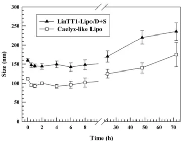
system (RES) organs (Monopoli et al., 2012). In these attempts, we studied the stability of LinTT1-lipo + D/S after incubation with human plasma and the resulting data was compared that obtained for Caelyx- like Lipo (control), which is similar to Doxil/Caelyx liposomal formu-lations currently approved from Food and Drug Administration and European Medicine Agency for the treatment of breast cancer (Monopoli et al., 2012, Bulbake et al., 2017). LinTT1-lipo + D/S and Caelyx-like Lipo had similar trend for average sizes after 72 h of incubation in human plasma (Fig. 4). They showed a slight decrease of particle sizes (~15 nm) in the 1 h of incubation, maybe due to the adsorption of a weak protein corona on the surface of PEGylated liposomes. This process resulted in an osmotic gradient on liposomal surface leading to the shrinkage of nanocarriers, and thus, the leakage of water content from the aqueous core of liposomes (Wolfram et al., 2014b). Conversely, the average sizes of LinTT1-lipo + D/S and Caelyx-like Lipo increased after 48 h of incubation and this phenomenon increased significantly after 72 h of incubation with resulting average sizes of ~ 230 nm and ~ 180 nm for LinTT1-Lipo + D/S and Caelyx-like Lipo (Fig. 4), respectively. The significant increase of particle sizes at 48 and 72 h of incubation, may depend on a biphasic process occurring during the formation of protein corona on nanocarriers surface. In fact, we can hypothesize that during the early stages of incubation, proteins having high concentrations but low affinity for liposomal bilayer, formed an unstable protein corona, which is in equilibrium and exchanges with the surface of liposomes without changing significantly particle sizes. Conversely, at long incu-bation times (48 and 72 h), circulating proteins, making protein corona, had a low concentration but high affinity for liposomes. The high affinity protein corona for liposomal surface replaced soft corona, adsorbed at short incubation times, with hard corona that stack to the surface of nanocarriers and thus increased their average size (Pozzi et al., 2015; Wolfram et al., 2014b). The resulting data also demonstrated that LinTT1-lipo + D/S and Caelyx-like Lipo were stable and did not change their average sizes up to 24 h (Fig. 4). The stability tests supported our hypothesis that LinTT1-Lipo + D/S is stable in human plasma and circulating proteins do not affect nanocarrier stability, and fostered a potential in vivo translation of LinTT1-Lipo + D/S.
Despite, the significant increment of particle size after 72 h of in-cubation with human plasma, average sizes of Caelix-like Lipo and LinTT1-Lipo + D/S were less than twice compared to the initial data (~112 nm (Caelix-like Lipo) and ~ 160 nm (LinTT1-Lipo + D/S) at time point 0 vs. ~ 180 nm (Caelix-like Lipo) and ~ 230 nm (LinTT1-Lipo + D/ S) at 72 h), thus supporting the hypothesis that nanocarriers did not make aggregates after 72 h of incubation in human plasma, but the
Fig. 4. Human plasma stability of LinTT1-Lipo + D/S and Caelyx-like Lipo. Results are the average of three independent experiments ± S.D. (n = 3). Error bars, if not shown, are within symbols.
increase of sizes depended on the resulting protein corona adsorbed on the surface of liposomes (Pozzi et al., 2015; Wolfram et al., 2014b). Our hypothesis was further supported by Turbiscan analysis, which demonstrated the lack of aggregation for LinTT1-Lipo + D/S and Caelix- like Lipo in saline solution (NaCl) 0.9%, w/v, (Figure S9) and medium (saline solution NaCl 0.9%/human plasma, 50:50 (v/v)) (Figure S10) at 37 ◦C. LinTT1-Lipo + D/S and Caelix-like Lipo had BS and T signal variations below 5% and 10% during the incubation time (1 h) and their relative lines were overlapped with the threshold baseline (Di Francesco et al., 2017a,b). The presence of negative or positive peak variations for BS and T signals at samples height over 8 mm and below 2 mm did not depend on the nanocarrier destabilization but was related to air bubbles present at the interfaces on the top or bottom of glass holder during the analysis (Celia et al., 2009). The long-term stability of LinTT1-Lipo + D/ S and Caelix-like Lipo in human plasma were further confirmed by destabilization kinetic profiles which were similar for both nanocarriers (Figure S11). These results were in agreement with data herein reported for DLS analysis (Fig. 4), and thus, demonstrate that LinTT1-Lipo + D/S is stable and has suitable physicochemical properties for a potential injection in the bloodstream.
3.5. In vitro cytotoxic effect on 2D cell models
DOX, as a free drug or formulated as PEGylated liposomes, is currently one of the most used chemotherapeutic drugs to treat breast cancers, and particularly TNB cancer (Waks and Winer, 2019). DOX is usually administereted in combination with other cytotoxic drugs, such as paclitaxel and cyclophosphamide (Waks and Winer, 2019; Tampaki et al., 2018), and these combinations have a synergistic effect that re-sults in a decreased effective dosage of drugs compared to mono-therapy (Fisusi and Akala, 2019). This approach decreases the side effects and modifies the induction rate of cancer resistant phenomena (Waks and Winer, 2019; Yardley, 2013). Despite controversial results about the use of SRF in combination with other chemotherapeutic agents in breast cancer therapies, some clinical trials are still ongoing (Chen et al., 2019a; Hwang et al., 2019). Moreover, several animal investigations have clearly shown the ability of SRF to increase the anticancer prop-erties of chemotherapic agents commonly used in clinic for breast cancer therapies, especially when the two drugs were co-loaded in the same nanocarriers. In these attempts, Sui et al. recently demonstrated the higher anticancer efficacy of SRF-loaded into pullulan-DOX conjugated nanoparticle than the same nanosystem containing only DOX, on murine breast cancer carcinoma (Sui et al., 2017). In this study, they also showed a great reduction of side effects when the two drugs were co- loaded into nanoparticles than the combination of free drugs adminis-tered in the free form. Similar results were also observed by Lei et al., which demonstrated a great synergistic effect of SRF and paclitaxel when the two drugs were co-loaded into the same liposomal carrier, on MCF-7/multidrug resistant cancer in mice (Lei et al., 2019).
In our study, the capability of SRF to potentiate the cytotoxic effect of DOX was tested in two different breast cancer cell lines. In particular, a positive estrogen receptor (MCF-7) and a triple negative (MDA-MB-231) breast cancer cell lines were used during the study. The cytotoxic effect was evaluated based on drug concentrations (0.01–10 µM) and incu-bation times (24, 48 and 72 h). Drug concentration, for the in vitro tests, is reported as a ratio to the DOX concentration, which is three-fold higher than SRF at different incubation time points. This difference depends on the final concentration of DOX and SRF, which are co-loaded inside liposomes, being 1600 and 540 µM for DOX and SRF, respectively. As the first step, the cytotoxic effect of DOX as a single agent was compared with the cytotoxic effect provided by the association of SRF and DOX, both in free form or loaded in bare liposomes (Fig. 5). The cytotoxic effect of SRF as a single agent was not investigated in this study, because despite its potential ability to increase the efficacy of breast cancer therapies when it is co-administered with other chemo-therapeutics, its efficacy as single agent has been demonstrated to be
poor in breast cancer (Bronte et al., 2017).
SRF significantly increased the cytotoxic effects of DOX when the two drugs were co-loaded in bare liposomes in positive estrogen re-ceptors (MCF-7) and in TNB cancer (MDA-MB-231) cell lines, compared to free drugs (Fig. 5).
This effect was similar in MCF-7 and MDA-MB-231 cells. In partic-ular, after 24 h of incubation, the cytotoxic effect of free DOX and combined free DOX and SRF was very similar for all drug concentrations in both investigated cell lines. On the contrary, at the same incubation time point, the cytotoxic effect of DOX and SRF co-loaded in bare lipo-somes (bare-Lipo/D + S) was significantly higher than bare lipolipo-somes containing only DOX (bare-Lipo/D), at the DOX concentration of 0.5 and 1 µM, and SRF concentration equivalent to 0.17 and 0.35 µM, respec-tively. The ability of SRF to improve the cytotoxic effect of DOX when the two drugs were co-loaded in bare liposomes in comparison with free drugs, became more evident after 48 and 72 h of incubation (Fig. 5). After 48 h of incubation, bare-Lipo/D + S showed a cell death of 45.8 ± 2.9% and 60.0 ± 3.6% on MCF-7 cell line at the DOX concentration of 0.5 µM (and SRF concentration equivalent to 0.17 µM) and at DOX concentration of 1 µM (and SRF concentration equivalent to 0.35 µM), respectively; while bare-Lipo/D demonstrated a cell death of 32.2 ± 4.5% and 44.3 ± 3.1% at the same DOX concentrations, respectively. Furthermore, the association of free drugs resulted in a slight reduction of cell viability compared with free DOX. After 48 h of incubation, MCF- 7 cells showed a cell death of 27.7 ± 4.9% and 38.3 ± 3.6% when treated with combined drugs at DOX concentration of 0.5 µM (and SRF con-centration equivalent to 0.17 µM) and at DOX concon-centration of 1 µM (and SRF concentration equivalent to 0.35 µM), respectively; and 22.5 ± 2.9% and 30.6 ± 4.8% for free DOX as single agent at the same DOX concentrations, respectively. Similar trend was observed in MDA-MB- 231 cells after 48 h of incubation with bare-Lipo/D, showing a cell death of 44.5 ± 2.1% at DOX concentration of 0.5 µM, while cell treated with bare-Lipo/D + S showed a cell death of 58.1 ± 4.4% at the same DOX concentration and SRF equivalent concentration of 0.17 µM. Also, in this cell line, the cytotoxic synergistic effect of combined free drugs was lower than DOX and SRF co-loaded liposomes. Indeed, after 48 h of incubation, the cell death was 32.8 ± 5.9% at DOX concentration of 0.5 and 39.9 ± 6.3% at the same DOX concentration (and SRF concentration equivalent to 0.17 µM), for free DOX and combined free drugs, respectively.
Moreover, after 72 h of incubation with bare-Lipo/D + S the cell death was of 59.2 ± 5.9% and 74.2 ± 4.6% at DOX concentration of 0.5 µM (and SRF concentration equivalent to 0.17 µM), for MCF-7 and MDA- MB-231 cell line, respectively (Fig. 5). Conversely, combinations of free drugs had a similar cytotoxic effect herein reported (61.5 ± 3.6% and 68.2 ± 5.2%) at DOX concentration equivalent to 1 µM (and SRF con-centration equivalent to 0.35 µM), for MCF-7 and MDA-MB-231 cell lines, respectively (Fig. 5). This effect highlighted the potentiality of liposomes to decrease the therapeutic drugs dosage for efficacious treatment in breast cancer (Meng et al., 2016).
The increased cytotoxic efficacy of bare-Lipo/D + S in comparison with the association of free drugs was probably due to the enhanced synergistic effect of the two chemotherapeutic drugs in response to their allocation in the same nanovesicle, which provided an increased uptake from breast cancer cells and reduced destabilization phenomena on drugs molecules (Sercombe et al., 2015). Based on these findings, for further analysis concerning the cytotoxic efficacy, liposomes containing both drugs were used. The cytotoxic effect of DOX and SRF, as free drugs or formulated as liposomes, was always higher in MDA-MB-231 cells than MCF-7 cells. This difference depends on the higher responsiveness of MDA-MB-231 cells than MCF-7 ones to DOX (Lovitt et al., 2018).
One of the main challenges in anticancer therapies is to target spe-cifically the pathological tissues, improving the efficacy of therapy and decreasing side effects on healthy cells (Yan et al., 2020; Pu et al., 2019). In these attempts, we functionalized liposomes by conjugating onto the surface of liposomes via PEG the LinTT1 peptide, a specific ligand for