Published Ahead of Print 9 June 2014.
10.1128/AAC.02594-14.
2014, 58(8):4782. DOI:
Antimicrob. Agents Chemother.
David Lembo
Volante, Elena Veccelli, Pasqua Oreste, Marco Rusnati and
Valeria Cagno, Manuela Donalisio, Andrea Civra, Marco
Histocultures
Cell Lines and Human Tracheal-Bronchial
Respiratory Syncytial Virus Infectivity in
Polysaccharide Derivatives Inhibit
http://aac.asm.org/content/58/8/4782
Updated information and services can be found at:
These include:
SUPPLEMENTAL MATERIAL
Supplemental material
REFERENCES
http://aac.asm.org/content/58/8/4782#ref-list-1
at:
This article cites 67 articles, 29 of which can be accessed free
CONTENT ALERTS
more»
articles cite this article),
Receive: RSS Feeds, eTOCs, free email alerts (when new
http://journals.asm.org/site/misc/reprints.xhtml
Information about commercial reprint orders:
http://journals.asm.org/site/subscriptions/
To subscribe to to another ASM Journal go to:
on July 16, 2014 by guest
http://aac.asm.org/
Downloaded from
on July 16, 2014 by guest
http://aac.asm.org/
Downloaded from
Respiratory Syncytial Virus Infectivity in Cell Lines and Human
Tracheal-Bronchial Histocultures
Valeria Cagno,aManuela Donalisio,aAndrea Civra,aMarco Volante,bElena Veccelli,cPasqua Oreste,cMarco Rusnati,dDavid Lemboa
Department of Clinical and Biological Sciences, University of Turin, Turin, Italya
; Department of Oncology, University of Turin, Turin, Italyb
; Glycores 2000 Srl, Milan, Italyc
; Department of Biomedical Sciences and Biotechnology, University of Brescia, Brescia, Italyd
Respiratory syncytial virus (RSV) exploits cell surface heparan sulfate proteoglycans (HSPGs) as attachment receptors. The in-teraction between RSV and HSPGs thus presents an attractive target for the development of novel inhibitors of RSV infection. In this study, selective chemical modification of the Escherichia coli K5 capsular polysaccharide was used to generate a collection of sulfated K5 derivatives with a backbone structure that mimics the heparin/heparan sulfate biosynthetic precursor. The screening of a series of N-sulfated (K5-NS), O-sulfated (K5-OS), and N,O-sulfated (K5-N,OS) derivatives with different degrees of sulfation revealed the highly sulfated K5 derivatives K5-N,OS(H) and K5-OS(H) to be inhibitors of RSV. Their 50% inhibitory concentra-tions were between 1.07 nM and 3.81 nM in two different cell lines, and no evidence of cytotoxicity was observed. Inhibition of RSV infection was maintained in binding and attachment assays but not in preattachment assays. Moreover, antiviral activity was also evident when the K5 derivatives were added postinfection, both in cell-to-cell spread and viral yield reduction assays. Finally, both K5-N,OS(H) and K5-OS(H) prevented RSV infection in human-derived tracheal/bronchial epithelial cells cultured to form a pseudostratified, highly differentiated model of the epithelial tissue of the human respiratory tract. Together, these features put K5-N,OS(H) and K5-OS(H) forward as attractive candidates for further development as RSV inhibitors.
H
uman respiratory syncytial virus (RSV) is an enveloped sin-gle-stranded negative-sense RNA virus belonging to the ge-nus Pneumovirus of the family Paramyxoviridae (1). It is the lead-ing cause of bronchiolitis and pneumonia in infants and young children worldwide. More than half of all children are seropositive for RSV by 1 year of age, and almost all children have been infected by 2 years of age (2). Moreover, RSV is a pathogen of considerable importance in immunocompromised adults and the elderly, par-ticularly in those with chronic obstructive pulmonary disease (3). In the United States alone, RSV is estimated to cause 120,000 hospitalizations each year and as many as 200 to 500 deaths in infants/young children, while around 160,000 fatalities occur an-nually worldwide (2,4,5). The economic burden related to RSV infection is approximately $500 million in the United States alone, without taking outpatient care into account (6,7).Currently, the treatment of RSV infections is mainly symptom-atic (8), and the development of a preventive vaccine is hampered by difficulties in eliciting long-lasting protective immunity (9). Antiviral therapy is limited to ribavirin, a nonspecific antiviral drug that interferes with viral transcription; however, the nonneg-ligible side effects of ribavirin and the recent recommendation of the American Academy of Pediatrics not to routinely use this drug in children with bronchiolitis (10) call for the development of more selective and safe therapeutics for the treatment of RSV in-fection (11,12). For immunoprophylaxis, a monoclonal human-ized antibody, palivizumab, is available, but it is administered only to high risk premature newborns due to its high cost (13,14). Another antibody, named motavizumab (an affinity-matured variant of palivizumab), was not provided with FDA approval due to safety concerns (15). Thus, in view of the continual rise world-wide in the morbidity and mortality of infants, the immunocom-promised (in particular AIDS patients), and elderly individuals resulting from RSV infection (16,17) and bearing in mind that no
antiviral drug exists to combat this pathogen, RSV constitutes an important target for the development of new antiviral molecules. The binding of RSV to cultured cells has been characterized at the molecular level: it involves an initial interaction between the positively charged basic amino acids present within the linear hep-arin-binding domain (HBD) (18) of the viral envelope proteins G and F (19,20) and the negatively charged sulfated/carboxyl groups of the cell surface heparan sulfate proteoglycans (HSPGs). RSV attachment to HSPGs is followed by a second interaction with nucleolin, a cellular protein which is involved in attachment and entry of several viruses, including human parainfluenza virus type 3, Crimean-Congo hemorrhagic fever virus, Japanese encephalitis virus, and HIV (20,21,22,23,24,25). Consequently, the interac-tion between the envelope glycoproteins of RSV and cellular HSPGs presents an attractive target for novel anti-RSV therapies. HSPGs are associated with the cell surface; they consist of a protein core and glycosaminoglycan (GAG) side chains of un-branched sulfated polysaccharides, known as heparan sulfates (HS), which are structurally related to heparin. Heparin and HS consist of a sequence of glucuronic (GlcA) or iduronic acid (IdoA) residues that are␣1¡4 linked to a glucosamine (GlcN) molecule that can be N-sulfated or N-acetylated. The disaccharide sequence
Received 11 March 2014 Returned for modification 14 April 2014 Accepted 1 June 2014
Published ahead of print 9 June 2014
Address correspondence to David Lembo, [email protected].
Supplemental material for this article may be found athttp://dx.doi.org/10.1128 /AAC.02594-14.
Copyright © 2014, American Society for Microbiology. All Rights Reserved. doi:10.1128/AAC.02594-14
on July 16, 2014 by guest
http://aac.asm.org/
can also be O-sulfated in different positions: positions 3 and 6 on GlcN and position 2 on uronic acid. HS show high structural heterogeneity along their chains, with specific regions responsible for binding to different ligands.
In respect to HS, heparin is endowed with a high degree of sulfation and a more homogeneous disposition of sulfated groups along its saccharidic chain (26), and consequently, it usually binds to cognate ligands (both viral and eukaryotic) with a higher affin-ity than HS, resulting in the strongest HSPG-antagonist activaffin-ity in competition experiments (27,28,29,30). This identifies heparin as an ideal reference compound in studies aimed at the identifica-tion of polyanionic HSPG antagonist compounds.
Besides the case of RSV, HSPGs have also been demonstrated to act as attachment receptors for human immunodeficiency virus (HIV) (31), herpes simplex virus (HSV) (32), human papilloma-virus (HPV) (33), human cytomegalovirus (HCMV) (34), dengue virus (35), and filoviruses (36); accordingly, several anti-HSPG strategies have been attempted for all of these viruses (29,37,38,
39,40). Despite that fact that this huge amount of in vitro exper-imentation initially provided promising results, few polyanionic, heparin-like compounds progressed to clinical trial for different viral diseases (41,42,43). These compounds were safe and well tolerated in phase I and II studies but were devoid of any impor-tant clinical benefit in phase III study. This failure of clinical trials of earlier polyanionic antiviral compounds calls for the design of newer compounds endowed with more controlled structures and biological properties.
A peculiar class of compounds, namely, the sulfated derivatives of capsular K5 polysaccharide from Escherichia coli, has emerged as a promising biotechnological candidate agent for the develop-ment of novel antiviral drugs (44). In brief, the capsular K5 poly-saccharide from Escherichia coli has the same structure as the hep-arin precursor N-acetyl heparosan. The chemical sulfation of K5 in various N and/or O positions along the polysaccharide results in the synthesis of K5 derivatives with different degrees of sulfa-tion and charge distribusulfa-tion that are endowed with specific bind-ing capacities and biological properties. These semisynthetic GAGs are devoid of the well-known anticoagulant activity that prevents the use of heparin and other sulfated polysaccharides as antivirals (26) and thus present a promising starting point for the development of new antiviral formulations.
In effect, K5 sulfated derivatives have been demonstrated to be endowed with inhibitory activity against different viruses, includ-ing herpes simplex virus (HSV) (38), human papilloma virus (HPV) (39), cytomegalovirus (CMV) (40), dengue virus (29), and HIV (37). Regarding this last virus, K5 polysaccharides have been demonstrated to classically act as antimicrobial agents, likely binding to the basic gp120 protein but also binding to and neu-tralizing other HIV proteins released by infected cells (i.e., Tat and p17) that contribute to HIV dissemination and to the onset of AIDS-associated infections (30,45). Taken together, these data point to K5 sulfated derivatives as an interesting class of antiviral compounds endowed with a multitarget activity that can be expli-cated at different levels (i.e., against different viruses but also aimed at different proteins of a given virus) (44). Relevant to this point, a tight relationship exists between HIV, HSV, and HPV infection, suggesting the possibility of generating K5-based drugs with a multitarget mechanism of action that can control and/or prevent HIV, HPV, and HSV infection simultaneously (44).
In-terestingly, a positive correlation has also been described for RSV and HIV infections (46).
The aim of the present work was to investigate whether the antiviral potential of K5 derivatives also extends to the respiratory virus RSV. To this purpose, a panel of N-sulfated (K5-NS), O-sul-fated (K5-OS), and N,O-sulO-sul-fated (K5-N,OS) derivatives was screened to identify compounds with RSV inhibitory activity; an-tiviral potency and the mode-of-action of the best-hit compounds were also investigated. Highly sulfated (H) K5 derivatives K5-N,OS(H) and K5-OS(H) emerged as nontoxic inhibitors of RSV infectivity in both cell culture and an in vitro tissue model of hu-man tracheal/bronchial epithelium.
MATERIALS AND METHODS
Heparin and K5 polysaccharide derivatives. Unmodified unfractionated
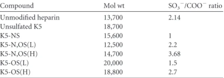
beef mucosal heparin was obtained from Laboratori Derivati Organici, Milan, Italy. K5 polysaccharide derivatives were obtained by N-deacety-lation/N-sulfation and/or O-sulfation of a single batch of K5 polysaccha-ride as previously described (47). The N-deacetylation/N-sulfation of K5 polysaccharide is performed in one step and has been scaled to a 10-g amount. The average yield of compound of the N-deacetylation/N-sulfa-tion is about 80%. The degree of N-sulfaN-deacetylation/N-sulfa-tion is determined by1H nuclear magnetic resonance (1H-NMR) at 500 MHz, and no signal of residual N-acetylation is detectable. The sulfate-to-carboxyl ratio of the final prod-ucts is measured according to the method of Casu et al. (48). The antiviral results have been reproduced with two different batches of compounds. The main chemical features of these GAGs are shown inTable 1.
Cells and viruses. The epithelial cell lines HEp-2 (ATCC CCL-23) and
A549 (ATCC CCL-185) were grown as monolayers in Eagle’s minimal essential medium (MEM) (Gibco/BRL, Gaithersburg, MD) supple-mented with heat-inactivated 10% fetal calf serum (FCS) (Gibco/BRL). RSV strain A2 (ATCC VR-1540) was propagated in HEp-2 cells by infect-ing a freshly prepared confluent monolayer grown in MEM supplemented with 2% of FCS. When the cytopathic effect involved the whole mono-layer, the infected cell suspension was collected and the viral supernatant was clarified. The virus stocks were aliquoted and stored at⫺80°C. The infectivity of virus stocks was determined on HEp-2 cell monolayers by standard plaque assay. The cell lines and the RSV were obtained from the American Type Culture Collection (Manassas, VA, USA).
Cell viability assay. Cells (A549 and HEp-2) were seeded at a density
of 5⫻ 104/well in 96-well plates and treated the next day with serially diluted GAGs to generate dose-response curves. After 72 h of incubation, cell viability was determined using the CellTiter 96 proliferation assay kit (Promega, Madison, WI, USA), according to the manufacturer’s instruc-tions. Absorbances were measured using a microplate reader (model 680; Bio-Rad) at 490 nm. Fifty percent cytotoxic concentration (CC50) values and 95% confidence intervals (CIs) were determined using Prism soft-ware (GraphPad Softsoft-ware, San Diego, CA).
Virus inactivation assay. Approximately 104PFU of RSV and 3.6 g/ml of each GAG (corresponding to 240 nM K5-N,OS(H), 191 nM K5-OS(H), and 263 nM heparin) were added to MEM and mixed in a total
TABLE 1 Molecular weights and degrees of sulfation of the GAGs used
in this work
Compound Mol wt SO3⫺/COO⫺ratio
Unmodified heparin 13,700 2.14 Unsulfated K5 18,700 K5-NS 15,600 1 K5-N,OS(L) 12,500 2.2 K5-N,OS(H) 14,700 3.68 K5-OS(L) 20,000 1.5 K5-OS(H) 18,800 2.7
on July 16, 2014 by guest
http://aac.asm.org/
Downloaded from
volume of 100l. Virus-GAG mixtures were incubated for 2 h at 37°C or 4°C and serially diluted to the noninhibitory concentration of each test compound, and the residual viral infectivity was determined by the viral plaque assay.
Binding assay. Each GAG (10M) was added to an aliquot of RSV
(5⫻ 104PFU) and administered directly to HEp-2 or A549 cell monolay-ers in MEM supplemented with 2% FCS, incubated for 2 h at 4°C, and washed three times to remove unbound virus. Cells were then fixed with 4% paraformaldehyde, air dried, and blocked with 5% bovine serum al-bumin (BSA) in phosphate-buffered saline(PBS)-Tween. Bound virus was detected using RSV monoclonal antibody (Ab35958; Abcam, Cam-bridge, United Kingdom) (diluted 1:400), incubated for 1 h at room tem-perature, washed three times with PBS-Tween, and incubated for 2 h at 37°C with goat anti-mouse IgG conjugated to horseradish peroxidase (HRP) (1:1,000). At the end of incubation, plates were washed three times with PBS-Tween before adding the ABTS substrate [2,2=-azinobis(3-eth-ylbenzthiazolinesulfonic acid)] (Thermo Scientific, Rockford, IL) and reading the absorbance at 405 nm. Percent inhibition of virus binding was determined by comparing the absorbance measured in the presence of the compound to that measured in untreated cultures.
Viral plaque assay. To evaluate the capacity to inhibit RSV infection,
GAGs were serially diluted to generate dose-response curves and added to RSV (multiplicity of infection [MOI], 0.01 PFU/cell). After 1 h of incuba-tion at 4°C, the mixture was added to cells grown as monolayers in a 96-well plate at a density of 5⫻ 104/well. After 3 h of incubation at 37°C, monolayers were washed and overlaid with 1.2% methylcellulose me-dium. Three days postinfection, cells were fixed with cold methanol and acetone for 1 min and subjected to RSV-specific immunostaining using an RSV monoclonal antibody (Ab35958; Abcam, Cambridge, United King-dom) and the UltraTech HRP streptavidin-biotin detection system (Beck-man Coulter, Marseille, France). Immunostained plaques were counted, and the percent inhibition of virus infectivity was determined by compar-ing the number of plaques in compound-treated wells with the number in untreated control wells. Fifty percent inhibitory concentration (IC50) val-ues and 95% CIs were determined using Prism software. All data were generated from duplicate wells in at least three independent experiments. To characterize the mechanism of the antiviral action of the K5 deriv-atives, the viral plaque assay was repeated, incorporating the following modifications.
Preattachment assay. HEp-2 and A549 cell monolayers in 96-well
plates were incubated with increasing concentrations of the various GAGs for 2 h at 37°C. After removal of the compound and two gentle washes, cells were infected with RSV (MOI, 0.01 PFU/cell) in the absence of com-pounds for 3 h at 37°C. Cells were then overlaid with 1.2% methylcellulose medium, incubated for 72 h at 37°C, and successively fixed and immuno-stained as described above. Plaques were then counted.
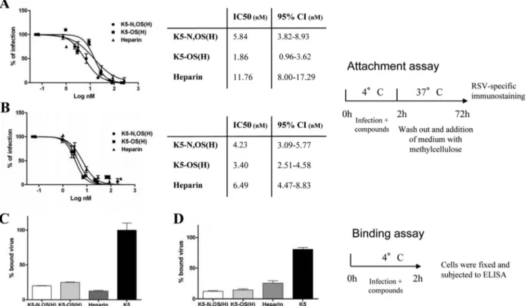
Attachment assay. Serial dilutions of the various GAGs were
preincu-bated with RSV (MOI, 0.05 PFU/cell) for 1 h at 4°C, added to cooled HEp-2 and A549 cells in 96-well plates, and incubated for 2 h at 4°C to ensure viral attachment but not entry. After two gentle washes, cells were overlaid with 1.2% methylcellulose medium, shifted to 37°C for 72 h, and successively fixed and immunostained as described above. Plaques were then counted.
Postattachment assay measuring viral yield. HEp-2 cell monolayers
in 24-well plates were infected with RSV (MOI, 0.005 PFU/cell) in MEM supplemented with 2% FCS for 3 h at 37°C and then subjected to two gentle washes to remove unbound virus. Increasing concentrations of the various GAGs in MEM supplemented with 2% FCS were then added to cultures after washout of the viral inoculum or after 1, 2, 3, or 24 h. Incubation continued until the cytopathic effect involved the whole monolayer in the untreated wells. The infected cell suspensions were col-lected, and the supernatants were clarified. RSV infectivity was deter-mined on A549 cell monolayers by standard plaque assay. Titrations were carried out at dilutions at which compounds were no longer active to
exclude the possibility that a carryover of tested polysaccharides into the titration culture would block virus attachment.
Percent inhibition was determined by comparing the viral titer mea-sured in the presence of the compounds to that meamea-sured in untreated wells.
Syncytium formation assay. The abilities of the various GAGs to
block RSV cell-to-cell spread were evaluated using a previously described method (49) with minor modifications. Cell monolayers in 96-well plates were infected with RSV (MOI, 0.01 PFU/cell) in MEM supplemented with 2% FCS for 3 h at 37°C and then subjected to two gentle washes to remove unbound virus. Following inoculum washout, increasing concentrations of each GAG in 1.2% methylcellulose medium were then added to cul-tures. Incubation continued for 72 h postinfection at 37°C; cells were then fixed and immunostained. The immunostained syncytia were visualized using a Leica inverted microscope equipped with a Bresser MikroCam microscope camera and MikroCamLab software (Rhede, Germany). Im-ageJ software was used to quantify plaque sizes. Untreated RSV-infected monolayers were used as the control.
Rotavirus infectivity assay. Rotavirus infectivity assays were
per-formed as previously described (50) with some modifications. Confluent MA104 cell monolayers in a 96-well plate were washed twice with MEM and then infected with human rotavirus strain Wa (ATCC VR-2018) at an MOI of 0.02 PFU/cell for 1 h at 37°C in the presence or absence of each test GAG. Virus was preactivated with 5g of porcine trypsin (Sigma)/ml for 30 min at 37°C. After the adsorption period, the virus inoculum was removed, cells were washed with MEM, and the cultures were maintained at 37°C for 16 h in medium with trypsin at 0.5g/ml. The infected cells were fixed and immunostained using an UltraTech HRP streptavidin-biotin detection system (Beckman Coulter).
EpiAirway tissues. EpiAirway tissues, cultured on collagen supports
under air-liquid interface conditions, were obtained from MatTek Corp. (Ashland, MA, USA). These tissues consisted of normal human-derived tracheal/bronchial epithelial cells that are highly differentiated (i.e., con-tain cilia, tight junctions, sodium and chloride channels, etc.) and recon-tain properties of normal respiratory tract epithelial tissue (i.e., actively secrete mucus, electrogenicity, etc.). Upon delivery, the EpiAirway tissue inserts were processed according to the supplier’s protocol. Briefly, each tissue insert was transferred to a well in a 6-well plate prefilled with 900l prewarmed serum-free medium (Air-100-MM; MatTek Corp.) and incu-bated at 37°C in 5% CO2overnight (16 to 18 h). EpiAirway tissues were then used in the following three assays.
Cytotoxicity assay. The cytotoxicity of K5 derivatives on mucous
membranes was assessed using the 3-(4,5-dimethyl-2-thiazolyl)-2,5-di-phenyl-2H-tetrazolium bromide (MTT) ET50tissue viability assay ac-cording to the manufacturer’s instructions. K5 derivatives (10M) were applied to the cell culture insert on top of the EpiAirway tissue samples and incubated for 1, 4, or 18 h at 37°C in duplicate. At the end of the incubation, any liquid on top of the EpiAirway tissue was decanted, and inserts were gently rinsed with PBS to remove any residual material. Tis-sues were then processed according to the MTT kit protocol (MatTek Corporation) and read using an enzyme-linked immunosorbent assay (ELISA) plate reader at a wavelength of 570 nm. Tissues incubated with assay medium were used as negative controls. The ET50is the time re-quired to reduce tissue viability to 50% and was determined using Prism software (GraphPad Software, San Diego, CA). According to the informa-tion provided by the supplier, ET50values of⬎18 h indicate that a tested compound is not irritating.
Antiviral assay. To assess the antiviral activity of K5 derivatives on
EpiAirway cultures, aliquots of 100l of medium containing 50,000 PFU of RSV with or without K5 derivatives (10M) were preincubated for 1 h at 4°C and then added to the apical surface of the tissues. After 3 h of incubation at 37°C, the medium was removed, and the cultures were washed apically with 100l of medium and then fed each day via the basolateral surface with 1 ml medium. To harvest the virus, 100l me-dium was added to the apical surface, and the tissues were allowed to
on July 16, 2014 by guest
http://aac.asm.org/
equilibrate for 30 min at 37°C. The suspension was then collected and stored at⫺80°C until viral titers were determined by plaque assay in A549 cell monolayers as described above. Collection of virus was performed sequentially from the same wells on each day postinfection.
Detection of RSV in EpiAirway tissue by immunohistochemistry.
RSV was detected immunohistochemically using a specific mouse mono-clonal antibody against RSV (Ab35958; Abcam, Cambridge, United King-dom). Briefly, EpiAirway tissue cultures exposed to RSV in the absence or presence of K5 derivatives (10M) were fixed in buffered formalin and embedded (properly oriented) in paraffin together with adherent collagen membranes. Immunohistochemical sections were processed for antigen retrieval in citrate buffer using a dedicated pressure cooker (1 cycle for 5 min at 125°C, followed by 10 s at 90°C) in parallel with sections stained with conventional hematoxylin and eosin. Following incubation with the primary antibody (1:500 dilution), the reaction product was visualized using a biotin-free polymer-conjugated secondary antibody (Envision; Dako, Glostrup, Denmark). In the presence of a positive reaction, the antibody showed cytoplasmic and nuclear immunoreactivity, mostly rec-ognizable in the cells of the superficial layers. Ten sections were analyzed for each experimental condition.
Statistical analysis. Inhibition of infectivity and formation of syncytia
in the presence and absence of the putative antiviral compounds were compared by analysis of variance (ANOVA) followed by a Bonferroni posttest, if P values showed significant differences, using the GraphPad Prism 5.00 program (GraphPad Software). Results are expressed as means⫾ CI or standard errors of the means (SEM) or standard deviations (SD), as appropriate.
RESULTS
Screening of derivatives of E. coli K5 polysaccharide for RSV antiviral activity. Knowing that heparin is structurally related to
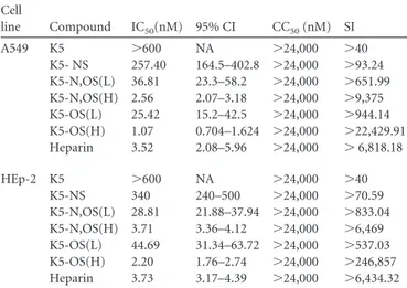
HSPG and prevents RSV adsorption (19), we exploited the viral plaque assay to screen a panel of E. coli K5 polysaccharide deriva-tives, which have structures similar to those of heparin and HSPG (26). As reported inTable 2, all GAGs, except unmodified K5, showed a half-maximal inhibitory concentration (IC50) in the
nanomolar range. To exclude the possibility that the antiviral ac-tivity of K5 derivatives might be due to cytotoxicity, the GAGs were evaluated by MTT assays with uninfected HEp-2 and A549 cells. As reported inTable 2, none of the GAGs tested exhibited toxic effects in the range of the examined concentrations, hence
the nondeterminable 50% cytotoxic concentrations (CC50) and
very favorable selectivity indexes (SI) for each active compound. K5-N,OS(H) and K5-OS(H) were endowed with the highest anti-viral activities and were therefore selected for further investiga-tion. Thus, the effect of K5-N,OS(H) and K5-OS(H) on cell via-bility was investigated with HEp-2 and A549 cells at higher doses than those reported inTable 2(i.e., up to 200M) in order to determine the CC50values. As shown in Fig. S1 in the
supplemen-tal material, K5-N,OS(H) and K5-OS(H) exerted a moderate dose-dependent reduction in cell viability only in HEp-2 cells at 50 M, 100 M, and 200 M, which did not allow the calculation of CC50values
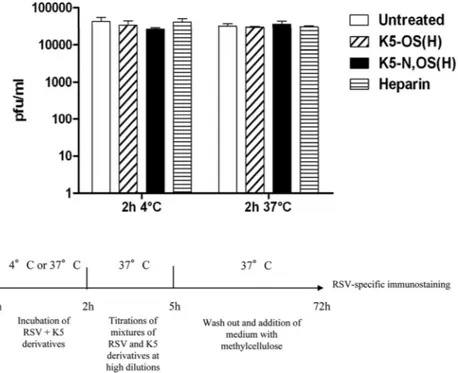
K5 derivatives do not inactivate RSV particles. Since certain
sulfated polysaccharides have been shown to exhibit direct viru-cidal activity (51), the K5 derivatives used in the present study were first subjected to a virus inactivation assay in our pursuit to understand their mechanism(s) of antiviral action. As shown in
Fig. 1, the virus titers of samples treated with N,OS(H), K5-OS(H), or heparin did not significantly differ from those deter-mined in untreated samples (P⬎ 0.05), indicating that the two K5 derivatives do not exert their antiviral activity via the direct inac-tivation of RSV particles.
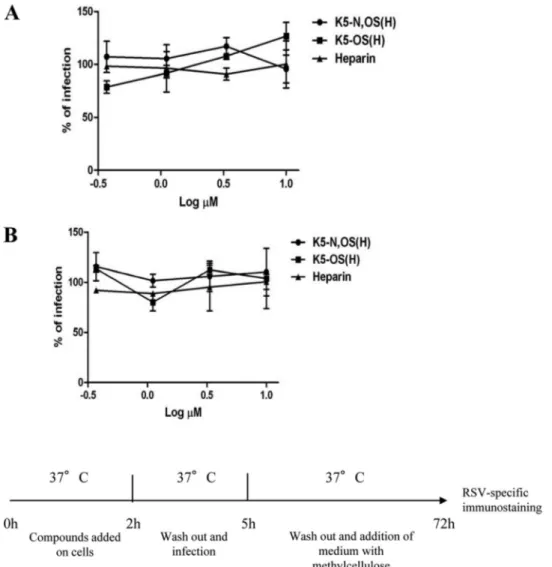
K5 derivatives do not affect cell susceptibility to RSV infec-tion. Some antiviral compounds are known to lower cell
suscep-tibility to viral infection by downregulating or directly masking virus receptors. In particular, we recently demonstrated that the compound SB-105A10 exerts its anti-RSV activity by masking HSPGs on the cell surface (49). To investigate whether the K5 derivatives affect cell susceptibility to RSV infection, preattach-ment assays were performed as described above. To this end, HEp-2 and A549 cells were preincubated for 2 h with different concentrations of K5-N,OS(H) or K5-OS(H) or with heparin as a control. After incubation, cells were washed to remove unbound GAGs from the medium and infected with RSV. As shown inFig. 2, under these experimental conditions, K5 derivatives and hepa-rin do not exert any antiviral activity. This indicates that K5 de-rivatives do not affect cell susceptibility to RSV infection.
K5 derivatives block RSV binding to host cells. The antiviral
activities of many sulfated polysaccharides correspond to their capacity to bind to and sequester the virus in the extracellular environment, thus hampering its attachment to the target cell (51). This possible mechanism was therefore investigated in rela-tion to the K5 derivatives and RSV using the attachment assay described above, the conditions of which allow for the attachment of the virus to the cell surface but prevent its entry. As shown in
Fig. 3, under these experimental conditions, K5 derivatives and heparin strongly inhibited RSV, with IC50s that are comparable to
those measured in the classical viral plaque assay (seeTable 2), suggesting that the antiviral activities of these GAGs depend on their capacity to inhibit the attachment of the virus to the cell surface.
To substantiate this interpretation, binding assays in which we directly evaluated the amounts of virus particles bound to the cells in the presence or absence of the active GAGs were performed. Consistent with previous results, N,OS(H), K5-OS(H), and heparin significantly reduced (P ⬍ 0.05) the amount of bound virus on HEp-2 and A549 cells (Fig. 3Cand
D, respectively), while unsulfated K5, which does not exhibit any antiviral activity, did not.
Taken together, these results indicate that the main
mecha-TABLE 2 Screening of K5 derivatives on A549 and HEp-2 cellsa
Cell line Compound IC50(nM) 95% CI CC50(nM) SI A549 K5 ⬎600 NA ⬎24,000 ⬎40 K5- NS 257.40 164.5–402.8 ⬎24,000 ⬎93.24 K5-N,OS(L) 36.81 23.3–58.2 ⬎24,000 ⬎651.99 K5-N,OS(H) 2.56 2.07–3.18 ⬎24,000 ⬎9,375 K5-OS(L) 25.42 15.2–42.5 ⬎24,000 ⬎944.14 K5-OS(H) 1.07 0.704–1.624 ⬎24,000 ⬎22,429.91 Heparin 3.52 2.08–5.96 ⬎24,000 ⬎ 6,818.18 HEp-2 K5 ⬎600 NA ⬎24,000 ⬎40 K5-NS 340 240–500 ⬎24,000 ⬎70.59 K5-N,OS(L) 28.81 21.88–37.94 ⬎24,000 ⬎833.04 K5-N,OS(H) 3.71 3.36–4.12 ⬎24,000 ⬎6,469 K5-OS(L) 44.69 31.34–63.72 ⬎24,000 ⬎537.03 K5-OS(H) 2.20 1.76–2.74 ⬎24,000 ⬎246,857 Heparin 3.73 3.17–4.39 ⬎24,000 ⬎6,434.32 a
IC50, 50% inhibitory concentration; 95% CI, 95% confidence interval; CC50, 50%
cytotoxic concentration; NA, not assessable. Values are means and CIs from three separate determinations.
on July 16, 2014 by guest
http://aac.asm.org/
nism of action of the K5 derivatives consists in their capacity to hamper the virus’s interaction with an entry receptor(s) expressed on the surface of target cells.
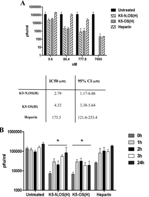
K5 derivatives reduce viral yield for 24 h postinfection. To
evaluate whether the reduction of RSV attachment and infection exerted by K5-N,OS(H) and K5-OS(H) is maintained in the long term, thus leading in a decrease in viral progeny production, post-attachment assays using virus yield were performed as described above; this stringent test allows multiple cycles of viral replication to occur before measuring the production of infectious viruses. In the first set of experiments, increasing concentrations of K5-N,OS(H), K5-OS(H), and heparin were added immediately after the removal of the viral inoculum in order to generate dose-re-sponse curves and to determine the IC50s (Fig. 4A). Under these
experimental conditions, the two K5 derivatives strongly reduced the RSV yield, with efficiencies that are similar to those measured in the classic viral plaque assay and in the attachment assay for K5-N,OS(H) and K5-OS(H), respectively. Interestingly, heparin exerted only modest inhibitory activity. Of note, K5-N,OS(H), K5-OS(H), and heparin were not cytotoxic even at the highest concentration tested (7M), as shown in Fig. S1 in the supple-mental material.
In the second set of experiments, a single concentration of K5-N,OS(H), K5-OS(H), or heparin was added 1 h, 2 h, 3 h, and 24 h after the removal of the virus inoculum. The results shown in
Fig. 4Bdemonstrate that reduction in viral yield is effective when the compounds are added up to 24 h postinfection. Once again, the inhibition profiles of K5-N,OS(H) and K5-OS(H) were better than that of heparin.
Taken together, these data indicate that the K5 derivatives but not heparin retain an antiviral activity for at least 24 h and are able to exert their inhibitory action over virions produced directly by
the cell, thereby preventing further cell infection and viral yield production.
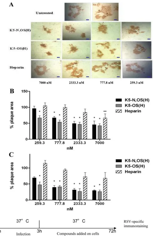
K5 derivatives inhibit cell-to-cell spread of RSV and syncy-tium formation. Massive viral production by infected cells
trig-gers cell-to-cell spread of RSV that in turn trigtrig-gers the formation of syncytia, the characteristic cytopathic effect of RSV; the forma-tion of these large, multinucleated epithelial cells helps the infect-ing virus avoid antibodies present in nasal secretions (52,53). We thus decided to investigate whether K5-N,OS(H), K5-OS(H), and heparin were able to block the cell-to-cell transmission of RSV. To this end, HEp-2 and A549 cells were infected with RSV in the absence of any GAG and then treated with different concentra-tions of K5-N,OS(H), K5-OS(H), or heparin after the removal of the virus inoculum. Three days postinfection, the cell-to-cell spread of RSV was evaluated by analyzing the size of the infec-tion foci. As shown inFig. 5A, all the compounds were able to reduce the transmission of RSV in a dose-dependent manner. A statistically significant reduction in syncytium dimension was observed in both A549 and HEp-2 cells treated with doses of K5-N,OS(H) ranging between 7M and 777.8 nM, and with doses of K5-OS(H) ranging between 7M and 259.3 nM. In contrast, a significant reduction in plaque size following treat-ment with heparin was observed in HEp-2 cells only at a dose of 7M (P ⬍ 0.01).
K5 derivatives do not exhibit antiviral activity against rota-virus. To date, a number of K5 derivatives that exhibit antiviral
activity against a panel of HSPGs-dependent viruses, including HSV, HIV, and HPV (see the introduction), have been identified. Moreover, work from our own group has revealed the HSPG-binding dendrimer SB105A10 to be active against RSV infection (49), and the present study identifies additional K5 derivatives with capacities to block RSV infection. To provide further
evi-FIG 1 K5 derivatives are not active in a virus inactivation assay. RSV was incubated with 3.6g/ml of K5-N,OS(H) (240 nM), K5-OS(H) (191 nM), or heparin (263 nM) for 2 h at 4°C or 37°C. The mixtures were then titrated on A549 cells at high dilutions at which the concentrations of compounds were not active. The titers, expressed as PFU/ml, are means and SEM for triplicates.
on July 16, 2014 by guest
http://aac.asm.org/
dence corroborating the hypothesis that the anti-RSV abilities of these K5 derivatives depend specifically on their capacity to pre-vent RSV from interacting with HSPGs on the target cell, the com-pounds were tested against human rotavirus, whose attachment and entry depend on interaction with integrins and heat shock proteins but not HSPG (54). Neither the K5 derivatives nor hep-arin could prevent rotavirus infection in MA104 cells when tested at doses up to 7M (Fig. 6), strongly indicating that these com-pounds are not active against viruses that do not bind to cell sur-face HSPGs.
Antiviral activities of K5 derivatives in EpiAirway tissue. The
EpiAirway system consists of human derived tracheal/bron-chial epithelial cells grown on a collagen-coated membrane to form a highly differentiated organotypic model with many of the features of respiratory mucosa, thus providing a useful in
vitro means of assessing respiratory virus infections. We
as-sessed the effect of K5-N,OS(H) and K5-OS(H) on RSV infec-tion by measuring the titer of virus emerging from the apical surface of tissues infected with mixtures containing 50,000 PFU of RSV in the presence or absence of 10M K5-N,OS(H)
or K5-OS(H) preincubated for 1 h at 4°C prior to virus appli-cation. At 72 h postinfection, the titer of virus in untreated control tissues was 1.45⫻ 103PFU/ml. In tissues treated with
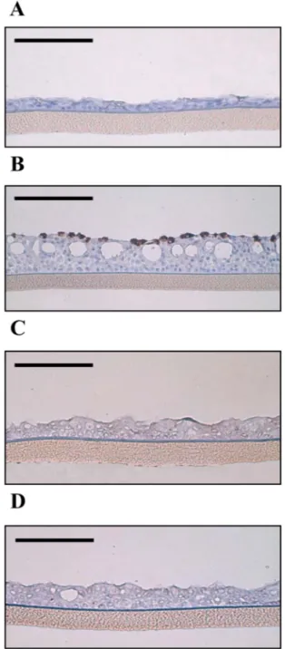
K5-N,OS(H), the detected titer was 40 PFU/ml, whereas in the samples treated with K5-OS(H), the virus titer was undetect-able (Fig. 7). Thus, the compounds inhibited the viral titer by 97.3% and 100%, respectively. The same tissues were fixed im-mediately after the virus harvest at 72 h postinfection and sub-jected to immunohistochemistry using an RSV-specific mono-clonal antibody. All the sections derived from the infected tissue consistently showed the presence of cells expressing the RSV antigen in the upper cellular layer (Fig. 8B). No RSV-positive cells could be observed in sections from uninfected tissue (Fig. 8A), demonstrating the specificity of the signal. Furthermore, no RSV-positive cells could be identified in tis-sues treated with K5-N,OS(H) (Fig. 8C) or K5-OS(H) (Fig. 8D), corroborating the virus titer results. To verify that the antiviral action was not due to a cytotoxic effect, an MTT assay was performed with tissues treated with 10M (each) K5 de-rivative for 1, 4, or 18 h at 37°C. The results shown inTable 3
FIG 2 Preattachment assay with HEp-2 and A549 cells. HEp-2 (A) or A549 (B) cells were pretreated with increasing concentrations of K5 derivatives or heparin
for 2 h at 37°C, washed, and infected. Three days postinfection, the cells were fixed and subjected to RSV-specific immunostaining, the plaques were counted, and the percent infection was calculated by comparing treated and untreated wells. The results are means and SEM for triplicates.
on July 16, 2014 by guest
http://aac.asm.org/
demonstrate that these K5 derivatives are not cytotoxic, and the time required to reduce tissue viability to 50% (ET50) was
greater than 18 h.
DISCUSSION
To infect target cells successfully, RSV needs to bind to HSPGs located on the cell membrane, and this interaction provides a tar-get for the development of new anti-RSV compounds. Inhibition of the RSV/HSPG interaction can be achieved by two distinct ap-proaches: the first involves receptor masking, usually achieved by means of polycationic compounds able to bind to the negatively charged sulfate groups present on the GAG side chains of HSPGs, and the second involves the use of polyanionic compounds that bind to and antagonize the virus. We recently confirmed the fea-sibility of the first approach by demonstrating that a highly posi-tively charged dendrimer effecposi-tively binds to HSPGs, inhibiting RSV infection (49). Accordingly, positively charged peptides de-rived from the HBD of the RSV G protein also block virus infec-tion (18). The feasibility of the second approach, on the other hand, has been supported by the demonstration that heparin (19,
55) and other negatively charged polysaccharides, such as chon-droitin sulfate (56) and dextran sulfate (57,58), are able to bind RSV, preventing its cell attachment and infection.
Due to their structural heterogeneity, heparin, heparan sulfate, and other GAGs are able to bind to a wide range of molecules and exert a number of biological activities that can interfere with one
another, leading to the risk of toxicity and undesired side effects. The solution therefore lies in the production of tailor-made hep-arin-like compounds endowed with specific antiviral activities; however, this requires detailed knowledge of the molecular basis of the heparin/HSPG interaction with viral envelope proteins. In the case of RSV, we know that the glycoproteins G and F are responsible for the heparin/HSPG interaction, and specific basic amino acid sequences acting as HBDs have even been identified in the each of these proteins (18,19,20). Nevertheless, little has been done to date to characterize the structural features of heparin/ HSPGs that mediate their binding to RSV protein, although it is very likely that the negatively charged sulfated groups of the GAG chains are those involved in the interaction, as demonstrated for almost all the other viral heparin-binding proteins (59).
The capsular K5 polysaccharide from Escherichia coli can be chemically sulfated in selected positions, resulting in the synthesis of completely N-sulfated compounds with different amounts of O-sulfation in different positions or completely N-acetylated molecules differing only in the position and degree of O-sulfation (30). Due to these features, sulfated K5 derivatives have been use-ful in the study of the structure-activity relationship of the inter-actions of several viral proteins with heparin and used in the de-sign of specific antiviral polysaccharides.
Here, we found that selected K5 sulfated derivatives exert a strong anti-RSV effect. Experiments aimed at elucidating their mechanism(s) of anti-RSV action indicate that the inhibitory
ef-FIG 3 Investigation of inhibitory mechanisms of the hit compounds. In the attachment assay, RSV and compounds were added to HEp-2 (A) or A549 (B) cells
for 2 h at 4°C. Cells were shifted to 37°C, and at 72 h postinfection, they were subjected to RSV-specific immunostaining, the plaques were counted, and the percentage of infection was calculated by comparing treated to untreated wells. In the binding assay, the virus bound to HEp-2 (C) or A549 (D) was detected by ELISA immediately after the removal of the virus inoculum. Each absorbance was mock subtracted, and the percentage of infection was calculated by comparing treated to untreated wells. The results are means and SEM from triplicates.
on July 16, 2014 by guest
http://aac.asm.org/
fect is due mainly to the capacity of K5-N,OS(H) and K5-OS(H) to interact with the virus particles, rather than with cell compo-nents, and thereby prevent virus attachment to the cell surface. Several lines of evidence support this. First, cells pretreated with K5 derivatives remained susceptible to RSV infection, thus ex-cluding the possibility that these compounds form stable interac-tions with one or more cellular components, preventing their in-teraction with viral glycoproteins. Second, the results of the binding and attachment assays demonstrate that K5-N,OS(H) and K5-OS(H) block the adsorption of RSV virions to the cell surface with a potency similar to that of heparin, which has been shown to prevent RSV infection by competing with cellular HSPGs for binding to virion components (60,61) Third, preincu-bation of RSV virions with the active sulfated K5 derivatives did not result in a loss of infectivity, suggesting that the antiviral
ac-tivity of the compounds does not rely on inactivation of a virion component(s). A similar mechanism of action was previously ob-served for heparin when it was tested against HSV-1 and RSV (49,
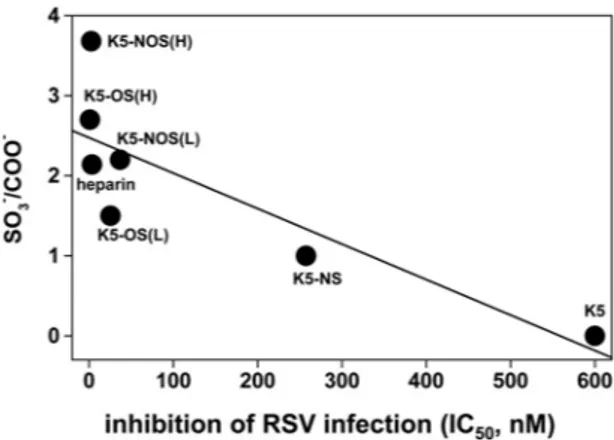
50) and when K5 derivatives were tested against HCMV (40). Unsulfated K5 polysaccharide, unlike N,OS(H) and K5-OS(H), did not show any significant RSV antagonist activity, in-dicating that the sulfate groups, rather than the backbone struc-ture, mediate the interaction with RSV. Moreover, a good correlation exists between the degree of sulfation of the GAGs tested and their capacities to inhibit RSV infection (Fig. 9). How-ever, this correlation is lost in the highly sulfated GAGs (see the left part ofFig. 9); thus, in addition to the degree of sulfation, the position of the sulfate groups along the polysaccharide chain is also important. Furthermore, complete sulfation of the N posi-tions confers very limited RSV antagonist activity to K5-NS, while
FIG 4 Reduction of viral yield. (A) HEp-2 cells were infected and subsequently treated with different concentrations of compounds. When the cytopathic effect
involved the whole monolayer of untreated wells, supernatant were harvested and titrated. The results are means and SEM from triplicates. The table in panel A shows the IC50and 95% CI values for each compound tested. (B) The same procedure was followed, with a fixed dose of 10g/ml added to infected cells at different times postinfection, ranging from 0 h to 24 h. The results are means and SEM for triplicates.ⴱ, P ⬍ 0.05 in a 2-way ANOVA.
on July 16, 2014 by guest
http://aac.asm.org/
sulfation of the O position confers an inhibitory capacity to K5-OS(L) that is almost 10 times higher than that of K5-NS (Table 2) despite a similar SO3⫺/COOH⫺ratio (1 and 1.5, respectively)
(Table 1). Similarly, K5-OS(H) is endowed with an inhibitory capacity that is 30 times higher than that of K5-N,OS(L) (Table 2) despite their similar SO3⫺/COOH⫺ratios (2.7 and 2.2,
respec-tively) (Table 1). Finally, the greater SO3⫺/COOH⫺ratio of
K5-OS(H) than that of K5-NK5-OS(H) (from 2.7 to 3.68) (Table 1) does not confer any additional anti-RSV potency.
Taken together, these data suggest that O- rather than N-sul-fated groups mediate the binding of RSV to K5 polysaccharides. In apparent contrast with our findings, Hallak and coworkers dem-onstrated that N-sulfation of heparin is necessary for RSV infec-tion (55). In this regard, it must be pointed out that heparin (but
FIG 5 Inhibition of RSV-induced syncytium formation by K5 derivatives and heparin. The images in panel A show representative syncytia in HEp-2 cells, with
horizontal bars corresponding to 20m. HEp-2 cells (B) or A549 cells (C) were infected with RSV in the absence of compounds. The inoculum was removed at 3 h postinfection, and cells were left untreated or incubated in the presence of the following concentrations of substances in 1.2% methylcellulose medium: 7,000 nM, 2,333.3 nM, 777.8 nM, and 259.3 nM. Formation of syncytia was assessed 72 h after infection, by immunostaining. The histograms show the percentage of plaque area of treated wells compared to that of untreated wells as a function of compound concentration. The pictures and histograms shown are representative of many analyzed plaques, ranging from 15 to 25 per condition.ⴱ, P ⬍ 0.001; ⴱⴱ, P ⬍ 0.01.
on July 16, 2014 by guest
http://aac.asm.org/
not K5 derivatives) is epimerized and that the presence of iduronic acid instead of glucuronic acid residues confers on heparin greater flexibility (62) that in turn may allow a better presentation of the N-sulfated groups to the RSV envelope proteins G and F. In ac-cordance with this hypothesis, when the RSV antagonist capacities of the K5 derivatives are compared to that of heparin, it is evident that their activities are enhanced by the presence of IdoA, since K5 N,OS(L) is much less active despite a similar sulfate-to-carboxyl ratio (2.2 and 2.4). Thus, the epimerization of K5 derivatives rep-resents a promising approach for the design of even more active and specific anti-RSV compounds.
K5-OS(H) and K5-N,OS(H) are also revealed as exhibiting more potent anti-RSV activity than heparin in viral yield reduc-tion assays (Fig. 4) and in limiting RSV cell-to-cell spread (Fig. 5). However, in the classic viral plaque assay and in the attachment assays, K5-OS(H) and K5-N,OS(H) show IC50s that are
compara-ble to or only 2 to 5 times higher than that of heparin. It should be mentioned, however, that these two assays somehow “favor” the HSPG antagonist action of GAGs that are allowed to bind to the
virus before its administration to cells. Indeed, although these assays are useful in their own right and are widely used for screen-ing purposes, they do not resemble the in vivo situation, which is characterized by the continuous release by infected cells of virions that promptly interact with neighboring cells, often resulting in direct cell-to-cell spread and syncytium formation.
Interestingly, we found that when K5-OS(H) and K5-N,OS(H)
FIG 6 Antiviral assay with MA104 cells infected with human rotavirus.
MA104 cells were infected in the presence of K5 derivatives or heparin. Sixteen hours postinfection, the cells were fixed and subjected to rotavirus-specific immunostaining. The infected cells were counted, and the percent infection was calculated by comparing treated and untreated wells. The results are means and SEM from triplicates.
FIG 7 Reduction of viral yield on EpiAirway tissue. Fifty thousand PFU and 10
M K5-N,OS(H) or K5-OS(H) were preincubated for 1 h at 4°C and subse-quently added to the apical surface of the EpiAirway tissues. After 3 h of incu-bation at 37°C, the medium was removed and the cultures were washed api-cally with 100l of medium. At 72 h postinfection, 100 l of medium was added to the apical surface, and the tissues were allowed to equilibrate for 30 min at 37°C. The suspension was then collected and titrated on A549 cells. The results are means and SEM from triplicates.
FIG 8 Reduction of RSV-infected cells in EpiAirway tissue by K5-N,OS(H)
and K5-OS(H). (A) Immunohistochemistry of control tissue; (B)RSV-in-fected tissue (50,000 PFU); (C) RSV-in(B)RSV-in-fected tissue treated with 10M K5-N,OS(H); (D) RSV-infected tissue treated with 10M K5-OS(H). Three days postinfection, RSV-infected cells were identified using a RSV-specific mono-clonal antibody (brown signal). The pictures shown are representative of many analyzed sections, ranging from 5 to 12 per condition. Horizontal bars corre-spond to 100m.
on July 16, 2014 by guest
http://aac.asm.org/
were assayed in the more stringent postinfection assay using HEp-2 cells, they retained a long-lasting RSV-inhibitory capacity comparable to that measured in the viral plaque assay, while hep-arin was indicated to be less effective, remaining active for only for short periods of time at concentrations that are 40 to 60 times lower than those of the two K5 derivatives (Fig. 4). Accordingly, K5-OS(H) and K5-N,OS(H) also presented significantly better inhibitory profiles than heparin when assayed for their capacity to inhibit RSV-induced syncytium formation (Fig. 5). K5-OS(H) and K5-N,OS(H) have a backbone structure more similar to that of HS than heparin, since they contain only GlcA, the presence of which, along with their high sulfate contents, might make these molecules more efficient than heparin in preventing electrostatic interactions between the RSV glycoproteins G and F and HSPGs at the cell surface. Alternatively, the peculiar structure of K5 deriva-tives may render these molecules more stable than heparin, thus contributing to their persistent RSV-inhibitory activity.
In conclusion, not only are the active K5 derivatives able to interfere with the virus adsorption process, but they also limit the cell-to-cell spread of RSV in a dose-dependent manner at non-toxic concentrations. These antiviral properties may be useful in the clinical setting, where K5-OS(H) and K5-N,OS(H) might be able to block both cell-to-cell spread and cell-free virus within the extracellular space—the two predominant routes of dissemina-tion for RSV in vivo (63,64,65,66).
As mentioned above, heparin and heparan sulfates cannot be used as anti-RSV drugs due to their anticoagulant activity and/or aspecific activities. K5-OS(H) and K5-N,OS(H), on the other
hand, are endowed with a significantly lower anticoagulant activ-ity (67). Moreover, since their structure is very similar to those of natural heparan sulfates, they can be metabolically recognized and easily catabolized without inducing toxicity, and they are expected to be tolerated by the immune system. Accordingly, recent results have shown that proinflammatory cytokines are not mobilized in the presence of K5 derivatives (67) but rather can even exert an anti-inflammatory effect (68).
Besides viral proteins (44), K5 derivatives are known to bind a wide array of eukaryotic proteins (26), implying possible adverse effects associated with their therapeutic administration. Relevant to this point, this class of molecules can be suitably tailored to produce countless compounds endowed with peculiar structural features (degree of sulfation, disposition of sulfated groups, length of GAG chain, and epimerization) (26) whose modulation im-pacts their binding capacity (see the discussion above), thus sug-gesting the possibility of producing selected K5 sulfated deriva-tives with specific binding capacities and biological effects.
With regard to a potential administration of K5 derivatives for the prevention or treatment of RSV infections, we assessed their antiviral activities in human tracheal/bronchial histocultures (EpiAirway). This model system avoids species extrapolation and the use of animal models at the early preclinical phase of drug development and provides a better simulation of the human re-spiratory tract than the cell monolayers used in standard antiviral assays. It carries the same cell type composition and polarity, mu-cus-secreting function, and mucociliary movements as the airway epithelium in vivo. Moreover, the HSPG composition and expres-sion level in vivo are expected to be well duplicated in the EpiAir-way tissue. In agreement with previous literature, we observed that RSV infects the lumenal ciliated columnar airway epithelial cells via the apical surfaces of the cultures, as shown inFig. 8B(69). Both the virus yield assays and the immunohistochemical analysis of histological cross sections showed that OS(H) and K5-N,OS(H) exhibit clear antiviral activity in the EpiAirway tissue at a dose of 10M with no signs of cytotoxic effect, indicating that this inhibitory strategy may well be effective in vivo. Studies to assess the clinical potential of these inhibitors against RSV infec-tions are ongoing in animal models.
ACKNOWLEDGMENT
This work was supported by a grant from Ricerca Finanziata dall’Università degli Studi di Torino (ex 60%) 2012 to D.L.
REFERENCES
1. Collins PL, Crowe JE, Jr. 2007. Respiratory syncytial virus and metap-neumovirus, p 1601–1646. In Knipe DM, Howley PM (ed), Fields virol-ogy, 5th ed. Lippincott Williams and Wilkins, Philadelphia, PA. 2. Shay DK, Holman RC, Roosevelt GE, Clarke MJ, Anderson LJ. 2001.
Bronchiolitis-associated mortality and estimates of respiratory syncytial virus-associated deaths among US children, 1979 –1997. J. Infect. Dis.
183:16 –22.http://dx.doi.org/10.1086/317655.
3. Falsey AR, Formica MA, Hennessey PA, Criddle MM, Sullender WM,
Walsh EE. 2006. Detection of respiratory syncytial virus in adults with
chronic obstructive pulmonary disease. Am. J. Respir. Crit. Care Med.
173:639 – 643.http://dx.doi.org/10.1164/rccm.200510-1681OC. 4. Leader S, Kohlhase K. 2002. Respiratory syncytial virus-coded pediatric
hospitalizations, 1997 to 1999. Pediatr. Infect. Dis. J. 21:629 – 632.http: //dx.doi.org/10.1097/00006454-200207000-00005.
5. World Health Organization. 2009. Initiative for vaccine research (IVR). Acute respiratory infections. World Health Organization, Geneva, Swit-zerland.http://www.who.int/vaccine_research/diseases/ari/en/. 6. Hall CB, Weinberg GA, Iwane MK, Blumkin AK, Edwards KM, Staat
TABLE 3 Viability on EpiAirway tissue
Conditions % of viabilitya Untreated [1 h] 100 K5-N,OS(H) [1 h] 129⫾ 9.8 K5-OS(H) [1 h] 127.2⫾ 11.2 Untreated [4 h] 100 K5-N,OS(H) [4 h] 89.2⫾ 7.9 K5-OS(H) [4 h] 90.8⫾ 8.5 Untreated [18 h] 100 K5-N,OS(H) [18 h] 80.8⫾ 10.2 K5-OS(H) [18 h] 81.3⫾ 6.8
aThe results presented are means and SD from triplicate tissues.
FIG 9 Correlation between the IC50s of K5 derivatives and heparin with their degree of sulfation (SO3⫺/COO⫺). Correlation coefficient,⫺0.83829; P ⬍ 0.01848.
on July 16, 2014 by guest
http://aac.asm.org/
MA, Auinger P, Griffin MR, Poehling KA, Erdman D, Grijalva CG, Zhu Y, Szilagyi P. 2009. The burden of respiratory syncytial virus infection in
young children. N. Engl. J. Med. 360:588 –598.http://dx.doi.org/10.1056 /NEJMoa0804877.
7. Pelletier AJ, Mansbach JM, Camargo CA, Jr. 2006. Direct medical costs of bronchiolitis hospitalizations in the United States. Pediatrics 118:2418 – 2423.http://dx.doi.org/10.1542/peds.2006-1193.
8. Corsello G. 2007. Bronchiolitis: the new American Academy of Pediatrics guidelines. J. Chemother. 19(Suppl 2):12–14.
9. Castilow EM, Varga SM. 2008. Overcoming T cell-mediated immuno-pathology to achieve safe RSV vaccination. Future Virol. 3:445– 454.http: //dx.doi.org/10.2217/17460794.3.5.445.
10. American Academy of Pediatrics Subcommittee on Diagnosis and
Management of Bronchiolitis. 2006. Diagnosis and management of
bronchiolitis. Pediatrics 118:1774 –1793.http://dx.doi.org/10.1542/peds .2006-2223.
11. Leyssen P, De Clercq E, Neyts J. 2008. Molecular strategies to inhibit the replication of RNA viruses. Antiviral Res. 78:9 –25.http://dx.doi.org/10 .1016/j.antiviral.2008.01.004.
12. Sidwell RW, Barnard DL. 2006. Respiratory syncytial virus infections: recent prospects for control. Antiviral Res. 71(2–3):379 –390.http://dx .doi.org/10.1016/j.antiviral.2006.05.014.
13. Johnson S, Oliver C, Prince GA, Hemming VG, Pfarr DS, Wang SC,
Dormitzer M, O’Grady J, Koenig S, Tamura JK, Woods R, Bansal G, Couchenour D, Tsao E, Hall WC, Young JF. 1997. Development of a
humanized monoclonal antibody (MEDI-493) with potent in vitro and in vivo activity against respiratory syncytial virus. J. Infect. Dis. 176:1215– 1224.http://dx.doi.org/10.1086/514115.
14. Wu H, Pfarr DS, Losonsky GA, Kiener PA. 2008. Immunoprophylaxis of RSV infection: advancing from RSV-IGIV to palivizumab and motavi-zumab. Curr. Top. Microbiol. Immunol. 317:103–123.
15. Welliver RC. 2010. Pharmacotherapy of respiratory syncytial virus infec-tion. Curr. Opin. Pharmacol. 10:289 –293. http://dx.doi.org/10.1016/j .coph.2010.04.013.
16. van Drunen Littel-van den Hurk S, Watkiss ER. 2012. Pathogenesis of respiratory syncytial virus. Curr. Opin. Virol. 2:300 –305.http://dx.doi .org/10.1016/j.coviro.2012.01.008.
17. King, JC, Jr. 1997. Community respiratory viruses in individuals with human immunodeficiency virus infection. Am. J. Med. 102:19 –24.http: //dx.doi.org/10.1016/S0002-9343(97)80005-8.
18. Crim RL, Audet SA, Feldman SA, Mostowski HS, Beeler JA. 2007. Identification of linear heparin-binding peptides derived from human respiratory syncytial virus fusion glycoprotein that inhibit infectivity. J. Virol. 81:261–271.http://dx.doi.org/10.1128/JVI.01226-06.
19. Feldman SA, Audet S, Beeler JA. 2000. The fusion glycoprotein of human respiratory syncytial virus facilitates virus attachment and infectivity via an interaction with cellular heparan sulfate. J. Virol. 74:6442– 6457.http: //dx.doi.org/10.1128/JVI.74.14.6442-6447.2000.
20. Feldman SA, Hendry RM, Beeler JA. 1999. Identification of a linear heparin binding domain for human respiratory syncytial virus attachment glycoprotein G. J. Virol. 73:6610 – 6617.
21. Tayyari F, Marchant D, Moraes TJ, Duan W, Mastrangelo P, Hegele
RG. 2011. Identification of nucleolin as a cellular receptor for human
respiratory syncytial virus. Nat. Med. 17:1132–1135.http://dx.doi.org/10 .1038/nm.2444.
22. Bose S, Basu M, Banerjee AK. 2004. Role of nucleolin in human parain-fluenza virus type 3 infection of human lung epithelial cells. J. Virol. 78: 8146 – 8158.http://dx.doi.org/10.1128/JVI.78.15.8146-8158.2004. 23. Xiao X, Feng Y, Zhu Z, Dimitrov DS. 2011. Identification of a putative
Crimean-Congo hemorrhagic fever virus entry factor. Biochem. Biophys. Res. Commun. 411:253–258. http://dx.doi.org/10.1016/j.bbrc.2011.06 .109.
24. Thongtan T, Wikan N, Wintachai P, Rattanarungsan C, Srisomsap C,
Cheepsunthorn P, Smith DR. 2012. Characterization of putative
Japa-nese encephalitis virus receptor molecules on microglial cells. J. Med. Vi-rol. 84:615– 623.http://dx.doi.org/10.1002/jmv.23248.
25. Nisole S, Krust B, Callebaut C, Guichard G, Muller S, Briand JP,
Hovanessian AG. 1999. The anti-HIV pseudopeptide HB-19 forms a
complex with the cell-surface-expressed nucleolin independent of heparin sulfate proteoglycans. J. Biol. Chem. 274:27875–27884.http://dx.doi.org /10.1074/jbc.274.39.27875.
26. Rusnati M, Oreste P, Zoppetti G, Presta M. 2005. Biotechnological engineering of heparin/heparan sulphate: a novel area of multi-target drug
discovery. Curr. Pharm. Des. 11:2489 –2509.http://dx.doi.org/10.2174 /1381612054367553.
27. Matos PM, Andreu D, Santos NC, Gutiérrez-Gallego R. 2014. Structural requirements of glycosaminoglycans for their interaction with HIV-1 en-velope glycoprotein gp120. Arch. Virol. 159:555–560.http://dx.doi.org/10 .1007/s00705-013-1831-3.
28. Rusnati M, Coltrini D, Oreste P, Zoppetti G, Albini A, Noonan D,
D’Adda di Fagagna F, Giacca M, Presta M. 1997. Interaction of HIV-1
Tat protein with heparin. Role of the backbone structure, sulfation, and size. J. Biol. Chem. 272:11313–11320.
29. Vervaeke P, Alen M, Noppen S, Schols D, Oreste P, Liekens S. 2013. Sulfated Escherichia coli K5 polysaccharide derivatives inhibit dengue vi-rus infection of human microvascular endothelial cells by interacting with the viral envelope protein E domain III. PLoS One 8:e74035.http://dx.doi .org/10.1371/journal.pone.0074035.
30. Bugatti A, Giagulli C, Urbinati C, Caccuri F, Chiodelli P, Oreste P,
Fiorentini S, Orro A, Milanesi L, D’Ursi P, Caruso A, Rusnati M. 2013.
Biochemical characterization of HIV-1 matrix protein p17 interaction with heparin. J. Biol. Chem. 288:1150 –1161.http://dx.doi.org/10.1074/jbc.M112 .400077.
31. Patel M, Yanagishita M, Roderiquez G, Bou-Habib DC, Oravecz T,
Hascall VC, Norcross MA. 1993. Cell-surface heparan sulfate
proteogly-can mediates HIV-1 infection of T-cell lines. AIDS Res. Hum. Retrovi-ruses 9:167–174.http://dx.doi.org/10.1089/aid.1993.9.167.
32. Shieh MT, Dunn DW, Montgomery RI, Esko JD, Spear PG. 1992. Cell surface receptors for herpes simplex virus are heparan sulfate proteoglycans. J. Cell Biol. 116:1273–1281.http://dx.doi.org/10.1083/jcb.116.5.1273. 33. Giroglou T, Florin L, Schafer F, Streeck RE, Sapp M. 2001. Human
papillomavirus infection requires cell surface heparan sulfate. J. Virol.
75:1565–1570.http://dx.doi.org/10.1128/JVI.75.3.1565-1570.2001. 34. Compton T. 2004. Receptors and immune sensors: the complex entry
path of human cytomegalovirus. Trends Cell Biol. 14:5– 8.http://dx.doi .org/10.1016/j.tcb.2003.10.009.
35. Chen Y, Maguire T, Hileman RE, Fromm JR, Esko J, Linhardt RJ,
Marks RM. 1997. Dengue virus infectivity depends on envelope protein
binding to target cell heparan sulfate. Nat. Med. 3:866 – 871.http://dx.doi .org/10.1038/nm0897-866.
36. Salvador B, Sexton NR, Carrion R, Jr, Nunneley J, Patterson JL, Steffen
I, Lu K, Muench MO, Lembo D, Simmons G. 2013. Filoviruses utilize
glycosaminoglycans for their attachment to target cells. J. Virol. 87:3295– 3304.http://dx.doi.org/10.1128/JVI.01621-12.
37. Vicenzi E, Gatti A, Ghezzi S, Oreste P, Zoppetti G, Poli G. 2003. Broad spectrum inhibition of HIV-1 infection by sulfated K5 Escherichia coli polysaccharide derivatives. AIDS 17:177–181.http://dx.doi.org/10.1097 /00002030-200301240-00006.
38. Pinna D, Oreste P, Coradin T, Kajaste-Rudnitski A, Ghezzi S, Zoppetti
G, Rotola A, Argnani R, Poli G, Manservigi R, Vicenzi E. 2008.
Inhi-bition of herpes simplex virus types 1 and 2 in vitro infection by sulfated derivatives of Escherichia coli K5 polysaccharide. Antimicrob. Agents Chemother. 52:3078 –3084.http://dx.doi.org/10.1128/AAC.00359-08. 39. Lembo D, Donalisio M, Rusnati M, Bugatti A, Cornaglia M, Cappello
P, Giovarelli M, Oreste P, Landolfo S. 2008. Sulfated K5 Escherichia coli
polysaccharide derivatives as wide-range inhibitors of genital types of hu-man papillomavirus. Antimicrob. Agents Chemother. 52:1374 –1381. http://dx.doi.org/10.1128/AAC.01467-07.
40. Mercorelli B, Oreste P, Sinigalia E, Muratore G, Lembo D, Palù G,
Loregian A. 2010. Sulfated derivatives of Escherichia coli K5 capsular
polysaccharide are potent inhibitors of human cytomegalovirus. Antimi-crob. Agents Chemother. 54:4561– 4567.http://dx.doi.org/10.1128/AAC .00721-10.
41. Ludwig M, Enzenhofer E, Schneider S, Rauch M, Bodenteich A,
Neu-mann K, Prieschl-Grassauer E, Grassauer A, Lion T, Mueller CA. 2013.
Efficacy of a carrageenan nasal spray in patients with common cold: a randomized controlled trial. Respir. Res. 14:124. http://dx.doi.org/10 .1186/1465-9921-14-124.
42. Marais D, Gawarecki D, Allan B, Ahmed K, Altini L, Cassim N,
Gopolang F, Hoffman M, Ramjee G, Williamson AL. 2011. The
effec-tiveness of Carraguard, a vaginal microbicide, in protecting women against high-risk human papillomavirus infection. Antivir. Ther. 16: 1219 –1226.http://dx.doi.org/10.3851/IMP1890.
43. Pirrone V, Wigdahl B, Krebs FC. 2011. The rise and fall of polyanionic inhibitors of the human immunodeficiency virus type 1. Antiviral Res.
90:168 –182.http://dx.doi.org/10.1016/j.antiviral.2011.03.176.
on July 16, 2014 by guest
http://aac.asm.org/
44. Rusnati M, Vicenzi E, Donalisio M, Oreste P, Landolfo S, Lembo D. 2009. Sulfated K5 Escherichia coli polysaccharide derivatives: a novel class of candidate antiviral microbicides. Pharmacol. Ther. 123:310 –322.http: //dx.doi.org/10.1016/j.pharmthera.2009.05.001.
45. Urbinati C, Bugatti A, Oreste P, Zoppetti G, Waltenberger J, Mitola
S, Ribatti D, Presta M, Rusnati M. 2004. Chemically sulfated
Esche-richia coli K5 polysaccharide derivatives as extracellular HIV-1 Tat protein antagonists. FEBS Lett. 568(1–3):171–177.http://dx.doi.org /10.1016/j.febslet.2004.05.033.
46. Moyes J, Cohen C, Pretorius M, Groome M, von Gottberg A, Wolter N,
Walaza S, Haffejee S, Chhagan M, Naby F, Cohen AL, Tempia S, Kahn K, Dawood H, Venter M, Madhi SA, South African Severe Acute Respiratory Illness Surveillance Group. 2013. Epidemiology of
respira-tory syncytial virus-associated acute lower respirarespira-tory tract infection hos-pitalizations among HIV-infected and HIV-uninfected South African children, 2010 –2011. J. Infect. Dis. 208:S217–S226.http://dx.doi.org/10 .1093/infdis/jit479.
47. Leali D, Belleri M, Urbinati C, Coltrini D, Oreste P, Zoppetti G, Ribatti
D, Rusnati M, Presta M. 2001. Fibroblast growth factor-2 antagonist
activity and angiostatic capacity of sulfated Escherichia coli K5 polysaccha-ride derivatives. J. Biol. Chem. 276:37900 –37908.http://dx.doi.org/10 .1074/jbc.M105163200.
48. Casu B, Gennaro U. 1975. A conductimetric method for the determina-tion of sulphate and carboxyl groups in heparin and other mucopolysac-charides. Carbohydr. Res. 39:168 –176.http://dx.doi.org/10.1016/S0008 -6215(00)82654-3.
49. Donalisio M, Rusnati M, Cagno V, Civra A, Bugatti A, Giuliani A, Pirri
G, Volante M, Papotti M, Landolfo S, Lembo D. 2012. Inhibition of
human respiratory syncytial virus infectivity by a dendrimeric heparan sulfate-binding peptide. Antimicrob. Agents Chemother. 56:5278 –5288. http://dx.doi.org/10.1128/AAC.00771-12.
50. Graham KL, Zeng W, Takada Y, Jackson DC, Coulson BS. 2004. Effects on rotavirus cell binding and infection of monomeric and polymeric peptides containing alpha2beta1 and alphaxbeta2 integrin ligand sequences. J. Virol.
78:11786 –11797.http://dx.doi.org/10.1128/JVI.78.21.11786-11797.2004. 51. Ghosh T, Chattopadhyay K, Marschall M, Karmakar P, Mandal P, Ray
B. 2009. Focus on antivirally active sulfated polysaccharides: from
struc-ture-activity analysis to clinical evaluation. Glycobiology 19:2–15.http: //dx.doi.org/10.1093/glycob/cwn092.
52. Morton CJ, Cameron R, Lawrence LJ, Lin B, Lowe M, Luttick A, Mason
A, McKimm-Breschkin J, Parker MW, Ryan J, Smout M, Sullivan J, Tucker SP, Young PR. 2003. Structural characterization of respiratory
syncytial virus fusion inhibitor escape mutants: homology model of the F protein and a syncytium formation assay. Virology 311:275–288.http://dx .doi.org/10.1016/S0042-6822(03)00115-6.
53. Black CP. 2003. Systematic review of the biology and medical manage-ment of respiratory syncytial virus infection. Respir. Care 48:209 –231. 54. Lopez S, Arias CF. 2004. Multistep entry of rotavirus into cells: a
Ver-saillesque dance. Trends Microbiol. 12:271–278. http://dx.doi.org/10 .1016/j.tim.2004.04.003.
55. Hallak LK, Spillmann D, Collins PL, Peeples ME. 2000. Glycosaminoglycan sulfation requirements for respiratory syncytial virus infection. J. Virol. 74: 10508 –10513.http://dx.doi.org/10.1128/JVI.74.22.10508-10513.2000.
56. Hallak LK, Collins PL, Knudson W, Peeples ME. 2000. Iduronic acid containing glycosaminoglycans on target cells are required for efficient respiratory syncytial virus infection. Virology 271:264 –275.http://dx.doi .org/10.1006/viro.2000.0293.
57. Kimura K, Ishioka K, Hashimoto K, Mori S, Suzutani T, Bowlin TL,
Shigeta S. 2004. Isolation and characterization of NMSO3-resistant
mu-tants of respiratory syncytial virus. Antiviral Res. 61:165–171.http://dx .doi.org/10.1016/j.antiviral.2003.09.008.
58. Hosoya M, Balzarini J, Shigeta S, De Clercq E. 1991. Differential inhib-itory effects of sulfated polysaccharides and polymers on the replication of various myxoviruses and retroviruses, depending on the composition of the target amino acid sequences of the viral envelope glycoproteins. Anti-microb. Agents Chemother. 35:2515–2520. http://dx.doi.org/10.1128 /AAC.35.12.2515.
59. Rusnati M, Urbinati C. 2009. Polysulfated/sulfonated compounds for the development of drugs at the crossroad of viral infection and onco-genesis. Curr. Pharm. Des. 15:2946 –2957.http://dx.doi.org/10.2174 /138161209789058156.
60. Krusat T, Streckert HJ. 1997. Heparin-dependent attachment of respira-tory syncytial virus (RSV) to host cells. Arch. Virol. 142:1247–1254.http: //dx.doi.org/10.1007/s007050050156.
61. Bourgeois C, Bour JB, Lidholt K, Gauthray C, Pothier P. 1998. Heparin-like structures on respiratory syncytial virus are involved in its infectivity in vitro. J. Virol. 72:7221–7227.
62. Mulloy B, Forster MJ. 2000. Conformation and dynamics of heparin and heparan sulfate. Glycobiology 10:1147–1156.http://dx.doi.org/10.1093 /glycob/10.11.1147.
63. Delage G, Brochu P, Robillard L, Jasmin G, Joncas JH, Lapointe N. 1984. Giant cell pneumonia due to respiratory syncytial virus. Occurrence in severe combined immunodeficiency syndrome. Arch. Pathol. Lab. Med. 108:623– 625.
64. Neilson KA, Yunis EJ. 1990. Demonstration of respiratory syncytial virus in an autopsy series. Pediatr. Pathol. 10:491–502.http://dx.doi.org/10 .3109/15513819009067138.
65. Collins PL, Graham BS. 2008. Viral and host factors in human respiratory syncytial virus pathogenesis. J. Virol. 82:2040 –2055.http://dx.doi.org/10 .1128/JVI.01625-07.
66. Richardson LS, Belshe RB, Sly DL, London WT, Prevar DA, Camargo
E, Chanock RM. 1978. Experimental respiratory syncytial virus
pneumo-nia in cebus monkeys. J. Med. Virol. 2:45–59.http://dx.doi.org/10.1002 /jmv.1890020108.
67. Oreste P, Zoppetti G. 2012. Semi-synthetic heparinoids. Handb. Exp. Phar-macol. 207:403– 422.http://dx.doi.org/10.1007/978-3-642-23056-1_18. 68. Collino M, Castiglia S, Manoni M, Salsini L, Chini J, Masini E, Fantozzi
R. 2009. Effects of a semi-synthetic N-,O-sulfated glycosaminoglycan K5
polysaccharide derivative in a rat model of cerebral ischaemia/reperfusion injury. Thromb. Haemost. 102:837– 845.http://dx.doi.org/10.1160/TH09 -01-0012.
69. Zhang L, Peeples ME, Boucher RC, Collins PL, Pickles RJ. 2002. Respira-tory syncytial virus infection of human airway epithelial cells is polarized, specific to ciliated cells, and without obvious cytopathology. J. Virol. 76:5654 – 5666.http://dx.doi.org/10.1128/JVI.76.11.5654-5666.2002.