POLITECNICO DI MILANO
Scuola di Ingegneria Industriale e dell’Informazione
Corso di Laurea Magistrale in Ingegneria Biomedica
A MULTI
-
OMICS APPROACH TO STUDY
MECHANISMS OF ACTION OF
ALL
-
TRANS RETINOIC ACID IN BREAST CANCER
Relatore: Prof. Linda Pattini
Correlatori: Dr. Marco Bolis
Dr. Maddalena Fratelli
Tesi di Laurea di:
Arianna Vallerga
Matr.: 814668
Et rien n'est tel que le rêve pour engendrer l’avenir. Utopie aujourd'hui, chair et os demain. Victor Hugo
Table of Contents
V
Table of Contents
Table of Contents ... VList of Figures ... VIII List of Tables ... IX Summary ... X Sommario ... XIX Introduction ... 1 Background ... 4 1 BREAST CANCER ... 4
1.1 BREAST CANCER EPIDEMIOLOGY ... 4
1.2 BREAST CANCER CLASSIFICATION ... 5
1.3 CLINICAL AND MOLECULAR CLASSIFICATION OF BREAST CANCER ... 10
2. RETINOIDS ... 12
2.1 RETINOIDS IN ONCOLOGY ... 15
2.2 RETINOIDS IN BREAST CANCER ... 16
3 ATRA SENSITIVITY ... 20
3.1 ATRA – SCORE ... 20
Materials and methods ... 22
1. EXPERIMENTAL SETUP ... 22
2. RNA-SEQUENCING DATA ANALYSIS ... 23
2.1 LOW LEVEL PROCESSING ... 23
2.2 DIFFERENTIAL GENE EXPRESSION ANALYSIS ... 25
Table of Contents
VI
2.2.2 VARIATION COEFFICIENT ... 28
2.3 GENE SET ENRICHMENT ANALYSIS ... 28
3. CHIP-SEQUENCING DATA ANALYSIS ... 32
3.1 LOW-LEVEL PROCESSING ... 32
3.2 PEAK CALLING ... 35
3.3 PEAK ANNOTATION ... 37
4. METHYLATION ARRAYS DATA ANALYSIS ... 38
4.1 LOW LEVEL PROCESSING ... 38
4.2 PROBE DESIGN BIAS CORRECTION ... 39
4.3 CORRELATION ANALYSYS ... 40
4.4 PROBE ANNOTATION ... 40
5. RETROVIRAL TRANSCRIPTS QUANTIFICATION ... 40
6. NETWORK GENERATION ... 41
Results ... 43
1. DATA QUALITY ASSESSMENT ... 43
2. RETINOIC ACID - INDUCED TRANSCRIPTIONAL PERTURBATIONS .... 45
3. RETINOIC ACID-INDUCED PATHWAYS PERTURBATIONS ... 50
4. IDENTIFICATION OF DIRECT TARGETS THROUGH CHIP-SEQUENCING DATA ANALYSIS ... 53
5. CORRELATION BETWEEN METHYLATION LEVELS AND ATRA-SCORE ... 54
6. GENOME-WIDE REACTIVATION OF ENDOGENOUS RETROVIRUSES . 56 7. PROTEIN-PROTEIN INTERACTION NETWORK ... 58
Discussion ... 60
Concluding remarks ... 64
Table of Contents VII Appendices ... 74 Appendix 1 ... 74 Appendix 2 ... 82 Appendix 3 ... 88 Ringraziamenti ... 91
List of Figures
VIII
List of Figures
Figure 1 Incidence rates of breast cancer worldwide [1] ... 4
Figure 2 Normal breast structure ... 6
Figure 3 Histological classification of breast cancer [6] ... 7
Figure 4. Kaplan-Meier relapse free survival and overall survival of intrinsic subtypes of breast cancer [15]. ... 9
Figure 5. - Correspondence between PAM50 and traditional classification of breast-cancer [17]. ... 11
Figure 6. Structure of the RAR/RXR genes and relative mRNA/protein products. ... 14
Figure 7. ATRA-score metrics [38]. ... 21
Figure 8. STAR alignment algorithm ... 24
Figure 9. MACS model for ChIP-Seq [69]. ... 36
Figure 10. Classification and Organization of Repetitive Elements in the Human Genome [81]. ... 41
Figure 11. Principal components Analysis ... 43
Figure 12. Unsupervised hierarchical clustering. ... 44
Figure 13. Network of protein-protein interaction based on positive-correlated genes. ... 47
Figure 14. Network of protein-protein interaction based on negative-correlated genes. ... 49
Figure 15.Hallmark collection. ... 51
Figure 16. KEGG collection. ... 52
Figure 17. Venn diagram of overlapping peaks between RARA and RARG. ... 53
Figure 18 Induction of endogenous retroviruses. ... 56
Figure 19 Basal Gene expression of RARA ... 57
Figure 20. Correlation between induction of retroviral elements and basal expression of RARA. ... 58
Figure 21. Protein – protein interaction network of genes perturbed by ATRA-treatment ... 59
List of Tables
IX
List of Tables
Table 1. Selected clinical trials of retinoids in breast cancer ... 19
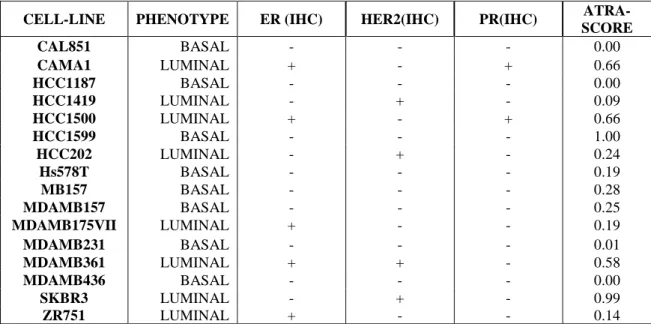
Table 2. Panel of 16 sensitive and resistant cell lines. ... 22
Table 3. 3 MSigDB collections ... 29
Summary
X
Summary
Breast cancer is the most commonly diagnosed malignancy and the leading cause of cancer death in women worldwide. The highest incidence rates were registered in Western and Northern Europe, Australia/New Zealand and North America; intermediate rates in South America, the Caribbean and Northern Africa; low rates in Sub-Saharan Africa and Asia. Factors that influence this variation in incidence rates are related to differences in reproductive and hormonal status (women in more developed countries have fewer children and a late age at first birth, use oral contraceptives or hormone replacement therapies) or in lifestyle (sedentary lifestyle, higher levels of obesity and higher alcohol consumption). Moreover in developed countries the screening programs have raised the rate of incidence, by permitting diagnoses of cancer that otherwise would have remained undiagnosed. The rate of survival for female breast cancer in developed countries is higher than for most of other types of cancer and this is due both to earlier diagnosis, made possible by screening programs, and to improvement in therapy. Despite the decrease in mortality rate registered during the last years, breast carcinomas still remain the leading cause of cancer death in female worldwide, accounting for about 14% of all cancer deaths.At present, the broad heterogeneity observed among breast cancer reflects the well-accepted notion that there is not just one disease with disparate variant subtypes, but that breast cancer instead represents a collection of distinct neoplastic diseases of the breast and the cells composing it. Behind this complexity, several systems have been developed to classify this very highly heterogeneous disease and possibly to get information about tumour behaviour and provide more effective therapies. Histological classification categorizes breast cancer either in “in situ” , which do not grow into or invade normal tissues within or beyond the breast, or “invasive”, which do grow into normal, healthy tissues. However, this classification relies only on histological characteristics, without taking into account molecular markers or morphological features that can be helpful for prognosis or therapy. In addition to this classification, it is crucial to assess the receptor status of a tumour, as it may determine the possibility of using targeted treatments in cancer therapy.
On this basis, various subgroups can be identified, according to their profile of gene expression and positivity to oestrogen receptor (ER), progesterone receptor (PR) and the
Summary
XI tyrosine kinase receptor, HER2. Moreover, breast tumours may be endowed with a different complement of the three above mentioned receptors (ER+/PR+/HER2+; ER+/PR+/HER2-; ER+/PR-/HER2+; ER+/PR-/HER2-; ER-/PR+/HER2-; ER-/PR-/HER2+; ER-/PR-/HER2-). Tumours that lack expression of all three receptors are defined as Triple Negative Breast Cancer (TNBC).
In recent years several studies on gene expression profiles have been conducted using high-throughput technologies, in order to identify molecular subtypes of breast cancer, to allow a better understanding of the complexity of the disease.
Based on hierarchical clustering of gene expression microarrays, six subtypes of breast cancers have been identified: the luminal A breast cancer is the most common subtype accounting for 50 – 60% of all diagnoses; it’s characterized by the expression of ER and the absence of HER2 over-expression. The luminal B breast cancer accounts for 10–20% of total, it often expresses HER2 and may express low level of oestrogen receptor. The HER2 positive subtype represents about 15-20% of all breast tumours and is characterized by expression of the HER2 gene, genes associated to its pathway and genes associated to cellular proliferation. The basal-like subtype, which represents 10-20% of total breast cancers, is characterized by expression of genes characteristic of the myo-epithelial (or basal) cells. In general, this subtype does not express ER, PR and HER2 and in clinical practice, it is often referred to as triple negative breast cancer (TNBC). The normal breast subtype is characterized by the expression of genes typical of adipose tissue and accounts for 5-10% of breast cancers; it lacks the expression of hormonal receptors and HER2 and so tumours belonging to this subtype are TNBC, but they are not basal-like cancers since they lack the expression of some genes characteristic of that category. Finally, the claudin-low subtype has been recently identified with a low expression of genes that encode for proteins involved in the formation of tight junctions, including Claudins and E-cadherin. It is rare and characterized by the absence of the oestrogen receptor, progesterone receptor and HER2 expression.
In 2009, Parker and colleagues introduced a new system of analysis, PAM50 (Prediction Analysis of Microarrays), to select a minimum set of genes, whose expression can predict the molecular subtype of a tumour. The PAM50 gene sets allows to obtain a classification similar to the one described above; it is based on a selection of gene sets consisting of a
Summary
XII large number of "intrinsic" genes, and therefore can be used in the clinics to define the molecular phenotype of the tumours.
Molecular classification of breast cancer based on gene expression patterns provides a connection between molecular biology and the behaviour of cancer cells in the corresponding subtypes. However, molecular classification of breast cancer has not yet reached clinical implementation as a routine aspect of patient management.
Although an immunohistochemical staining proxy can be used to stratify and classify breast cancers in a clinical setting, the correspondence between clinical and molecular is not yet remarkable.
The term retinoids refers to a group of compounds comprising metabolites and analogues of vitamin A, both natural and synthetic. The natural retinoids are essential components of diet and physiological regulators of many essential biological processes, such as embryonic development, metabolism and haematopoiesis. In adult mammals, retinoids such as All-Trans Retinoic Acid (ATRA), control homeostasis of different organs and tissues.
All-trans retinoic acid (ATRA) is a small lipophilic molecule and an important regulator of gene expression. The biological action of ATRA and its derivatives is mediated by two classes of nuclear receptors for retinoids called Retinoic Acid Receptor (RAR) and Retinoic X Receptor (RXR). The receptors are ligand-dependent transcription factors that control the activity of several target genes either through a direct or indirect mechanism. Both receptor subtypes exist in three different forms known as alpha, beta and gamma, encoded by different genes (RARA, RARB, RARG/ RXRA, RXRB, RXRG). Each subtype comprises two or more isoforms, which differ in the N-terminal region and are generated by a different promoter or by alternative splicing mechanisms. RAR and RXR receptors form stable hetero-dimers (RAR/RXR) that bind to specific sequences on DNA, called Retinoic Acid Responsive Elements (RAREs), localized within the promoter of target genes. ATRA modulates transcription through different mechanisms: it can directly modulate expression of target genes, through the interaction of the RAR/RXR with a group of co-activators and co-repressors or, in the absence of ligand, the RAR/RXR dimer is bound to the RAREs sequences on DNA and is associated with a complex of co-repressors that inhibit the transcription of target genes.
Tumorigenesis is a multistep process characterized by a series of inherited or acquired genetic changes (mutations, chromosomal rearrangements, epigenetic phenomena), leading
Summary
XIII to a disruption of cellular homeostasis and development of the neoplastic process. Several lines of evidence indicate an important role of retinoids in homeostasis.
Mechanisms by which retinoids exert their antitumor activity have not yet been completely clarified, although studies in vitro and in vivo have shown the ability of retinoids to inhibit proliferation, induce differentiation and apoptosis, making these molecules of therapeutic interest. It is clear that ATRA is able to act through different genomic mechanisms as well as to interact with other intracellular signalling systems, that provide the basis for its pleiotropic action.
Currently, the best example of the anticancer action of retinoids is the use of ATRA in the treatment of patients suffering from acute promyelocytic leukaemia (APL).
The retinoids have been investigated extensively for the prevention and treatment of cancer, predominantly because of their ability to induce cellular differentiation and to arrest proliferation. Systemic retinoids are approved by the U.S. Food and Drug Administration (FDA) also for treatment of cutaneous T-cell lymphoma, other than acute promyelocytic leukemia. The anti-leukemic action of ATRA is not primarily cytotoxic and it is the result of a direct cyto-differentiating action followed by a secondary apoptotic response rendering ATRA the first example of clinically useful cyto-differentiating agent. The use of ATRA in APL is also an example of targeted therapy, as the retinoids’ primary target is PML-RARα, the aberrant retinoid receptor expressed into the leukemic cell. To date, more than 85% of patients with APL achieve complete remission following treatment with ATRA in combination with chemotherapy. The unique mechanism of action and the results obtained in APL has raised enthusiasm in generalizing the use of retinoids to other types of cancers, including breast cancer. Pre-clinical data support the idea that ATRA is a promising agent in the treatment and chemoprevention of certain subgroups of breast cancer, with particular reference to ER+ and HER2+ tumours characterized by co-amplification of the retinoic acid receptor alpha gene. There is also a low proportion of triple negative breast cancers which show sensitivity to this unusual anti-tumour agent.
To evaluate the response of breast cancer cell lines to the anti-proliferative effect exerted by retinoic acid, Bolis and colleagues first defined the profile of ATRA-sensitivity in a panel of 48 breast cancer cell lines of the Cancer Cell Lines Encyclopedia (CCLE), well- representing the heterogeneity of the disease. The drug response of each cell line has been quantified by computation of a sensitivity score (ATRA-score).
Summary
XIV
ATRA-score was computed on cell-lines that have been treated with vehicle (DMSO) and 5
logarithmically increasing concentration of ATRA (0.001-10.0 μM) for 9 days and its value is calculated from the relative growth-inhibition (GI) data (ATRA vs. vehicle).
To define the ATRA-score sensitivity metric, they fitted growth-inhibition curves relative to DMSO-treated controls and computed the area under the curve (AUC) and the maximal inhibitory effect (Amax). At this point, ATRA-score values, which are equal to the log2 transformation of AUC x Amax , are rescaled in a range between 0 and 1, zero indicating total resistance and one standing for maximum sensitivity.
Moreover, Bolis and colleagues developed a tool capable of predicting ATRA-sensitivity, exploiting the association between this in vitro profiling and basal gene-expression data. In previous studies, a large panel of breast cancer cell lines (>50 lines) representative of the heterogeneity of the disease, have been profiled for their sensitivity to the anti-proliferative action of ATRA. They used a network-guided approach to develop a generalized model based on 21 genes (ATRA-21) capable of predicting ATRA-sensitivity across tumour types other than breast cancer.
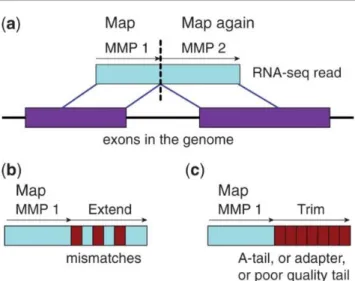
To identify gene-networks and gene pathways involved in the anti-proliferative action of ATRA, in this study we performed total RNA-sequencing experiments in a panel of 16 sensitive and resistant cell lines, before and after treatment with the retinoid (1.0 µM) for 24 hours. To better represent breast cancer heterogeneity, cell lines have been chosen based on their phenotype (8 luminal, which includes luminal A, luminal B and HER2+, and 8 basal-like) and their widely variable sensitivity to pharmacological treatment with retinoic acid. Alignment of high-throughput paired-end reads derived from RNA-sequencing experiments to the reference genome has been performed. Genome-generation was performed using the comprehensive gene annotations present in Gencode; in particular, the v27 release of the GTF file has been used. As many RNA-sequencing aligners suffer from high mapping error rates, read length limitation or mapping biases, sequence–alignment to reference human genome (hg38) has been performed using STAR (Spliced Transcript Alignment to a Reference) sequence-aligner, which was designed specifically to align non-contiguous sequences directly to the reference genome, using a novel strategy for these spliced alignments.
Summary
XV Differential gene expression analysis was conducted exloiting the R package DESeq2, which provides methods to test for differential expression by the use of a negative binomial generalized linear model.
After this first phase of differential expression analysis, we performed a test to verify the correlation between fold changes of differentially expressed genes and the predicted response of cell lines to pharmacological treatment, in terms of ATRA-score. To this aim, we computed both Pearson’s product moment correlation coefficient and Spearman’ rank correlation coefficient. We selected only those genes with a Pearson’s coefficient R or a Spearman’s coefficient RHO <0.01. To further select only those genes showing a variation across samples that is sufficient to result in a biologically significant action, we computed a variation coefficient, defined as 𝑆𝑑{𝑀𝑎𝑡𝑟𝑖𝑥[𝑖, ] 𝑚𝑒𝑎𝑛(𝑀𝑎𝑡𝑟𝑖𝑥[𝑖, ])⁄ } ∗ 100.
On the basis of this additionally parameter, a more restrictive selection of genes has been performed, considering only those genes with VC>50%.
Selected genes have been organized in networks based on protein – protein interactions, to a more precise visualization of possible interaction mechanisms induced.
Moreover, gene set enrichment analysis (GSEA) on sequencing data led to the identification of ATRA-dependent pathways and gene-networks with significance for the anti-tumour activity of the retinoid: “interferon-dependent” and immune modulatory pathways are found to be strictly up- regulated after treatment with ATRA. Genes and pathways that are down-regulated specifically in ATRA-sensitive cell lines, are linked to cell proliferation and cell cycle progression which are tightly connected with the antiproliferative effect exerted by retinoic acid, and thus can be considered part of a downstream mechanism of action. We inspected ChIP-sequencing data from a public database of two forms of RARs transcription factors (RARA, RARG) in one breast cancer cell line treated with retinoic acid (MCF-7): we wanted to evaluate which of the more central genes in our response network were directly perturbated by the binding in the regions of their promoters of the ATRA-activated transcription factors. To this purpose, raw data obtained in “FASTQ” file format have been aligned to the reference genome (hg38), using the Burrows-Wheeler Alignment Tool (BWA), a read alignment algorithm that is based on the backward search with Burrows-Wheeler Transform (BWT). This represents an effective method to align short sequencing reads (50 bp) against a large reference sequence, such as the human genome.
Summary
XVI Next, we used a command line tool designed by Zhang and Liu, MACS (Model-based Analysis of ChIP-Sequencing), to analyse pre-processed ChIP-Sequencing data. Given the ChIP-Sequencing data with the correspondent control sample, this peak-finding algorithm
can be used to identify transcription factor binding sites (or even histone modifications, if necessary): it uses a dynamic Poisson distribution, which captures local biases in the genome, allowing for robust predictions and giving fewer false positives than the other available methods. Last, we annotated the identified peaks with an Ensembl based annotation package for Homo Sapiens, Ensembl version 86 (EnsDb.Hsapiens.v86).
As result, we obtained a list of genes that are part of the above-mentioned interferon signalling, which have been identified as directs targets for RARA or RARG transcription factors; however, some of the most crucial genes involved in such pathways cannot be included in the list. Among the genes identified as RARA direct targets, is of particular interest the presence of “interferon- related” genes, such as DTX3L and PARP9, such as the presence of one of the genes that encodes a protein that is part of the MHC-I complex, HLA-E. In contrast, one of the more important transcription factors involved in the regulation of the interferon signalling, IRF1, is not a direct target, neither for RARA transcription factor, nor for RARG.
Methylation data available for a panel of almost 40 un-treated breast cancer cell lines have been investigated, to find out whether there is a correlation between the basal methylation levels of genes necessary to trigger the mechanism of response to retinoids, and the sensitivity of cell lines to ATRA. All the data were obtained through the HumanMethylation450 BeadChip Array Platform: after a few normalization steps, methylation data have been tested for association with a defined parameter, the ATRA-score. To this aim, dmpFinder function implemented in the package minfi in R environment has been used, which tested each genomic position for association between methylation and a “phenotype”, our defined parameter. Given the ATRA-score as a continuous parameter, association has been tested with linear regression. Finally, differentially methylated probes have been annotated, exploiting the information stored in the Bioconductor package “IlluminaHumanMethylation450kanno.ilmn12.hg19.
Again, a few genes involved in the interferon-related mechanism have found to have a correlation between their methylation levels and the activation of the response to retinoids.
Summary
XVII In particular, 73 out of the total 298 identified genes are well interesting, because of their highly interconnected position in the network defined during the differential expression analysis. In particular, among the genes with a higher methylation level we can find TLR3, a crucial part of innate immune response, whose transcription is deeply inducted after treatment with retinoic acid. Moreover, two other genes involved in the innate immune response and inducted by treatment with retinoic acid, HLA-E and PSMB8, have high methylation level in basal condition, correlated with the ATRA-score.
In the second part of the study we took again into account the RNA-sequencing data to quantify possible transcription of repetitive elements from retroviral DNA, which are known to be widely distributed in the human genome: we hypostasized that they can be the cause of the above-mentioned interferon-driven immune system reactivation.
To quantify expression of these transposable elements, we retrieved their genomic positions from RepeatMasker database (http://www.repeatmasker.org/). These coordinates were assembled into a customized annotation file (gene transfer file, GTF), which was used to determine the abundance of all retroviral-derived transcripts, by using FeatureCounts. To avoid detection of false positives, we discarded all transposable elements that show any overlap to known gene-associated exons, according to Gencode annotations. Afterwards, viral RNA abundance was normalized for library size and tested for differential expression between ATRA-treated and untreated samples, using the same approach as described in Material and Methods.
A widely distributed up-regulation of these transcripts can be observed: induction (fold change) of the transcriptomic regulation slightly correlates with the sensitivity to ATRA-treatment. Cell lines which are completely resistant to the pharmacological treatment, display no induction; then transcriptomic up-regulation grows with an increasing
ATRA-score. Despite the presence of a general trend of correlation between the induction of the
retroviral elements transcription and the sensitivity to retinoids, of particular interest is the transcriptional effect on a few cell lines (CAMA1, ZR751), which doesn’t follow the global behaviour. To better understand the reasons of such tendency, we proceeded with further investigations: it has been shown that this general course tightly correlates with the expression levels of RARA in each cell line.
All things considered, we identified gene-networks whose expression is selectively modulated by ATRA in retinoid-sensitive luminal and triple-negative cell lines as well as
Summary
XVIII other gene-networks which are commonly regulated in both cell groups. Among the networks stimulated by ATRA, the group of genes involved in interferon- responses is of particular interest, as it indicates that the retinoid exerts a strong and specific immuno-modulatory action in sensitive breast cancer cell lines.
We are evaluating the functional significance of specific elements of these gene-networks for the anti-tumour-metastatic action of ATRA with the use of silencing and over-expression approaches.
The results obtained in our cellular models provide insights into the molecular mechanisms underlying the anti-tumour action of ATRA in breast cancer. In addition, the sequencing data led to the identification of ATRA-dependent pathways and gene-networks with significance for the anti-tumour activity of the retinoid. Finally, the approach provides information as to potential new molecular targets for the design of rational therapeutic combinations based on ATRA for the treatment and secondary chemo-prevention of certain types of breast cancer.
Beside the anti-proliferative effect described above, our data suggest that the pharmacological treatment with ATRA might also have an immunoregulatory effect on these cells. In particular, it has been observed that there is a dramatic up-regulation of the “Antigen-presentation and assembly/loading of class I MHC” pathways, as well as “Inflammatory responses”: this may result in an increased antigen presentation mechanism which may activate innate immune response.
All things considered, this study provides a strong rationale for the combination of ATRA with the immune checkpoint inhibitors.
Sommario
XIX
Sommario
Il carcinoma della mammella è il tumore più frequentemente diagnosticato tra le donne nel mondo, sia nei Paesi economicamente più avanzati che in quelli in via di sviluppo. Nel 2018 sono stati stimati 2 milioni di nuovi casi in tutto il mondo.La più alta incidenza è stata osservata in Nord America, in Europa del Nord e Ovest e in Oceania, dove sono in aumento la prevalenza dei fattori di rischio e le rilevazioni di tumori allo stadio iniziale. Nell'ultimo decennio in questi Paesi si è registrato però anche un aumento della sopravvivenza, grazie alla disponibilità di programmi di prevenzione primaria e diagnosi precoce (mammografia) e di nuove strategie terapeutiche efficaci, che permettono di migliorare la prognosi delle pazienti. Attualmente il tumore della mammella è la quinta causa di morte per tumore (552,000, il 6,4%). Nei Paesi in via di sviluppo, come Sud e Centro America, Africa e Asia, sono invece in aumento sia l’incidenza che la mortalità; questa tendenza riflette il recente cambiamento nello stile di vita (dieta, obesità) insieme a cambiamenti relativi allo stato ormonale e riproduttivo (minor numero di gravidanze e in età più adulta, allattamento di minore durata, utilizzo di contraccettivi), mancanza di programmi di screening efficaci e, in alcuni casi, limitato accesso ai trattamenti.
In Italia, secondo i dati presentati dall'Associazione Italiana di Oncologia (AIOM) e dall'Associazione Italiana dei Registri Tumori (AIRTUM), si registra un aumento dell'incidenza di tumore della mammella nelle donne, con 52,300 nuovi casi stimati nel 2018. Questo aumento può essere in parte ricondotto all'ampliamento della fascia di screening mammografico in alcune Regioni. È il secondo tipo di tumore più frequente nelle donne, ma la sopravvivenza a 5 anni si avvicina al 90%, con percentuali ancora più alte se diagnosticato ad uno stadio precoce. La mortalità è quindi in diminuzione, seppure con differenze tra le Regioni del Nord e Sud Italia, come risultato di una maggiore prevenzione primaria e del miglioramento delle strategie terapeutiche
Il carcinoma della mammella è una malattia eterogenea che comprende entità distinte in termini di istologia, caratteristiche molecolari, prognosi clinica e risposta ai trattamenti. Questa diversità ha reso più complesso lo sviluppo di classificazioni clinicamente utili per determinare il comportamento di un tumore sulla base delle sue caratteristiche biologiche .
Sommario
XX La classificazione istopatologica si basa sulle differenti caratteristiche morfologiche dei tumori. Il tumore della mammella può essere innanzitutto classificato come in situ o invasivo. In situ significa che il tumore rimane confinato all’interno del tessuto epiteliale in cui si sviluppa, mentre è definito invasivo quando invade il tessuto circostante, diffonde nei linfonodi e vasi sanguigni ed eventualmente in altre aree del corpo.
Negli ultimi decenni è stata dimostrata la fondamentale importanza di due recettori di ormoni steroidei, il recettore degli estrogeni (ER) e del progesterone (PR), e del recettore tirosin-chinasico HER2 (human epidermal growth factor receptor 2) per l’eziologia, la prognosi e la terapia dei tumori della mammella. Accanto alla classificazione istopatologica, i tumori della mammella si possono distinguere sulla base dell’espressione dei suddetti recettori, valutata mediante analisi immunoistochimica, che permette di rilevare la presenza della proteina. Inoltre, I tumori della mammella possono essere caratterizzati da diverse complementazioni dei tre recettori sopra citati: (ER+/PR+/HER2+; ER+/PR+/HER2-; ER+/PR-/HER2+; ER+/PR-/HER2-; ER-/PR+/HER2-; ER-/PR-/HER2+; ER-/PR-/HER2-).
Infine, i tumori della mammella tripli negativi (TNBC, triple-negative breast cancer) costituiscono un gruppo di tumori molto eterogenei, caratterizzati dalla mancanza di espressione del recettore degli estrogeni e del progesterone e dalla mancanza di amplificazione/sovra-espressione di HER2.
L’analisi dell’espressione genica, resa possibile dallo sviluppo di tecniche basate su microarray a cDNA, ha permesso di suddividere i tumori in diversi sottotipi molecolari sulla base della somiglianza del profilo di espressione genica. In questo modo è stata definita una classificazione, detta intrinseca, che ha individuato sei diversi sottotipi.
Il sottotipo luminale A (50 -60% di tutti i carcinomi della mammella) comprende tumori caratterizzati da alti livelli di espressione del recettore degli estrogeni e dall’assenza di over-espressione del recettore HER2. Il sottotipo luminale B (10-20% di tutti i carcinomi della mammella) comprende tumori
che esprimono alti livelli di HER2 e spesso bassi livelli del recettore per l’estrogeno. Il sottotipo HER2-arricchito (15-20% di tutti i carcinomi della mammella) è costituito da tumori che sono prevalentemente HER2-amplificati, mostrano elevati livelli di espressione di numerosi geni dell'amplicone ERBB2 e geni associati a meccanismi di proliferazione cellulare. I tumori basal-like (10-20% di tutti i carcinomi della mammella) sono caratterizzati da alti livelli di espressione di marcatori delle cellule mioepiteliali basali e di geni che
Sommario
XXI regolano il ciclo cellulare. Sono inoltre caratterizzati dalla mancanza o da bassi livelli di espressione di ER e dei geni correlati ad ER, incluso PR, e dalla frequente assenza della sovra-espressione/amplificazione di HER2. Per questo motive, in pratica clinica, si fa riferimento a questo tipo di tumore come sottotipo triplo negativo.
Il sottotipo normal breast-like (5-10% di tutti i carcinomi della mammella) è costituito da tumori caratterizzati da elevati livelli di espressione di geni tipici del tessuto adiposo e di altri tipi cellulari non epiteliali. Sono caratterizzati dalla mancanza di espressione di ER, PR ed HER2, (sottotipo triplo negativo, ma non basal like, in quanto non esprimono geni caratteristici di quella categoria.
Infine, il sottotipo Claudine-low (12-14% dei carcinomi della mammella) è costituito per la maggior parte da carcinomi invasivi, caratterizzati da bassi livelli di espressione di geni coinvolti nelle giunzioni e nell’adesione cellula-cellula, come quelli codificanti per le Claudine 3/4/7 e la E-caderina. Inoltre, sono tumori rari e caratterizzati dall’assenza di recettore per l’estrogeno, per il progesterone ed anche HER2 negativi (tripli negativi). Nel 2009, Parker e colleghi hanno implementato un sistema di analisi, PAM50 (prediction
analysis of microarrays), per selezionare un set minimo di geni (50 geni) la cui espressione
fosse predittiva di uno specifico sottotipo molecolare. Il set di geni PAM50 permette di ottenere una classificazione in accordo con quella precedente, che si basava sulla selezione di set costituiti da un numero maggiore di geni "intrinsechi", e può essere utilizzato in clinica per definire il fenotipo molecolare del tumore.
La classificazione molecolare del tumore della mammella basata sui pattern di espressione genica costituisce un elemento di connessione tra la biologica molecolare del tumore e il conseguente progredire delle cellule tumorali del corrispondente sottotipo. Tuttavia, la classificazione molecolare del tumore del seno non ha ancora raggiunto un’implementazione standardizzata a livello clinico, poiché la corrispondenza tra gli aspetti clinici e quelli molecolari non è ancora completamente definita.
Con il termine retinoidi ci si riferisce a tutte le molecole strutturalmente e funzionalmente analoghe al retinolo (Vitamina A), sia naturali che sintetiche. La vitamina A e i suoi metaboliti biologicamente attivi (acido retinoico tutto-trans, acido 9-cis retinoico e acido 13-cis retinoico) sono molecole essenziali per lo sviluppo embrionale, il meccanismo della visione e l’omeostasi di numerosi tessuti e sistemi, tra cui il sistema nervoso, immunitario e riproduttivo. A livello cellulare, regolano la proliferazione, il differenziamento e l’apoptosi.
Sommario
XXII L'acido retinoico tutto-trans (ATRA) è una piccolo vitamina liposolubile, importante regolatrice dell’espressione genica. L’acido retinoico e i suoi derivati regolano infatti l’espressione di geni coinvolti nella crescita e nel differenziamento cellulare attraverso specifici recettori nucleari, RAR (retinoic acid receptor) e RXR (retinoid X receptor). Questi recettori, in forma di omo- o etero-dimeri (RAR-RXR o RXR-RXR), agiscono da fattori di trascrizione. Sia RAR che RXR presentano ciascuno tre sottotipi recettoriali (α, β, γ) codificati da geni distinti. Per ciascun sottotipo esistono più isoforme, generate per splicing alternativo, che possono avere una differente affinità per i vari retinoidi e mediare differenti funzioni biologiche.
L’acido retinoico tutto-trans è un agonista di tutte le isoforme recettoriali RAR, a cui si lega con la stessa affinità, modulando la trascrizione attraverso diversi meccanismi. In assenza di ligando, il dimero RAR-RXR è costitutivamente legato alle sequenze RARE contenute nei promotori dei geni bersaglio ed è associato a co-repressori , che inibiscono la trascrizione genica. Il legame di ATRA a RAR induce modificazioni conformazionali che possono determinare il rilascio di co-repressori e il reclutamento di co-attivatori con attivazione della trascrizione genica (meccanismo genomico diretto). Inoltre, i geni regolati direttamente codificano per proteine coinvolte nel trasporto, metabolismo e trasduzione del segnale dell’acido retinoico stesso e per i fattori di crescita, a loro volta, modulano l’espressione di geni coinvolti nella proliferazione cellulare (meccanismo genomico indiretto). In questo modo, ATRA inibisce la crescita arrestando il ciclo cellulare e guida la cellula verso un programma di differenziamento.
La carcinogenesi è un processo caratterizzato dal graduale accumulo di alterazioni genetiche ed epigenetiche responsabile della deregolazione dell’omeostasi cellulare. In questo processo, le cellule sane vanno incontro ad una serie di trasformazioni neoplastiche con formazione di lesioni pre-maligne e sviluppo di carcinomi in situ e metastatici.
I retinoidi svolgono un ruolo importante nel mantenimento dell’omeostasi: attraverso la regolazione dell’espressione genica, garantiscono il corretto equilibrio tra crescita e differenziamento cellulare. La perdita della loro attività o la diminuzione dei loro livelli intracellulari è associata ad una crescita cellulare aberrante e allo sviluppo di un’ampia varietà di tumori.
I meccanismi con cui i retinoidi esercitano la loro attività antitumorale non sono ancora stati completamente chiariti, sebbene gli studi in vitro e in vivo abbiano dimostrato la capacità dei retinoidi di inibire la proliferazione, indurre differenziazione e apoptosi, rendendo queste molecole di interesse terapeutico. È chiaro che ATRA sia in grado di agire attraverso diversi
Sommario
XXIII meccanismi genomici e di interagire con altri sistemi di segnalamento intracellulare, che forniscono la base per la sua azione pleiotropica.
L’acido retinoico tutto-trans è stato il primo agente anti-proliferativo e cito-differenziante ad essere utilizzato in clinica nel trattamento di un raro sottotipo di leucemia mieloide acuta (AML, acute myeloid leukemia), la leucemia promielocitica acuta (APL, acute
promyelocytic leukemia). L'azione anti-leucemica di ATRA non è citotossica, ma è il risultato di un'azione diretta anti-proliferativa e cito-differenziante, seguita da una risposta apoptotica secondaria che rende ATRA il primo esempio di agente cito-differenziante correntemente utilizzato in clinica. L'uso di ATRA in APL è inoltre un esempio della cosiddetta “targeted” therapy, poiché l'obiettivo primario del retinoide è la fusione genica PML-RARα, che determina un’alterazione della funzione recettoriale di RARα.
In combinazione con la chemioterapia (antracicline), questa terapia permette a più dell’85% dei pazienti con APL di andare incontro a remissione completa della malattia.
Grazie al successo di ATRA nell’ambito della leucemia promielocitica acuta, l’interesse per il potenziale utilizzo terapeutico dei retinoidi si è esteso anche ad altri tipi di carcinomi, come il tumore della mammella. Ciò ha portato allo sviluppo di numerosi analoghi sintetici dell’acido retinoico, promettenti agenti cito-differenzianti e pro-apoptotici, e alla disponibilità di una serie di dati ottenuti da numerosi studi preclinici. Questi ultimi, tuttavia, si sono tradotti in un numero molto limitato di studi clinici. Tale insuccesso potrebbe essere dovuto al fatto che gli studi sono stati condotti senza tenere in considerazione l’eterogeneità del tumore della mammella e senza una selezione dei sottotipi tumorali.
Si è quindi reso necessario definire i determinanti molecolari della sensibilità e resistenza ad ATRA nei diversi sottotipi di tumore della mammella.
Per valutare la risposta delle linee cellulari di carcinoma mammario all'effetto antiproliferativo esercitato dall'acido retinoico, Bolis e colleghi hanno definito il profilo di sensibilità ad ATRA in un pannello di 48 linee cellulari di carcinoma mammario, che ben rappresentasse l'eterogeneità della malattia. La risposta farmacologica di ciascuna linea cellulare è stata quantificata per il calcolo finale di un punteggio di sensibilità
(ATRA-score).
Tale punteggio è stato calcolato su linee cellulari che sono state trattate con veicolo (DMSO) e 5 una concentrazione logaritmicamente crescente di ATRA (0,001-10.0 μM) per 9 giorni.
Sommario
XXIV Il puntegio finale è stato calcolato a partire dai dati relativi alle curve di crescita-inibizione (ATRA vs veicolo).
Per definire la metrica di sensibilità dell'ATRA-score, è stato eseguito un fitting delle curve di inibizione della crescita e per ciascuna è stata calcolata l'area sottesa alla curva (AUC) e l'effetto inibitorio massimo (Amax). A questo punto, i valori di ATRA-score, pari alla
trasformazione logaritmica del prodotto AUC x Amax, sono stati riscalati in un range
compreso tra 0 e 1, dove zero indica la resistenza totale e uno la massima sensibilità. Inoltre, Bolis e colleghi hanno sviluppato uno strumento in grado di predire la sensibilità al trattamento farmacologico con ATRA, sfruttando l'associazione tra questo profilo di sensitività in vitro e i dati disponibili di espressione genica basale. A partire da questi dati, utilizzando un approccio “network-guided” è stato sviluppato un modello basato sui dati di espressione basale di 21 geni (ATRA-21), in grado di predire la sensibilità ad ATRA anche in tipi di tumori diversi dal cancro al seno.
Per identificare i meccanismi molecolari coinvolti nell'azione anti-proliferative di ATRA, in questo studio sono stati condotti esperimenti di sequenziamento di RNA in un pannello di 16 linee cellulari di carcinoma della mammella, sensibili e resistenti ad ATRA, prima e dopo il trattamento con acido retinoico (1,0 µM) per 24 ore. Per meglio rappresentare l'eterogeneità del cancro al seno, le linee cellulari sono state scelte in base al loro fenotipo (8 di fenotipo luminale, comprendenti luminali A, luminali B e HER2 +, e 8 di fenotipo basale) e la loro variabile sensibilità al trattamento farmacologico con acido retinoico. Le sequenze ottenute dagli esperimenti di sequenziamento di RNA sono state allineate al genoma di riferimento. La generazione del genoma è stata effettuata usando le annotazioni genomiche presenti in GENCODE; in particolare, è stata utilizzata la versione 27 (v27) del file GTF( gene transfer file).
Poiché molti allineatori di sequenze di RNA soffrono di alti tassi di errore di mappatura, limitazioni nella lunghezza delle sequenze che possono essere allineate o biases nella mappatura, l’allineamento delle sequenze al genoma di riferimento (hg38) è stato eseguito utilizzando STAR (Spliced Transcripts Alignment to a Reference), progettato in modo specifico per l’allineamento di sequenze non contigue, utilizzando una strategia innovativa per sequenze soggette a splicing.
L’analisi di espressione differenziale è tata poi successivamente condotta in ambiente R, attraverso l’utilizzo del pacchetto DESeq, in grado di testare l’espressione differenziale di
Sommario
XXV geni tra due diverse condizioni sperimentali (controlli e trattamenti con acido retinoico) attraverso l’utilizzo di un modello lineare (GLM) binomiale negativo.
Dopo questa prima fase di analisi, è stato eseguito un test di correlazione tra l’induzione (fold change) dei geni differenzialmente espressi e la risposta predetta di ciascuna linea cellulare, in termini di ATRA-score. Per fare ciò, sono stati calcolati sia il coefficiente di correlazione secondo Pearson (R), sia il coefficiente di correlazione secondo Spearman (RHO). Sono stati selezionati solo quei geni con R<0.01 o RHO<0.01.
Da ultimo, sono stati ulteriormente selezionati solo quei geni che mostrassero una variabilità nei diversi campioni sufficiente a determinarne un’ azione biologicamente significativa. Per fare ciò, è stato calcolato il coefficiente di variazione tra i campioni, definito in termini percentuali. Solo i geni con una variazione superiore al 50% sono stati tenuti in considerazione per analisi successive.
I geni selezionati sono stati organizzati in reti basate sulle interazioni proteina-proteina, per una visualizzazione più precisa dei possibili meccanismi di interazione indotti.
Successivamente, l'analisi di arricchimento di espressione genica (GSEA) sui dati di sequenziamento ha condotto all'identificazione dei pathways molecolari la cui attivazione o repressione sia dipendente dall’attività antitumorale dell’acido retinoico. Le vie dipendenti dall’attivazione di interferone e quelli relative all’attivazione della risposta immunitaria sono fortemente up-regolate dopo il trattamento con ATRA.
Al contrario, le vie connesse con la proliferazione e la progressione del ciclo cellulare, sono fortemente down-regolate: questo meccanismo sembra connesso con l'effetto antiproliferativo dell’ acido retinoico, e quindi si ipotizza essere parte di un meccanismo di azione a valle.
Abbiamo ispezionato i dati di sequenziamento di immuno-precipitazione di cromatina (ChIP), depositati in un database pubblico, di due forme di fattori di trascrizione RAR -dipendenti (RARA, RARG) in una linea cellulare di cancro al seno trattata con acido retinoico (MCF-7): abbiamo voluto valutare quale dei geni più interconnessi nella rete di risposta al trattamento, siano stati direttamente perturbati dal legame nelle regioni del loro promotore dei fattori di trascrizione attivati da acido retinoico. A questo scopo, i dati grezzi ottenuti dal sequenzaimento sono stati allineati al genoma di riferimento (hg38), utilizzando lo strumento di allineamento Burrows-Wheeler (BWA), un algoritmo basato sull’utilizzo della trasformata di Burrows-Wheeler (BWT). Si tratta infatti di un metodo efficace per
Sommario
XXVI
allineare brevi sequenze (50 paia di basi) su sequenze di riferimento molto lunghe, come appunto il genoma umano.
Successivamente, abbiamo utilizzato un algoritmo implementato da Zhang e Liu, MACS (Model-based Analysis of ChIP-Sequencing), per analizzare i dati di sequenziamento di immuno-precipitazione di cromatina pre-processati. Una volta associato il dato di sequenziamento del trattamento con il corrispondente campione di controllo, questo algoritmo di ricerca può essere utilizzato per identificare i siti di legame dei fattore di trascrizione (o anche le modifiche istoniche, se necessario): utilizzando un modello basato su una distribuzione di Poisson dinamica, permette di ottenere robuste previsioni e un basso numero di falsi positivi, confrontato con altri algoritmi disponibili. Infine, i siti di legame identificati sono stati annotati, con un pacchetto di annotazione che fa riferimento alla release
Ensembl 86 (https://www.ensembl.org/index.html).
Come risultato, abbiamo ottenuto una lista di geni, alcuni dei quali associabili al signalling di interferone, che sono stati identificati come bersagli diretti per fattori di trascrizione RARA o RARG. Tuttavia, alcuni dei geni cruciali coinvolti nelle vie molecolari interferoniche non possono essere inclusi nella lista. Tra i geni identificati come bersagli diretti di RARA, è di particolare interesse la presenza di geni correlati alla via interferonica come DTX3L e PARP9, insieme ad uno dei geni codificanti una proteina parte del complesso MHC-I, HLA-E. Al contrario, uno dei fattori di trascrizione più importanti coinvolti nella regolazione del segnale dell'interferone, IRF1, non è un bersaglio diretto, né per il fattore di trascrizione RARA, né per RARG.
I dati di metilazione disponibili per un pannello di quasi 40 linee cellulari di carcinoma della mammella in condizioni basali sono stati indagati, per scoprire se esistesse una correlazione tra i livelli di metilazione basale di geni necessari per innescare il meccanismo di risposta ai retinoidi, e la sensibilità delle linee cellulari ad ATRA.
Tutti i dati utilizzati, sono stati ottenuti attraverso la piattaforma HumanMethylation450 BeadChip: dopo alcuni passaggi di normalizzazione, i dati di metilazione sono stati testati per l'associazione con un parametro definito, l'ATRA-score. A questo scopo è stata utilizzato l’algoritmo implementato nella funzione dmpFinder, implementata nel pacchetto minfi in ambiente R, in grado di verificare, per ogni posizione genomica, l’associazione tra la metilazione e un "fenotipo" o parametro definito. Dato l'ATRA-score come parametro continuo, l'associazione è stata testata con un modello di regressione lineare.
Sommario
XXVII Infine, le zone genomiche identificate sono state annotate, sfruttando le informazioni contenute nel pacchetto "IlluminaHumanMethylation450kanno. ilmn12. hg19.” In
Bioconductor (https://bioconductor.org/).
Anche in questo caso, per alcuni geni coinvolti nel meccanismo molecolare di azione dipendente dall’attivazione interferonica, è stata trovata una correlazione fra i livelli di metilazione e l'attivazione della risposta all’acido retinoico.
In particolare, tra i geni più rilevanti per il loro ruolo centrale all’interno dei network di interazione identificati, possiamo trovare TLR3, fondamentale nei meccanismi di risposta immunitaria innata, il cui livello di metilazione basale correla con una trascrizione profondamente indotta dopo il trattamento con acido retinoico. Inoltre, altri due geni coinvolti nella risposta immunitaria innata e indotti dal trattamento con acido retinoico, HLA-E e PSMB8, hanno un alto livello di metilazione in condizioni basali, che ben correla con l'ATRA-score.
Nella seconda parte di questo studio, abbiamo preso nuovamente in considerazione i dati di sequenziamento di RNA per quantificare la possibile trascrizione di sequenze ripetute da DNA retrovirale, note per essere ampiamente distribuite nel genoma umano: l’ipotesi di partenza dell’analisi, è la possibilità che la perturbazione trascrittomica di queste sequenze possa essere la causa della riattivazione delle vie interferoniche. Per quantificare l'espressione di questi elementi trasponibili, abbiamo ottenuto le loro posizioni genomiche dal database RepeatMasker (http://www.RepeatMasker.org/). Queste coordinate sono state assemblate in un file di annotazione personalizzato (gene transfer file, GTF), che è stato utilizzato per quantificare l’induzione nella trascrizione di queste sequenze retrovirali, utilizzando l’algoritmo implementato in FeatureCounts. Per evitare falsi positivi, tutti gli elementi trasponibili che mostrano la sovrapposizione con esoni noti associati a geni codificanti, sono stati scartati. In seguito, dopo una fase di normalizzazione, la quantificazione dell’espressione differenziale di queste sequenze fra campioni trattati con ATRA e campioni non trattati, è stata condotta usando un approccio analogo a quello usato per i geni codificanti. Ciò che si è potuto osservare è un’induzione ampiamente distribuita di tutte queste sequenze: l'induzione della regolazione trascrittomica è inoltre correlata con la sensibilità all’acido retinoico. Le linee cellulari che sono completamente resistenti al trattamento farmacologico, non mostrano alcuna induzione; linee maggiormente sensibili mostrano un maggiore livello di attivazione. Nonostante la presenza di una tendenza
Sommario
XXVIII generale di correlazione tra l'induzione della trascrizione degli elementi retrovirali e la sensibilità ai retinoidi, di particolare interesse è l'effetto trascrizionale su alcune linee cellulari (CAMA1, ZR751), che non segue il comportamento globale. Per meglio comprendere le ragioni di tale tendenza, sono state effettuate ulteriori indagini: è stato dimostrato che questo andamento generale è strettamente correlato con i livelli di espressione basale di RARA in ogni linea cellulare.
I risultati ottenuti in questi modelli cellulari forniscono informazioni sui meccanismi molecolari che sottendono l'azione anti-tumorale di ATRA nel tumore della mammella. Inoltre, i dati di sequenziamento hanno condotto all'identificazione delle vie e delle reti genichedipendenti dall’azione dell’acido retinoico, legate alla sua attività anti-tumorale. Infine, l'approccio fornisce informazioni sui potenziali nuovi bersagli molecolari per la progettazione di combinazioni terapeutiche razionali basate su ATRA per il trattamento e la prevenzione di una chemioterapia secondaria su alcuni tipi di carcinoma.
Al di là dell’attività antiproliferativa, che potrebbe essere il risultato di una combinazione di fattori diversi, i dati presentati suggeriscono che il trattamento con acido retinoico possa avere un effetto immuno-regulatorio.
Infatti, il possibile aumento nella presentazione di antigeni, unito alla up-regolazione di pathway molecolari legati all’attivazione del sistema immunitario innato, avrebbe effetto significativo sulla rilevazione dei tumori da parte sistema immunitario e la sua conseguente attivazione.
Secondo questa osservazione, questo studio fornisce un forte razionale per lo studio della combinazione di ATRA con farmaci recentemente introdotti nel trattamento immuno-oncologico, cioè gli inibitori del checkpoint immunitario.
Introduction
1
Introduction
Breast cancer is the most common malignancy and the leading cause of cancer deaths in women in the Western Hemisphere [1]. This is a very complex and heterogeneous disease and numerous efforts have been made to identify histological and molecular characteristics associated to clinical outcome. On the basis of gene expression data, different subtypes have been identified that show significant differences in incidence, survival and response to drug treatment. The most important determinants of these subtypes are the presence or absence of the estrogen receptor (ER) and the progesterone receptor (PgR), and the over-expression of tyrosine kinase receptor ERBB2 [7]. Given the heterogeneity of the disease, the diversity of the molecular mechanisms activated in different subgroups of this tumor and the developing of resistance to classical therapies, it would be helpful to use combinations of different drugs. All-trans-retinoid acid (ATRA) is the active metabolite of vitamin A and a promising agent in the prevention and treatment of breast cancer. In view of the development of ATRA-based therapeutic strategies aimed at personalized treatment of mammary tumours, a recent study demonstrated that approximately 70% of oestrogen-receptor-positive (ER+) breast cancer cell lines and primary tumours are sensitive to anti-proliferative effects of ATRA [20]. In contrast, only 10-20% of the HER2-positive and triple-negative counterparts respond to the retinoid.Mechanisms by which retinoids exerts their antitumor activity have not yet been completely clarified, although studies in vitro and in vivo have shown the ability of retinoids to inhibit proliferation, induce differentiation and apoptosis, making these molecules of therapeutic interest. Therefore, it would be important to decipher retinoids’ transcriptionally mechanisms of action, since it could lead to the development of targeted therapeutic strategies, able to implement new drug treatments.
At present, on the basis of the data and the available basal gene-expression profiles of breast cancer cell lines and primary tumors, Bolis and colleagues [38] have developed a model consisting of 21 genes (ATRA-21) which correctly predicts ATRA-sensitivity in the context of breast cancer.
The present study is aimed at getting insights into the molecular mechanisms underlying the anti-tumor action of ATRA in the specific subsets of breast cancer identified. In addition,
Introduction
2 we intend to identify specific genes and gene-networks modulated by ATRA which may represent pharmacological targets for the design and development of rational combinations between the retinoid and unrelated therapeutic agents to be used in the personalized treatment of breast cancer agents. A final goal is the identification of potential bio-markers of the anti-tumor response to ATRA and potentially pharmacological targets to be used in the clinics.
To address all these points, we used a multi-omics approach to investigate various aspects of the molecular mechanisms that can be involved in the response of the breast tumour cells to treatment with retinoic acid.
In the first part of the study, we analysed data obtained after performing deep-sequencing experiments on a panel of sixteen cell lines recapitulating the heterogeneity of the breast cancer phenotype and characterized for their anti-proliferative response to ATRA. Each cell line has been exposed to ATRA (1 µM) for 24 hours and total RNA was extracted and subjected to high throughput sequencing. The global gene-expression data were analyzed to
Introduction
3 evaluate the transcriptomic profile induced by the pharmacological treatment, with a number of complementary bio-informatic tools.
To complement this analysis, we obtained data from available databases, such as the NCBI GEO (Gene Expression Omnibus), deriving from different techniques, such as Chromatin Immunoprecipitation (ChIP) sequencing and Methylation arrays, performed on a wide number of breast cancer cell lines. We analysed these data and then complemented our results to the data obtained from the first part of the study.
In the second part of the study RNA-sequencing data were analysed to quantify transcription of possible retroviral elements that are part of the human genome, in order to asses if a phenomenon called “viral mimicry” could trigger the mechanism of response to pharmacological treatment with retinoids.
This multi-omics approach gave us a more comprehensive view of the transcriptional and molecular mechanisms that are implicated in the sensitivity or resistance of breast cancers to the treatment with retinoic acid.
It is worth noticing that the huge amount of data to be processed and analysed for this project required a massive computational time and power: for many processing steps we had to exploit CINECA supercomputers.
Background
4
Background
1 BREAST CANCER
1.1 BREAST CANCER EPIDEMIOLOGY
Breast cancer is the most commonly diagnosed malignancy among females and the leading cause of cancer death in women worldwide, accounting for an estimated 2 million new cancer cases in 2018 [1]. The highest incidence rates were registered in Western and Northern Europe, Australia/New Zealand and North America; intermediate rates in South America, the Caribbean and Northern Africa; low rates in Sub-Saharian Africa and Asia (Figure 1).
Figure 1 Incidence rates of breast cancer worldwide (adapted from [1])
Despite the increasing number of breast cancer diagnoses over recent decades, the rate of mortality has become stable (or decreasing), reflecting both increased screening programs and improvements in treatment’s efficacy.
In Italy breast cancer is the most frequently diagnosed malignancy (excluding non-melanoma skin cancers) in women, with about 52.300 new cases expected in 2018, the 29% of the total, with 12.274 estimated deaths at different stages of life [2].
The main risk factor for developing breast cancer is age, together with female gender and individual hormonal status. Inherited genetic factors, such as hereditary mutations in BRCA1 and BRCA2 tumour suppressor genes, PTEN, p53, CDH1, CHEK2, ATM and a few others
Background
5 also play an important role, significantly increasing lifetime risk of developing breast cancer [3].
Hormonal and reproductive factors, such as a long menstrual history (early menarche and/or late menopause), use of contraceptives or menopausal hormone replacement therapy, or never having children, are related to an increased risk of developing breast cancer. Lifestyle also affects the risk of developing breast cancer: never breastfed, physical inactivity, overweight/obesity, elevated alcohol consumption, high energy diet and smoking can increase the risk.
1.2 BREAST CANCER CLASSIFICATION
Breast cancer is a heterogeneous disease, characterized by several pathological features, different response to therapeutics, and substantial differences in long-term patient survival. The broad heterogeneity observed among breast cancer reflects the now well-accepted notion that there is not just a one disease with disparate variant subtypes, but that breast cancer instead represents a collection of distinct neoplastic diseases of the breast and the cell composing it [4].
To better understand breast cancer and its heterogeneity it is necessary to briefly explain breast structure.
Human breast is a complex secretory organ, made of two main types of tissue: supporting (stromal) tissue and glandular tissue (mammary gland).
The supporting tissue, or stroma, is composed of collagenous connective tissue and adipose tissue, which sustain the mammary gland with blood and lymphatic vessels associated. The mature mammary gland consists of 15-20 lobes, each of them further divided into several lobules containing the alveoli, the milk producing units, and the ducts, tubes carrying the milk from the alveoli to the nipple.
The alveoli are hollow cavities, delimited by a basement membrane lined by luminal milk secreting cells. Lobules and ducts are formed respectively by alveolar and ductal luminal epithelial cells, which are surrounded by a basal layer of myo-epithelial cells, that contract to excrete the milk through the duct toward the nipple [5]. These cell types form a bi-layer that lies adjacent to the basement membrane and lines both the ducts and cluster of lobules known as terminal ductal lobules (TDLUs) (Figure 2).
Background
6
Figure 2 Normal breast structure [www.cancer.org/cancer/breastcancer]
Behind this complexity, several systems have been developed to classify a very highly heterogeneous disease and possibly to get information about tumour behaviour and provide more effective therapies.
Histological classification categorizes breast cancer either in “in situ” or “invasive”.
The term in situ refers to a type of cancer that has developed within the epithelial tissue. It can be divided either in ductal (ductal carcinoma in situ, DCIS) or lobular (lobular
carcinoma in situ, LCIS), depending on the original site of the cancer, ducts or lobules. The
lobular in situ breast cancer is not considered as a real cancer, but a risk factor for developing it. [6]
The invasive or infiltrating breast cancer has overcome the basement membrane and invaded nearby tissue and vessels and possibly spread to other organs. It can be further sub-classified into multiple sub-groups [6]:
• Invasive Ductal Carcinoma (IDC) (70-80%): it is the most common type of breast cancer, that starts into the duct and then spread and grows into the surrounding tissue. At this point, it may be able to spread (metastasize) to other parts of the body through the lymph system and the bloodstream.
Background
7 • Invasive Lobular Carcinoma (ILC) (10%): it is the second most common type of
breast cancer; it originates in the milk producing lobules and canspread (metastasize) to other parts of the body.
Other sub-types of invasive carcinomas much less common than the breast cancers listed above (less than 5%) are adenocystic carcinoma, medullary carcinoma, mucinous carcinoma, papillary carcinoma and tubular carcinoma.
Figure 3 Histological classification of breast cancer (adapted from [6])
This classification relies only on histological characteristics without taking into account molecular markers or morphological features that can be helpful for prognosis or therapy. In clinical practice breast cancer classification also relies on clinical-histopathological features, on the presence or absence of hormonal receptors and on the Ki67 proliferation index.
In particular, it is critical to assess the receptor status of a tumour, as it determines the possibility of using targeted treatment.
Hormonal receptors for oestrogen (ER) or progesterone (PR) are usually identified through immunohistochemistry (IHC). Cancer cells expressing ER need oestrogen to grow, so ER positive breast cancer can be treated with drugs that reduce the effects of the production of oestrogens.
Human epidermal growth factor receptor 2 (HER2) is a membrane receptor that can be over-expressed in breast cancer. HER2 positive tumours are characterized by a DNA-amplification of the region containing this tyrosine-kinase receptor which leads to its
Background
8 overexpression and abnormal functioning. These cancer cells can be treated with the monoclonal antibody Trastuzumab, aimed at blocking HER2 receptor activity, in combination with chemotherapy [7].
The proliferation marker Ki67 is a nuclear protein associated with cellular proliferation, as it is expressed only in proliferating and not in quiescent cells. This factor is associated with histopathological parameters, as it has been shown that poorly differentiated cancers have a high proliferation index [8] and can be considered as an independent prognostic factor for overall survival (OS).
Moreover, breast tumours may be endowed with a different complement of the three above mentioned receptors (ER+/PR+/HER2+; ER+/PR+/HER2-; /HER2+; ER+/PR-/HER2-; ER-/PR+ER+/PR-/HER2-; ER-/PR-/HER2+; ER-/PR-/HER2-). Tumours that lack expression of all three receptors are defined as Triple Negative Breast Cancer (TNBC). In recent years several studies on gene expression profiles have been conducted using high-throughput technologies, in order to identify molecular subtypes of breast cancer, to allow a better understanding of the complexity of the disease.
Based on hierarchical clustering of gene expression microarray data from 65 tumours and normal breast samples, six subtypes of breast cancers have been identified [9].
The luminal A breast cancer is the most common subtype accounting for 50 – 60% of all diagnoses. It’s characterized by the expression of ER, the absence of HER2 over-expression, together with a low Ki67 proliferation rate [10].
The luminal B breast cancer accounts for 10–20% of total. This subtype of cancer often expresses HER2 and may express low level of oestrogen receptor. Moreover, it has a higher proliferative rate (measured by Ki67).
The HER2 positive subtype represents about 15-20% of all breast tumours and is characterized by expression of the HER2 gene, genes associated to its pathway and genes associated to cellular proliferation [11].
The basal-like subtype, which represents 10-20% of total breast cancers, is characterized by expression of genes characteristic of the myo-epithelial (or basal) cells. In general, this subtype does not express ER, PR and HER2 and often, in clinical practice, it is referred to
Background
9 as triple negative breast cancer, even if the terms are not equivalent since a discordance between the two groups has been observed [12].
The normal breast subtype is characterized by the expression of genes typical of adipose tissue and accounts for 5-10% of total breast cancers [13]. These tumours lack the expression of hormonal receptors and HER2, so they are TNBC, but they are not basal-like cancers since they lack the expression of some genes characteristic of that category. Moreover, this category is still poorly characterized, and its clinical relevance has yet to be determined.
The claudin-low subtype has been recently identified [14] with a low expression of genes that encode for proteins involved in the formation of tight junctions, including Claudins and E-cadherin. It is rare and characterized by the absence of the oestrogen receptor, progesterone receptor and HER2 expression. Furthermore, tumours belonging to this subtype over-express a set of genes related to the Epithelial to Mesenchymal Transition (EMT), that are not present neither in the basal tumours nor in other subtypes.
The molecular subtypes identified show significant differences in incidence, survival and response to treatment (Figure 4), providing a wide range of information that greatly expand the knowledge obtained from the classical pathological markers [15].
Figure 4. Kaplan-Meier relapse free survival and overall survival of intrinsic subtypes of breast cancer (adapted from [15]).
![Figure 1 Incidence rates of breast cancer worldwide (adapted from [1])](https://thumb-eu.123doks.com/thumbv2/123dokorg/7499480.104384/32.892.134.702.525.787/figure-incidence-rates-breast-cancer-worldwide-adapted.webp)
![Figure 2 Normal breast structure [www.cancer.org/cancer/breastcancer]](https://thumb-eu.123doks.com/thumbv2/123dokorg/7499480.104384/34.892.130.436.149.522/figure-normal-breast-structure-www-cancer-cancer-breastcancer.webp)
![Figure 3 Histological classification of breast cancer (adapted from [6])](https://thumb-eu.123doks.com/thumbv2/123dokorg/7499480.104384/35.892.131.719.350.655/figure-histological-classification-breast-cancer-adapted.webp)
![Figure 4. Kaplan-Meier relapse free survival and overall survival of intrinsic subtypes of breast cancer (adapted from [15])](https://thumb-eu.123doks.com/thumbv2/123dokorg/7499480.104384/37.892.134.727.733.992/figure-kaplan-relapse-survival-overall-survival-intrinsic-subtypes.webp)
![Figure 5. - Correspondence between PAM50 and traditional classification of breast-cancer (adapted from [17])](https://thumb-eu.123doks.com/thumbv2/123dokorg/7499480.104384/39.892.134.746.155.416/figure-correspondence-pam-traditional-classification-breast-cancer-adapted.webp)


![Figure 7. ATRA-score metrics (adapted from[38]).](https://thumb-eu.123doks.com/thumbv2/123dokorg/7499480.104384/49.892.133.629.150.615/figure-atra-score-metrics-adapted-from.webp)