Mitochondrial Dysfunction in Skeletal Muscle of Amyloid
Precursor Protein-overexpressing Mice
*
Received for publication, March 9, 2012, and in revised form, April 6, 2012Published, JBC Papers in Press, April 19, 2012, DOI 10.1074/jbc.M112.359588
Simona Boncompagni‡1, Charbel E.-H. Moussa§1, Ezra Levy¶, Matthew J. Pezone¶, José R. Lopez储, Feliciano Protasi‡, and Alexander Shtifman¶2
From the‡CeSI-Centro Scienze dell’Invecchiamento and DNI-Department of Neuroscience and Imaging, University Gabriele d’Annunzio, I-66100 Chieti, Italy, the§Department of Neuroscience, Department of Biochemistry and Molecular & Cell Biology, Georgetown University School of Medicine, Washington, D.C. 20057, the储Department of Anesthesia. Brigham & Women’s Hospital, Boston, Massachusetts 02115, and the¶Department of Neurology, Steward St. Elizabeth’s Medical Center, Tufts University School of Medicine, Boston, Massachusetts 02135
Background:Intracellular accumulation of-amyloid is a key step in pathogenesis of the inclusion body myositis (IBM).
Results: Intramyofiber accumulation of -amyloid in MCK-APP mice leads to structural and functional mitochondrial abnormalities.
Conclusion:Mitochondrial abnormalities precede IBM-related histological and motor deficits in MCK-APP mice.
Significance:The diminished mitochondrial function may play a key role during the-amyloid mediated pathogenesis in IBM.
Inclusion body myositis, the most common muscle disorder in the elderly, is partly characterized by abnormal expression of amyloid precursor protein (APP) and intracellular accumula-tion of its proteolytic fragments collectively known as -amy-loid. The present study examined the effects of-amyloid accumulation on mitochondrial structure and function of skeletal muscle from transgenic mice (MCK-APP) engi-neered to accumulate intramyofiber -amyloid. Electron microscopic analysis revealed that a large fraction of myofi-bers from 2–3-month-old MCK-APP mice contained numerous, heterogeneous alterations in mitochondria, and other cellular organelles. [1H-decoupled]13C NMR spectros-copy showed a substantial reduction in TCA cycle activity and indicated a switch from aerobic to anaerobic glucose metab-olism in the MCK-APP muscle. Isolated muscle fibers from the MCK-APP mice also exhibited a reduction in cytoplas-mic pH, an increased rate of ROS production, and a partially depolarized plasmalemma. Treatment of MCK-APP muscle cells with Ru360, a mitochondrial Ca2ⴙuniporter antagonist, reversed alterations in the plasmalemmal membrane poten-tial (Vm) and pH. Consistent with altered redox state of the cells, treatment of MCK-APP muscle cells with glutathione reversed the effects of-amyloid accumulation on Ca2ⴙ tran-sient amplitudes. We conclude that structural and functional alterations in mitochondria precede the reported appearance of histopathological and clinical features in the MCK-APP mice and may represent key early events in the pathogenesis of inclusion body myositis.
Inclusion body myositis (IBM)3is the most common muscle
disease in the aging population (1). A cardinal feature of IBM is the presence of endomysial inflammation characterized by the invasion of myofibers by CD8⫹cytotoxic T cells. In addition to the immunopathogenic events, there is an equally strong degenerative process in IBM muscle evidenced by the presence of sparsely occurring vacuoles, atrophic muscle fibers, and loss of muscle mass. Although the pathogenesis of IBM is still unknown, it appears that skeletal muscle tissue accumulates numerous misfolded proteins that are also associated with Alzheimer disease (2–5). Among these proteins, an abnormal expression of the amyloid precursor protein (APP) and the intracellular accumulation of its proteolytic products, collec-tively known as-amyloid (A), may represent key events in the pathogenic process of IBM (6 – 8). Ultrastructurally, -am-yloid deposits are localized in vacuolated muscle fibers often associated with multiprotein inclusions (2, 9). In addition to vacuoles and inclusions, mitochondrial abnormalities and decreased cytochrome c oxidase activity have been shown to be mediated by A accumulation in cultured muscle fibers driven to overexpress APP (10, 11). Although the role of impaired mitochondrial function in pathophysiology of IBM has not been fully defined, there are indications of mitochondrial par-ticipation in the development of muscle disease (12, 13).
The consequences of intracellular A accumulation in skel-etal muscle can be now studied with transgenic mice (MCK-APP) that are engineered to selectively overexpress holo-APP, thus accumulating a substantial amount of APP proteolytic fragments including C99, A1– 40, and A1– 42 in the muscle tissue (14 –16). The MCK-APP transgenic mice present age-dependent myopathic features of IBM, including angulated
*This work was supported, in whole or in part, by National Institutes of Health Grants K01-AR053114 and R03-AR054519 (to A. S.) and AG30378 (to C. M.). This work was also supported by Research Grant GGP08153 from the Italian Telethon ONLUS Foundation (to F. P.).
1These authors contributed equally to this work.
2To whom correspondence should be addressed: Dept. of Neurology,
Stew-ard St. Elizabeth’s Medical Center, Tufts University School of Medicine, 736 Cambridge St., CBR-406, Boston, MA 02135. Tel.: 789-2678; Fax: 617-789-3111; E-mail: [email protected].
3The abbreviations used are: IBM, inclusion body myositis; APP, amyloid
pre-cursor protein; A, -amyloid; EDL, extensor digitorum longus; FDB, flexor digitorum brevis; DCF, 2⬘,7⬘-dichlorofluorescein; RyR, ryanodine receptor Ca2⫹release channel; SR, sarcoplasmic reticulum; SERCA, sarcoplasmic/ endoplasmic reticulum Ca2⫹-ATPasel; ROS, reactive oxygen species; TMRE,
tetramethylrhodamine, ethyl ester; DHPR, dihydropyridine receptor. THE JOURNAL OF BIOLOGICAL CHEMISTRY VOL. 287, NO. 24, pp. 20534 –20544, June 8, 2012 © 2012 by The American Society for Biochemistry and Molecular Biology, Inc. Published in the U.S.A.
fibers, inflammation, and deficits in motor performance (15, 16). In addition to gross structural and functional abnormali-ties, it has now been established that age-dependent dysregula-tion of cytoplasmic Ca2⫹ homeostasis and alterations in the
function of the excitation-contraction coupling process repre-sent presymptomatic events in A-affected skeletal muscle (17). Although the downstream effects of Ca2⫹dysregulation
mediated by A are still under investigation, potential targets could include processes that are modulated by Ca2⫹inside the
mitochondria. It is well documented that disruption of cyto-plasmic Ca2⫹handling can have unfavorable effects on mito-chondrial function (18). Under normal physiological condi-tions, an increase in Ca2⫹transport from the cytoplasm into the mitochondrial matrix exerts a positive effect on mitochondrial function (19, 20), whereas excessive Ca2⫹uptake by
mitochon-dria can lead to an overload of Ca2⫹within the mitochondrial matrix. Long term mitochondrial Ca2⫹overload can ultimately
lead to swelling of the matrix, rupture of the outer mitochon-drial membrane, and a loss of mitochonmitochon-drial membrane poten-tial (⌬⌿mito) (21, 22). Combined effects of mitochondrial Ca
2⫹
overload and direct, deleterious effects that A exerts on mito-chondria (23–28) could have detrimental implications on cel-lular energy production as well as overall celcel-lular function (18). In the current study, we investigated the effects of intracel-lular A accumulation on skeletal muscle mitochondria from 2–3-month-old MCK-APP mice. A young age group was spe-cifically chosen because, based on previous investigations, young MCK-APP mice do not manifest IBM-like features such as inflammation and motor deficits (15, 16) but already exhibit alterations in Ca2⫹ homeostasis and
excitation-con-traction coupling (17). Using the MCK-APP mouse model, we demonstrate that structural and functional alterations in mito-chondria precede the reported appearance of histopathological and clinical features in the MCK-APP mice and may represent key early events in the pathogenesis of IBM.
EXPERIMENTAL PROCEDURES
Mice—Age-matched, hemizygous MCK-APP (15) and WT (control) mice were used in this study following a protocol approved by the St. Elizabeth’s Medical Center Institutional Animal Care and Use Committee. All of the mice were gener-ated by breeding a WT female with a heterozygous MCK-APP transgenic male, thus producing litters containing WT and MCK-APP mice with an isogenic background. The mice were euthanized by pentobarbital-based euthanasia solution over-dose delivered by intraperitoneal injection followed by a cervi-cal dislocation.
Fixation and Embedding for Electron Microscopy—The skin was removed from both legs, and intact extensor digitorum longus (EDL) muscles were dissected, pinned through the ten-dons in a Sylgard-coated (Dow Corning) dish at resting length and fixed with 3.5% glutaraldehyde in 0.1Msodium cacodylate
buffer, pH 7.2, at room temperature. Specimens were then stored in fixative at 4 °C. Small bundles of fixed muscle fibers were post-fixed in 2% OsO4in the same sodium cacodylate
buffer for 1–2 h at 4 °C and block-stained in saturated uranyl acetate. After dehydration, the specimens were embedded in an epoxy resin (Epon 812). Ultrathin sections (⬃50 nm) were cut
in an ultramicrotome Leica Ultracut R (Leica Microsystem, Vienna, Austria) using a Diatome diamond knife (Diatome Ltd., CH-2501, Biel, Switzerland). Later, sections were stained in 4% uranyl acetate and lead citrate solutions. All of the sections were examined with a FP 505 Morgagni Series 268D electron micro-scope (Philips, Brno, Czech Republic) at 60 kV equipped with a Megaview III digital camera and Soft Imaging System (Mun-ster, Germany).
A ELISA—Human specific A1– 40 and A1– 42 ELISA (BIOSOURCE) was performed using 50l (1 g/l) of whole muscle lysates detected with 50l of human A1– 40or A1– 42
primary antibody (3 h) and 100l of anti-rabbit antibody (30 min) at room temperature. The extracts were incubated with stabilized Chromogen for 30 min at room temperature, and the solution was stopped and read at 450 nm (Multiskan FC; Thermo Fisher) according to the manufacturer’s protocol.
High Frequency13C NMR Spectroscopy—The animals were
fasted overnight with free access to tap water and were intra-peritoneally injected with [1-13C]pyruvate solution (0.5 mol/L) over 10 s (0.3 ml/25–30 g of body weight; 200 mg/kg). 45 min later, the animals were sacrificed, and the hamstring muscles were excised and immediately frozen in N2. Muscle tissue was homogenized in 5% ice-cold perchloric acid (50 mMNaH2PO4).
After homogenization and lyophilization, the extracts were resuspended in 0.65 ml of D2O containing 2 mM sodium
[13C]formate as internal intensity and chemical shift reference
(␦ 171.8). Metabolite pool size was identified on1H[13 C-de-coupled] NMR spectra. Peak areas were adjusted for nuclear Overhauser effect, saturation, and natural abundance effects and quantified by reference to [13C]formate. Metabolite pool
sizes were determined by integration of resonances in fully relaxed 400 MHz [13C-decoupled]1H spectra using
N-acetylas-partate as internal intensity reference. Incorporation of13C into
isotopomers was measured in reference to [13C]formate. All of
the data were collected on a 9.7-Tesla Varian Spectrometer with dual 13C/1H probe. [13C-decoupled]1H spectra was
acquired with 3000 scans: pulse width, 45°; relaxation delay, 1 s; line broadening, 0.5 Hz; acquired data points, 13.132; and trans-formation size, 32,000 at room temperature. [1H-decoupled]13C
spectra were acquired with 30000 scans and 31,875 data points. The spectra were integrated and quantified using MestReNova (Master Lab Research).
Fluorescence Recording—Isolated flexor digitorum brevis (FDB) muscle fiber preparations were carried out as described previously (29). FDB muscles were removed, placed in DMEM (Invitrogen) containing 2 mg/ml collagenase A (Roche Applied Science), and incubated under gentle agitation for 3 h at 37 °C. Thereafter, the muscle bundles were transferred to DMEM supplemented with 10% bovine growth serum, 1% penicillin, 1% streptomycin, and 1% glutamine and gently triturated with a polished glass pipette until a significant portion was dissociated to single cells. Myofibers were plated onto ECM-coated (Sigma) glass-bottom dishes (MatTek, Ashland, MA) and allowed to settle to the bottom of the dish.
Whole cell fluorescence changes were detected using an Ion-Optix fluorescence system (IonIon-Optix, Milton, MA) interfaced with an inverted Zeiss Axiovert 200 microscope equipped with a Neofluar 40⫻ oil immersion objective. Changes in
fluores-cence were detected by a photo multiplier tube recording signal at 5 kHz. Ca2⫹transients were elicited by applying
suprathresh-old rectangular pulses (1-ms duration) through two platinum electrodes placed on opposite sides of the experimental cham-ber. All of the experiments were conducted at room tempera-ture. The detected changes in fluorescence within each cell were analyzed using IonOptix imaging software (IonOptix, Milton, MA).
Doubled-barreled pH Selective Microelectrode Preparation— The plasma membrane potential (Vm) and intracellular pH were
recorded simultaneously using double-barreled microelec-trodes that were prepared from thin-walled borosilicate glass capillaries (WPI PB150F-4, Sarasota, FL). Prior to pulling, all of the capillaries were washed with HCl followed by a rinse with distilled water and then dried at 150 °C for 3 h. The capillaries were pulled to make short taper microelectrodes with an out-side tip diameter of⬃0.6m. After being pulled, the double-barreled microelectrodes were heated in a oven (200 °C) for 1 h, and then a small droplet of dimethyldichlorosilane was added into the back of the 1.5-mm (outer diameter) barrel and imme-diately placed in the oven (200 °C) for another 1 h to polymerize the silane. The Vmbarrel (0.75-mm outer diameter) was
back-filled with 3MKCl (tip resistance, 10 –15 M⍀), and the was
silanized. The pH barrel was filled with neutral proton carrier (ETH-1907; Fluka). All pH-selective microelectrodes were cal-ibrated at room temperature in solutions of known pH and an ionic background mimicking the intracellular milieu before and after the measurements. The pH microelectrodes used in the experiments had a linear dynamic response between pH 9 and 4 and an average slope of 56 mV/pH unit. The potentials from the
Vmand the pH barrel were recorded via high impedance ampli-fier (WPI FD-223, Sarasota, FL), and the Vmpotential was
elec-tronically subtracted from pH potential to obtain the pH-spe-cific potential, which equivalent to the intracellular pH. Acceptable cellular impalements were characterized by an abrupt negative deflection that remained at a stable value for more than 1 min and that returned to the original base line when the microelectrode was withdrawn. The recordings were conducted at room temperature.
Data Analysis—Statistical analysis was performed using Stu-dent’s t test for two independent populations. Differences were considered to be statistically significant at p⬍ 0.05. All of the data are presented as the means⫾ S.E.
RESULTS
Severe Ultrastructural Alterations in MCK-APP Muscle Fibers—Using EDL muscle from 2–3-month-old MCK-APP and age-matched WT (control) mice, we performed ultrastruc-tural analysis using EM. Analysis of samples from MCK-APP animals revealed that at least 30 – 40% of muscle fibers exhib-ited numerous and heterogeneous ultrastructural alterations.
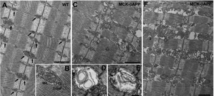
In WT EDL muscle fibers, mitochondria were typically round or slightly oval in shape and located at the I band, on both sides of the Z-line (Fig. 1A, arrows). In this position, the mito-chondria are specifically placed next to Ca2⫹release units or
triads (Fig. 1B). This is similar to what has been previously reported for healthy muscle (30). The majority of mitochondria in WT fibers presented a healthy appearance that was charac-terized by electron-dense matrix, well organized internal cris-tae, consistent morphology, and an intact outer membrane (as in Fig. 1B). Conversely, muscle from MCK-APP animals con-tained numerous fibers with abnormal appearance and many damaged mitochondria (Fig. 1, C and F). This was rarely observed in WT fibers. The abnormal mitochondria appeared
FIGURE 1. EDL muscle fibers from MCK-APP mice showed severe mitochondrial alterations. A and B, in WT EDL fibers from 2–3-month-old animals (n ⫽ 3), mitochondria were primarily located in correspondence to the I band, forming two rows on both sides of each Z line (A, arrows). Mitochondria (Mit.) were located in the close proximity of Ca2⫹release units or triads (B) and presented a healthy appearance (see “Results” for more detail). C–E, muscle fibers from 2–3-month-old MCK-APP mice (n ⫽ 3) contained a large number of mitochondria with severely altered morphology (open arrows). These mitochondria were usually swollen and contained vacuoles, myelin figures, or lamellar structures surrounded by an almost completely clear matrix (D and E). F, in some fibers mitochondria were abnormally enlarged and formed columns or clusters between myofibrils and/or under the sarcolemma. Bars, A, C, and F, 1m; B, D, and E, 0.2m.
swollen and variable in size and shape (Fig. 1, C–F, open
arrows). Other changes included alterations in the internal cris-tae that appeared fragmented or, at times, completely missing. In addition, the mitochondrial matrix was translucent and devoid of electron density (Fig. 1F). In several instances, mito-chondria contained vacuoles, myelin figures, or lamellar struc-tures surrounded by an almost completely clear matrix (Fig. 1,
Dand E). Finally, in the more severely affected organelles, the outer membrane was also frequently disrupted. Similar altera-tions in mitochondrial structure have been described in several human diseases and pathological mouse models (30 –32).
Large Areas of Severely Degenerated Mitochondria and SR Membrane Generate Regions of Vacuolization—In some MCK-APP, but never in WT fibers, structurally compromised and degenerating mitochondria formed large accumulations (Fig. 2A, open arrows) that were found either just beneath the sarco-lemma or deeper in the cell, between the myofibrils. In these areas, there were also alterations in the SR membranes, which appeared fragmented and compromised (Fig. 2C, arrows). In these MCK-APP fibers, areas with large accumulations of damaged mitochondria coexisted with apparently empty vacu-oles (Fig. 2, B–D, asterisks). These vacuvacu-oles closely resemble modifications characteristic of IBM and may represent an early
event in the formation of larger vacuoles observed in this dis-ease (33).
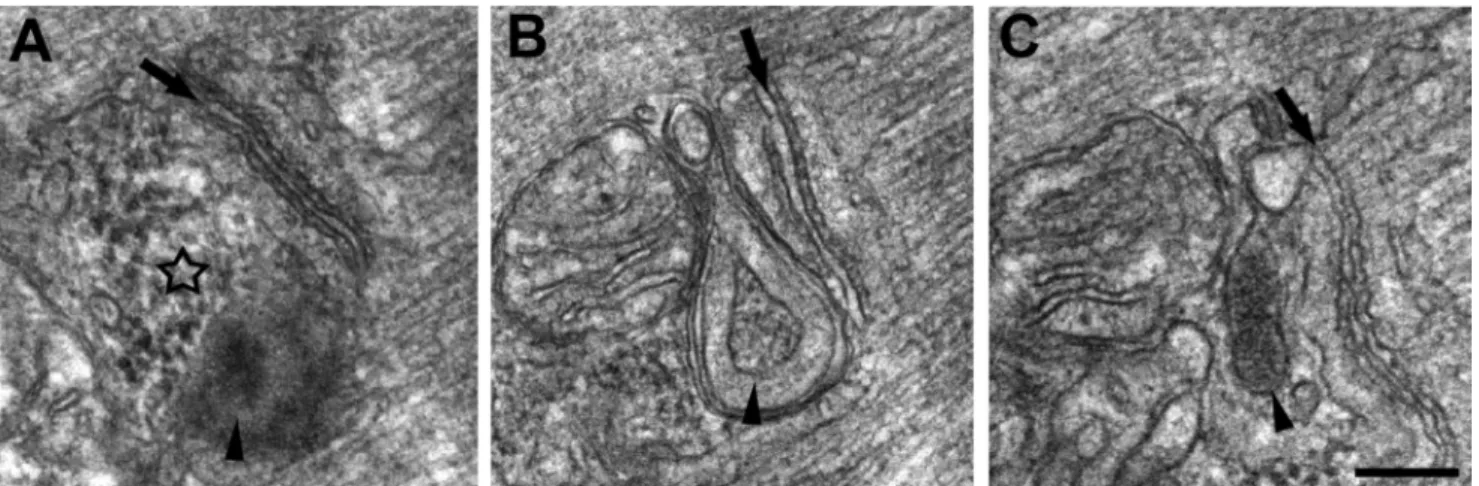
Accumulation of Amorphous Electron-dense Material and Formation of Membrane-bound Granular Inclusions—The regions of the cell characterized by accumulation of degenerat-ing mitochondria and vacuolization (Fig. 3, open arrows) also contained accumulations of amorphous electron-dense mate-rial (Fig. 3, asterisk) that was granular in appearance (Figs. 3, B and C, and 4A). There were instances where this material appeared with (Fig. 3C, arrowhead) and without (Fig. 3C,
aster-isk) a membrane envelope. These granular inclusions were also frequently seen in close proximity to mitochondria and triads and in some instances between them (Fig. 4, B and C).
Altered TCA Cycle in A-affected Muscle—To correlate the
effects of intramyofiber accumulation of A with potential alterations in the mitochondrial TCA cycle activity, we per-formed parallel ELISA and high frequency [1H-decoupled]13C
NMR spectroscopy with the muscle tissue from the 2-month-old animals. Because MCK-APP mice are engineered to express human APP transgene, we performed ELISA specifi-cally for human A1– 40and A1– 42to determine whether toxic
A fragments are present at an early age in the muscle of MCK-APP mice. No differences in the levels of A1– 42 were FIGURE 2. Accumulation of degenerated mitochondria and SR membranes produce areas of vacuolization. A, in some instances, damaged mitochondria formed large clusters that were located either under the sarcolemma or between myofibrils (open arrows). B–D, mitochondrial degeneration was often associated with SR membrane fragmentation (C, arrows) and with formation of different size vacuoles (asterisks). There were also frequent accumulation of stack-like aggregations of the SR membranes (wavy arrow). Bars, A, 2m; B–D, 1 m.
detected between the genotypes. However, the levels of A1– 40
were significantly greater in MCK-APP compared with con-trol mice (Fig. 5A). To determine whether there is a correlation between A accumulation and mitochondrial activity at this early age, mice were injected with [13C]pyruvate, and muscle tissue was collected 45 min after injection. With this approach,
13C served as a label for succinate, a TCA cycle intermediate.
NMR analysis of 5% perchloric acid muscle extracts showed a 3-fold reduction in the level of13C labeling of succinate C2/C3
(Fig. 5B) and a 4-fold reduction in its total pool size (Fig. 5C), indicating a diminished activity of the mitochondrial TCA cycle. To further investigate the possibility of reduced TCA
cycle activity, we determined the levels of glutamate, a by-prod-uct of the TCA cycle metabolism. Our results revealed a signif-icant decrease of13C flux into glutamate C4 (33%) and C2 (27%)
consistent with a decrease (34%) in total glutamate levels. This further indicates that TCA cycle metabolism is compromised at an early age in the muscle of MCK-APP mice. Because the reduction in mitochondrial metabolism suggests a possible switch to anaerobic metabolism, which is expected to lead to increased accumulation of lactic acid, we also compared the levels of lactate between control and A-affected muscle. Flux of the13C label into lactate C3 was significantly increased (94%)
in MCK-APP compared with control mice. This was
congru-FIGURE 3. Accumulation of amorphous materials and membrane bound inclusions. A, regions of the cell characterized by accumulation of degenerating mitochondria and vacuolization (open arrows) also contained accumulations of amorphous electron-dense material (asterisks). B and C, the electron-dense material was granular in appearance and was observed either with (C, arrowhead) or without (C, asterisk) a membrane envelope. In certain MCK-APP fibers, stack-like aggregations of the SR membranes were also observed (wavy arrows). Bars, A, 1m; B and C, 0.5 m.
FIGURE 4. Accumulation of amorphous electron-dense material and formation of membrane-bound granular inclusions in proximity of triads and
mitochondria of MCK-APP muscle fibers. Electron-dense bodies of granular electron-dense material were found in MCK-APP muscle fibers, often in
proximity of triads and mitochondria. Arrows mark the central elements of triads, the T-tubule, whereas the star in A points to an accumulation of electron-dense material, which is not yet as electron-dense and not enclosed in a membrane as those indicated by arrowheads in B and C. Bar, A–C, 0.2m.
ent with an increase (70%) in total lactate levels and further indicates an impairment of mitochondrial TCA cycle activity concurrent with the intracellular accumulation of A.
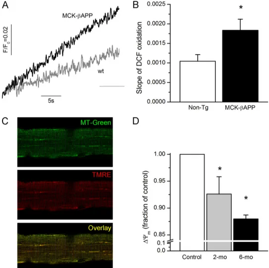
Increased ROS Production and Partial Loss of⌬⌿min
-Am-yloid Affected Muscle Cells—Because cytoplasmic [Ca2⫹] ([Ca2⫹]
i) is augmented in MCK-APP muscle cells (17, 29), a
consequent progressive increase in mitochondrial Ca2⫹
accu-mulation could result in increased production of ROS as well as increased frequency of PT pore opening ultimately, leading to a loss of the mitochondrial membrane potential (⌬⌿m).
To compare the ROS production between healthy and A-affected muscle, enzymatically dissociated FDB fibers from 2-month-old WT and MCK-APP mice were loaded with the ROS probe, 5-(and 6-)chloromethyl-2 ⬘,7⬘-dichlorodihydro-fluorescein diacetate. Upon cleavage of the acetate groups by intracellular esterases and oxidation, the nonfluorescent 2 ⬘,7⬘-dichlorodihydrofluorescein diacetate is converted to the highly fluorescent 2⬘,7⬘-dichlorofluorescein (DCF). As demonstrated
in Fig. 6, the rate of DCF oxidation was much greater in resting MCK-APP fibers compared with the WT cells, suggesting that A-producing cells were generating a greater amount of ROS. Mitochondrial membrane potential was monitored in iso-lated FDB fibers loaded with TMRE, a fluorescent indicator whose accumulation in mitochondria is driven by ⌬⌿m. For mitochondrial localization, fibers were simultaneously loaded with MitoTracker Green FM (MTG), which accumulates within mitochondria regardless of⌬⌿m. The distribution pat-terns for MitoTracker Green and TMRE were uniform across the genotypes (Fig. 6C). However, further analysis of TMRE fluorescence within individual mitochondria showed that MCK-APP cells contained a large population of mitochondria that exhibited diminished TMRE fluorescence (Fig. 6D) sug-gesting that these organelles were partially depolarized. The partial loss of⌬⌿mappears to be an age-dependent
phenome-non because muscle cells from older animals (6 months old), which exhibit greater dysregulation of cytoplasmic Ca2⫹(17), contained an even larger fraction of depolarized mitochondria.
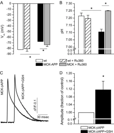
Acidification of Cytoplasm and Plasma Membrane Depolar-ization—The increased level of lactate shown in Fig. 5 is expected to cause acidification of the cytoplasm. The combination of acidic pH and increased ROS production can lead to excessive lipid peroxidation and subsequent depolarization of the plasma membrane. Our previous results demonstrate that intramyofi-ber accumulation of A results in partial depolarization of the plasma membrane (17, 29). Using ion selective microelec-trodes, we simultaneously measured pH and Vmin WT and
MCK-APP muscle fibers (Fig. 7). Consistent with previous observations, MCK-APP myofibers exhibited a partially depolarized plasma membrane in comparison with the WT fibers (Fig. 7A). The pH in the same cells was also more acidic compared with the controls (Fig. 7B). To determine whether these phenomena were driven by alterations in the mitochon-drial Ca2⫹ homeostasis, the cells were treated for 24 h with Ru360 (1 nM), a highly selective blocker of the mitochondrial
Ca2⫹uniporter (34). These treatments resulted in partial rever-sal of changes in Vm(Fig. 7A) and complete reversal of changes in pH (Fig. 7B) in MCK-APP cells without affecting these parameters in the control cells. These results suggest that func-tional modifications of mitochondria and subsequent cytoplas-mic alterations are potentially mediated by Ca2⫹ transport through the mitochondrial Ca2⫹ uniporter and subsequent mitochondrial Ca2⫹overload.
Because ryanodine receptor Ca2⫹ release channels (RyR) contain reactive thiol residues, the functional state of these channels is modulated by the cellular redox state. Based on these factors, it is feasible that alterations in depolarization evoked Ca2⫹release previously reported in MCK-APP muscle
(17, 29) arise, in part, from altered redox state via augmented mitochondrial ROS production and decreased cytoplasmic pH. To assess whether oxidation of RyRs is responsible for reduc-tion in Ca2⫹release from the SR, MCK-APP cells were treated with glutathione (GSH), a potent antioxidant. A 24-h treatment with GSH (5 mM) increased the peak amplitude of single square
electrical pulse-elicited Ca2⫹release in the MCK-APP muscle fibers (Fig. 7, C and D) without affecting the peak amplitudes of Ca2⫹transients in the WT cells (not shown). Together, these
FIGURE 5. Increased levels of A1– 40and altered TCA cycle metabolism in
MCK-APP muscle. A, ELISA measurement of the levels of human A1– 42and
A1– 40in hamstring muscle extracts from MCK-APP mice in 30% formic acid.
B, metabolic flux of13C label from 1-[13C]pyruvate into isotopomers of lactate
at C3, glutamate at C4 and C2, and succinate at C2/C3 as measured by [1
H-decoupled]13C NMR spectroscopy. C, total metabolite pool size of lactate,
glutamate, and succinate as measured by [13C-decoupled]1H NMR
results demonstrate that alterations in Ca2⫹release in MCK-APP muscle were partly due to oxidation of RyRs.
DISCUSSION
The main goal of the current study was to determine the effects of intracellular A accumulation on mitochondrial structure and function in skeletal muscle. Our results demon-strate that severe structural alterations in mitochondria and other subsarcolemmal organelles, as well as disruption of TCA cycle activity, coincide with accumulation of A in skeletal muscle. This study utilized MCK-APP transgenic mice that accumulate A specifically in skeletal muscle and reportedly exhibit age-dependent appearance of numerous features anal-ogous to those observed in IBM such as intracellular inclusions, angulated fibers, inflammation, and deficits in motor perform-ance. However, none of the myopathic features have been pre-viously reported in the MCK-APP mice at ages chosen for this study (15). We specifically chose to study young MCK-APP mice to explore the effects of the A prior to the appearance of IBM disease features.
The alterations in mitochondrial structure and function were likely caused by the initial overexpression of the APP and
accu-mulation of its proteolytic fragments. Our results point out that at the current stage of the disease progression, MCK-APP muscle predominantly accumulates the A1– 40 species of -amyloid rather than A1– 42, which is considered to be the
more toxic species. Although the specific effects of these pep-tides on mitochondria are still under investigation, A has been demonstrated to accumulate into this organelle and destabilize its membrane properties (35, 36). Deleterious effects of A on the mitochondrial function include reduced cytochrome c oxi-dase activity (35, 37) caused by inhibition of its gene expression (37), decreased respiration and ATP synthesis, and cytochrome
crelease (35, 38).
Despite the existence of many fibers presenting normal ultra-structure in the MCK-APP muscle, a significant number of fibers showed compromised mitochondrial apparatus. These alterations are exemplified by a large number of mitochondria with altered morphological internal cristae, suggesting that these organelles were at varying stages of degeneration. Fur-thermore, these degenerated mitochondria often accumulated in large clusters under the sarcolemma or, less frequently, between myofibrils. Interestingly, under high magnification,
FIGURE 6. Increased rate of ROS production and reduced⌬⌿min MCK-APP muscle fibers. A, temporal change in DCF fluorescence in WT and MCK-APP muscle fibers. B, rate of change (slope) in DCF fluorescence in WT (n⫽ 14) and MCK-APP (n ⫽ 16) muscle fibers. C, confocal images of FDB muscle fibers loaded with MitoTracker Green (MT-Green, top panel) and TMRE (middle panel). The bottom panel presents a merged image of MitoTracker Green and TMRE fluores-cence. D, mean intensity of TMRE fluorescence of individual mitochondria presented as fraction of control in 2-month-old (2-mo) WT (nmito⫽ 626) and
MCK-APP (nmito⫽ 796) and 6-month-old (6-mo) WT (nmito⫽ 1127) and MCK-APP (nmito⫽ 1288) muscle fibers. *, p ⬍ 0.05 (t test).
Mitochondrial Dysregulation by
-Amyloid in Skeletal Muscle
areas of mitochondrial clusters appear to be analogous to what has been described in vacuolated fibers of IBM patients (39). In these areas, there are several additional characteristic features of A-accumulating fibers that include the presence of what appear to be autophagic vacuoles, myelin bodies, membrane whorls, and granular and filamentous membrane-bound inclu-sions. The EM data also show mitochondrial impairment and ultrastructural modifications that are consistent with previous findings, which demonstrated alterations in lysosomal enzymes and up-regulation of autophagic activity in IBM and cultured human muscle fibers, thus collectively suggesting that IBM muscle may have autophagic defects (40). The results presented here point to a possibility that the mitochondrial impairments and ultrastructural modifications could be mediated by autophagic removal of defective mitochondria via mitophagy in MCK-APP muscle. In addition to mitochondrial alterations, we also observed degeneration of the SR membranes, which could contribute to the previously described impairment in the excitation-contraction coupling mechanism (17, 29). All of the above described alterations in the MCK-APP muscle are likely
to have a negative impact on muscle contractility and may con-tribute to the development of muscle weakness.
The disruption of mitochondrial ultrastucture can poten-tially account for the diminished TCA cycle activity and the reduced⌬⌿mthat were observed in MCK-APP muscle.
Mito-chondrial abnormalities have been described in IBM (12, 13). Similarly, mitochondrial alterations are found in a mouse model for amyotrophic lateral sclerosis (32) and in core-like myopathies such as the malignant hyperthermia (MH) (41). Interestingly, in each case, these alterations have been associ-ated with a high level of oxidative stress.
In MCK-APP mice, the functional consequences of altered mitochondrial activity is manifested by the reduced production of the TCA cycle intermediates and by-products, increased ROS generation, and increased lactic acid production, which at least partially contributed to a more acidic cytoplasm. The increase in the lactic acid accumulation reflects the fact that there is a shift in the equilibrium between the aerobic and the anaerobic glucose metabolism. The reduced TCA cycle activity is expected to lead to a reduced mitochondrial generation of ATP. However, because MCK-APP animals do not exhibit muscle weakness at this age (16), it seems that a potential reduction in ATP levels at this early stage of the disease pro-gression is not sufficient to appreciably affect ATP-dependent processes. Nevertheless, the progressive nature of A accumu-lation will likely lead to greater mitochondrial dysfunction that could ultimately result in a substantial reduction of cellular ATP levels, thus influencing the muscle strength and other ATP-dependent signaling pathways.
The acidification of the cytoplasm and enhanced ROS pro-duction in resting A-affected myofibers could further result in altered cellular Ca2⫹handling. Oxidation of the reactive thiols of the RyR1 is known to increase channel activity. This is partly due to altered sensitivity of the channel to ATP, Ca2⫹, and Mg2⫹and modified interactions of RyRs with the auxiliary pro-teins (42, 43). Our previous investigation has shown that A can also directly increase RyR channel activity (29). The current results suggest that oxidation of the RyRs may be partially responsible for the reduced SR Ca2⫹release in the MCK-APP
muscle. An acidic cytoplasm may affect the rate of Ca2⫹uptake into SR by reducing the affinity of the SR Ca2⫹ ATPase
(SERCA) pump to Ca2⫹(44, 45). Although we did not observe any changes in Ca2⫹reuptake parameters (not shown), which in part reflect Ca2⫹uptake by the SERCA, in the mice at ages
chosen for this study, the rate of Ca2⫹ uptake is significantly slower in the older MCK-APP mice (29). Thus, a cumulative effect of RyR oxidation, enhanced RyR channel activity caused by modulation by A and reduced activity of the SERCA, could be responsible for increased [Ca2⫹]
i at rest and diminished
excitation-contraction coupling in A affected cells in the later stages of disease progression.
The precise mechanism of the functional interplay between A accumulation, mitochondrial dysfunction, and altered Ca2⫹remains to be determined. However, based on the
cur-rently available information, we put forth the following working model for the effects of A accumulation on mitochondrial function, intracellular Ca2⫹ handling, and development of
muscle weakness in IBM (Fig. 8). Studies have shown that A
FIGURE 7. Alterations in Vmand pH in MCK-APP muscle fibers. A, record-ing of restrecord-ing membrane potential (Vm) with microelectrodes in 2-month-old
WT (n⫽ 3) and MCK-APP (n ⫽ 4) muscle fibers before and after 24 h of treatment with Ru360 (1 nM). MCK-APP cells exhibited depolarized plasma membrane. This alteration could be partially reversed by Ru360 treatment.
B, recording of intracellular pH with microelectrodes in the same WT and
MCK-APP muscle fibers as in A before and after 24 h of treatment with Ru360 (1 nM). MCK-APP cell exhibited reduced pH compared with WT cells. Treat-ment with Ru360 had no effect on the WT cell but increased pH to near normal level. C, mean MagFluo-4 fluorescence transient from untreated (n⫽ 17) and GSH-treated (n⫽ 21, 5 mMfor 24 h) MCK-APP muscle fibers. Ca2⫹transients were elicited by 1-ms single square electrical pulses. D, peak amplitude of Ca2⫹release in untreated and GSH treated MCK-APP muscle fibers. GSH treatment increased peak amplitudes of Ca2⫹transients in DJ-1 null cells. *,
accumulations can lead to modulation of L-type Ca2⫹channel
properties (46, 47). Whether these effects result from direct modification of these channels by A or whether this modula-tion is indirect remains to be determined. There is recent evi-dence that A can interact with the  subunit of CaV1.2 and
CaV1.3 L-type Ca
2⫹channels (48). Additional studies need to
be carried out to determine whether this interaction takes place between A and CaV1.1 in the skeletal muscle. Studies have shown that A-accumulating cells exhibit a nifedipine-sensi-tive augmentation of the cytoplasmic [Ca2⫹] in muscle (17) and neurons (49), thus suggesting a contribution of L-type Ca2⫹
channels to changes in cytoplasmic Ca2⫹handling.
It has been demonstrated that both the extracellular and the intracellular compartments contribute to the augmented [Ca2⫹] in MCK-APP muscle (17). It is likely that RyR-mediated Ca2⫹efflux could be a major intracellular contributor to this
phenomenon. Direct binding of A to RyR has not been dem-onstrated; however, previous investigations have shown that A can effectively modulate RyR function when these channels are reconstituted in the planar lipid bilayers and in SR vesicles prepared from the skeletal muscle (29).
Concurrently with alterations in the cytoplasmic [Ca2⫹], A
can accumulate within the mitochondria (35, 36) and possibly lower the threshold for activation of the mitochondrial Ca2⫹
transporters, such as the Ca2⫹uniporter, and lead to
mitochon-drial Ca2⫹ overload. Effects of mitochondrial Ca2⫹ overload include, among others, swelling of the mitochondria and acti-vation of the mitochondrial permeability transition pore
com-plex that leads to the loss of the ⌬⌿m and a burst of ROS.
Similar features are observed in MCK-APP muscle and are presented in this study. The increased mitochondrial permea-bility transition pore complex opening could affect the plasma membrane integrity,which is manifested by the lipid peroxida-tion and depolarizaperoxida-tion of plasmalemma. Increased ROS pro-duction could also enhance Ca2⫹leak from the SR. The
dimin-ished cellular ATP levels that likely arise from mitochondrial impairment can later lead to a reduction of the stimulated SR Ca2⫹release and affect Ca2⫹sequestration by the SR after
mus-cle contraction. Based on this hypothetical model, it is conceiv-able that A can be a catalyst that initiates a vicious cycle whereby disrupted Ca2⫹homeostasis exerts a negative effect on the mitochondrial function and vice versa in the pathogenesis of IBM.
In summary, the present results demonstrate that A accu-mulation within skeletal muscle leads to considerable altera-tions in mitochondrial structure and function that precede other reported aspects of IBM in the MCK-APP transgenic mice. The functional interplay between the disrupted cytoplas-mic Ca2⫹handling and diminished mitochondrial function in A-affected muscle could potentially serve as therapeutic tar-get for the treatment of IBM and other A-mediated disorders. Acknowledgment—We thank the Drug Discovery Unit at Georgetown University for the use of the NMR equipment.
FIGURE 8. Proposed model of-amyloid mediated muscle dysfunction in IBM. Increased expression of APP and accumulation of its proteolytic A fragments affects numerous intracellular regulatory complexes. Modulation of the intracellular Ca2⫹release channels (RyR), dihydropyridine receptors (DHPR) and possibly other surface membrane Ca2⫹channels leads to Ca2⫹efflux and influx, respectively, which contribute to augmentation of the resting cytoplasmic Ca2⫹concentration ([Ca2⫹]i). A accumulation also leads to decreased Ca2⫹release by RyRs. Augmented [Ca2⫹]itogether with modification of mitochondrial function by A lead to enhanced Ca2⫹flux into the mitochondrial matrix and subsequent mitochondrial Ca2⫹overload. Mitochondrial Ca2⫹overload
promotes enhanced generation of ROS, mitochondrial permeability transition pore complex opening, and disruption of⌬⌿m. Diminished⌬⌿mresults in
inefficient TCA cycle and reduction in mitochondrial ATP production. Increased ROS production in combination with acidic cytoplasm promotes lipid peroxi-dation, further increase in [Ca2⫹]i, and reduction in Ca2⫹release from the SR through modulation of the RyRs. All of these events combine to lead to diminished muscle contractility. Solid arrows represent observed, whereas dashed arrows represent unconfirmed effects of A.
REFERENCES
1. Askanas, V., Serratrice, G., and Engel, W. K. (1998) Inclusion-Body
Myo-sitis and Myopathies, Cambridge University Press, Cambridge
2. Askanas, V., Alvarez, R. B., and Engel, W. K. (1993)-Amyloid precursor epitopes in muscle fibers of inclusion body myositis. Ann. Neurol. 34, 551–560
3. Askanas, V., Serdaroglu, P., Engel, W. K., and Alvarez, R. B. (1991) Immu-nolocalization of ubiquitin in muscle biopsies of patients with inclusion body myositis and oculopharyngeal muscular dystrophy. Neurosci. Lett.
130,73–76
4. Askanas, V., Engel, W. K., Bilak, M., Alvarez, R. B., and Selkoe, D. J. (1994) Twisted tubulofilaments of inclusion body myositis muscle resemble paired helical filaments of Alzheimer brain and contain hyperphosphory-lated tau. Am. J. Pathol. 144, 177–187
5. Fidzian´ska, A., and Glinka, Z. (2006) Rimmed vacuoles with-amyloid and Tau protein deposits in the muscle of children with hereditary myop-athy. Acta Neuropathol. 112, 185–193
6. Schmidt, J., Barthel, K., Wrede, A., Salajegheh, M., Bähr, M., and Dalakas, M. C. (2008) Interrelation of inflammation and APP in sIBM. IL-1 in-duces accumulation of -amyloid in skeletal muscle. Brain 131, 1228 –1240
7. Vattemi, G., Nogalska, A., King Engel, W., D’Agostino, C., Checler, F., and Askanas, V. (2009) Amyloid-42 is preferentially accumulated in muscle fibers of patients with sporadic inclusion-body myositis. Acta
Neuro-pathol. 117,569 –574
8. Nogalska, A., D’Agostino, C., Engel, W. K., Klein, W. L., and Askanas, V. (2010) Novel demonstration of amyloid- oligomers in sporadic inclu-sion-body myositis muscle fibers. Acta Neuropathol. 120, 661– 666 9. Askanas, V., Engel, W. K., and Alvarez, R. B. (1992) Light and electron
microscopic localization of-amyloid protein in muscle biopsies of pa-tients with inclusion-body myositis. Am. J. Pathol. 141, 31–36
10. Askanas, V., McFerrin, J., Alvarez, R. B., Baqué, S., and Engel, W. K. (1997) APP gene transfer into cultured human muscle induces inclusion-body myositis aspects. Neuroreport 8, 2155–2158
11. Askanas, V., McFerrin, J., Baqué, S., Alvarez, R. B., Sarkozi, E., and Engel, W. K. (1996) Transfer of-amyloid precursor protein gene using adeno-virus vector causes mitochondrial abnormalities in cultured normal hu-man muscle. Proc. Natl. Acad. Sci. U.S.A. 93, 1314 –1319
12. Oldfors, A., Moslemi, A. R., Jonasson, L., Ohlsson, M., Kollberg, G., and Lindberg, C. (2006) Mitochondrial abnormalities in inclusion-body myo-sitis. Neurology 66, S49 –55
13. Horvath, R., Fu, K., Johns, T., Genge, A., Karpati, G., and Shoubridge, E. A. (1998) Characterization of the mitochondrial DNA abnormalities in the skeletal muscle of patients with inclusion body myositis. J. Neuropathol.
Exp. Neurol. 57,396 – 403
14. Sugarman, M. C., Kitazawa, M., Baker, M., Caiozzo, V. J., Querfurth, H. W., and LaFerla, F. M. (2006) Pathogenic accumulation of APP in fast twitch muscle of IBM patients and a transgenic model. Neurobiol. Aging
27,423– 432
15. Sugarman, M. C., Yamasaki, T. R., Oddo, S., Echegoyen, J. C., Murphy, M. P., Golde, T. E., Jannatipour, M., Leissring, M. A., and LaFerla, F. M. (2002) Inclusion body myositis-like phenotype induced by transgenic overexpression ofAPP in skeletal muscle. Proc. Natl. Acad. Sci. U.S.A.
99,6334 – 6339
16. Moussa, C. E., Fu, Q., Kumar, P., Shtifman, A., Lopez, J. R., Allen, P. D., LaFerla, F., Weinberg, D., Magrane, J., Aprahamian, T., Walsh, K., Rosen, K. M., and Querfurth, H. W. (2006) Transgenic expression of-APP in fast-twitch skeletal muscle leads to calcium dyshomeostasis and IBM-like pathology. FASEB J. 20, 2165–2167
17. Lopez, J. R., and Shtifman, A. (2010) Intracellular-amyloid accumulation leads to age-dependent progression of Ca2⫹dysregulation in skeletal mus-cle. Muscle Nerve 42, 731–738
18. Brookes, P. S., Yoon, Y., Robotham, J. L., Anders, M. W., and Sheu, S. S. (2004) Calcium, ATP, and ROS. A mitochondrial love-hate triangle. Am. J.
Physiol. Cell Physiol. 287,C817–C833
19. Denton, R. M., and McCormack, J. G. (1985) Physiological role of Ca2⫹ transport by mitochondria. Nature 315, 635
20. Denton, R. M., and McCormack, J. G. (1986) The calcium sensitive dehy-drogenases of vertebrate mitochondria. Cell Calcium 7, 377–386 21. Wang, W., Fang, H., Groom, L., Cheng, A., Zhang, W., Liu, J., Wang, X., Li,
K., Han, P., Zheng, M., Yin, J., Wang, W., Mattson, M. P., Kao, J. P., Lakatta, E. G., Sheu, S. S., Ouyang, K., Chen, J., Dirksen, R. T., and Cheng, H. (2008) Superoxide flashes in single mitochondria. Cell 134, 279 –290
22. Chen, Q., Chai, Y. C., Mazumder, S., Jiang, C., Macklis, R. M., Chisolm, G. M., and Almasan, A. (2003) The late increase in intracellular free radical oxygen species during apoptosis is associated with cytochrome c release, caspase activation, and mitochondrial dysfunction. Cell Death Differ 10, 323–334
23. Casley, C. S., Canevari, L., Land, J. M., Clark, J. B., and Sharpe, M. A. (2002) -Amyloid inhibits integrated mitochondrial respiration and key enzyme activities. J. Neurochem. 80, 91–100
24. Casley, C. S., Land, J. M., Sharpe, M. A., Clark, J. B., Duchen, M. R., and Canevari, L. (2002)-Amyloid fragment 25–35 causes mitochondrial dys-function in primary cortical neurons. Neurobiol. Dis. 10, 258 –267 25. Eckert, A., Hauptmann, S., Scherping, I., Rhein, V., Müller-Spahn, F.,
Götz, J., and Müller, W. E. (2008) Soluble-amyloid leads to mitochon-drial defects in amyloid precursor protein and Tau transgenic mice.
Neu-rodegener Dis. 5,157–159
26. Grant, S. M., Shankar, S. L., Chalmers-Redman, R. M., Tatton, W. G., Szyf, M., and Cuello, A. C. (1999) Mitochondrial abnormalities in neuroecto-dermal cells stably expressing human amyloid precursor protein (hAPP751). Neuroreport 10, 41– 46
27. Keil, U., Bonert, A., Marques, C. A., Scherping, I., Weyermann, J., Stro-sznajder, J. B., Müller-Spahn, F., Haass, C., Czech, C., Pradier, L., Müller, W. E., and Eckert, A. (2004) Amyloid-induced changes in nitric oxide production and mitochondrial activity lead to apoptosis. J. Biol. Chem.
279,50310 –50320
28. Pereira, C., Santos, M. S., and Oliveira, C. (1998) Mitochondrial function impairment induced by amyloid-peptide on PC12 cells. Neuroreport 9, 1749 –1755
29. Shtifman, A., Ward, C. W., Laver, D. R., Bannister, M. L., Lopez, J. R., Kitazawa, M., LaFerla, F. M., Ikemoto, N., and Querfurth, H. W. (2010) Amyloid- protein impairs Ca2⫹release and contractility in skeletal mus-cle. Neurobiol. Aging 31, 2080 –2090
30. Boncompagni, S., Rossi, A. E., Micaroni, M., Beznoussenko, G. V., Polish-chuk, R. S., Dirksen, R. T., and Protasi, F. (2009) Mitochondria are linked to calcium stores in striated muscle by developmentally regulated tether-ing structures. Mol. Biol. Cell 20, 1058 –1067
31. Chung, M. J., and Suh, Y. L. (2002) Ultrastructural changes of mitochon-dria in the skeletal muscle of patients with amyotrophic lateral sclerosis.
Ultrastruct. Pathol. 26,3–7
32. Dobrowolny, G., Aucello, M., Rizzuto, E., Beccafico, S., Mammucari, C., Bonconpagni, S., Belia, S., Wannenes, F., Nicoletti, C., Del Prete, Z., Rosenthal, N., Molinaro, M., Protasi, F., Fanò, G., Sandri, M., and Musarò, A. (2008) Skeletal muscle is a primary target of SOD1G93A-mediated toxicity. Cell Metab. 8, 425– 436
33. Yunis, E. J., and Samaha, F. J. (1971) Inclusion body myositis. Lab. Invest.
25,240 –248
34. Matlib, M. A., Zhou, Z., Knight, S., Ahmed, S., Choi, K. M., Krause-Bauer, J., Phillips, R., Altschuld, R., Katsube, Y., Sperelakis, N., and Bers, D. M. (1998) Oxygen-bridged dinuclear ruthenium amine complex specifically inhibits Ca2⫹uptake into mitochondria in vitro and in situ in single car-diac myocytes. J. Biol. Chem. 273, 10223–10231
35. Aleardi, A. M., Benard, G., Augereau, O., Malgat, M., Talbot, J. C., Mazat, J. P., Letellier, T., Dachary-Prigent, J., Solaini, G. C., and Rossignol, R. (2005) Gradual alteration of mitochondrial structure and function by -amyloids. Importance of membrane viscosity changes, energy depriva-tion, reactive oxygen species producdepriva-tion, and cytochrome c release. J.
Bio-energ. Biomembr. 37,207–225
36. Khalifat, N., Puff, N., Dliaa, M., and Angelova, M. I. (2012) Amyloid- and the failure to form mitochondrial cristae: a biomimetic study involving artificial membranes. J. Alzheimers Dis. 28, 33– 48
37. Hong, W. K., Han, E. H., Kim, D. G., Ahn, J. Y., Park, J. S., and Han, B. G. (2007) Amyloid--peptide reduces the expression level of mitochondrial cytochrome oxidase subunits. Neurochem. Res. 32, 1483–1488
38. Rosen, K. M., Veereshwarayya, V., Moussa, C. E., Fu, Q., Goldberg, M. S., Schlossmacher, M. G., Shen, J., and Querfurth, H. W. (2006) Parkin pro-tects against mitochondrial toxins and-amyloid accumulation in skeletal muscle cells. J. Biol. Chem. 281, 12809 –12816
39. Askanas, V., Engel, W. K., and Nogalska, A. (2009) Inclusion body myosi-tis. A degenerative muscle disease associated with intra-muscle fiber multi-protein aggregates, proteasome inhibition, endoplasmic reticulum stress and decreased lysosomal degradation. Brain Pathol. 19, 493–506 40. Nogalska, A., D’Agostino, C., Terracciano, C., Engel, W. K., and Askanas,
V. (2010) Impaired autophagy in sporadic inclusion-body myositis and in endoplasmic reticulum stress-provoked cultured human muscle fibers.
Am. J. Pathol. 177,1377–1387
41. Boncompagni, S., Rossi, A. E., Micaroni, M., Hamilton, S. L., Dirksen, R. T., Franzini-Armstrong, C., and Protasi, F. (2009) Characterization and tem-poral development of cores in a mouse model of malignant hyperthermia.
Proc. Natl. Acad. Sci. U.S.A. 106,21996 –22001
42. Zissimopoulos, S., Docrat, N., and Lai, F. A. (2007) Redox sensitivity of the ryanodine receptor interaction with FK506-binding protein. J. Biol. Chem.
282,6976 – 6983
43. Zissimopoulos, S., and Lai, F. A. (2006) Redox regulation of the ryanodine receptor/calcium release channel. Biochem. Soc. Trans. 34, 919 –921 44. Allen, D. G., Lännergren, J., and Westerblad, H. (1995) Muscle cell
func-tion during prolonged activity. Cellular mechanisms of fatigue. Exp.
Physiol. 80,497–527
45. Wolosker, H., Rocha, J. B., Engelender, S., Panizzutti, R., De Miranda, J., and de Meis, L. (1997) Sarco/endoplasmic reticulum Ca2⫹-ATPase iso-forms. Diverse responses to acidosis. Biochem. J. 321, 545–550
46. Webster, N. J., Ramsden, M., Boyle, J. P., Pearson, H. A., and Peers, C. (2006) Amyloid peptides mediate hypoxic increase of L-type Ca2⫹ chan-nels in central neurones. Neurobiol. Aging 27, 439 – 445
47. Scragg, J. L., Fearon, I. M., Boyle, J. P., Ball, S. G., Varadi, G., and Peers, C. (2005) Alzheimer’s amyloid peptides mediate hypoxic up-regulation of L-type Ca2⫹channels. FASEB J. 19, 150 –152
48. Kim, S., and Rhim, H. (2011) Effects of amyloid- peptides on voltage-gated L-type CaV1.2 and CaV1.3 Ca2⫹channels. Mol. Cells 32, 289 –294
49. Lopez, J. R., Lyckman, A., Oddo, S., Laferla, F. M., Querfurth, H. W., and Shtifman, A. (2008) Increased intraneuronal resting Ca2⫹in adult Alzhei-mer’s disease mice. J. Neurochem. 105, 262–271