ISSN 2044-4648 DOI: 10.5567/pharmacologia.2016.103.113
Research Article
Antioxidant Activities and Chemical Constituents of Extracts
from Cordyline fruticosa (L.) A. Chev. (Agavaceae) and
Eriobotrya japonica (Thunb) Lindl, (Rosaceae)
1
Romuald Tematio Fouedjou,
2Elvine Pami Nguelefack-Mbuyo,
1Beaudelaire Kemvoufo Ponou,
2
Telesphore Benoit Nguelefack,
3Luciano Barboni and
1Léon Azefack Tapondjou
1Laboratory of Environmental and Applied Chemistry, Department of Chemistry, Faculty of Science, University of Dschang, P.O. Box 67,
Dschang, Cameroon
2Laboratory of Animal Physiology and Phytopharmacology, Department of Animal Biology, Faculty of Sciences, University of Dschang,
P.O. Box 67, Dschang, Cameroon
3School of Science and Technology, Chemistry Division, University of Camerino, Via S. Agostino 1, I-62032 Camerino, Italy
Abstract
Background and Objective: Cordyline fruticosa (Agavaceae) and Eriobotrya japonica (Rosaceae) are two medicinal plants used for the treatment of various diseases such as infections of mammary glands, sore throat and neck pain for the first plant, diabetes, cough, ulcers, protection against oxidative stress and cognitive deficits for the latter. The present study was designed to evaluate the antioxidant activity of the different extracts of these two plants as well as to isolate and identify their chemical constituents. Materials and Methods: The plant extract was prepared by maceration in methanol, compounds were isolated from EtOAc and n-BuOH extracts of the two plants using column chromatography and their structures were determined by means of NMR and MS analysis as well as in comparison with published data. Antioxidant tests (DPPH, ferric reduction antioxidant power and anti-hemolytic) were performed over the MeOH, EtOAc and n-BuOH extracts of the plants. Results: The antioxidant-guided phytochemical investigation of the MeOH extracts of the two plants led to the isolation of twelve compounds identiWed as: Farrerol 1, quercetin helichrysoside 2, apigenin 8-C-$-D-glucopyranoside 3, isoquercitrin 4 and rutin 5 from C. fruticosa, $-sitosterol 6, catechin 7, oleanolic acid 8, lyoniresinol 9, cinchonain IIb 10, lyoniresinol 2-a-O-$-D-xylopyranoside 11 and $-sitosterol-3-O-$-D-glucopyranoside 12 from E. japonica.
Amongst the isolated compounds, the most important antioxidant ones were identified as helichrysoside and rutin from C. fruticosa, catechin, cinchonain IIb, lyoniresinol 2-a-O-$-D-xylopyranoside from E. japonica with EC50 of 8.73, 9.91, 4.11, 3.14 and 10.61 µg mLG1,
respectively. Conclusion: Based on the obtained results, it can be concluded that the high ability to scavenge free radicals, reducing power of Fe3+ and hemolysis activity exerted by extracts of C. fruticosa and E. japonica were due to their high content of phenolic compounds,
thus the structure-activity relationships of the isolated flavonoids were discussed. The results of this study suggest that the extracts from these two plants could serve as potential source of antioxidant compounds.
Key words: Medicinal plant, extracts, phenolic compounds, antioxidant activity
Received: December 14, 2015 Accepted: March 01, 2016 Published: March 15, 2016
Citation: Romuald Tematio Fouedjou, Elvine Pami Nguelefack-Mbuyo, Beaudelaire Kemvoufo Ponou, Telesphore Benoit Nguelefack, Luciano Barboni and Léon Azefack Tapondjou, 2016. Antioxidant activities and chemical constituents of extracts from Cordyline fruticosa (L.) A. Chev. (Agavaceae) and Eriobotrya japonica (Thunb) Lindl, (Rosaceae). Pharmacologia, 7: 103-113.
Corresponding Author: Léon Azefack Tapondjou, Laboratory of Environmental and Applied Chemistry, Department of Chemistry, Faculty of Science, University of Dschang, P.O. Box 67, Dschang, Cameroon Tel: +237-675004826 Fax: +237-233451735
Copyright: © 2016 Romuald Tematio Fouedjou et al. This is an open access article distributed under the terms of the creative commons attribution License, which permits unrestricted use, distribution and reproduction in any medium, provided the original author and source are credited.
INTRODUCTION
The presence of active Reactive Oxygen Species (ROS) in excess of the available antioxidant buffering capacity defines the oxidative stress1. The oxidation of biological
molecules such as DNA, lipids and proteins can be cause by excessive amount of ROS. Therefore, ROS are highly involved in cellular damages and in several human diseases such as cancer, inflammation, atherosclerosis, diabetes, hypertension, neurodegenerative diseases, acute respiratory and pulmonary oedema and cataracts. As such, much attention is being paid to antioxidant substances because it is believed that they can prevent and cure diseases or their subsequent complications related to oxidative stress. Besides, the antioxidant capacity is widely used as a parameter to characterize food, medicinal plants and their bioactive components2.
The genus Cordyline with approximately 20 species having a Southern hemisphere distribution with the greatest diversity concentrated in Australia and New Zealand.
Cordyline fruticosa is a woody plants dont la graine n a qu un seul cotylédon3. The plant is traditionally used for the
treatment of various diseases; the leaves are used as haemostatic4 and to induce abortion5 and the roots are used
for toothache, laryngitis and mammary glands infections6.
Cordyline fruticosa is used in Malaysia to cure cough, bloody cough, dysentery, fever, headache, inflammation of the digestive tract, kidney diseases, bloody urine and also difficulties to urinate7.
The genus Eriobotrya includes about 26 species8,9,
distributed in tropical and sub-tropical Eastern and Southern Asia. The E. japonica is a well-known medicinal plant in Japan and China and its astringent leaves have been used since long time to treat chronic bronchitis, cough, fever and gastroenteric disorders. A decoction of the leaves has also been applied locally to wounds, ulcers and cancers10. Phytochemical studies
have shown that the flowers of E. japonica are rich sources of phenolic compounds and flavonoids11,12.
Currently a variety of synthetic antioxidant supplements are marketed to balance oxidants/antioxidants but unfortunately these substances have suspected toxic effects. Some synthetic antioxidant such as butylated hydroxyanisole, gallic acid esters and butylated hydroxytoluene are suspected to have negative health effects13. Consequently, strict
restrictions on the use of these substances have been implemented in several countries14. For these reasons,
researchers are currently focusing their attention on natural antioxidants, particularly those obtainable from plants. According to Cook and Samman15, the antioxidant activity of
plants is mainly due to the presence of secondary metabolites
from the polyphenol class. In the continuous search of potential antioxidant substances from Cameroonian medicinal plants16-18, a bio-guided investigation was conducted for two
medicinal plants growing in the Western region of Cameroon, namely Cordyline fruticosa and Eriobotrya japonica.
MATERIALS AND METHODS
Plant materials: The leaves of Cordyline fruticosa (L.) A. Chev. (Agavaceae) were collected in Dschang city (west region of Cameroon) in December, 2011. The plant was identified by Mr. J.P. Dondjang, botanist at the Department of Forestry, University of Dschang, under the voucher specimen (Ref: LACAPE 0001) kept in our Laboratory.
The stem barks of Eriobotrya japonica (Thunb) Lindl (Rosaceae) were collected in Bafou village near Dschang (west region of Cameroon) in March, 2013. The plant was identified at the Cameroon National Herbarium, Yaoundé, Cameroon by Mr. Tadjouteu Fulbert where a voucher specimen was deposited under the reference number 44164/HCN.
Extraction and isolation
Preparation of Cordyline fruticosa extracts: The dried and
pulverized leaves of C. fruticosa (3 kg) were extracted three times (each time for 24 h) with MeOH. The combined filtrate was concentrated under reduced pressure to give a dark residue (503 g) which was suspended in distilled water and partitioned successively with n-hexane, EtOAc and n-BuOH, yielding 58, 69 and 60 g of dry extracts, respectively. Part of the EtOAc extract (59 g) was fractionated by silica gel column chromatography using a gradient of EtOAc in n-hexane and then a gradient of MeOH in EtOAc to give ten main fractions (A-J). Part of the n-BuOH extract was also fractionated prior to the purification of different fractions using silica gel column chromatography.
Isolation of compounds from Cordyline fruticosa:
Fraction C (0.5 g) (eluted with Hexane-EtOAc (6-4)) was chromatographed on a silica gel column using hexane-EtOAc (70-30) as eluent to mainly yield compound 1 (27 mg). Fraction F (7.12 g) (eluted with EtOAc) was submitted to silica gel CC eluted with EtOAc-MeOH (98:2) and on sephadex LH-20 CC using MeOH as eluent to give compound 2 (7 mg). Compound 3 (57 mg) was obtained upon recrystallization from fraction G (2.6 g) and the resulting filtrate was chromatographed on silica gel column eluted with EtOAc-MeOH-H2O (97:3:1) and then on sephadex LH-20
Part of the n-BuOH extract (50 g) was fractionated by column chromatography eluted with EtOAc containing increased amounts of MeOH. The fraction obtained by eluting with EtOAc-MeOH (75:25) (9.04 g) was rechromatographed on a silica gel column using EtOAc-MeOH (90:5) as eluent to afford six sub-fractions. One of these sub-fractions (157 mg) was purified on a sephadex LH-20 column eluted with MeOH to yield compound 5 (57 mg).
Preparation of Eriobotrya japonica extract: The dried and
ground stem bark of E. japonica (3 kg) was extracted three times (each time for 24 h) with MeOH. The combined filtrate was concentrated under reduced pressure to give a dark residue (368 g), after removal of the solvent by evaporation, part of this residue (358 g) was suspended in distilled water and partitioned successively with n-hexane, EtOAc and n-BuOH yielding 35.0, 38.5 and 46.5 g of dry extracts, respectively. Part of the EtOAc extract (35 g) was absorbed with silica gel and then fractionated on a silica gel column using a gradient of EtOAc in n-hexane and then a gradient of MeOH in EtOAc, affording twelve fractions (A-L).
Isolation of compounds from Eriobotrya japonica:
Compound 6 (11 mg) was obtained upon recrystallization of fraction C (1.4 g) (eluted with hexane-EtOAc (90:10)). Fraction I (9.7 g) (eluted with EtOAc-MeOH (95:5) was applied on silica gel column with EtOAc as eluent to give 3 sub-fractions (I1, I2 and I3). Sub-fraction I1 (3.2 g) was purified on a silica gel
column with hexane-EtOAc (70:30) as eluent to yield compounds 7 (21 mg) and 8 (51 mg). Similarly, sub-fraction I2 (3.2 g) was subjected to silica gel column eluted with
hexane-EtOAc (80:20) to yield compound 9 (6 mg) and sub-fraction I3 (2.3 g) was purified on a silica gel column
using hexane-EtOAc (70:30) as eluent to yield compound 10 (22 mg). Fraction J (6 g) eluted with EtOAc-MeOH (90:10) was subjected to silica gel CC eluting with EtOAc to give compounds 11 (18 mg) and 12 (27 mg).
Structure elucidation and identification of the isolated compounds: Samples for NMR experiments were dissolved
in deteurated solvents (CD3OD and DMSO-d6) and analyzed
on a varian mercury plus spectrometer (400 MHz for 1H and
100 MHz for 13C). The ESI-MS were carried out on an agilent
Technologie LC/MSD Trap SL (G2445D SL). The CC was performed on silica gel 60 (0.040-0.063 or 0.063-0.200 mm) and sephadex gel LH-20. Fractions were monitored by TLC using Merck pre-coated silica gel sheets (60 F254) and spots
were visualized under UV light (254 and 365 nm) and by spraying with 50% H2SO4 and heating at 110EC.
Farrerol 1: Yellow powder, 13C NMR (CD
3OD, 100 MHz)
δ 78.6 (C-2), 42.7 (C-3), 196.9 (C-4), 157.9 (C-5), 102.5 (C-6), 162.7 (C-7), 101.8 (C-8), 103.3 (C-9), 158.8 (C-10), 130.0 (C-1 ), 127.4 (C-2 ), 114.8 (C-3 ), 157.4 (C-4 ), 114.8 (C-5 ), 127.4 (C-6 ), 6.7 (6-Me), 5.9 (8-Me). The 1H NMR (CD
3OD, 400 MHz)
δ 5.30 (1H, dd, J = 12.9, 3.1 Hz, H-2), 3.05 (1H, dd, J = 17.1, 12.9 Hz, H-3b), 2.73 (1H, dd, J = 17.1, 3.1 Hz, H-3a) 7.32 (2H, d, J = 8.3 Hz, H-2 , 6 ), 6.81 (2H, d, J = 8.8 Hz, H-3 , 5 ), 1.99 (3H, s, 6-Me), 2.00 (3H, s, 8-Me).
Quercetin 3-O-[6 trans-p-coumaroyl-$-D-glucopyranoside] (Helichrysoside) 2: Yellow powder,
13C NMR (CD 3OD, 100 MHz) δ 158.5 (C-2), 135.3 (C-3), 179.6 (C-4), 161.3 (C-5), 100.1 (C-6), 166.1 (C-7), 94.9 (C-8), 159.3 (C-9), 105.7 (C-10), 123.5 (C-1 ), 114.9 (C-2 ), 146.1 (C-3 ), 149.9 (C-4 ), 117.0 (C-5 ), 123.2 (C-6 ) for aglycone; 104.0 (C-1 ), 75.9 (C-2 ), 78.2 (C-3 ), 71.9 (C-4 ), 76.0 (C-5 ), 64.5 (C-6 ), 127.3 (C-1 ), 131.3 (C-2 ), 116.1 (C-3 ), 163.1 (C-4 ), 116.1 (C-5 ), 131.3 (C-6 ), 146.6 (C-7 ), 117.4 (C-8 ), 169.0 (C-9 ) for sugar moiety. The 1H NMR (CD
3OD, 400 MHz)
δ 6.12 (1H, d, J = 1.9 Hz, H-6), 6.30 (1H, d, J = 1.9 Hz, H-8), 7.58 (1H, d, J = 8.3 Hz, H-2 ), 6.80 (1H, dd, J = 2.4 Hz, 8.3 Hz, H-5 ), 7.56 (1H, d, J = 2.4 Hz, H-6 ) for aglycone. About 5.26 (1H, d, J = 7.6 Hz, H-1 ), 3.46 (1H, m, H-2 ), 3.44 (1H, m, H-3 ), 3.32 (1H, m, H-4 ), 3.42 (1H, m, H-5 ), 4.29 (1H, dd, J = 1.8, 2.3 Hz, H-6a), 4.18 (1H, dd, J = 11.8, 6.7 Hz, H-6b), 7.30 (2H, d, J = 8.8 Hz, H-2 , H-6 ) for sugar moiety, 6.06 (2H, d, J = 8.8 Hz, H-3 , H-5 ), 7.38 (1H, d, J = 16.1 Hz, H-7 ), 6.10 (1H, d, J = 16.1 Hz, H-8 ).
Apigenin 8-C-glucoside 3: Yellow powder, 13C NMR
(DMSO-d6, 100 MHz) δ 164.3 (C-2), 102.8(C-3), 182.5 (C-4),
161.5 (C-5), 98.5 (C-6), 163.0 (C-7), 105.0 (C-8), 156.0 (C-9), 104.2 (C-10), 122.0 (C-1 ), 129.4 (C-2 ), 116.2(C-3 ), 160.8 (C-4 ), 116.2 (C-5 ), 160.8(C-6 ) for aglycone; 73.8 (C-1 ), 71.2 (C-2 ), 79.0 (C-3 ), 70.9 (C-4 ), 82.2 (C-5 ), 61.6 (C-6 ) for sugar moiety. The 1H NMR (DMSO-d
6, 400 MHz) δ 6.79 (1H, s, H-3)
6.27 (1H, s, H-6), 8.03 (2H, d, J = 8.3 Hz, H-2 , 6 ), 6.89 (2H, d, J = 8.6 Hz, H-3 , 5 ), for aglycone; 4.69 (1H, d, J = 10.0 Hz, H-1 ), 3.84 (1H, overlapped, H-2 ), 3.26 (1H, overlapped, H-3 ), 3.37 (1H, overlapped, H-4 ), 3.24 (1H, m, H-5 ), 3.75 (1H, m, H-6a ), 3.52 (1H, m, H-6b ) for sugar moiety.
Quercetin 3-O-$-D-glucopyranoside 4: Yellow powder,
13C NMR (CD 3OD, 100 MHz) δ 157.6 (C-2), 134.2 (C-3), 178.1 (C-4), 161.1 (C-5), 98.5 (C-6), 164.7 (C-7), 93.3 (C-8), 157.6 (C-9), 104.2 (C-10), 121.7 (C-1 ), 116.1 (C-2 ), 144.5 (C-3 ), 148.4 (C-4 ), 114.6 (C-5 ), 121.6 (C-6 ) for aglycone; 102.8 (C-1 ), 76.7 (C-2 ), 77.0 (C-3 ), 69.8 (C-4 ), 74.3 (C-5 ), 61.1(C-6 ) for sugar moiety. The 1H NMR (CD
3OD, 400 MHz) δ 6.20 (1H, d,
J = 2.1 Hz, H-6), 6.39 (1H, d, J = 2.1 Hz, H-8), 7.70 (1H, d, J = 2.2 Hz, H-2 ), 6.86 (1H, d, J = 8.5 Hz, H-5 ), 7.58 (1H, dd,
J = 2.2, 8.5 Hz, H-6 ) for aglycone; 5.26 (1H, d, J = 7.5 Hz, H-1 ), 3.42 (1H, overlapped, H-2 ), 3.21 (1H, overlapped, H-3 ), 3.34 (1H, overlapped, H-4 ), 3.47 (1H, overlapped, H-5 ), 3.70 (1H, d, J = 11.9, 2.4 Hz, H-6 a), 3.57 (1H, d, J = 11.9, 5.3 Hz, H-6 b) for sugar moiety.
Quercetin 3-O-"-l-rhamnopyranosyl-(166)-$-d-glucopyranoside (rutin) 5: Yellow powder,13C NMR (CD
3OD, 100 MHz) δ 158.5 (C-2), 135.6 (C-3), 179.4 (C-4), 162.9 (C-5), 99.9 (C-6), 166.0 (C-7), 94.8 (C-8), 159.3 (C-9), 105.7 (C-10), 123.5 (C-1 ), 116.0 (C-2 ), 145.8 (C-3 ), 149.8 (C-4 ), 117.6 (C-5 ), 123.1 (C-6 ) for aglycone 104.7 (C-1 ), 75.7 (C-2 ), 78.1 (C-3 ), 71.3 (C-4 ), 77.2 (C-5 ), 68.5 (C-6 ), 102.4 (C-1 ), 72.1 (C-2 ), 72.2 (C-3 ), 73.9 (C-4 ), 69.7 (C-5 ), 17.8 (C-6 ) for sugar moiety. The 1H NMR (CD 3OD, 400 MHz) δ 6.25 (1H, d, J = 2.1 Hz, H-6), 6.35 (1H, d, J = 2.1 Hz, H-8), 6.80 (1H, d, J = 8.5 Hz, H-2 ), 7.70 (1nonoH, d, J = 2.2 Hz, H-5 ), 7.65 (1H, dd, J = 8.5, 2.2 Hz, H-6 ) for aglycone. 5.15 (1H, d, J = 7.6 Hz, H-1 ), 3.45 (1H, m, H-2 ), 3.42 (1H, m, H-3 ), 3.27 (1H, m, H-4 ), 3.30 (1H, m, H-5 ), 3.40 (1H, broad single, H-6a ), 3.80 (1H, m, H-6b ), 4.40 (1H, d, J = 1.6 Hz, H-1 ), 3.62 (1H, m, H-2 ), 3.52 (1H, m, H-3 ), 3.28 (1H, m, H-4 ), 3.45 (1H, m, H-5 ), 1.10 (3H, d, J = 6.2 Hz, H-6 ) for sugar moiety.
$-Sitosterol 6: White powder, 13C (CD
3OD, 100 MHz) δ 37.3 (C-1), 31.6 (C-2), 71.8 (C-3), 42.2 (C-4), 140.8 (C-5), 121.7 (C-6), 31.9 (C-7), 31.9 (C-8), 51.2 (C-9), 36.5 (C-10), 21.1 (C-11), 39.8 (C-12), 42.3 (C-13), 56.8 (C-14), 24.3 (C-15), 28.3 (C-16), 56.0 (C-17), 11.9 (C-18), 19.4 (C-19), 36.2 (C-20), 18.8 (C-21), 33.9 (C-22), 26.1 (C-23), 45.9 (C-24), 29.2 (C-25), 19.8 (C-26), 19.3 (C-27), 23.1 (C-28), 12.2 (C-29). The 1H (CD 3OD, 400 MHz) δ 3.52 (1H, m, H-3), 5.35 (1H, brs, H-6), 0.92 (3H, d, J = 6.4 Hz, H-21), 0.81 (3H, d, J = 6.5 Hz, H-26), 0.83 (3H, d, J = 6.5 Hz), 0.85 (3H, t, H-29).
Catechin 7: Yellow powder, 13C NMR (CD
3OD, 100 MHz) δ 80.0 (C-2), 67.6 (C-3), 29.4 (C-4), 157.8 (C-5), 96.5 (C-6), 158.2 (C-7), 96.0 (C-8), 146.0 (C-9), 100.2 (C-10), 132.4 (C-1 ), 115.4 (C-2 ), 149.5 (C-3 ), 157.5 (C-4 ), 116.0 (C-5 ), 119.5 (C-6 ). The 1H NMR (CD 3OD, 400 MHz): δ 4.80 (1H, brs, H-2), 4.18 (1H, m, H-3), 2.74 (1H, dd, J = 16.8, 2.8 Hz, H-4a), 2.86 (1H, dd, J = 16.8, 4.6 Hz, H-4b), 5.95 (1H, d, J = 2.3 Hz, H-6), 5.90 (1H, d, J = 2.3 Hz, H-8), 6.97 (1H, d, J = 2.0 Hz, H-2 ), 6.76 (1H, d, J = 8.6 Hz, H-2 ), 6.78 (1H, dd, J = 8.6, 2.0 Hz, H-6 ).
Oleanolic acid 8: White powder, 13C NMR (CD 3OD, 100 MHz) δ 38.5 (C-1), 27.4 (C-2), 78.7 (C-3), 38.7 (C-4), 55.2 (C-5), 18.3 (C-6), 36.6 (C-7), 39.8 (C-8), 47.6 (C-9), 37.0 (C-10), 23.1 (C-11), 122.1 (C-12), 143.4 (C-13), 41.6 (C-14), 27.7 (C-15), 23.4 (C-16), 46.6 (C-17), 41.3 (C-18), 45.8 (C-19), 30.6 (C-20), 33.8 (C-21), 32.3 (C-22), 28.1 (C-23), 15.6 (C-24), 15.3 (C-25), 16.8 (C-26), 26.0 (C-27), 181.0 (C-28), 33.1 (C-29), 23.6 (C-30).
Lyoniresinol 9: White powder, 13C NMR (CD
3OD, 100 MHz)
δ 33.7 (C-1), 40.9 (C-2), 66.9 (C-2a), 47.0 (C-3), 64.2 (C-3a), 42.5 (C-4), 147.7 (C-5), 139.0 (C-6), 148.7 (C-7), 107.8 (C-8), 130.3 (C-9), 126.4 (C-10), 139.4 (C-1 ), 106.9 (C-2 ), 149.1 (C-3 ), 134.5 (C-4 ), 106.9 (C-5 ), 60.3 (C-6 ), 56.6 (5-OMe), 56.7 (7-OMe), 56.7 (3 , 5 -OMe). The 1H NMR (CD
3OD, 400 MHz): δ 2.70 (1H, dd, J = 15.2, 4.9 Hz, H-1a), 2.59 (1H, J = dd, 15.2, 11.4 Hz, H-1b), 1.62 (1H, m, H-2), 3.49 (1H, m, Ha-2a), 4.59 (1H, m, Hb-2a), 1.95 (1H, m, H-3), 3.49 (2H, m, H-3), 4.30 (1H, d, J = 5.7 Hz, H-4), 6.58 (1H, s, H-8), 6.38 (2H, s, H-2 , 6 ), 3.36 (3H, s, 5-OMe), 3.85 (3H, s, 7-OMe), 3.73 (6H, s, 3 , 5 -OMe).
Cinchonain IIb 10: Yellow powder, 13C NMR (CD 3OD, 100 MHz) δ 77.1 (C-2), 73.2 (C-3), 37.1 (C-4), 156.4 (C-5), 95.5 (C-6), 151.1 (C-7), 105.5 (C-8), 153.8 (C-9), 109.1 (C-10), 133.0 (C-1 ), 115.6 (C-2 ), 145.8 (C-3 ), 145.2 (C-4 ), 116.4 (C-5 ), 119.5 (C-6 ), 80.5 (C-2 ), 67.6 (C-3 ), 30.1 (C-4 ), 156.2 (C-5 ), 96.5 (C-6 ), 155.7 (C-7 ), 108.9 (C-8 ), 146.2 (C-9 ), 100.2 (C-10 ), 132.8 (C-1 ), 116.4 (C-2 ), 145.6 (C-3 ), 145.0 (C-4 ), 116.0 (C-5 ), 119.7 (C-6 ), 136.0 (C-1 ), 119.5 (C-2 ), 145.5 (C-3 ), 144.3 (C-4 ), 115.2 (C-5 ), 119.8 (C-6 ), 37.8 (C-"), 35.3 (C-$), 171.8 (C=O). The 1H NMR (CD3OD, 400 MHz) δ 5.64 (1H, H-2), 3.99 (1H, s, H-3), 4.65 (1H, s, H-4), 5.94 (1H, s, H-6), 6.73 (1H, d, J = 1.9 Hz, H-2 ), 6.68 (1H, d, m, H-5 ), 6.52 (1H, dd, J = 8.2, 2.0 Hz, H-6 ), 4.33 (1H, s, H-2 ), 3.81 (1H, s, H-3 ), 2.82 (1H, brs, H-4 ), 6.12 (1H, brs, H-6 ), 6.68 (1H, m, H-2 ), 6.70 (1H, m, H-5 ), 6.62 (1H, dd, J = 8.21, 2.2Hz, H-6 ), 6.69 (1H, m, H-2 ), 6.85 (1H, brs, H-5 ),6.70 (1H, m, H-6 ), 2.00 (1H, dd, J = 6.6, 15.6, H-"1), 2.56 (1H, dd, J = 6.6, 15.6, H-"2), 4.12 (1H, dd, J = 6.6, 15.6, H-$).
Lyoniresinol 2a-O-$-D-xylopyranoside 11: Pink powder,
13C NMR (CD
3OD, 100 MHz) δ 33.9 (C-1), 40.4 (C-2), 65.9 (C-2a),
43.5 (C-3), 69.2 (C-3a), 41.6 (C-4), 146.2 (C-5), 137.0 (C-6), 147.2 (C-7), 106.3 (C-8), 128.7 (C-9), 125.6 (C-10), 138.9 (C-1 ), 105.3 (C-2 , 6 ), 147.5 (C-3 , 5 ), 132.5 (C-4 ), 58.6 (5-OMe), 55.1 (7-OMe), 55.2 (3 , 5 -OMe) for aglycone 103.9 (C-1 ), 73.4 (C-2 ), 76.6 (C-3 ), 69.7 (C-4 ), 65.5 (C-5 ) for sugar moiety. The 1H NMR (CD 3OD, 400 MHz) * 2.70 (1H, dd, J = 15.2, 4.9 Hz, H-1a), 2.63 (1H, dd, J = 15.2, 11.2 Hz, H-1b), 1.70 (1H, m, H-2), 3.56 (1H, m, Ha-2a), 3.65 (1H, m, Hb-2a), 2.04 (1H, m, H-3), 3.84 (1H, m, Ha-3a), 3.41 (1H, m, Hb-3a), 4.37 (1H, d, J = 6.7 Hz, H-4), 6.56 (1H, s, H-5), 6.42 (2H, s, H-2 , 6 ), 3.31 (3H, s, 6-OMe), 3.85 (3H, s, 8-OMe), 3.31 (6H, s, 3 , 5 -OMe) for aglycone 4.20 (1H, d, J = 7.5 Hz, H-1 ), 3.23 (1H, overlapped, H-2 ), 3.31 (1H, overlapped, H-3 ), 3.46 (1H, overlapped, H-4 ), 3.16 (1H, overlapped, H-2 ) for sugar moiety.
$-Sitosterol-3-O-$-D-glucopyranoside 12: White powder,
13C (CD
39.2 (C-4), 140.8 (C-5), 121.7 (C 6), 31.9 (C-7), 31.9 (C-8), 51.2 (C-9), 36.5 (C-10), 21.1 (C-11), 39.8 (C-12), 42.3 (C-13), 56.8 (C-14), 24.3 (C-15), 28.3 (C-16), 56.0 (C-17), 11.9 (C-18), 19.4 (C-19), 36.2 (C-20), 18.8 (C-21), 33.9 (C-22), 26.1 (C-23), 45.9 (C-24), 29.2 (C-25), 19.8 (C-26), 19.3 (C-27), 23.1 (C-28), 12.2 (C-29) for aglycone, 102.5 (C-1'), 75.3 (C-2'), 78.6 (C-3'), 71.6 (C-4'), 78.1 (C-5'), 62.5 (C-6') for sugar moiety. The
1H (CD
3OD, 400 MHz) δ 0.66-1.25 (three CH3 groups),
3.50 (1H, m, H-3), 5.35 (1H, m, H-5) for aglycone; 5.02 (1H, d, J = 7.5 Hz, H-1') representing the anomeric proton of the sugar.
Antioxidant activity
DPPH radical scavenging test: The antioxidant activity of
the extracts was determined in terms of hydrogen donating or radical scavenging ability, using the stable radical DPPH (2, 2-diphenyl-1-picrylhydrazyl), according to the procedure described by Nguelefack-Mbuyo et al.18.
Ferric Reduction Antioxidant Power (FRAP): This experiment
was conducted as previously described by Athukorala et al.19. AAPH-induced Red Blood Cells (RBC) hemolysis assay: A
modified method of Zhang et al.20 was used to assess the
antihemolytic effect of extracts, fractions and compounds. Blood samples were collected from healthy male wistar rats through abdominal aorta into tubes containing EGTA (Ethylene Glycol tetra acetic). The RBCs were separated from plasma by centrifugation at 1500 rpm for10 min. The RBCs were then washed three times with five volumes phosphate buffer saline (PBS, pH 7.4). Thereafter, the packed RBCs were suspended into PBS solution to obtain a 5% cell suspension. 100 µL of RBCs suspension were mixed with 100 µL solution of extracts, fractions, isolated compounds or ascorbic acid. 100 µL of 200 mM 2', 2'-Azobis (2-Methylpropionamidine) dihydrochloride (AAPH) were then added to the mixture and incubated at 37EC for 3 h with gentle shaking. At the end of the incubation period, 4 mL of PBS were added into the reaction milieu followed by centrifugation at 3500 rpm for 10 min. The absorbance of the supernatant was read at 540 nm. To yield a complete hemolysis, an aliquot of RBC suspension was treated with equal volume of ice-cold distilled water. The percentage inhibition was calculated as follow:
control sample control A A I (%) = 100 A
Statistical analysis: All the pharmacological test data are
expressed as the Mean±Standard Error of the Mean
(SEM). About 50% Effective Concentrations (EC50) were
determined after logarithmic transformation of the concentration-response curve using GraphPad Prism 5.0 software. The Efficiency Index (EI) was expressed as EC50/Emax.
RESULTS AND DISCUSSION
Phytochemical analysis: When the methanol extract of C. fruticosa and E. japonica were developed on the silica gel TLC, the spots showed not only the UV absorbance at 254 or 365 nm but also a yellow colorization by spraying 10% H2SO4
solution and then heating the TLC plate, indicating the presence of flavonoids in the extracts.
The MeOH extracts of the leaves of C. fruticosa and the stem bark of E. japonica were suspended into distilled H2O
and then partitioned successively with hexane, EtOAc and n-BuOH to yield the hexane, EtOAc and n-BuOH extracts, respectively. Fractionation and puriWcation of the above EtOAc and n-BuOH extracts by column chromatography led to the isolation of twelve compounds. The structural identification of these compounds was carried out by interpretation of their spectral data especially, NMR spectra in conjunction with 2D experiments (1H-1H COSY, HSQC, HMBC), mass spectrometry
in comparison with published information and also by co-TLC comparison with some authentic samples previously obtained in our research group for some cases.
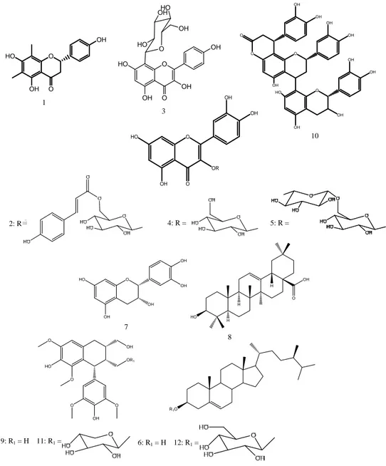
The isolated compounds (Fig. 1) were identiWed as: farrerol 121, quercetin
3-O-[6-trans-p-coumaroyl]-$-D-glucopyranoside 2 (helichrysoside)22, apigenin
8-C-$-D-glucopyranoside 323, quercetin 3-O-$-D glucopyranoside
(isoquercitrin) 424 and quercetin
3-O-"-l-rhamnopyranosyl-(166)-ß-d-glucopyranoside 525 from C. fruticosa. $-sitosterol
626, catechin 714, oleanolic acid 827, Lyoniresinol 928, cinchonain
IIb 1029, Lyoniresinol 2-a-O-$-D-xylopyranoside 1130 and
$-sitosterol-3-O-$-D-glucopyranoside 1226 from E. japonica. Antioxidant activity
DPPH radical scavenging test: As depicted in Fig. 2a-d crude
extracts (MeOH extracts) and their sub-sequent extracts (n-BuOH and EtOAc extracts) and isolated compounds from
C. fruticosa and E. japonica exhibited a good radical scavenging activity on DPPH. It can be noticed that the activity increases with the fractionation of crude extracts.
Eriobotrya japonica appeared to be more potent than
C. fruticosa and compound 4 was the less active of all the substances tested. More importantly compounds 7 and 10 isolated from E. japonica acted similarly to ascorbic acid used as the reference drug. Indeed these compounds were as powerful as ascorbic acid with respective efficiency index (EI) of 0.04, 0.03 and 0.03 (Table 1).
Table 1: Radical scavenging activity of extracts, fractions and isolated compounds from C. fruticosa and E. japonica on DPPH
Test substances EC50 (µg mLG1) Emax (%) EI
Cordyline fruticosa MeOH extract 181.30 61.86 2.93 n-BuOH extract 49.34 90.69 0.54 EtOAc extract 50.68 97.99 0.52 1 11.28 96.23 0.12 2 8.73 94.70 0.09 3 45.22 93.45 0.48 4 ND 63.83 ND 5 9.91 93.49 0.11
Eriobotrya japonica MeOH extract 16.55 93.59 0.18 EtOAc extract 7.19 94.56 0.08 7 4.11 95.25 0.04 10 3.14 94.61 0.03 11 10.61 95.11 0.11 Ascorbic acid 3.11 96.51 0.03 ND: Not determined
The antioxidant activity of phenolic compounds is due to their ability to scavenge free radicals, donate hydrogen atoms or electron or chelate metal cations. The structure of phenolic compounds is a key determinant of their radical scavenging and metal chelating activity and this is referred to as structure-activity relationships (SAR)31.
Comparing the structures (Fig. 1) and activities of compounds 2, 4 and 5 (Fig. 2c), the main differences are the substituents of the C-3 glucose moiety at position C-6''. It is inferred that the presence of an additional p-coumaroyl moiety (compound 2) or sugar group (compound 5) at position C-6'' seems to have some little influence on antioxidant activity compare to compound 4. By comparing the activity of compounds 3 and 4, it is inferred that the
Fig. 1: Chemical structures of compounds isolated from the leaves of Cordyline fruticosa and stem bark of Eriobotrya japonica
O O OH HO OH O OH OH O OH OH O O OH OH HO OH OH OH O OH OH OR OH HO O O O OH HO OH OH O HO OH HO OH O OH OH OH OH HO H H H OH O HO OR1 OH O O O HO O OH R1O 1 3 2: R 4: R = 5: R = 10 8 7 9: R1 = H 11: R1 = 6: R1 = H 12: R1 =
120 100 80 60 40 20 0 Log concentration (µg mL )G1 0 1 2 3 In h ib itio n ( % ) Ascorbic acid 7 10 10 (d) 130 110 90 70 50 30 10 -10 Log concentration (µg mL )G1 0 1 2 3 Inh ibi ti on ( % ) Ascorbic acid 1 2 3 4 5 (c) 130 110 90 70 50 30 10 -10 Inhi b it io n (% ) Ascorbic acid MeOH extract EJ EtOAc extract EJ (b) 130 110 90 70 50 30 10 -10 In h ib iti o n (% ) Ascorbic acid MeOH extract CF n-BuOH extract CF EtOAc extract CF (a)
Fig. 2(a-d): DPPH scavenging activity of, (a) Cordyline fruticosa extracts, (b) Eriobotrya japonica extracts, (c) Some compounds isolated from C. fruticosa and (d) Some compounds isolated from E. japonica and reference antioxidants (Ascorbic acid)
C-glycosylation (compound 3) or O-glycosylation and catechol group (compound 4) are the key factor that affects the activities, Although, compound 4 bears a catechol group in B-ring, which is very important for radical scavenging activity in flavonoides32 compare to compound 3, the later was more
active than the former owing to the free hydroxyl residue at C-3 position of C-ring in compound 3 which is also a key factor for radical scavenging activity in flavonol derivatives33-34. Thus,
the high activity of compound 10 could be attributed to its greater number of catechol and free hydroxyl groups.
Ferric Reduction Antioxidant Power (FRAP): The ability of
plant extracts and isolated compounds to induce the reduction of ferric cyanide complex to the ferrous form is associated with increase optical density. Figure 3a-d shows that crude and sub-sequent extracts, pure isolated compounds induced a concentration-dependent antioxidant activity. As observed with DPPH, E. japonica was more active than C. fruticosa in this assay. Interestingly, the reducing power of the EtOAc extract of E. japonica was higher than that of ascorbic acid.
In both methods, compounds 2, 7 and 10 showed potent activity, while compound 3 showed very weak antioxidant activity, which is consistent with the reported results. Furthermore, for compound 3 and 4 quantitative structure-activity relationship analysis also suggested that compound 3 would not show effective activity because of the absence of a catechol residue in the B ring and less free-OH groups in its structure, which are required for high antioxidant activity. Comparing the structures and activities of compounds 1 and 7, it is inferred that the presence of methyl group at C-6 and C-8 seems to have little influence on the activity.
AAPH-induced Red Blood Cells (RBC) hemolysis assay: All
extracts and isolated compounds potentiated the effect of AAPH at lower concentrations. A weak antioxidant activity was observed at a higher concentration (300 µg mLG1)
with n-BuOH, EtOAc extracts and compound 5 derived from
C. fruticosa (Fig. 4a, c). The E. japonica failed to inhibit AAPH-induced hemolysis (Fig. 4b, d).
R e d u ci n g p o w er ( O D) (a) 7.5 5.5 3.5 1.5 -0.5 Ascorbic acid MeOH extract CF EtOAc extract CF n-BuOH extract CF R e d u c ing p o w e r (O D ) (b) 7.5 5.5 3.5 1.5 -0.5 Ascorbic acid MeOH extract EJ EtOAc extract EJ Log concentration (µg mL )G1 0 1 2 3 R e du ci n g po w e r ( O D ) (d) 7.5 5.5 3.5 1.5 -0.5 Ascorbic acid 7 10 11 R e du c ing p o w er (O D ) (c) 7.5 5.5 3.5 1.5 -0.5 Ascorbic acid 2 3 4 5
Fig. 3(a-d): Ferric Reducing Ability Power (FRAP) at various concentrations of ascorbic acid, (a) Cordyline fruticosa extracts, (b) Eriobotrya japonica extracts, (c) Some compounds isolated from C. fruticosa and (d) Some compounds isolated from E. japonica
100 75 50 25 0 -25 -50 In hi b it io n (% ) (d) Log concentration (µg mL )G1 0 1 2 3 Ascorbic acid 7 10 11 100 50 0 -50 -100 In h ibi ti o n ( % ) (a) Ascorbic acid MeOH extract CF EtOAc extract CF n-BuOH extract CF 100 50 0 -50 -100 In hi bi ti o n ( % ) (c) Ascorbic acid 1 2 3 4 5 100 50 0 -50 In h ib itio n ( % ) (b) Ascorbic acid MeOH extract CF EtOAc extract CF
Fig. 4(a-d): Effects of, (a) Cordyline fruticosa extracts, (b) Eriobotrya japonica extracts, (c) Some compounds isolated from
C. fruticosa and (d) Some compounds isolated from E. japonica and reference antioxidants (Ascorbic acid) on AAPH-induced hemolysis
The SAR of Xavonoids is generally more complicated due to the relative complexity of the Xavonoid molecules. Some of the structural features and nature of substitutions on rings B and C which determine the antioxidant activity of Xavonoids include the following:
C The degree of hydroxylation and the positions of the -OH groups in the B ring, in particular an ortho-dihydroxyl structure of ring B (catechol group) results in higher activity as it confers higher stability to the aroxyl radical by electron delocalization32 or acts as the preferred
binding site for trace metals35
C A double bond between C-2 and C-3 conjugated with the 4-oxo group in C-ring enhances the radical scavenging capacity of Xavonoids35
C A double bond between C-2 and C-3, combined with a 3-OH in ring C, also enhances the active radical scavenging capacity of Xavonoids, as seen in the case of kaempferol32. Substitution of the 3-OH results in increase
in torsion angle and loss of coplanarity and subsequently reduced antioxidant activity36
CONCLUSION
At the end of this study, it is considered that the bio-guided purification of C. fruticosa and E. japonica
revealed six compounds with excellent proven antioxidant and scavenging activities. The results indicated that
C. fruticosa and E. japonica fractions exhibit excellent radical scavenging ability in all assays employed and the EtOAc fraction of E. japonica was the most active fraction among them. Phytochemical investigation of the fractions led to the isolation of twelve compounds and the radical scavenging assays indicated that all compounds have stronger antioxidant capacity but less than the positive control ascorbic acid. Overall, C. fruticosa and E. japonica are promising sources of natural antioxidants ingredients.
It is well established that the efficacy of flavonoids as antioxidants depend on the number and position of the hydroxyl substitutions on the basic structure; an increase in number of hydroxyl groups is directly correlated with increasing activity and the 3, 4-dihydroxy substitution is significant.
ACKNOWLEDGMENTS
The authors are grateful to the Italian Ministry of Education (MIUR) for supporting this research through the COOPERLINK 2011 (Prot.CII113PPUC ''Tesi di Dottoratosullo studio di molecule biologicamenteattiveestratte da
piantedellamedicina tradizionale del Camerun''. Project Manager L. Barboni) and the IFS (International Foundation for Science, Stockholm, Sweden) program through grant to Prof. A. Léon TAPONDJOU (RGA No.F/3976-3F).
REFERENCES
1. Adly, A.A.M., 2010. Oxidative stress and disease: An updated review. Res. J. Immunol., 3: 129-145.
2. Al Mashkor, I.M.A., 2015. Evaluation of antioxidant activity of clove (Syzygium aromaticum). Int. J. Sci., 13: 23-30.
3. Conran, J.G., 1998. Lomandraceae. In: The Families and Genera of Vascular Plants, Vol. 3, Kubitzki, K., H. Huber, P. Rudall, P. Stevens and T. Stutzel (Eds.). Springer, New York, USA.
4. Dalimartha, S., 2007. Atlas Tumbuhan Obat Indonesia. Vol. 4, Puspa Swara, Jakarta.
5. Nugent, J., 2006. Permaculture Plants: Agaves and Cacti. 2nd Edn., Sustainable Agriculture Research Institute, German. 6. Nombo, P. and J. Leach, 2010. Reite Plants: An Ethnobotanical Study in Tok Pisin and English. ANU E Press, Canberra, ISBN-13: 9781921666018.
7. Kulip, J., 2003. An ethnobotanical survey of medicinal and other useful plants of Muruts in Sabah, Malaysia. Telopea, 10: 81-98.
8. Vidal, J.E., 1965. Notes sur quelques Rosacees asiatiques (III). Revision du genre Eriobotrya (pomoideae). Adansonia, 5: 537-580.
9. Liao, W.B., Y. Ren and M.J. Zhong, 1997. On the variation patterns of morphological characteristics and geographical distribution of Eriobotrya (Rosaceae). J. North West Univ., 27: 57-60.
10. Perry, L.M. and J. Metzger, 1980. Medicinal Plants of East and Southeast Asia: Attributed Properties and Uses. MIT Press, Cambridge, USA., pp: 342-343.
11. Rashed, K.N. and M. Butnariu, 2014. Isolation and antimicrobial and antioxidant evaluation of bio-active compounds from Eriobotrya japonica stems. Adv. Pharmaceut. Bull., 4: 75-81.
12. Ito, H., E. Kobayashi, Y. Takamatsu, S.H. Li and T. Hatano et al., 2000. Polyphenols from Eriobotrya japonica and their cytotoxicity against human oral tumor cell lines. Chem. Pharm. Bull., 48: 687-693.
13. Davia, M.L. and F. Gnudi, 1999. Phenolic compounds in surface water. Water Res., 33: 3213-3219.
14. Philippe, B.A., N. Karine, A.K. Barthelemy, Z.J. Noel, D.A. Joseph and K. Hosttetmann, 2010. Bio-guided isolation of antioxidant compounds from Chrysophyllum perpulchrum, a plant used in the Ivory coast pharmacopeia. Molecules, 15: 6386-6398.
15. Cook, N.C. and S. Samman, 1996. Flavonoids-chemistry, metabolism, cardioprotective effects and dietary sources. Nutr. Biochem., 7: 66-76.
16. Nono, N.R., L. Barboni, R.B. Teponno, L. Quassinti and M. Bramucci et al., 2014. Antimicrobial, antioxidant, anti-inflammatory activities and phytoconstituents of extracts from the roots of Dissotis thollonii Cogn. (Melastomataceae). South Afr. J. Bot., 93: 19-26.
17. Nzowa, K.L., L. Barboni, R.B. Teponno, M. Ricciutelli and G. Lupidi et al., 2010. Rheediinosides A and B, two antiproliferative and antioxidant triterpene saponins from
Entada rheedii. Phytochemistry, 71: 254-261.
18. Nguelefack-Mbuyo, P.E., T. Dimo, T.B. Nguelefack, A.G.B. Azebaze, A.B. Dongmo, P. Kamtchouing and A. Kamanyi, 2010. In vitro antioxidant activity of extracts and coumarins from the stem bark of
Mammea africana Sabine. J. Complement. Integr. Med., Vol. 7. 10.2202/1553-3840.1447
19. Athukorala, Y., K.N. Kim and Y.J. Jeon, 2006. Antiproliferative and antioxidant properties of an enzymatic hydrolysate from brown alga, Ecklonia cava. Food Chem. Toxicol., 44: 1065-1074.
20. Zhang, Z., H.N. ElSohly, X.C. Li, S.I. Khan and S.E. Broeldon Jr. et al., 2003. Phenolic compounds from
Nymphaea odorata. J. Nat. Prod., 66: 548-550.
21. Chen, G., H. Jin, X. Li, Q. Zhang, Y. Shen, S. Yan and W. Zhang, 2009. Chemical constituents from
Rhododendron spinuliferum. Chem. Nat. Compounds, 45: 725-727.
22. Lavault, M. and P. Richomme, 2004. Constituents of
Helichrysum stoechas variety olonnense. Chem. Nat. Comp., 40: 118-121.
23. El-Toumy, S.A., E.A. Omara, S.A. Nada and J. Bermejo, 2011. Flavone C-glycosides from Montanoa bipinnatifida stems and evaluation of hepatoprotective activity of extract. J. Med. Plants Res., 5: 1291-1296.
24. Teponno, R.B., A.L. Tapondjou, J.D. Djoukeng, E. Abou-Mansour and R. Tabacchi et al., 2006. Isolation and NMR assignment of a pennogenin glycoside from
Dioscorea bulbifera L. var sativa. Nat. Prod. Sci., 12: 62-66. 25. Kazuma, K., N. Noda and M. Suzuki, 2003. Malonylated
flavonol glycosides from the petals of Clitoria ternatea. Phytochemistry, 62: 229-237.
26. Saeidnia, S., A. Gohari, M. Malmir, F.M. Afrapoli and Y. Ajani, 2011. Tryptophan and sterols from Salvia limbata. J. Med. Plant, 10: 41-47.
27. Mahato, S.B and A.P. Kundu, 1994. 13C NMR Spectra of
pentacyclic triterpenoids-A compilation and some salient features. Phytochemistry, 37: 1517-1575.
28. Tung, Y.T., K.C. Cheng, S.T. Ho, Y.L. Chen, T.L. Wu, K.C. Hung and J.H. Wu, 2011. Comparison and characterization of the antioxidant potential of 3 wild grapes-Vitis thunbergii,
V. flexuosa and V. kelungeusis. J. Food Sci., 76: C701-C706. 29. Matsuo, Y., Y. Fujita, S. Ohnishi, T. Tanaka, H. Hirabaru and
T. Kai et al., 2010. Chemical constituents of the leaves of rabbiteye blueberry (Vaccinium ashei) and characterisation of polymeric proanthocyanidins containing phenylpropanoid units and A-type linkages. Food Chem., 121: 1073-1079. 30. Szakiel, A., V.L. Voutquenne-Nazabadioko and M. Henry,
2011. Isolation and biological activities of lyoniside from rhizomes and stems of Vaccinium myrtillus. Phytochem. Lett., 4: 138-143.
31. Rice-Evans, C.A., N. Miller and G. Paganga, 1997. Antioxidant properties of phenolic compounds. Trends Plant Sci., 2: 152-159.
32. Van Acker, S.A.B.E., D.J. Van Den Berg, M.N.J.L. Tromp, D.H. Griffioen, W.P. Van Bennekom, W.J.F. Van Der Vijgh and A. Bast, 1996. Structural aspects of antioxidant activity of flavonoids. Free Radic. Biol. Med., 20: 331-342.
33. Hirano, R., W. Sasamoto, A. Matsumoto, H. Itakura, O. Igarashi and K. Kondo, 2001. Antioxidant ability of various flavonoids against DPPH radicals and LDL oxidation. J. Nutr. Sci. Vitamonol., 47: 357-362.
34. Haraguchi, H., H. Ishikawa, Y. Sanchez, T. Ogura, Y. Kubo and I. Kubo, 1997. Antioxidative constituents in
Heterotheca inuloides. Bioorg. Med. Chem., 5: 865-871. 35. Pietta, P.G., 2000. Flavonoids as antioxidants. J. Nat. Prod.,
63: 1035-1042.
36. Seeram, N.P. and M.G. Nair, 2002. Inhibition of lipid peroxidation and structure-activity-related studies of the dietary constituents anthocyanins, anthocyanidins and catechins. J. Agric. Food Chem., 50: 5308-5312.