1
Università degli Studi di Ferrara
DOTTORATO DI RICERCA IN
BIOCHIMICA, BIOLOGIA MOLECOLARE E BIOTECNOLOGIE
CICLO XXVII
COORDINATORE Prof. Francesco Bernardi
Cellular and biomolecular technologies for
stratification of β thalassemia patients:
applications in theranostics
Settore Scientifico Disciplinare BIO/10
Dottoranda Tutore
Dott.ssa Lucia Carmela Cosenza Prof. Roberto Gambari
Secondo Relatore Dr. Nicoletta Bianchi
2
Introduction
41. Hemoglobin and hemoglobinopathies 4
1.1 Hemoglobinopathies 5
1.2 Specific Hemoglobinpathies: HbS and sicle cells disease 5
1.3 Thalassemia 7
1.4 Globin clusters 7
1.5 The β-thalassemia 14
1.6 The β-globin genes 15
2. HPFH phenotype 18
2.1 The γ-globin genes 18
2.2 The γ-globin gene expression 19
3. The hematopoiesis and erythroid cell methods of culture. 27 4. Therapeutic approaches for β-thalassemia and sickle cell anemia 35
4.1 Definitive therapies 35
4.2 Maintenance therapies 36
4.3 Gene therapy 41
5. Collection of biological samples in Biobank and Biobanks around the world
50
Aim of the thesis
57Material and Methods
591. Biological samples collection 59
2. ErPCs Isolation and culture 59
3. Freezing 61
4. Thawing 61
5. Benzidine staining 61
6. Treatment with HbF inducers. 61
7. RNA extraction 62
8. Synthetic oligonucleotides 62
9. RT-PCR and RT qPCR analysis 62
10. High Performance Liquid Chromatography (HPLC) 64
3
12. Agarose gel electrophoresis 64
13. Quantification of DNA spectrophotometer 65
14. Polymerase Chain Reaction (PCR) 65
15. Purification of PCR products with MicroCLEAN (Microzone Limited)
65
16. Sequencing Reaction: "Sanger" method 66
Results
671. Sample collection 67
2. CD34+ cells: isolation, expansion and freezing 70
3. Kinetics of EPO-induced erythroid differentiation following
subculturing of cryopreserved ErPCs from β-thalassemia patients.
72
4. Validation of cell samples stored in the Biobank 75
4.1. Biobanked samples of a same β-thalassemia patient frozen and subcultured at different time maintain the same hemoglobin pattern.
75 4.2. Biobanked samples originated from different blood sampling of a
same β-thalassemia patient maintain the same hemoglobin pattern.
76 4.3. Biobanked samples thawed and subcultured in different
laboratories exhibit a similar pattern of hemoglobin production.
76
4.4. Conclusions. 78
5. Induction of biobanked CD34+ cells versus two liquid phases Fibach protocol.
79 6. Use of protocol A and protocol C with different HbF inducers. 83
7. Analysis with HbF hemoglobin inducers. 84
8. HbF hemoglobin inducers tested in cellular Biobank. 85
Discussion
87Appendix
95Appendix 1 96
Appendix 2 105
4
Introduction
1. Hemoglobin and hemoglobinopathies
The hemoglobin (Hb) is a globular chromoprotein (Kendrew 1962; Bianco 1998), in red blood cells, that deals with transportation of oxygen from the lungs to various tissues through the bloodstream. It is a tetramer composed of two types of subunits designated α and β, with a stoichiometry α2β2 (Silvestroni and Bianco
1948; Straus, Gordon et al. 1969; Weatherall 1974). The hemoglobin consists of a fraction of high molecular weight, globin, and a fraction of non-protein low molecular weight, the heme group binding oxygen molecules in the pulmonary alveoli and then disposes in various tissues (Efimov 1979). Each individual heme molecule contains one Fe2+ atom. In the lungs where oxygen is abundant, an oxygen molecule binds to the ferrous iron atom of the heme molecule. This interaction produces a conformational change creating a three-dimensional structure, where the different subunits of hemoglobin can interact in different ways with other subunits and neighboring molecules.
Figure 1 Structure of adult hemoglobin. Hemoglobin is a globular protein consisting of two α
chains and two β-chains . It is located within the red cells where It has the function to transport oxygen from the lungs to the tissues due to the presence of a prosthetic group called heme containing an iron ion.
5
These additional interactions determine the formation of the quaternary structure of the protein (Ranney HM. and Beutler E 1991; Fronticelli, Sanna et al. 1995). The variations in the interactions between the different subunits are trasmitted from the surface to the heme group of a second subunit. This cause a more facilitated access of the oxygen to the ion iron leading to a greater affinity of the haemoglobin for a second molecule of oxygen. (Figure1) (Saroff 1970).
Defects in this protein can lead to various pathological complications broadly documented and characterized as hemoglobinopathies or thalassemias.
1.1 Hemoglobinopathies
The hemoglobinopathy is a genetic defect that results in an abnormal structure in one of the globin chains of the hemoglobin protein. The genetic defect may be due to the substitution of one amino acid for another eg. HbS, HbC and other altered hemoglobins, delection of a portion of the amino acid sequence, uncorrect hybridization between two chians, or abnormal elogation of the globin chian (Old 1996; Giardine, Borg et al. 2014). The resultulting chain may be the α-chain (eg. HbGPhiladelphia), β-chain (eg. HbS, HbC), γ-chian (HbFTexas), or δ-chain (HbA2Flatbush)
(Weatherall 1974). These abnormal hemoglobins may involve a wide variety of physiological effects, but the most severe (HbS or HbC) are characterized by hemolysis (Weatherall, Clegg et al. 1974).
1.2 Specific Hemoglobinpathies: HbS and sicle cells disease
The HbS gene was detected for the first time in population of native African origin. The incidence of the gene in this population reaches 40%, in native Afro-Americans the incidence is 8%. Its presence, although at much lower frequency, was also documented in the non-Indo-European aboriginal populations of India as well as Middle East. In rare case have been reported in Caucasians of Mediterranean descent. In 1944 in Italy, Silvestroni and Bianco, submitted the first case in the world of β-thalassemia association with HbS, which they called micro-drepanocytosis. In Italy, the most affected region is Sicily, where HbS is present with a frequency of 2-5% followed immediately by Calabria. Its’ frequency in other areas of Italy appears to be very low (Silvestroni and Bianco 1948). However, the new migratory waves from Africa and the Middle East could, in the next ten years
6
significantly change the geographical distrubution of this gene and related diseases (Old 1996; Patrinos, Kollia et al. 2005).
The expression of the HbS gene is mostly present under certain populations, perhaps given more to climatic and environmental conditions. In fact, its expression in heterozygosity (sickle cell trait) represents a kind of protection against the infestation of Plasmodium Falciparum and reduces the severity of related clinical manifestations (Flint, Harding et al. 1998; Clegg and Weatherall 1999). Unfortunately, however homozygous expression produces sickle cell disease characterized by a chronic hemolytic anemia and vaso-occlusive condition that in most cases leads to the death of the patient (Andemariam, Owarish-Gross et al.; Nur, Biemond et al.; Kato, McGowan et al. 2006).
From the molecular perspective, the HbS is characterized by a point mutation GAG→GTG in codon 6 of the β-globin gene that results in the substitution in 6 position on the β chain of a glutamic acid residue with one of valine (Rees, Williams et al.). This structural variation changes the surface of hemoglobin causing in deoxygenation conditions hydrophobic interaction between the hemoglobin tetramers, leading to the formation of polymers organized in parallel structures of fiber bundles (tactoids). The polymerization of hemoglobin is associated with significant changes in the membrane erythrocytes, which become less malleable and more fragile (Ballas 2002; Vekilov 2007). These alterations of the membrane, initially reversible, culminate with the formation in the red blood cells of the classical sickle form (Samuel, Salmon et al. 1990). This gives the name to the pathology associated with the presence of HbS, sickle cell anemia.
Clinical manifestations associated with the presence of Hb are associated with different genotypes: i) homozygosity for Hb(S/S) that causes sickle cell anemia classic; ii) double heterozygosity with β-thalassemia (S/β such), (the form that most frequently found in Italy (Silvestroni, Bianco et al. 1950); iii) double heterozygosity with some hemoglobin variants eg. S/C, D/D Los Angeles, S/O-Arab. The clinical demonstrations include both acute and chronic manifestations. In individuals homozygous for the gene Hbs, the 97% of hemoglobin is Hbs in childhood, the remainder being the shortest normal hemoglobin, HbA2 (α2 δ2).
Several coexisting genetic abnormalities prevalent in African populaitons may ameliorate the course of the disease:
1. α-thalassemia carriers, which comprise 20% of African Americans, have a lower Mean Corpuscolar Haemoglobin Concentration (MCHC) than normal individuals. It
7
has been suggested that a low MCHC produce a benefit in decreasing of the vasoocclusive properties of sickled cells. These sickle cell patients live longer and have a milder disease with respect to non-thalassemic patients (Akinsheye, Alsultan et al.; Steinberg 2009).
2. Hereditary Persistence of Fetal Hemoglobin (HPFH) has established itself prominently in the black population. These people in the fetal hemoglobin (HbF) gene does not "turn off" in infancy but persists indefinitely. With the accurance of the HbF gene, there is a dilution of HbS, which reduces sickling in population (Thein, 2009)
3. G6PD deficiency has been suggested as an ameliorative condition for sickle cell disease (Badens, Martinez di Montemuros et al. 2000; van Schaftingen and Gerin 2002). This is however controversial.
1.3 Thalassemia
The thalassemia is a genetic defect that results in the production of an abnormally low quantity of a given hemoglobin chains (Silvestroni and Bianco 1948; Clegg and Weatherall 1999; Canali 2008). The defect may affect the α, β, γ, δ chain, or may affect some combination of thre β, γ and δ in the same patient. The result is an imbalanced production of globin chians and the production of an inadequate number of red cells. These cells are hypochromic/microcytic and contain a surfeit of the unaffected chains, which cannot bind the thalassemic chains in stoichiometrically manner (Williams Hematology Ernest Beutler 1995). These unrelated chains produce various kinds of damage inside the red blood cells and which leads to destruction of red cells within the marrow (ineffective erythropoiesis) and in bloodstream (hemolysis) (Cazzola 1996).
1.4 Globin clusters
To better understand thalassemia, the genes that code for the different globin chains should identify and characterize.
The gene coding for the different types of globin chains present in the hemoglobin are organized in two groups known as globin clusters (α and β); each cluster consists of structural genes and some regulatory regions constituted by highly conserved nucleotide sequences (Bianco 1998).
8
This α-cluster of about 30 Kb is located in the distal short arm of chromosome 16 and includes an embryonic gene (ζ), a fetal-adult block consisted of the α1 and α2
genes although producing different quantities of mRNA, which have identical globin chains (Myers, Tilly et al. 1986), a few pseudo-genes (ψξ, ψα1, ψα2) and the
θ gene, which is considered a pseudo-gene, because while producing a mRNA does not express any globin .
The cluster β (or named non-α), approximately 70 kb is located in the distal portion of the short arm of chromosome 11and contains five structural genes ε, Gγ, Aγ, δ and β and the pseudo gene ψβ (Efstratiadis, Posakony et al. 1980). The genes present in β cluster produce during the embryonic period the haemoglobins Gower I (ζ2ε2), Portland (ζ2γ2) and Gower II (α2ε2); in the fetal period HbF (α2γ2) and in the
adult period HbA (α2β2) and HbA2 (α2δ2) (Figure2). (Lawn, Efstratiadis et al. 1980;
Thein 2004; Thein 2005).
The expression of the globin chains at different stages of development depends on the activation of several genes and them switching off through methylation and demethylation processes (Ho and Thein 2000; Harju, McQueen et al. 2002; Levings and Bungert 2002; Qiliang Li 2002). The first gene switch already happens at the fifth week of gestation and produce the switching off of embryonic hemoglobin genes in favor of the activation of fetal hemoglobin genes. In this step, zeta globin (ζ) gene expression decrease and the levels of alpha globins gene (α) increase. Alpha globins get to the maximum level at the twenty-fourth week of conception and remain constant throughout life. Epsilon globins (ε), instead, are replaced by the gamma globins (γ). The second gene switch determines a drastic reduction of γ-globins, which are replaced by beta-globins (β) within the first year of age. However, during adulthood, a small amount of γ-globins are expressed, so there are three types of hemoglobins: HbA (α₂β₂) that represents 97% of hemoglobin total, HbA₂ (α₂δ₂) and 0-2% HbF present in very low amounts. The HbF content depend on several factors such as age, sex and genetic characteristics and on the presence of point mutations in the β cluster, which could produce an increase of HbF (Bank 2006; Sankaran and Nathan 2010).
The fetal hemoglobin, compared to the adult form, has greater affinity for oxygen and this makes it more efficient oxygen transfer from the maternal to fetal blood (Efstratiadis, Posakony et al. 1980).
9 Fi gu re 2 H e m o gl o b in swi tc h in g . A ) S che ma tic r ep res en tat ion of the produc tion of th e he mo gl o bi ns du ri n g the s ta ge s of de ve lo pm en t fr o m th e em bry on ic s tag e to ad u lth o od : B ) S che m ati c rep res e n tati o n of th e α -c lus ter o n chromos o me 1 6 an d β -c lus ter on c hromos o me 1 1. T h ere are a ls o rep res en te d th e tet ra me rs of he mo g lo bi ns s ynth es iz e d du ri ng t he d ifferen t st ag es of de ve lo pm e nt. C) T h e orga ns an d tis sue s, w h e re oc cur the he mo gl ob ins s ynth es is du ri ng th e d ifferent de ve lo pm e nt st ag es , ar e rep orted wi th di ffere nt col ors , a s i nd ic a te d
10
In each clusters the structural genes are sorted in the same order to reflect the gene of expression during ontogenesis (Hanscombe, Whyatt et al. 1991). Their transcription is finely regulated by different protein elements (transcription factors) that activate or repress the globin genes through specific regulatory sequences placed within the globin cluster: i) promoters, ii) enhancer sequences, iii) the Locus Control Region (LCR). In particular the latter is located upstream of the gene for the ε-globin and it is able to modulate the gene transcription interacting with the promoters of the single globin genes. LCR region contains five domains, hypersensitive to DNase 1, named HS1, HS2, HS3, HS4, HS5, only the first four are specific to erythroid cells (Hardison, Slightom et al. 1997; Wilber, Nienhuis et al.). Among these the most important is certainly HS2, which performs the function of the enhancer or activator of transcription, it contains the binding sites for several transcription factors including: Sp1 (Specificity Protein 1), NF-E₂ (nuclear factor E2), GATA-1 (erythroid cell and megakaryocyte specific trascription factor 1) and USF (Upstream Stimolatory Factor) (Elnitski, Miller et al. 1997; Harju, McQueen et al. 2002). The domains HS3 and HS4 are involved in chromatin remodeling so as to facilitate the transcription factors binding. The role of HS5 is still uncertain. Some studies attribute to him the role of silencer or insulator (Wai, Gillemans et al. 2003). The insulator is an element able to block the activity of the histone deacetylase (Hardison, Slightom et al. 1997), so LCR plays an important role in chromatin remodeling (Levings and Bungert 2002; Qiliang Li 2002).
Initial studies about the regulation of the globin genes were mainly focused on the relationship between the LCR and the β-like genes, through the analysis of the function of the locus large fragments in transgenic mice (Myers, Tilly et al. 1986). Experiments carried out on transgenic mice, transduced with constructs containing both portions that the entire locus for the human β-globin gene, demostrated the presence of a cis-control of globin switching (Dillon, Trimborn et al. 1997). Studies about factors which control the globin gene expression have revealed a variety of activators and repressors factors that act at different stages of development. Unlike the LCR, the cis-acting elements play local function on neighboring regions of chromatin; in contrast, LCR acts on long distances to activate the expression of the globin genes (Dillon, Trimborn et al. 1997). These elements cis-elemnt could be:
1. Silencer. It binds protein complexes interfering with the promoter activity and causing the reduction of gene expression (Ramchandran, Bengra et al. 2000). A
11
silencer element positioned distally in the promoter for the ε-globin gene controls the repression of its expression during the fetal and adult stage. The GATA1 and YY1 proteins constitute at least two of the components included in the repression complex (Raich, Clegg et al. 1995; Ramchandran, Bengra et al. 2000) In addition, two elements DR (Direct Repeat) in the ε-globin promoter, bind a protein recently identified DRED (direct repeat definitive erythroid-binding protein), which interferes with the factor EKLF (erythroid Kruppel-like factor) binding causing the final silencing of the ε-globin gene in adulthood (Nuez, Michalovich et al. 1995).
2. Insulator. It forms independent functional domains blocking the adverse effects of heterochromatin surrounding without stepping or activate gene transcription. It disturbs the interaction between the promoter region and other regulatory element, interposing between these sequences. The elements insulators can also block the activity of histone deacetylase. Finally, the insulators facilitate activity of elements enhancers located into the region of open chromatin. One hypothesis is that the LCR could have function of insulator, in particular, the hypersensitive 5'HS5 LCR site (Harju, McQueen et al. 2002).
3. MARs (matrix attachment region) and SARs (scaffold attachment region).These DNA elements promote the binding to the nuclear matrix obtaining the formation of loops in contiguous sequences of DNA. These items can be a barrier protecting the locus from the effects of surrounding chromatin and impose a restriction structural chromatin remodeling; for these reasons, the loop of DNA could be a transcriptional target. The MARs have the function to protect DNA from the effects mediated by the elements, which act in cis in adjacent loops when the chromatin it widens, or have the function of keeping together with the cis-regulatory elements of the neighboring loops. They can also promote the juxtaposition of cis-regulatory elements and gene promoters in the same loop. Since the 5'HS-5 region presents homologies with the regions MARs can attribute this activity; this hypothesis is further supported by the fact that, when the LCR is positionally inverted, 5'HS5 could isolate the globin genes by interaction with the LCR, causing the silencing of their expression. Although the role of 5'HS5 remains controversial, one can compare its function to that of a silencer element rather than insulator.
4. Boundary elements. They can be positioned at various levels inside the locus and take the limiting gene expression when associated proteins. These elements are characterized by some main properties: their association with insulator elements, the maintenance of a balance between the open and closed
12
conformation of chromatin. (Harju, McQueen et al. 2002). Multiple mechanisms of action have been proposed to explain the function of the LCR; the first models based on experiments conducted on transgenic mice, suggesting that switching globin happen following mechanisms of competition and the that genes proximal had preferential interaction with the LCR sequence.
Currently have been proposed four different models of interaction of the LCR region with the different globin genes (Harju, McQueen et al. 2002).
Looping model. LCR and regulatory elements for transcription are folded to form a loop interacting with the interested gene promoter.
Tracking model. The transcription factors and cofactors bind LCR to form an active complex, which slides along the DNA helix, until reaching the gene of interest. At this point other cofactors bind the protein complex activating gene transcription.
Facilitated traking model. Transcription factors bind the LCR sequence and form a loop which slides along the DNA until it reaches the correct gene.
Linking model. Transcription factors in precise sequential order are arranged to reach the promoter region of a particular gene of interest. The chromatin structure is maintained in an open conformation so the different transcription factors may interact activating gene transcription. (Qiliang Li 2002).Below the most important transcription factors are reported involved in the globin expression regulation. (Table1)
A possible deletion of LCR region inevitably produces an inhibition of the expression of all proteins encoded in the globin loci resulting in severe forms of thalassemia. (Curtin, Pirastu et al. 1985; Driscoll, Dobkin et al. 1989).
The thalassemias have been described for all four chains α, β, γ, δ we will focus on β-thalassemia.
13
Table 1 Main transcription factors involved in the regulation of the globin genes expression Transcription factor Mechanism of Action Effects
NF-E₂
(nuclear-factor erythroid derived2)
It binds to the AP-1 domain of the
HS2 LCR. It remodels the chromatin.
GATA-1
(Erythroid cell and megacaryocyte specific trascription factor 1)
It binds to the promoter of ε and γ globin genes with activator activity. It binds to the region of the silencer ε gene in the presence of the protein YY1 with repressor activity.
It check the process of erythroid cells maturation.
It controls the expression of the ε and γ globins.
GATA-2
(erythroid cell and megacaryocyte specific trascription factor 2)
Recognizes the same DNA sequences of GATA-1.
It regulates the erythroid differentiation process.
FOG
(Friend of GATA1)
It presents zinc-finger domains with which binds to GATA-1 without binding directly to DNA.
It checks the globin expression.
NF-E₄
(nuclear-factor erythroid derived 2)
p22 binds to a ubiquitous transcription factor (CP2) recalling the polymerase 2.
p14 binds to CP2 and the transcription factor NF-E₂ preventing the formation of the protein complex that activates γ-globin gene transcription.
It regulates the expression of γ-globin gene.
SOX 6
(Sex Determining Region Y)-Box 6)
It binds the promoter of the y-globin gene inhibiting the transcription of the latter. It binds to the promoter of the γ-globin gene only in the presence of BCL11A.
It checks the maturation and proliferation of erythroid cells.
FOP
(friend of protein arginine methyltransferase 1)
It works by reducing levels of the transcription factor FOP, indirectly increasing the levels of fetal hemoglobin.
It controls the expression of γ-globin genes.
COUP-TF2
(chicken ovalbumin upstream promoter trascription factor interacting protein)
It binds to the γ-globin promoter. Represses transcription of γ-globin gene.
DRED-TR₂-TR₄ It binds to the promoter of the ε-globin in the vicinity of a sequence referred to as DR.
It is involved in silencing of the fetal globins.
KLF-1
(erythroid Kruppel-like factor)
It binds to the CACCC sequence present in the β-globin gene. It binds to the BCL11A, activating gene transcription and consequently reducing expression of fetal hemoglobin.
It checks chromatin remodeling and gene transcription
It is necessary to adjust hematopoiesis process.
It check γ-globin expression
SP1
(Specificity Protein 1)
It recognizes and binds to GC-rich regions through zinc-finger motifs that bind to highly conserved regions.
It takes part in the differentiation of erythroid cells processes. It regulates the globin switching process
BCL11A
(B cell limphomaleukemia 11A)
In combination with other transcription factors, it binds to the γ-globin promoter.
It regulates the expression of the β-globin gene and consequently the levels of fetal hemoglobin.
14 1.5 The β-thalassemia
The β-thalassemia is an autosomal recessive disorder caused by mutations within and near the β-globin gene. All the mutations result in either the absence of the synthesis (β0-thalassemia) or reduction in synthesis (β+-thalassemia) of β-globin chains. (Olivieri 1999).
The first description was written by Dr. Cooley in 1925. The name thalassemia was coined to reflect the geografic origin of the target population (“thalassa” is the classical Greek name for the Mediterranean Sea) (Haldane 1949) (Weatherall, Clegg et al. 1974; Weatherall and Clegg 1996). It is estimated that currently about 1.5% of the world's population is healthy carrier (Figus, Lampis et al. 1989; Ristaldi, Pirastu et al. 1989). The patients are approximately 400,000 and there are about 60,000 new cases each year (Galanello and Origa 2010). The β-thalassemia is present in several countries of Mediterranean, Middle east, Central Asia, India, Southern China, Far East as well as countries of Africa and South America (Figure3). The highest incidence is reported in Cyprus (14%). In Italy, the most affected areas are Sicily, the river Po delta and Sardinia, where there is an incidence of approximately 95.7% (Silvestroni, Bianco et al. 1950; Hill 1987; Rosatelli, Dozy et al. 1992; Canali 2008). Migration flows and marriages between different ethnic groups are introducing the thalassemia in new geographies such as North Europe (Flint, Harding et al. 1998).
Figure 3 . β-thalassemia distribution. The areas with a high incidence of this disease are
highlighted in red. In Italy stand out the areas of the Po Delta, Sardinia, Sicily and the coast of Southern Italy, where the incidence is very high.
15
In general, β-thalassemia can be summarized in four main clinical conditions: 1) Thalassemia silent: this term is used to identify a condition of β thalassemia minor, which is hardly identified as hematological parameters and levels of HbA2 and the HbF are almost normal in heterozygousity; it can be shown only a slight imbalance in the synthesis of the chains in vitro (Gonzalez-Redondo et al, 1989; Moi et al, 2004)
2) Thalassemia Minor or microcytemia, also called thalassemia carrier or β-thalassemia trait. It is caused by a single mutated allele (β°/β or β+/β). Clinically asymptomatic, patients show a slight anemia and high levels of HbA2 associated
with increased HbF. However, in most cases, patients show clinical signs asymptomatic and are defined 'carriers' (Bianco 1998; Moi, Faa et al. 2004).
3) Thalassemia Intermediate: affected individuals with significant anemia which does not require blood transfusions, however. This anemia is caused by abnormalities of both genes β, but one or both mutations are mild (β°/β or β+/β+) (Bianco 1998; Thein 2004).
4) Thalassemia Major or Cooley's disease also known as Mediterranean Anemia: the sick people have a severe clinical presentation that implies regular blood transfusions. This form of β-thalassemia is caused by total lack of adult hemoglobin HbA. There are both mutated alleles (β°/β°, β+/β+ or β°/β+) (Thein
2004).
1.6 The β-globin genes
The β-globin gene has a size of 1600 bp and coding for 146 amino acids. The gene consists of three exons (coding regions) and two introns or IVS (non-coding sequences) (Efstratiadis, Posakony et al. 1980; Bianco 1998).
The general structure of the β-globin gene (Figure4) is typical of other globin loci and like all other globin genes consists of:
- three exons (coding regions) and two introns or IVS ( non-coding sequences). - promoter region: placed upstream of the gene, essential for transcription, is comprised of several groups of nucleotides (box). Also, in humans there are two other hexanucleotide sequence located downstream of the transcription start site involved in the transduction of mRNA (messenger RNA).
- untraslated Region (UTR) present at the 5' and 3' ends. UTRs are essential for translation of mRNA, they are transcribed but never translated. The 3’UTR constitutes the region between the termination codon and the poly(A) tail.
16
This consensus hexanucleotide serves as a signal for Figu
re 4 β -clust er and s chemat ic st ru ctu re o f th e β -g lob in g ene. β -clust er is locat ed o n Ch ro mo s o me 11. In the s tr uc ture o f β -c lus ter , the L CR reg io n is l oc at ed up str ea m of the gl o bi n g en es an d c on ta ins th e hy p ers en si tiv e si tes ( HS ) di sp lay ed as p in k do ts . T he g en es are loc ate d refl ec tin g th e ac tiv ati on order du ri ng th e de vel op m en t. . A t the b ott o m of p ic ture, th e rep res en tat io n of the β -gl o bi n g en e is r ep orte d. In bl u e, the thre e ex o n s sp ac ed by two i ntron ic r eg ion s.
17
cleavage of the 3’ end of the primary transcript and addition of the poly(A) tail which confers stability on the processed mRNA and enhances translation (Weatherall DJ 1991). The 5’UTR occupies a region of 50 nucleotides between ‘cap’ site of the β-globin mRNA and the initiation (ATG) codon. There are two prominently conserved sequences in the 5’UTR of the various globin genes, one is the CTTCTG hexanucleotide found 8 through 13 nucleotides downstream from the cap site, and the second conserved sequence is the CACCATG, in which the last three nucleotides form the starting codon (Bianco 1998).
There are more than 800 mutations (Giardine, Borg et al. 2014) in the β globin gene. They are mainly point mutations (involving a single nucleotide) and deletions (removal of gene sequence or regulatory regions) (Smetanina, Gu et al. 1997; Higgs 2004; Patrinos, Giardine et al. 2004; Patrinos, Kollia et al. 2005).
Promoter mutations. In the great majority of cases there are point mutations in the promoter region. These kinds of mutations involve a reduction of efficiency of transcription. In particular, the β-globin chains are present, but much smaller than those produced in healthy subject (β+-thalassemia). Such changes may be described as "slight" (Meloni, Rosatelli et al. 1992; Rosatelli, Dozy et al. 1992). Non sense mutations: involve an incomplete translation, and therefore the production of a no functional protein. They consists in the substitution of a base in a DNA codon with premature stop codon generation and, thus, termination of translation (β°-thalassemia) (Liebhaber, Trecartin et al. 1981; Trecartin, Liebhaber et al. 1981).
Frameshift mutations: consist of deletions or inserrtion of one or more nucleotides, which causes a shift in the reading frame of the mRNA (frameshift) alterating the termination of translation (Bianco 1998).
Defects in the mRNA maturation: splicing defects and polyadenilation defects (Olivieri 1999).
- Splicing mutations: they are a very large and important group of mutations which are responsible of a lot of β-thalassemias cases. Usually the defects consist in a point mutations that, with various mechanisms, alter the normal process of splicing. These mutations can occur either within an intron or within an exon. In the first case, they may cancel a regular site of splicing destroying the donor site GT or the acceptor site AG, reduce the efficiency of a splicing site altering its consensus sequence or create a new site of splicing, producing a new signal GT or AG in a cryptic site. When, however, mutations affecting the exons, the only thing that can
18
happen is the creation of a new donor or acceptor site of splicing, thus producing an abnormal mRNA (Orkin, Kazazian et al. 1982; Dominski and Kole 1993).
- Mutations in the cut and polyadenylation site of the pre-mRNA: if the AATAAA sequence is mutated, the cut and polyadenilation process occurs much more downstream. It produces mRNA much longer (several hundred nucleotides) and very unstable. The resulting phenotype, in heterozygous, is a mild β+ -thalassemia with only a little lower HbA together with an increase of HbA2
(Huisman 1997).
Deletion of the β-globin gene: this type of mutation in β-thalassemia is quite rare. Individuals with this kind of mutation present hematologic characteristics very similar to a person with microcytemia. In these cases we can have levels of HbF normal or slightly higher, and a ratio α/β>1 produced by large deletions removing the β-globin gene, but leaving intact δ-globin gene. With few exceptions, the deletion usually involves the region immediately upstream of the β-globin gene increasing the HbA2 content (Bianco 1998).
2. HPFH phenotype
The clinical picture resulting in patients affected by β+-thalassemia or β0 -thalassemia may be improved by co-heritance of mutations related to other genes of the β-globin cluster (Orkin, Kazazian et al. 1982; Thein 2005).
Particularl conditions have observed in subjects affected by hematological disorders with HPFH phenotype (Hereditary Persistence of Fetal Hemoglobin), observed for the first time in some African patients affected by sickle cell anemia (Thein and Craig 1998). High levels of HbF significantly improve the health status in patients (Thein and Menzel 2009; Galanello and Origa 2010; Cao, Moi et al. 2011). In the HPFH condition the HbF, normally expressed at high levels only during the fetal stage of human development, continues to be expressed at high levels in adult erythroid cells. The synthesis of γ-chains in the post fetal period allows to recruit α-chains to perform the HbF hemoglobin (α2γ2) (Weatherall DJ
1991). This greatly reduces the number of free alpha chains therefore decrease the α/non α-globin chain imbalance. In patients affected by thalassemia or other hemoglobinopathies, the presence of HbF reduces the severity of the disease and of all its related clinical complications.
19
The γ-globin genes (HBG1 and HBG2) are normally expressed in the fetal liver, spleen and bone marrow. The transcripts of the two γ-globin genes differ only by one amino acid residue in 136 position. In fact, the Glycine present in this position in the G-γ chain (HBG2 gene) is replaced with an Alanine in the A-γ chain (HBG1). The G-γ chains are the predominant transcripts at birth.
The expression of the HBG1 and HBG2 genes, which encode the γ isoforms of HbF, is normally suppressed shortly before birth and replaced by expression of the β or δ chains, which encode HbA. Normally adults have less than 1% HbF, whereas the thalassemic patients and in other subjects affected by hematological disease, but heterozygotes for HPFH, have variable levels (5-30%) of HbF. Homozygotes for HPFH can express HbF in up to 100% of red blood cells (Thein and Craig 1998).
The ratio of G-γ to A-γ is fairly constant (about 7:3) during the fetal period. The ratio decrease progressively during the postnatal γ-to-β switch, leading to an average value of 2:3 in the small residual amount of HbF detectable in normal adult blood. This switch in the γ ratio occurs together with the γ-β switch (Comi, Giglioni et al. 1980). Since the 60-70 years many researchers have studied about γ-globin genes extending the understanding of the different molecular mechanisms that regulating the activation, the expression and regulation of these genes. However, many of these regulatory mechanisms appear still unclear.
In 2004 Li et al. have identified the minimum promoter sequence for the down regulation of γ-globin gene (Li, Han et al. 2004). By a series of γ promoter truncations the CACCC box was largely responsible for the down regulation of the γ-globin gene in adult erythropoiesis. The CACCC box is a common element in the proximal promoters of many housekeeping and lineage-specific genes. All mutations or deletions of this box alter the expression of the involved genes suggesting the CACCC box functions as a positive transcriptional element.
2.2 The γ-globin gene expression
The expression of the globin genes is regulated by several transcription factors. As for the β-globin genes two transcription factors, named KLF1 (Krupper-like factor 1) and BCL11A (B cell lymphoma-leukemia 11A), have a crucial role (Armstrong, Bieker et al. 1998). However, BCL11A and KLF1 cooperate with numerous other erythroid-specific and chromatin modifiers factors to carry out their action.
20
BCL11A, also known as EV19 (Ecotropic Viral Integration Site 19) is a zinc finger transcription factor necessary for B lymphopoiesis and T (Liu, Keller et al. 2003). Several studies demonstrated that BCL11A is a negative regulator of the expression of γ-chains in human erythroid precursors and knockout mice (Sedgewick, Timofeev et al. 2008). Currently are known some BCL11A isoforms generated by alternative splicing. Four are the main and best characterized: isoform BCL11AXL (835 amino acids), isoform BCL11AL (773 amino acids), isoform BCL11AS (243 amino acids), isoform BCL11AXS (142 amino acids). The amino acids encoded by the triplets of the first three exons are the same in these isoforms, but they contain different carboxy-terminal end.
BCL11AXL and BCL11AL act as repressors of the γ-globin expression in human adult erythroid cells (Uda, Galanello et al. 2008). BCL11A binds the upstream locus control region (LCR), ɛ-globin, and the intergenic regions of the β-globin locus, but does not results associated with the promoter of the γ-globin genes indicating that its regulation depends on a complex process. In fact, BCL11A interacts with transcription factors such GATA1, FOG1 and SOX6 and it is required for recruitment of the NuRD and LSD1/CoREST chromatin remodeling complexes to the β-globin locus in primary erythroid cells. BCL11A tied SOX6 DNA inducing curvature. Therefore, SOX6 may perform its function as an architectural factor by organizing local chromatin structure and assembling other DNA-bound transcriptional factors into biologically active and sterically defined multiprotein transcriptional complexes (Xu, Sankaran et al.; Sankaran, Menne et al. 2008; Sankaran, Xu et al. 2009; Sankaran and Nathan 2010). KLF1 is a member of a large gene family encoding erythroid-specific transcription factors, characterized by the presence of zinc-finger domains. The zinc fingers is an amino acid sequence containing four conserved amino acids including cysteine and/or histidine binding a zinc atom (Armstrong, Bieker et al. 1998). KLF1 binds specific consensus site (CACCC) on the β-globin gene promoter. Mutations in this sequence prevent the binding of KLF1 to the promoter and determine the phenotype of a mild β-thalassemia (Orkin, Kazazian et al. 1982; Moi, Faa et al. 2004). Finally, recent studies in knockout mice for KLF1 gene and in families with persistence hereditary HbF (HPFH) have demonstrated that KLF1 plays a role in the regulation of HbF, also through an other mechanism. Studies about the regulation of the encoding KLF1 gene in human erythroid precursor cells carry out using shRNA KLF1 (short interfering RNA, RNA molecule double helix containing
21
21-22 nucleotides blocking the translation of messenger RNA target), demonstrated an increased expression of the γ-globin genes associated to a strong reduction of BCL11A, which is an inhibitor of the γ-globin gene expression. In addiction, it was observed in studies carried out using specific antibody versus transcriptional factor lied to DNA ChIP quantitative, and playing a direct rule in the regulation of BCL11A expression (Zhou, Liu et al.).
Studies of G-WAS (genome-wide association studies) identified other loci able to influence the production of HbF. Among these we note the region HBS1L-Myb located on chromosome 6q23.3. Myb is a proto-oncogene that is encoding the c-MYB transcription factor, which plays a crucial role in erythroid differentiation. The absence of c-MYB in mice is lethal due to failure to develop hematopoiesis in fetal liver. Disruption of the HBS1L-Myb intergenic region in mice suppresses Myb levels and leads to elevated expression of embryonic globins and hematopoietic parameters similarly to human HPFH phenotype. Moreover, down regulation of MYB transcription factor through over-expression of microRNAs, as miR-15a and 16-1, results in an increase of HbF levels. The HBS1L-Myb intergenic polymorphisms (HMIP) are present in three linkage disequilibrium (LD) blocks with most of the effect on HbF levels and numbers of F cells contributed by the second block (Thein, Menzel et al. 2007; Wahlberg, Jiang et al. 2009).
So, to summarize, in the fetal period the expression of KLF1 is reduced in a manner to not be enough to stimulate for induce the β-globin gene and BCL11A expression (Zhou, Liu et al.). This is the typical profile of fetal β-globin gene and expression with low levels of β-globin and BCL11A, together with high levels of γ-globin genes. Similar situation occurs in the presence of molecular defects of the KLF1 gene. In postnatal life we observe an increase of expression of KLF,which determines the activation of both the β-globin gene and BCL11A, inhibiting γ-globin expression.
Recent experiments carried out in CD34+ cells from patients with β-thalassemia transduced with shRNA versus the BCL11A mRNA showed that its silencing can determine a significant increase of HbF, equal to 30% of the total (Wilber, Hargrove et al.). In summary the pharmacological modulation of BCL11A can activate the production of HbF and could be represent a therapeutic approach in β-thalassemia and sickle cell anemia.
We can resume two potential alternatives for pharmacologically activation of HbF production:
22
- expression inhibition of KLF1 to reduce the of BCL11A levels and to increase the HbF production;
- gene silencing of BCL11A to increased the expression of γ-globin genes.
Currently here are two possible startegie for the down regulation of BCL11A. One involves the use of siRNA (small interference RNA). The siRNA binding to the target mRNA are able to degrade the mRNA reducing the gene expression and the protein production. Another strategy to down regulate BCL11A is using microRNAs, small molecules of single-stranded RNA, physiologically present in the cells. These molecules are able to negatively regulate the expression of several genes. MicroRNA binds the 3'UTR of the mRNA encoding target protein. This binding produces in some cases the inhibition of protein synthesis, in others, the mRNA degradation. In both cases there is a decrease of protein product. So far we have described some of the molecular mechanisms that are involved in the γ-globin expression. We can see as an alteration in each of these systems leads to a deregulation of gene expression and in this case the increase of fetal hemoglobin content.
We can briefly distinguish three situations which involve increase of HbF:
- The β-thalassemia mutation per se increase the γ-glonbin chain output. This occurs in the following situations: 1) δβ0-thalassemia is caused by large deletions in the HBB cluster; 2) deletion remove only 5’ upstream region of the HBB promoter, which results in high levels of HbA2.
- Co-transmission of HPFH phenotype, which is the result of single point mutation in the hemoglobin Gγ (HBG2) and hemoglobin Aγ (HBG1) gene promoter.
- Co-heritance of heterocellular HPFH phenotype linked or not linked to the HBB gene cluster. As we have previously described, recent studies using G-WAS have identified two quantitative trait loci (QTLs), BCL11A on chromosome 2p16 and HBS1L-MYB intergenic region on chromosome 6q23, that account 20%-30% of the common variation in HbF levels in healthy adults and subjects affected by β-thalassemia and sickle cells anemia (Thein, Menzel et al. 2007; Uda, Galanello et al. 2008; Thein, Menzel et al. 2009).
Higher expression of HbF is often termed 'pancellular,' whereas lower expression of HbF is often termed 'heterocellular’(Forget 1998).
However HPFH phenotype can also be caused by other genetic alterations:
- 'deletional': HPFH result from deletions within the β-globin gene cluster on chromosome 11p15 (Ottolenghi and Giglioni 1982; Forget 1998).
23
- 'non-deletional': HPFH result from point mutations in the promoter regions of the γ globin genes HBG1 and HBG2, these alterations keep intact β cluster (Forget 1998).
Regarding HBG1 gene about 58 mutation have been identified. Ten of these are particularly interesting because they are related to the HPFH phenotype. In HBG2 gene there are about 73 mutations reported in HbVar (Giardine, Borg et al. 2014). Also in this case, some mutation seem to be related to the HPFH.
These mutations occur especially in the regulatory regions at the binding sites of transcription factors, that are ubiquitous or erythro-specific. They alter the binding of the proteins to their consensus sequence and could cause an increased affinity for transactivating factors; an affinity reduction for transcriptional repressors, or a combination of the two mechanisms. Through structural studies it was possible to note that non-deletion HPFH point mutations are grouped in three regions of the γ- globin promoter. They are located close to the positions -200, -175 and -115 upstream to transcription starting site. The region -200, particularly rich in GC, is known to be the target of the different point mutations, which affect Gγ promoter in the position -202 (C→G) and the Aγ promoter respectively in -202 (C→T), -198 (C→G) and -195 (C→G) positions. In vitro studies have investigated as these mutations influence the binding of some proteins to DNA. Indeed, mutations at position -202, -198, -196, -195 increase the Sp1 transcriptional factor binding affinity (Motum, Deng et al. 1994). In vitro experiments have shown that -175 Aγ/Gγ point mutation prevents the binding with Otc-1 protein (Octamer Binding Factor 1) and modifies the binding site for GATA-1 (erythroid cell and megakaryocyte-specific transcription factor 1); in both cases activating the γ-globin gene expression (Liu Lr and JW. 2004).
Similar point mutations are identified in subjects with HPFH phenotype in different ethnic groups. For example, the -196 (C→T) Aγ mutation was found in Sardinia and its association with the β039 mutation in the β-globin gene produces an F-thalassemia. This condition presents HbF levels increase up to 10-20% by improving the clinical status of the disease (Galanello and Origa 2010) (Cao, Moi et al. 2011).
The -117 Aγ/Gγ, -114 Aγ/Gγ mutations and a deletion of 13 bp in Aγ promoter involve several sites for transcription factors including the CCAAT sequence present in duplicate; in this case, are involved sites of different proteins: CP1
24
(poly(C)-binding protein-1), CDP (CCAAT displacement protein), GATA-1 and NF-E3 (Nuclear Factor erytroid derived 3) (Ronchi, Bottardi et al. 1995).
The -158 (C→T) point mutation in the Gγ promoter is known also as the XmnI G-γ polymorphism (rs7482144). This sequence is recognized and cut by restriction enzyme XmnI. This mutation is localized in Saudi Arabia and in other ethnic groups such as Americans and Africans. It is an example of a genetic alteration which is expressed only in hematopoietic stress condition. In fact, an high level of HbF is present in patients who have a chronic hemolytic stress condition, but it is completely absent in healthy parents (Thein 2005). Unlike the other non deletional HPFH mutations, transcription factors capable of recognizing the sequence XmnI are not yet known (Gilman and Huisman 1985; Labie, Pagnier et al. 1985).
So, mutations in the HBG1and HBG2 (Table 2) promoter regions are able to condition several transcription factors binding involved in the HbF versus HbA switch, that induce HbF expression favor HPFH phenotype in the adulthood. Recently, G-WAS conducted on different ethnic groups, have shown the involvement of outside β cluster region with HbF regulation.
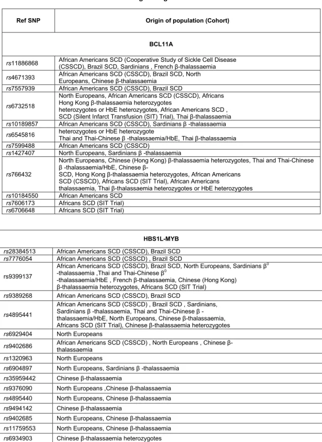
Table 2 Selected BCL11A and HBS1L-MYB intergenic region SNPs
A γ-globin gene–HBG1
nd-HPFH -113 (AG)
Georgia nd-HPFH -114(CT)
Greek-Sardinian HPFH -117 (GA) and IVS-I-110
Black-Greek nd-HPFH -117 (GA) and IVSII-745 (CG)
Cretan -158 (CT) Black nd-HPFH -175 (TC) Brazilian nd-HPFH -195 (CG) Italian nd-HPFH -196 (CT) British nd-HPFH -198(TC) Black nd-HPFH -202 (CT) Venezuelan nd-HPFH -211 (CT) 13bp deletion (-114 -102) Gγ-globin gene–HBG2 Czech nd-HPFH -110 (AC) Algerian nd-HPFH -114 (CA) Japanese nd-HPFH -114 (CT) Australian nd-HPFH -114 (CG) Black nd-HPFH (XmnI) -158 (CT) Black nd-HPFH -161 (AG) Black-Sardinia-British nd-HPFH -175 (TC) Black nd-HPFH -202 (CG) -250 (C-->T) -250 (CT) -386 (A->G) -386 (AG) Iranian American nd-HPFH -567 (TG)
25
Over the years various regions were investigated but two have led to interesting results. It was shown that on 6q23 SNPs in the intergenic region HBS1L-Myb influence HbF levels. Studies in Caucasians have identified three significant SNPs. As for 2p16 region, in the second intron of the BCL11A gene, several mapped SNPs were implicated strongly in the HbF modulation. In particular three SNPs rs11886868, rs4671393, rs7557939 have an higher frequency in different populations (Sankaran, Menne et al. 2008).
However, these SNPs often are present with a different frequency depending on the population taken into account. The links between SNPs and HbF has been amply demonstrated and validated in different groups of patients with hemoglobinopathies.
For example SNPs rs28384513, rs9399137, rs4895441 (HBS1L-Myb) and rs4671393 (BCL11A) were identified in sickle cell anemia patients (Lettre, Sankaran et al. 2008), while rs 11886868 (BCL11A) was recognized as a major factor amiliorating the status of homozygotes β039 thalassemia patients in the Sardinian population (Uda, Galanello et al. 2008). (Table3)
All aspects so far described are very interesting as they can help to clarify the molecular basis for the genotype/phenotype correlation and prove useful for the prognostic purposes.
A polymorph framework which modulating the HbF synthesis is able to influence the disease clinical condition, therefore can be considered a positive prognostic impact element for the patient. This offers numerous reflections on possible therapeutic strategies for the treatment of hemoglobinopathies.
The International HapMap Project is an organization which aims to develop a haplotype map (HapMap) of the human genome to describe the common patterns of human genetic variation. HapMap is a key resource for researchers to find genetic variants affecting health, disease and responses to drugs and environmental factors too. The information produced by the project is made freely available to researchers around the world (Project 2003). The International HapMap Project is a collaboration among researchers of academic centers, non-profit biomedical research groups and private companies in Canada, China, Japan, Nigeria, the United Kingdom, and the United States. It officially started with a meeting on October 2002, and was expected to take about three years.
26
Table 3 Selected BCL11A and HBS1L-MYB intergenic region SNPs
Ref SNP Origin of population (Cohort) BCL11A
rs11886868 African Americans SCD (Cooperative Study of Sickle Cell Disease (CSSCD), Brazil SCD, Sardinians , French β-thalassaemia
rs4671393 African Americans SCD (CSSCD), Brazil SCD, North Europeans, Chinese βthalassaemia rs7557939 African Americans SCD (CSSCD), Brazil SCD
rs6732518
North Europeans, African Americans SCD (CSSCD), Africans Hong Kong β-thalassaemia heterozygotes
heterozygotes or HbE heterozygotes, African Americans SCD , SCD (Silent Infarct Transfusion (SIT) Trial), Thai β-thalassaemia
rs10189857 African Americans SCD (CSSCD), Sardinians β -thalassaemia
rs6545816 heterozygotes or HbE heterozygote Thai and Thai-Chinese β -thalassaemia/HbE, Thai β-thalassaemia rs7599488 African Americans SCD (CSSCD)
rs1427407 North Europeans, Sardinians β -thalassaemia
rs766432
North Europeans, Chinese (Hong Kong) βthalassaemia heterozygotes, Thai and Thai-Chinese β thalassaemia/HbE, Chinese β
SCD, Hong Kong βthalassaemia heterozygotes, African Americans SCD (CSSCD), Africans SCD (SIT Trial), African Americans
thalassaemia, Thai β-thalassaemia heterozygotes or HbE heterozygotes
rs10184550 African Americans SCD
rs7606173 Africans SCD (SIT Trial)
rs6706648 Africans SCD (SIT Trial)
HBS1L-MYB
rs28384513 African Americans SCD (CSSCD), Brazil SCD
rs7776054 African Americans SCD (CSSCD) , Brazil SCD
rs9399137
African Americans SCD (CSSCD), Brazil SCD, North Europeans, Sardinians β0
-thalassaemia ,Thai and Thai-Chinese β0
-thalassaemia/HbE , French β-thalassaemia, Chinese (Hong Kong) β-thalassaemia heterozygotes, Africans SCD (SIT Trial)
rs9389268 African Americans SCD (CSSCD), Brazil SCD
rs4895441
African Americans SCD (CSSCD) , Brazil SCD , Sardinians, Sardinians β -thalassaemia, Thai and Thai-Chinese β - thalassaemia/HbE, North Europeans, Chinese β-thalassaemia, Africans SCD (SIT Trial), Chinese β-thalassaemia heterozygotes
rs6929404 North Europeans
rs9402686 African Americans SCD (CSSCD) , North Europeans , Chinese β- thalassaemia rs1320963 North Europeans
rs6904897 North Europeans, Sardinians β -thalassaemia
rs35959442 Chinese βthalassaemia
rs9376090 North Europeans ,Chinese β-thalassaemia
rs4895440 North Europeans, Chinese β-thalassaemia
rs9494142 Chinese βthalassaemia
rs9402685 North Europeans, Chinese β-thalassaemia
rs11759553 North Europeans, Chinese β-thalassaemia
rs6934903 Chinese βthalassaemia heterozygotes
It comprises two phases, the complete data obtained in Phase I were published on October 2005. The analysis of the Phase II dataset was published in October 2007. The Phase III dataset was released in spring 2009.
27
Galarneau et al. (2010) resequenced 175.2 kb to fine map HbF association signals at the BCL11A, HBS1L-Myb and β-globin loci investigated in 190 individuals including the HapMap European CEU, Nigerian YRI founders and 70 African Americans with sickle cell anemia. The authors discovered 1489 sequence variants, including 910 previously unreported variants. Using this information and data from HapMap, Galarneau et al. (2010) selected and genotyped 95 SNPs. The XmnI polymorphism rs7482144 in the proximal promoter of HBG2 marks the Senegal and Arab-Indian haplotypes and is associated with HbF levels in African Americans with sickle cell disease (Lettre et al., 2008). Galarneau et al. 2010) replicated the association between rs7482144 and HbF levels. p=3.7x10(-7). However, rs10128556, a T/C SNP located downstream of HBG1 (rs142200), was more strongly associated with HbF levels than rs7482144 by 2 orders of magnitude (p=1.3x10(-9). When conditioned on rs10128556, the HbF association result for rs7482144 was not significant, indicating that rs7482144 is not a causal variant for HbF levels in African Americans with sickle cell anemia. The results of a haplotype analysis of the 43 SNPs in the beta-globin locus using rs10128556 as a covariate were not significant (p=0.40), indicating that rs10128556 or a marker in linkage disequilibrium with it is the principal HbF-influencing variant at the beta-globin locus in African Americans with sickle cell anemia.
3. The hematopoiesis and erythroid cell methods of culture.
The cells forming the blood have a short relatively lifetime, for this reason, the human body contains systems to ensure a continuous cell turnover. The hematopoiesis is the process of production of all blood cells from stem cells pluri-potent and it takes place, in the adult, in the bone marrow. The site of hematopoiesis varies in different stages of our life. The bone marrow is about 5% of total body weight and it is distributed mainly in the flat bones and long in childhood and youth, while in later life you it happens at the level of the ribs, vertebrae, sternum and pelvis. During intrauterine life the hematopoiesis happens instead, first in the yolk sac and then in the liver and spleen, while from 7-8 months of gestation, the hematopoiesis occurs gradually in the bone marrow in special areas called "niches" represented by 1-2% of the population bone marrow.
As the site of erythropoiesis changes the process regarding the red cells production, the globin genes being expressed also change, progressing from
28
embryonic to fetal and finally to adult (Figure5). Thus, globin gene regulation serves both as a model for developmental gene regulation during ontogeny and for the molecular mechanism playing a role in cellular differentiation (Pope, Fibach et al. 2000).
Figure 5 Hemopoiesis. All lymphoid and myeloid cells are originated from a single multipotent
hematopoietic stem cell. At the bottom are represented the steps leading to the formation of a mature red blood cell.
Hematopoietic stem cells are also found in the blood, but in very low percentage, approximately 0.05% of peripheral blood cells (Isgro, Marziali et al. 2009). Hematopoietic stem cells are characterized by the presence of specific antigens located on the cell surface. In particular, the CD34+ antigen identifies all
29
hematopoietic stem cells and bone marrow progenitors; this is a transmembrane glycoprotein of 110 kDa single chain; the presence of many glycosylation sites, the lack of tyrosine kinase activity and the very low frequency of synthesis and degradation suggests that CD34+ is a receptor involved in cell or cell-extracellular matrix interaction. His expression varies with the degree of differentiation and proliferative potential of hematopoietic cellular elements: it is highest in the most primitive population of the bone, represented by the "long-term culture-initiating cells" (LTC-IC), and it is thought that it is lost gradually during the cell proliferation and differentiation. This marker is expressed by 1-3% of the cells of the bone, from 0.01-0.1% of peripheral blood cells and by 0.1-0.4% of the cells of the umbilical cord (Isgro, Marziali et al. 2009). One of the first steps of differentiation of hematopoietic stem cells is to continue the path of differentiation in myeloid or lymphoid sense. Together they will give rise to all blood cells. Stem cells still have the capacity to divide, but which are not able to differentiate along all the different ways and guide its development only to a specific line are defined hematopoietic committed, these cells are intended for differentiation according to one line of differentiation. They proliferate in differentiating morphologically identifiable precursors, which are subject to greater maturity, through which acquire the functions highly specialized and lose their ability to regenerate yourself (Amit and Itskovitz-Eldor 2006). The latter, in their turn, give rise to cells that can differentiate further producing (Gurdon, Byrne et al. 2003) myeloblasts, from which they originated granulocytes; erythroblasts, from which derive reticulocytes, and then the red blood cells; megakaryocytes, from which are formed platelets. The process of the differentiation, and in particular the specialization of cells is made possible by a system of gene regulation that repressing the not specific-genes of that cellular sector. Gene regulation is ensured by chemical signals internal to the same cell and by signals from outside the cell, for example from adjacent cells. The early stages of the hematopoietic process are controlled by a group of growth factors called cytokines, produced and secreted not only by the marrow cells, but also by cells of the immune system. Among the cytokines mainly involved in the differentiation process are the SCF (stem cells factor), interleukin-3, GM-CSF (Granulocyte-macrophage colony stimulating factor). Then there are growth factors such as EPO (erythropoietin) which acts on already specialized cells. EPO binds to protein receptors with tyrosine kinase activity activating a mechanism of phosphorylation of the target proteins (Box et al., 1996). The whole process
30
leading to the production of erythrocytes starting from stem cells is called erythropoiesis. This process requires in addition to the presence of cytokines and erythropoietin and the presence of folic acid, vitamin B₁₂, iron and trace elements such as copper, cobalt and nickel too.
The first erythroid-committed progenitors are defined functionally as burst-forming units (BFUe) (Clarke and Housman 1977). They can be isolated from bone marrow and peripheral blood. The BFU-E can be maintained in culture and after 10-15 days give rise to colonies of erythroid precursors. Erythroid units colony-forming (CFU-E) are more mature cells, capable of producing of small dimensions cellular clones. The formation of erythrocytes from the committed cells is regulated by the EPO that interacting with specific receptors present on the membrane of committed cells and leads to the formation of precursor said pro-normoblasts (also called pro-erythroblasts). The time required for this process is usually about a week (at least four cell divisions) and during this period occurs the reduction the size of the nucleus and the increase in the synthesis of hemoglobin (Cuneo, Balboni et al. 1994). Following the last cellular division, the nucleus is pyknotic ousted from erythroblast and in this way, creates the reticulocyte. Reticulocytes remain for about two or three days in the bone marrow and then be placed in the blood circuit, where they assume their final morphology (loss of mitochondria and ribosomes). After 24 hours the erythrocytes take a structure similar to biconcave and oval disks with a diameter of about 6-8 uM and a maximum thickness of 2 uM, is totally free of cellular organelles and has the ability to transfer large amounts of gas (especially oxygen and CO₂) facilited by ratio between surface and volume. The developmental aspects of erythropoiesis have been studied in culture using in vitro established cell lines that can be induced to express erythroid characteristics, or using primary cultures of cells derived from progenitors present in the bone marrow or in peripheral blood.
Erythroid cell lines have been used extensively as models to study β-thalassemia and to develop new therapeutic strategies. In addition, primary cultures of erythroid cells can be readily established from a lot of normal and individual patients and their growth in vitro forming blackberries. Until today, the erythroid cells were cultured either in semi-solid medium, where they develop into discrete colonies, or in liquid medium where they grow as single cells or clusters in suspension (Fibach, Manor et al. 1989) (Leimberg, Konijn et al. 2003)(Fibach and Prus 2005). In all these systems erythropoietin (EPO) is essential for full
31
development of hemoglobin containing erythroid cells (Fibach, Bianchi et al. 2003). Here will be describe these two cellular culture systems.
Semi-solid cultures. This method permit to evaluate the quantity and quality of the grown progenitors, but theis immobilization in the semisolid medium results in several disadvantages that make it technically difficult to carry out quantitative analyses of the developing cells. The culture of erythroid precursors performed in this manner could be provide for suspension of single hematopoietic cells, derived from bone marrow, peripheral blood or from other sources, such as the fetal liver, cord blood or neonatal spleen adults, and are dispersed in semi-solid media usually containing methyl cellulose or plasma clot. With this method it's possible to study the colonies grown from a single cell in vitro. Colonies begin to appear after 3-4 days of incubation, and reach their final size and haemoglobinization after 1-2 weeks. Each colony is a clone derived from a progenitor erythroid-committed. On the basis of the final size of the colonies and the time required for theircan be distinguished hemoglobinization various types of progenitors. The colony forming units erythroid late (CFUe) reach the final size and hemoglobinization after 1 week and then disappear, while the early erythroid burst-forming units (BFUe) develop after 2 weeks. Erythroid colonies can be distinguishes from other (myeloid) colonies by their red colour or by their positive reaction with heme-specific reagents. The fact that in this culture system the cells are immobilized in semi-solid medium results in several disadvantages. The cell yield per colony and the total cells per culture are low making it technically difficult to carry out biochemical, molecular and immunological characterizations of the developing cells. Also, it is a one-step continuous culture and addition of the tested agents during the culture is difficult. In addition, the HbF produced in colonies grown in semi-solid medium is significantly higher than that produced in vivo by the same donor of the cells (Fibach, Bianchi et al. 2003).
The two-phase liquid cultures. The culture procedure yields populations that are large, relatively pure, and synchronized (in terms of differentiation), and which mimic in vivo erythropoiesis. Since the cells are grown in suspension, samples of cells can be remuved every time without disturbing the cultures and assayed for various parameters, e.g., morphology, size, number, viability, apoptosis, cell cycle, surface antigens, and gene expression. Cultures can be initiated with cells derived from a whole unit of blood obtained from normal donors (or polycythemia patients undergoing phlebotomy). The starting material, i.e., the “buffy-coat” fraction, is