Full Terms & Conditions of access and use can be found at
https://www.tandfonline.com/action/journalInformation?journalCode=kccy20
ISSN: 1538-4101 (Print) 1551-4005 (Online) Journal homepage: https://www.tandfonline.com/loi/kccy20
Time-course analysis of nuclear events during
conjugation in the marine ciliate Euplotes vannus
and comparison with other ciliates (Protozoa,
Ciliophora)
Yaohan Jiang, Tengteng Zhang, Adriana Vallesi, Xianyu Yang & Feng Gao
To cite this article: Yaohan Jiang, Tengteng Zhang, Adriana Vallesi, Xianyu Yang & Feng Gao (2019) Time-course analysis of nuclear events during conjugation in the marine ciliate Euplotesvannus and comparison with other ciliates (Protozoa, Ciliophora), Cell Cycle, 18:3, 288-298, DOI:
10.1080/15384101.2018.1558871
To link to this article: https://doi.org/10.1080/15384101.2018.1558871
View supplementary material Accepted author version posted online: 19 Dec 2018.
Published online: 13 Jan 2019. Submit your article to this journal Article views: 215
View related articles View Crossmark data
RESEARCH PAPER
Time-course analysis of nuclear events during conjugation in the marine ciliate
Euplotes vannus and comparison with other ciliates (Protozoa, Ciliophora)
Yaohan Jianga,b, Tengteng Zhanga,b, Adriana Vallesic, Xianyu Yangd, and Feng Gaoa,b
aInstitute of Evolution and Marine Biodiversity, Ocean University of China, Qingdao, China;bMinistry of Education, Key Laboratory of Mariculture (Ocean University of China), Qingdao, China;cLaboratory of Eukaryotic Microbiology and Animal Biology, University of Camerino, Camerino, Italy;dCollege of Animal Science and Technology, Zhejiang A&F University, Hangzhou, China
ABSTRACT
Ciliates represent a morphologically and genetically distinct group of single-celled eukaryotes that segregate germline and somatic functions into two types of nuclei and exhibit complex cytoge-netic events during the sexual process of conjugation, which is under the control of the so-called “mating type systems”. Studying conjugation in ciliates may provide insight into our understand-ing of the origins and evolution of sex and fertilization. In the present work, we studied in detail the sexual process of conjugation using the model speciesEuplotes vannus, and compared these nuclear events with those occurring in other ciliates. Our results indicate that in E. vannus: 1) conjugation requires about 75 hours to complete: the longest step is the development of the new macronucleus (ca. 64h), followed by the nuclear division of meiosis I (5h); the mitotic divisions usually take only 2h; 2) there are three prezygotic divisions (mitosis and meiosis I and II), and two of the eight resulting nuclei become pronuclei; 3) after the exchange and fusion of the pronuclei, two postzygotic divisions occur; two of the four products differentiate into the new micronucleus and macronucleus, respectively, and the parental macronucleus degenerates completely; 4) comparison of the nuclear events during conjugation in different ciliates reveals that there are generally three prezygotic divisions while the number of postzygotic divisions is highly variable. These results can serve as reference to investigate the mating type system operating in this species and to analyze genes involved in the different steps of the sexual process.
ARTICLE HISTORY Received 25 August 2018 Revised 16 November 2018 Accepted 1 December 2018 KEYWORDS
Ciliate; Euplotes vannus; conjugation; mating type; life cycle
Introduction
Ciliates represent a monophyletic group of
eukar-yotic microorganisms characterized by the
pre-sence of two types of nuclei within the same
cytoplasm: a small diploid germline micronucleus
(MIC) which is transcriptionally silent in cell’s
vegetative life, and a large polyploid somatic
macronucleus (MAC) which is transcriptionally
active and regulates the cell phenotype [
1
–
6
]. In
cell reproduction, MIC undergoes mitosis while
MAC divides by
“amitosis”, a division which
does not involve spindle formation and
chromo-some condensation but is simply a split of the
DNA content into two equal halves [
7
–
12
].
The sexual process of ciliates is typically
repre-sented by conjugation, which involves a temporary
union of two mating partners, although in some
groups such as the peritrichs and chonotrichs the
mating partners merge permanently [
13
,
14
]. Once
mating partners are united in pairs, MIC
under-goes meiosis to generate two gametic pronuclei,
one resident and one migratory. The migratory
pronuclei are exchanged between the two mating
partners through a cytoplasmic bridge, and after
mutual fertilization between the resident and
migratory gametic pronuclei, the two cells
sepa-rate. The new nuclear apparatus eventually
devel-ops in each exconjugant cell from the mitotic
products of the synkaryon, while the old MAC
fragments and degrades [
15
]. The genome of the
MIC remains organized in large chromosomes,
while the genome of the new developing MAC is
subjected to extensive rearrangements which
includes chromosome fragmentation, DNA
elimi-nation and gene amplification to generate
uncon-ventional linear nano-chromosomes [
7
,
16
–
20
].
Mating
is
genetically
controlled
through
a mechanism known as the
“mating type system”.
CONTACTFeng Gao [email protected]; Xianyu Yang [email protected]
Supplementary material data for this article can be accessedhere. CELL CYCLE
2019, VOL. 18, NO. 3, 288–298
https://doi.org/10.1080/15384101.2018.1558871
Some species have only two mating types, such as
Paramecium aurelia [
21
]: mating type E depends on
expression of the transmembrane protein mtA, while
type O is determined during MAC development by
scnRNA-dependent excision of the mtA promoter
[
22
]. Others have more than two types. Tetrahymena
thermophila has seven mating types, each one
deter-mined by a specific mating type gene pair that is
stochastically assembled in the MAC during somatic
differentiation by homologous recombination of
incomplete mating type gene pairs that are linearly
arranged in the MIC [
23
]. Euplotes Ehrenberg, 1830,
one of the most highly diverse and cosmopolitan
ciliate genera, has evolved high-multiple mating type
systems [
4
,
24
–
29
]. In E. patella, E. octocarinatus,
E. raikovi, and E. crassus, each mating type is
deter-mined by the allele combination at the
“mating type
locus” of the germinal MIC [
30
–
32
]. These alleles are
converted into gene-size DNA molecules in the MAC,
where they are co-dominantly expressed to control the
synthesis of type-specific chemical markers usually
referred as pheromones [
13
,
32
–
35
].
Euplotes vannus is a cosmopolitan marine ciliate
that has been used as model organism in a wide range
of disciplines [
27
,
36
,
37
]. It has more than ten mating
types; however, the molecular mechanism of its
mat-ing type determination is still largely unknown.
Detailed knowledge of the different steps taking
place during conjugation in this species thus appears
to be crucial for analyzing genes controlling the
sex-ual process. In this paper, we describe the time-course
analysis of the nuclear events occurring during and
after conjugation in E. vannus strains collected along
the Yellow Sea coast of Qingdao, China, and compare
them with those of other species.
Results
The conjugation process step-by-step
Conjugation was induced by mixing cells of
dif-ferent mating types (
Figure 1(e–g)
), and the initial
formation of mating pairs was taken as time 0 of
the process (
Figure 1(l
)). A detailed description of
the different steps of nuclear events is as follows.
Step 1: mitosis of the MIC (first prezygotic
division).
Soon after cell-cell union, MIC migrates out of
the concavity of the
“C”-shaped macronucleus and
the concaved surface of MAC flattens (
Figure 2
(a
)). At this point, MIC divides by mitosis,
form-ing two nuclear products (
Figure 2(b
)). This step
lasts about 2 h (
Figure 1(l
)).
Step 2: meiosis (second and third prezygotic
divisions)
The two products of the first MIC division
undergo a classical two-step meiosis. The first
meiotic division (
Figure 2(c
–i)) (second prezygotic
division) takes about 5h (
Figure 1(l
)), during
which chromatin polymerizes assuming the
“bou-quet-like” shape, a typical zygotene stage when
chromosomes approximately line up with each
other into homologous chromosome pairs [
38
,
39
]
(
Figure
2(e–g)
).
The
four
products
enter
the second meiotic division (third prezygotic
divi-sion) which lasts about 1 hour (
Figure 1(l
)), with
the final formation of eight pronuclei (
Figure 2(j
)).
Step 3: pronuclei and synkaryon formation
At the end of meiosis, four pronuclei gather
together in the anterior part and four in the
poster-ior part of the cell. For each anterposter-ior and posterposter-ior
group of the pronuclei, one out of four swells while
the other three become pyknotic and degenerate
(
Figure 2(k
)). Of the two swollen nuclei, one is the
migratory pronucleus and the other is the stationary
pronucleus. After the reciprocal exchange of the
migratory pronuclei, the fusion of pronuclei
gener-ates the synkaryon in each mating cell (
Figure 2(l
)).
The degenerating meiotic products accumulated on
the edge of the cell are still visible at this stage. This
step takes about 1.5 hours (
Figure 1(l
)).
Step 4: first and second postzygotic divisions
The synkaryon is subjected to two successive
mitotic divisions generating four postzygotic
nuclei
(
Figure
2(m
,n)).
After
approximately
11 hours, mating cells complete the second
syn-karyon division and start to separate. Mating pairs
usually separate completely within 1 hour after
the second synkaryon division; at this point,
frag-ments of the old MAC and the four products of
the synkaryon division are visible inside each
mat-ing cell (
Figure 3(a
)).
Step 5: new MIC and MAC development
Soon after mate separation, one out of four
pro-ducts (usually the third one from anterior to posterior)
of the synkaryon divisions swells and differentiates
into the macronuclear anlagen (
Figure 3(b
)). In the
placed but moves to occupy the mid-region of the
posterior half of the cell and enlarges rapidly,
increas-ing in size to 40
μm within the next 24 hours, to reach
about 1/3 of the cell length (
Figure 3(c,d)
). The time of
anlagen development appears to be directly correlated
with the uptake of food by the cell: in the absence of
food, the anlagen will remain in the cell for more than
40 hours, whereas this time is reduced by at least a half
when food is present. The parental macronuclear
frag-ments degrade completely and the newly developing
macronucleus elongates to acquire the typical
“C”
shape (
Figure 3(e
–l)).
The new MIC derives from one of the three
resi-dual products of synkaryon divisions. The remaining
two eventually degenerate, gathering together with
fragments of the old macronucleus in the front part
of the cell (
Figure 3(d-f)
). On 200 exconjugant cells
analyzed at the stage of maximum expansion of
macronuclear anlagen, 66 cells had 3 residual nuclei,
51 cells had 2, and 83 cells had only 1. Residual nuclei
could be observed in some exconjugant cells even
after the new macronucleus is completely formed.
Fate of the parental macronucleus
From the beginning of conjugation to the end of
the first prezygotic division, the parental MAC is
“C”-shaped without obvious morphological
changes. From the second prezygotic division
(
Figure 2(c
)), the macronucleus begins to break
into several oval fragments, which then cluster in
the anterior part of the cell. With the development
of the macronuclear anlagen, these fragments
become irregular and move toward the center of
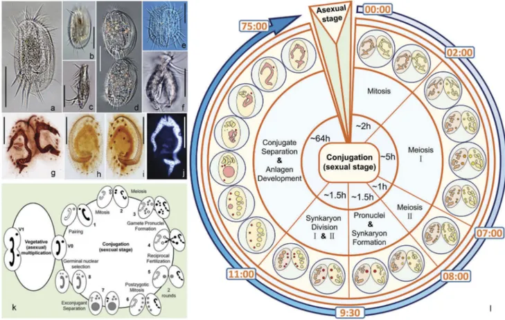
Figure 1.Morphology and life cycle of Euplotes vannus. (a–b) Ventral side of a representative vegetative cell; (c) Lateral view of a vegetative cell; (d) Ventral side of the divider at late divisional stage; (e–f) Ventral and lateral views of a conjugating pair. (g) Ventral view of a conjugating pair to show the infraciliature after protargol staining; (h–i) Dorsal and ventral views of the same specimen at vegetative stage to show the infraciliature; (j) A vegetative cell after Hoechst 33342 staining to show the micronucleus and macronucleus; (k) Life cycle of E. vannus. Left: vegetative (asexual) reproduction phase. V0: vegetative cell. V1: cell undergoing binary fission. Micronucleus and macronucleus divide mitotically and amitotically, respectively. Right: conjugation, the sexual stage of the life cycle. (l) Timing of nuclear events during conjugation. The initial formation of mating pairs was taken as time 0 of the process. Scale bars = 50μm.
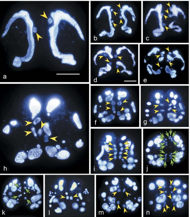
Figure 2.Hoechst 33342 stained conjugating pairs of Euplotes vannus to show the nuclear events before the pair separation. Yellow arrowhead: nucleus that has the same ploidy with the micronucleus. Green arrow: gamete nucleus. (a) Micronucleus migrates out of the concavity of macronucleus after the pair formation; (b) A conjugating pair after mitosis of the MIC; (c-d) Mitosis products become inflated. Meanwhile, the macronucleus begins to degenerate; (e) Zygotene stage of the first meiosis showing that chromatin polymerizes into a typical“bouquet” shape; (f–i) Various stages of the first meiosis division; (j) The second meiotic division resulting in eight pronuclei; (k) Gamete nuclei (indicated by green arrows) inflate while other division products degenerate; (l) Synkaryon formation after migration and fusion of pronuclei; (m) The first synkaryon division; (n) The second synkaryon division. Scale bars = 30μm.
the cell and they reduce in size (
Figure 3(a
–i)).
Finally they disappear when a new macronucleus
is completely developed (
Figure 3(j
-l)).
Discussion
Conjugation is unique to ciliates and has been
exten-sively studied since its discovery [
40
]. Cell mating
and restoration of the vegetative state (
Figure1(k
))
have been described in many different ciliates, but
mostly intensively in Tetrahymena, Paramecium,
Oxytricha, Chilodonella, and Euplotes, and primarily
focused on nuclear events [
41
–
43
] (
Figure 5
). In
E. vannus, we determined the time needed to
com-plete the prezygotic and postzygotic divisions by
using a fluorescent dye to visualize the nuclear
divi-sion products. Our results indicate that the
prezygo-tic nuclear division of meiosis I, which lasts about
five hours, is much longer than mitosis, which
requires about two hours. The three prezygotic
divi-sions are carried out in about eight hours. After
approximately 11 hours from mating pair formation,
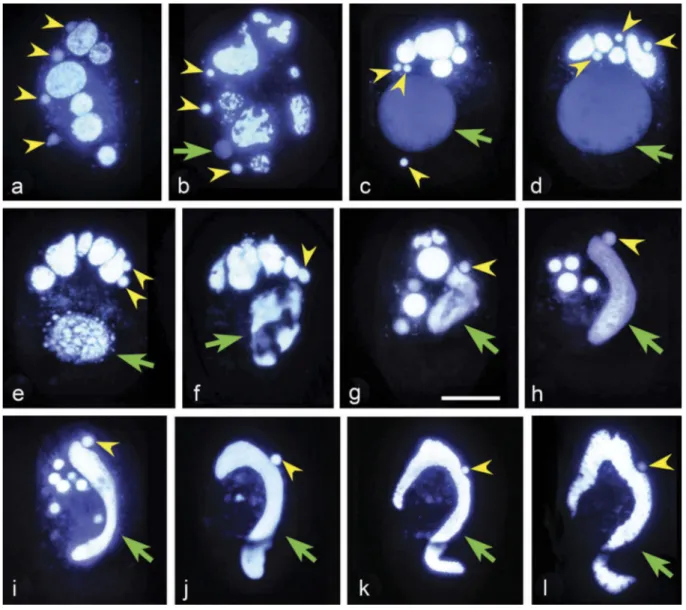
Figure 3.Hoechst 33342 stained exconjugants of Euplotes vannus to show the nuclear events. Yellow arrowhead: synkaryon division product or micronucleus. Green arrow: macronucleus anlagen or macronucleus. (a) There are four products of synkaryon division in each exconjugant (as indicated by the yellow arrowheads); (b–d) One out of four products (usually the third one from anterior to posterior) of the synkaryon divisions swells and differentiates into the macronucleus anlagen (indicated by the green arrow); (e–g) Complex DNA rearrangements during the development of macronucleus anlagen. (h–k) Development of macronucleus anlagen and micronucleus selection, while the old macronucleus and the residual nuclei gradually degrade; (l) Well-developed new macro-nucleus and micromacro-nucleus. Scale bars = 30μm.
most mating partners complete the second
synkar-yon division and start to separate. An additional ca.
64 hours are needed to complete the development of
the new nuclear apparatus in each exconjugant cell
(
Figure 1(l
)).
Our observations confirmed that, as previously
reported for the E. crassus-vannus-minuta group
[
45
], MIC undergoes only three prezygotic divisions
(mitosis, meiosis I and meiosis II) in E. vannus,
whreas
in
other
Euplotes
species,
such
as
E. woodruffi, E. charon, E. raikovi, E. octocarinatus
and E. cristatus, MIC divides four times (a
prelimin-ary mitotic division, meiotic I and II, and a second
mitotic division) (
Figure 4
) [
38
,
46
–
48
]. Therefore,
the stationary and migratory pronuclei in E. vannus
derive directly from two of the eight meiotic
pro-ducts and, as such, may be genetically different.
Similar to E. vannus, Chilodonella uncinata,
Halteria grandinella, Tetrahymena thermophila and
Paramecium caudatum have only one MIC and
three prezygotic divisions occur in these species
(
Figure
4(d–g)
).
In
P.
caudatum
[
49
]
and
T. thermophila [
15
], however, MIC first enters two
meiotic divisions (meiosis I and II) and generates four
haploid nuclei, only one of which divides mitotically
to form gametic nuclei. In H. grandinella [
50
] and
C. uncinata [
43
], one product is selected at the end of
each division to complete the process (
Figure 4(d–e)
).
Consequently, in these four species stationary and
migratory gametic pronuclei are genetically identical.
In Paramecium aurelia [
21
] and Oxytricha
tri-fallax [
51
], which have two MICs, there are three
prezygotic divisions (meiosis I and II, and
mito-sis). Conjugation in peritrichs such as Carchesium
polypinum [
52
] differs from most other ciliates
because the two mating cells are anisometric
com-prising a stationary macrogamete and a motile
microgamete. The MIC divides three times also
in the microgamete. Therefore, it appears that
three prezygotic divisions (mitosis, meiosis I and
meiosis II) represent a general rule, although in
each species different products are selected for
generating gametic pronuclei. Euplotes vannus
conforms to this rule, whereas the other Euplotes
species with four prezygotic divisions represent
exceptions.
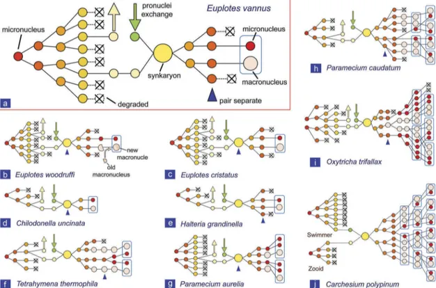
Figure 4.Comparison of the nuclear events during conjugation in Euplotes vannus with those in other ciliate species. (a) Euplotes vannus from present study; (b) Euplotes woodruffi [38]; (c) Euplotes cristatus [46]; (d) Chilodonella uncinata [43]; (e) Halteria grandinella [50]; (f) Tetrahymena thermophila [15]; (g) Paramecium aurelia [21,53]; (h) Paramecium caudatum [49]; (i) Oxytricha trifallax [42,51]; (j) Carchesium polypinum [52].
The number of postzygotic divisions is more
vari-able in different ciliates (
Figure 4
). In C. uncinata the
synkaryon divides only once to produce the new
MIC and MAC [
43
]; in H. grandinella and all
Euplotes spp. it divides twice [
38
,
46
,
50
]; in
T. thermophila and P. aurelia it divides twice
fol-lowed by one cell division [
15
,
21
,
53
]; in O. trifallax it
divides twice followed by two cell divisions [
42
,
51
];
in P. caudatum it divides three times followed by two
cell divisions [
49
]; in C. polypinum it divides three
times followed by three cell divisions [
52
]. Also the
selection of nuclei for MIC and MAC differentiation
appears to be species-specific.
In conclusion, conjugation in E. vannus requires
about 75 hours to complete all the different steps,
from mating pair formation to the development of
new MIC and MAC in exconjugant cells. Pre- and
postzygotic nuclear divisions occur rapidly and,
soon after the second postzygotic division, two of
the four nuclei are selected to generate the new MIC
and the MAC anlagen, while the remaining two
nuclei degenerate. Compared to the pre- and
post-zygotic nuclear divisions, a longer time is needed to
complete the development of a new MAC. Different
species of Euplotes, however, vary with regard to the
fate of the old macronucleus. In E. woodruffi and
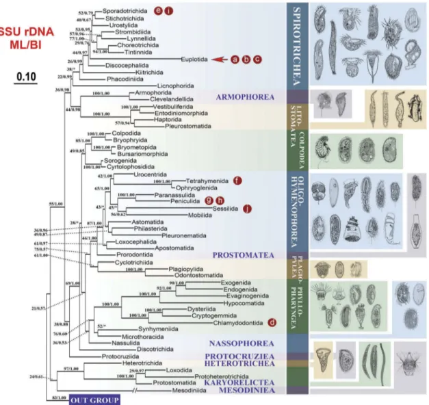
Figure 5.Maximum likelihood (ML) trees of the phylum Ciliophora based on the SSU rDNA to show the position of the ciliates that were compared in the present study. Numbers at nodes represent the bootstrap values of maximum likelihood (ML) out of 1000 replicates and the posterior probability of Bayesian analysis (BI). Asterisk (*) indicates disagreement between ML and BI analyzes. The scale bar corresponds to 10 substitutions per 100 nucleotide positions. The letters marked in the tree are corresponding to the taxa shown in Figure 4. (a) Euplotes vannus; (b) Euplotes woodruffi; (c) Euplotes cristatus; (d) Chilodonella uncinata; (e) Halteria grandinella; (f) Tetrahymena thermophila; (g) Paramecium aurelia; (h) Paramecium caudatum; (i) Oxytricha trifallax; (j) Carchesium polypinum. The illustrations on the right side are according to the previous study [44].
E. patella, the remnants of the old MAC reorganize
and fuse with the new MAC anlagen [
38
,
54
]. In
other species of Euplotes, including E. vannus, this
phenomenon has never been observed, and
frag-ments of the old MAC degenerate completely.
Conjugation in ciliates is equivalent to, but
more complicated than sexual reproduction in
higher eukaryotes. First, due to the nuclear
dimorphism, the daughter nuclei of the last
post-zygotic division have to differentiate into both
germline and somatic nuclei within a single cell.
Although recent studies indicate that nucleus
differentiation in ciliates requires biased
assem-bly of the nuclear pore complex [
55
], the entire
process has not yet been fully clarified. The most
complicated nucleus differentiation was observed
in
the
species
Blepharisma
japonicum
in
which macronucleus could differentiate through
both sexual and asexual paths proceeded
syn-chronously in each cell with one path eventually
dominated the other [
56
]. Second, conjugation is
performed under the control of the so-called
“mating type systems”, which exhibit significant
variety in terms of mating type number, mating
type determinants, and mating type inheritance
[
25
]. The mechanisms of mating type
determina-tion in Paramecium [
57
], Tetrahymena [
41
,
58
],
and Euplotes [
24
,
35
,
59
,
60
] are strikingly different
at the molecular level. Therefore, this study may
provide basic information to investigate the
evo-lution of mating type systems, sexual phenomena
and differentiation of dimorphic nuclei in ciliates
and, more in general, may shed new light on the
origins and evolution of sex and fertilization in
eukaryotes.
Materials and methods
Species identification and mating type
determination
Strains used in this study were collected in
July 2015 from the Silver Sand Beach of Qingdao,
China (35°55ʹN, 120°12ʹE), and have been
assigned to the species E. vannus on the basis of
morphological characters observed both in vivo
and after protargol staining (
Figure 1(a–d,h,i)
) as
previously described [
61
–
64
]. Single cells of the
exconjugants
were
separated
to
obtain
17
monocultures. Six mating types were identified
using pairwise mixtures of the 17 monocultures.
Cell culture and conjugation induction
Cells were maintained at room temperature (ca.
25°C) in sterilized seawater, using Escherichia coli
as food source. To obtain highly reactive cells, cells
were centrifuged (300 g, 3 min), re-suspended in
fresh seawater at a concentration of 4000 cell/ml,
and starved for two days. Conjugation was
induced by mixing reactive cells of different
mat-ing types. The rate of conjugation was usually
more than 80%.
Staining and observation
When the cells started to form pairs, the paired
cells were picked out and were considered as
synchronized (time 0). Cells were sampled
every 30 min or 1 hour after initial mating pair
formation (Table (S1)) and stained with Hoechst
33342 (Beyotime Institute of Biotechnology,
Haimen, Jiangsu, China) [
49
]. Hoechst stock
solutions (10x, 100 µg/ml) were prepared in
sterilized distilled water and kept at 4°C until
further processing. Aliquots of 2.5 µl Hoechst
stock solution were added to 200 µl cell
suspen-sions (final concentration: 1.1
μg/ml), and
incu-bated at room temperature for 15 min. Cells
were then transferred to a glass microscope
slide, covered by a coverslip, and observed
under a
“ZEISS AXIO Imager. D2” fluorescence
microscope, equipped with an Axiocom 506
camera for photographic documentation. For
each time point, about 20–50 pairs were
recorded.
Phylogenetic analyzes
In order to show the phylogenetic positions of
spe-cies used in the present study, phylogenetic analyzes
based on small subunit ribosomal DNA (SSU rDNA)
sequences (accession number as shown in Table
(S2)) were performed as previous described [
65
,
66
].
In brief, 59 SSU rDNA sequences (including the
representatives from 57 orders covering all the
major lineages of ciliates and two outgroup taxa)
were downloaded from the National Center for
Biotechnology Information (NCBI) database and
aligned using the GUIDANCE2 Server with default
parameters [
67
]. The alignment was manually
mod-ified using BioEdit v.7.0.1 [
68
] to generate a matrix
of 57 taxa with 1,484 nucleotide sites. Both
maxi-mum likelihood (ML) and Bayesian inference (BI)
analyzes were performed in CIPRES Science
Gateway (URL:
http://www.phylo.org/sub_sections/
portal
) [
69
]. ML tree was constructed using
RAxML-HPC2 on XSEDE v.8.2.10 [
70
] with GTR+I + G
model and 1000 bootstrap replicates. BI analysis
was performed using MrBayes on XSEDE v.3.2.6
with GTR+I + G model which was selected by
MrModeltest v.2.0 [
71
] and PAUP [
72
]. Markov
chain Monte Carlo (MCMC) simulations were run
for 10,000,000 generations with a frequency of 100
generations and a burn-in of 10,000 trees. A majority
rule consensus tree with posterior probabilities (PP)
was constructed by all remaining trees. Tree
topolo-gies were visualized with MEGA v.6.06 [
73
].
Acknowledgments
This research was funded by the Natural Science Foundation of China (No. 31772428), the Marine S&T Fund of Shandong Province for Pilot National Laboratory for Marine Science and Technology (Qingdao) (No. 2018SDKJ0406-1), Young
Elite Scientists Sponsorship Program by CAST
(2017QNRC001) and the Fundamental Research Funds for the Central Universities (201841013) to FG. We gratefully acknowledge Prof. Weibo Song, Dr. Alan Warren and Dr. Xiaotian Luo (OUC) for their help in revising this paper and species identification.
Disclosure statement
No potential conflict of interest was reported by the authors.
Funding
This work was supported by the Natural Science Foundation of China [31772428]; The Marine S&T Fund of Shandong Province for Pilot National Laboratory for Marine Science and Technology (Qingdao) [2018SDKJ0406-1]; Fundamental Research Funds for the Central Universities [201841013]; Young Elite Scientists Sponsorship Program by CAST [2017QNRC001].
References
[1] Juranek SA, Lipps HJ. New insights into the macro-nuclear development in ciliates. Int Rev Cytol.
2007;262:219–251.
[2] Nanney DL. Experimental ciliatology: an introduction to genetic and developmental analysis in ciliates. New York (NY): Wiley;1980.
[3] Wang YR, Wang YY, Sheng Y, et al. A comparative study of genome organization and epigenetic mechan-isms in model ciliates, with an emphasis on
Tetrahymena, Paramecium and Oxytricha. Eur
J Protistol.2017;61:376–387.
[4] Song WB, Warren A, Hu X. Free-living ciliates in the Bohai and Yellow Seas, China. Beijing (BJ): Science Press;2009.
[5] Gao F, Huang JA, Zhao Y, et al. Systematic studies on ciliates (Alveolata, Ciliophora) in China: progress and achievements based on molecular information. Eur J Protistol.2017;61:409–423.
[6] Wang YY, Chen X, Sheng Y, et al. N6-adenine DNA methylation is associated with the linker DNA of H2A. Z-containing well-positioned nucleosomes in Pol II-transcribed genes in Tetrahymena. Nucleic Acids Res.2017;45:11594–11606.
[7] Prescott DM. The DNA of ciliated Protozoa. Microbiol Rev.1994;58:233–267.
[8] Yan Y, Rogers AJ, Gao F, et al. Unusual features of non-dividing somatic macronuclei in the ciliate class Karyorelictea. Eur J Protistol.2017;61:399–408. [9] Chen XM, Lu XT, Luo XT, et al. Researches on
forma-tion of cortical patterns during morphogenesis in cili-ates supported by the IRCN-BC and NSFC projects. Eur J Protistol.2017;61:439–452.
[10] Raikov IB. The protozoan nucleus–morphology and evolution. Berlin: Springer-Verlag;1982.
[11] Zhao X, Wang YY, Wang YR, et al. Histone methyl-transferase TXR1 is required for both H3 and H3.3 lysine 27 methylation in the well-known ciliated protist
Tetrahymena thermophila. Sci China Life Sci.
2017;60:264–270.
[12] Chen X, Wang YR, Sheng Y, et al. GPS it: an auto-mated method for evolutionary analysis of noncultur-able ciliated microeukaryotes. Mol Ecol Resour.
2018;18:700–713.
[13] Luporini P, Alimenti C, Pedrini B, et al. Ciliate
com-munication via water-borne pheromones. In:
Witzany G, Nowacki M, editors. Biocommunication of ciliates. Cham: Springer;2016. p. 159–174.
[14] Orias E. Ciliate conjugation. In: Gall JG, editor. The molecular biology of ciliated protozoa. New Yok (NY): Academic Press;1986. p. 45–84.
[15] Orias E, Cervantes MD, Hamilton EP. Tetrahymena thermophila, a unicellular eukaryote with separate germline and somatic genomes. Res Microbiol.
2011;162:578–586.
[16] Zhang TT, Wang C, Katz LA, et al. A paradox: rapid evolution rates of germline-limited sequences are asso-ciated with conserved patterns of rearrangements in cryptic species of Chilodonella uncinata (Protista, Ciliophora). Sci China Life Sci.2018;61:1071–1078.
[17] Zheng W, Wang C, Yan Y, et al. Insights into an exten-sively fragmented eukaryotic genome: de novo genome sequencing of the multinuclear ciliate Uroleptopsis citrina. Genome Biol Evol.2018;10:883–894.
[18] Swart EC, Bracht JR, Magrini V, et al. The Oxytricha trifallax macronuclear genome: A complex eukaryotic genome with 16,000 tiny chromosomes. PLoS Biol. 2013;11: e1001473.
[19] Maurer-Alcalá XX, Knight R, Katz LA. Exploration of the germline genome of the ciliate Chilodonella unci-nata through single-cell omics (transcriptomics and genomics). mBio.2018;9:e01836–17.
[20] Chen X, Bracht JR, Goldman AD, et al. The architec-ture of a scrambled genome reveals massive levels of genomic rearrangement during development. Cell.
2014;158:1187–1198.
[21] Sonneborn TM. Mating types in Paramecium aurelia: diverse conditions for mating in different stocks; occurrence, number and interrelations of the types. Proc Am Philos Soc.1938;79:411–434.
[22] Singh DP, Saudemont B, Guglielmi G, et al. Genome-defence small RNAs exapted for epigenetic mating-type inheritance. Nature.2014;509:447–452.
[23] Cervantes MD, Hamilton EP, Xiong J, et al. Correction: selecting one of several mating types through gene
segment joining and deletion in Tetrahymena
thermophila. PLoS Biol.2013;11:e1001518.
[24] Dini F, Luporini P. Mating-type polymorphic variation in Euplotes minuta (Ciliophora: hypotrichida) 1. J Protozool.1985;32:111–117.
[25] Phadke SS, Zufall RA. Rapid diversification of mating systems in ciliates. Biol J Linn Soc.2009;98:187–197.
[26] Nobili R, Luporini P, Dini F. Breeding systems, species relationships and evolutionary trends in some marine species of Euplotidae (Hypotrichida, Ciliata). In: Battaglia B, Beardmore JA, editors. Marine organisms: genetics, ecology and evolution.New York: Plenum Press ;1978. p. 591–616.
[27] Zhao Y, Yi Z, Warren A, et al. Species delimitation for the molecular taxonomy and ecology of the widely distribu-ted microbial eukaryote genus Euplotes (Alveolata, Ciliophora). Proc R Soc B.2018;285:20172159.
[28] Lian C, Luo X, Fan X, et al. Morphological and molecular redefinition of Euplotes platystoma Dragesco & Dragesco-Kernéis, 1986 and Aspidisca lynceus (Müller, 1773) Ehrenberg, 1859, with reconsideration of a“well-known” Euplotes ciliate, Euplotes harpa Stein, 1859 (Ciliophora, Euplotida). J Eukaryot Microbiol.2018;65:531–543.
[29] Yan Y, Fan Y, Luo X, et al. New contribution to the species-rich genus Euplotes: morphology, ontogeny and systematic position of two species (Ciliophora; Euplotia). Eur J Protistol.2018;64:20–39.
[30] Vallesi A, Alimenti C, Federici S, et al. Evidence for gene duplication and allelic codominance (not hier-archical dominance) at the mating-type locus of the ciliate, Euplotes crassus. J Eukaryot Microbiol.
2014;61:620–629.
[31] Luporini P, Raffioni S, Concetti A, et al. The ciliate Euplotes raikovi heterozygous at the mat genetic locus coreleases two individual species of mating phero-mone: genetic and biochemical evidence. Proc Natl Acad Sci USA.1986;83:2889–2893.
[32] Luporini P, Pedrini B, Alimenti C, et al. Revisiting fifty years of research on pheromone signaling in ciliates. Eur J Protistol.2016;55:26–38.
[33] Luporini P, Miceli C. Mating pheromones. In: Gall JG, editor. The molecular biology of ciliated protozoa. New Yok (NY): Academic Press;1986. p. 263–299.
[34] Vallesi A, Alimenti C, Luporini P. Ciliate pheromones: primordial self-/nonself-recognition signals. In: Ballarin L, Cammarata M, editors. Lessons in
immu-nity: from single-cell organisms to mammals.
Amsterdam: Academic Press ;2016. p. 1–16.
[35] Heckmann K, Kuhlmann HW. Mating types and mat-ing inducmat-ing substances in Euplotes octocarinatus. J Exp Zool.1986;237:87–96.
[36] Zhou L, Li J, Lin X, et al. Use of RAPD to detect DNA damage induced by nitrofurazone in marine ciliate, Euplotes vannus (Protozoa, Ciliophora). Aquatic Toxicol.2011;103:225–232.
[37] Xu H, Song W, Warren A. An investigation of the toler-ance to ammonia of the marine ciliate Euplotes vannus (Protozoa, Ciliophora). Hydrobiologia.2004;519:189–195.
[38] Rao MVN. Nuclear behavior of Euplotes woodruffi during conjugation. J Eukaryot Microbiol.1964;11:296–304.
[39] Grell KG. Protozoology. Berlin: Springer-Verlag;1973. [40] Dini F, Nyberg D. Sex in ciliates. In: Jones JG, editor. Advances in microbial ecology. New York: Plenum Press..1993. p. 85–153.
[41] Orias E, Singh DP, Meyer E. Genetics and epigenetics of mating type determination in Paramecium and Tetrahymena. Annu Rev Microbiol.2017;71:133–156.
[42] Gregory LH. The conjugation of Oxytricha fallax. J Morphol.2010;37:555–581.
[43] Bellec L, Maureralcala XX, Katz LA. Characterization of the life cycle and heteromeric nature of the macronucleus of the ciliate Chilodonella uncinata using fluorescence microscopy. J Eukaryot Microbiol.2014;61:313–316.
[44] Gao F, Warren A, Zhang Q, et al. The all-data-based evolutionary hypothesis of ciliated protists with a revised classification of the phylum Ciliophora (Eukaryota, Alveolata). Sci Rep.2016;6:24874.
[45] Lueken WW. A marine Euplotes (Ciliophora,
Hypotrichida) with reduced number of prezygotic micronuclear divisions. J Protozool.1973;20:143–145.
[46] Wichterman R. Mating types, breeding system, conju-gation and nuclear phenomena in the marine ciliate Euplotes cristatus Kahl from the gulf of Naples. J Eukaryot Microbiol.2010;14:49–58.
[47] Miceli C, Luporini P, Bracchi P. Morphological description, breeding system, and nuclear-changes dur-ing conjugation of Euplotes-raikovi agamaliev from mediterranean-sea. Acta Protozool.1981;20:215. [48] Valbonesi A, Ortenzi C, Luporini P. Observations on
the biology of Euplotes charon (Hypotrichida,
Ciliophora). Ital J Zool.1987;54:111–118.
[49] Yang X, Gao X, Shi X. Detection of haploid nuclear death in living Paramecium caudatum. Jpn J Protozool.
2007;40:123–130.
[50] Agatha S, Foissner W. Conjugation in the spirotrich ciliate Halteria grandinella (Müller, 1773) Dujardin, 1841 (Protozoa, Ciliophora) and its phylogenetic impli-cations. Eur J Protistol.2009;45:51–63.
[51] Adl SM, Berger JD. Timing of life cycle morphogenesis in synchronous samples of Sterkiella histriomuscorum. II. The sexual pathway. J Eukaryot Microbiol.2000;47:443–449.
[52] Raikov I. Nuclear phenomena during conjugation and autogamy in ciliates. In: Chen T-T, editor. Research in protozoology. New York: Pergmon Press; 1972. p. 147–289.
[53] Diller WF. Nuclear reorganization processes in Paramecium aurelia, with descriptions of autogamy and“hemixis”. J Morphol.1936;59:11–67.
[54] Turner JP. Division and conjugation in Euplotes patella Ehrenberg with special reference to the nuclear phenomena. Univ Calif Publ Zool.1930;33:193–258.
[55] Iwamoto M, Koujin T, Osakada H, et al. Biased assem-bly of the nuclear pore complex is required for somatic and germline nuclear differentiation in Tetrahymena. J Cell Sci. 2015;128;1812–1813.
[56] Miyake A, Rivola V, Harumoto T. Double paths of
macronucleus differentiation at conjugation in
Blepharisma japonicum. Eur J Protistol.1991;27:178–200.
[57] Siegel R, Larison L. The genic control of mating types in Paramecium bursaria. Proc Natl Acad Sci USA.
1960;46:344–349.
[58] Nanney D, Caughey PA, Tefankjian A. The genetic control of mating type potentialities in Tetrahymena pyriformis. Genetics.1955;40:668.
[59] Akada R. Mating types and mating-inducing factors (gamones) in the ciliate Euplotes patella syngen 2. Genet Res.1985;46:125–132.
[60] Kimball R. The nature and inheritance of mating types in Euplotes patella. Genetics.1942;27:269.
[61] Song WB, Packroff G. Taxonomische untersuchungen an marinen ciliaten aus China mit beschreibungen von zwei neuen arten, Strombidium globosaneum nov. spec. and S. platum nov. spec. (Protozoa, Ciliophora). Arch Protistendk.1997;147:331–360.
[62] Yan Y, Fan Y, Chen X, et al. Taxonomy and phylogeny of three heterotrich ciliates (Protozoa, Ciliophora), with description of a new Blepharisma species. Zool J Linn Soc.2016;177:320–334.
[63] Pan H, Jiang J, Fan X, et al. Phylogeny and taxonomy of five poorly known species of cyrtophorian ciliates (Protozoa: Ciliophora: Phyllopharyngea) from China seas. Zool J Linn Soc.2016;180:475–492.
[64] Valbonesi A, Ortenzi C, Luporini P. An integrated study of the species problem in the Euplotes crassus-minuta-vannus Group 1. J Protozool.1988;35:38–45.
[65] Huang JB, Zhang TT, Zhang Q, et al. Further insights into the highly derived haptorids (Ciliophora, Litostomatea): phylogeny based on multigene data. Zool Scr.2018;47:231–242.
[66] Wang C, Zhang TT, Wang YR, et al. Disentangling sources of variation in SSU rDNA sequences from single cell analyses of ciliates: impact of copy number variation and experimental error. Proc R Soc B.
2017;284:20170425.
[67] Sela I, Ashkenazy H, Katoh K, et al. GUIDANCE2: accurate detection of unreliable alignment regions
accounting for the uncertainty of multiple
parameters. Nucleic Acids Res. 2015;43:W7–W14.
[68] Hall TA. BioEdit: a user-friendly biological
sequence alignment editor and analysis program for Windows 95/98/NT. Nucleic Acids Symp Ser;
London: Information Retrieval Ltd.; 1999. p.
c1979–c2000.
[69] Miller MA, Pfeiffer W, Schwartz T. Creating the CIPRES Science Gateway for inference of large phy-logenetic trees. Gateway Computing Environments Workshop (GCE), New Orleans, LA, USA; IEEE;
2010. p. 1–8.
[70] Stamatakis A. RAxML version 8: a tool for phyloge-netic analysis and post-analysis of large phylogenies. Bioinformatics.2014;30:1312–1313.
[71] Ronquist F, Teslenko M, Van Der Mark P, et al. MrBayes 3.2: efficient Bayesian phylogenetic inference and model choice across a large model space. Syst Biol.
2012;61:539–542.
[72] Nylander J MrModeltest, a program to evaluate the fit of several models of evolution to a given data and unrooted tree (version 2.2). Program distributed by the author. Sweden: Evolutionary Biology Centre, Uppsala University;2004. Available from: http://www abc se/~ nylander
[73] Tamura K, Dudley J, Nei M, et al. MEGA4: mole-cular evolutionary genetics analysis (MEGA) soft-ware version 4.0. Mol Biol Evol.2007;24:1596–1599.