cells
renders the
TCRx-/-
mouse
an
in-valuable
animal model for the study of yi
cell function. In
particular, these mice can
potentially
resolve whether
-yV
cells
pro-vide, as
hypothesized,
a
response to a
broad
range
of epithelial insults (10) and whether
-hey
respond, as suggested, to specific
pathogens such as
mycobacteria
(28),
or
both.
The
development
of B cells
in
the
ab-;ence
of ad+
T
cells
is
consistent
with
the
independent
development of the humoral
and
cell-mediated immune systems (29).
However, the de facto influence of T cells
on B cell
development
is
poorly
under-stood. Our
data
indicate that
TCRa-/-mice will
be useful
in
resolving this;
unlike
in
nude mice, in which
spleens
and Peyer's
patches
are
approximately normal
in
size,
the
complete elimination
of ad+
T
cells
has
dichotomous effects
on
B
cell
develop-ment in
the
spleen
and
Peyer's
patches.
Whether this
dichotomy
is a
result of
anat-omy or whether
it is a
result of direct effects
of ad+
T
cells
on B
cell
development
(for
example, negative
regulation)
that are
dif-ferent
in
the
two
organs
can now
be
directly
tested.
REFERENCES
AND
NOTES
1. H.Clevers, B.Alarcon,T.Wileman,C. Terhorst,
Annu. Rev. Immunol. 6, 629 (1988); R. D. Klaus-ner, J. Lippincott-Schwartz, J. S. Bonifacino,
Annu.Rev.Cell Biol.6, 403 (1990); M. B.Brenner
etal.,Nature322,145(1986).
2. D. R.Madden,J.C.Gorga,J. L.Strominger,D.C. Wiley,Nature 353, 321 (1991); A. Y.Rudensky, P.
Preston-Hurlburt, S-C. Hong, A. Barlow, C. A.
Janeway, Jr., ibid.,p. 622.
3. H. Kawanishi, L. Saltzman, W.Strober, J. Exp.
Med. 157, 433 (1983).
4. J.J.
Cebra,
J.A. Fuhrman, P.J.Gaerhart,
J.L.Horwitz, R. D. Shahin, in Recent Advances in
Mucosal Immunity, W. Strober, L. A. Hanson, K. W. Sell, Eds. (Raven, NewYork, 1982), pp. 155-171.
5. R. M.Ikedaand M. E.Gershwin,inAnimalModels ofComparativeandDevelopmental Aspectsof Immunity and Diseases, M. E.Gershwinand E. L.
Cooper,
Eds. (Pergamon, Elmsford, NY, 1978),pp.201-210.
6. J. J. Cebraet
al.,
Ann. N.YAcad. Sci. 409, 25(1983);P. K.Mongini,W. E.Paul,E.S. Metcalf,J.
Exp. Med.157, 69(1983).
7. H.Saitoetal.,Nature 309, 757(1984). 8. W. Bornetal., Science249, 67 (1990); L. A.Matis,
R.Cron,J. A.Bluestone,Nature330, 262(1987); D.Vidovi6et
al.,
ibid.340, 646(1989). 9. W. L. Havran and J. P.Allison, Nature335, 443(1988).
10. C. A.Janeway, Jr.,B.Jones, A. Hayday, Immunol.
Today9, 73(1988).
11. S.Itoharaetal.,Nature 343, 754 (1990).
12. W.A.Kuzieletal., ibid. 328, 263 (1987); G.
Stingl
et al., Proc. Nati. Acad. Sci. U.S.A. 84, 4586
(1987).
13. C. R. MacKay and W. R. Hein, Res. Immunol. 141, 611 (1990).
14. A.Carboneet
al.,
Immunol. Rev. 120, 35(1990);P. F. Mixter, B. C. Sydora, R. M. Hershberg,
M. Kronenberg, J. Immunol. 147, 4109 (1991). 15. M.Bonneville et
al.,
Nature342, 931 (1989). 16. The male ES cell line,GK129
derived from a1
29/Ola/Hsd
strainmouse(30) was maintained on mitomycin-treatedSTOfeeder cells supplement-edwith leukemiainhibitory factor (103U/mI)
(ES-GRO, Amrad, Australia). Cells (2 x 10⁷) at pas-sage8 weretrypsinized, washed with phosphate-buffered saline, andresuspended at 2.7 x 10⁷ cells permilliliter. The DNA construct shown in Fig. 1 was linearized at the Not site of the polylinker, and cells weretransformedwith 20
pg
of linearized plasmid with a Bio-Radelectropo-rater at 0.2 V and500
gF.
After electroporation, ES cells were plated onto a feeder layer of G418-resistantSTO cells, and resistantES cell clones wereselected by theaddition of G418 (Gibco, GrandIsland, NY) (200pg/ml)
beginning2days afterelectroporation. Cells were re-fed with G418-containing media every 2days. Three hundred eightyG418-resistant colonies (1 in 5x10⁴) were visible 10 days afteraddition of selective medium.Colonies were picked into 24-well plates, and pools of 10 were generated from half of each colony for DNAanalysis. Pools that contained homologousintegration events were identified by PCR analysis with the primers shown in Fig. 1.ES
clonesfrompositive pools were analyzed
individ-ually. Positive clones were grown for two further passagesbeforebeing injected into blastocysts. 17. K. L. Philpott, J. L.Viney, G. Kay,M. J. Owen,
unpublisheddata.
18. R. Kubo, W. Born, J. Kappler, P. Marrack, M.Pigeon, J. Immunol.142, 2736(1989).
19. F. Hochstenbach and M.B.Brenner,Nature340, 562(1989).
20. C. J. Guidos,J.S. Danska,C. G. Fathman, I. L. Weissman, J.Exp.Med. 172, 835(1990).
21. H.S.Teh etal., Nature 335, 229(1988); L. J.Berg etal., Cell58, 1035(1989).
22. E.C.Butcher and 1.L.Weissman, in Fundamental Immunology, W. E.Paul, Ed. (Raven, New York, ed.2,1989) pp. 117-138.
23. W.Stroberand D. Jacobs, Adv. Host Def. Mech. 4, 1 (1985).
24. A.C. Hayday and S. Roberts, unpublished data. 25. W. L. Havran and J. P. Allison, Nature 344, 68 (1990); S. Carding et al., Genes Dev. 4, 1304 (1 990).
26. M.C. Raft, E. R. Abney, J. Fok-Seang,Cell 42, 61 (1985).
27. A.C. Hayday, in Encyclopedia of Immunology, I. Roitt and P. Delves Eds. (Saunders, Philadelphia, in press).
28. E. M. Janis,S.E.Kaufmann, R. H.Schwartz,D. M. Pardoll, Science 244, 713 (1989).
29. B.Glick, T. S. Chang, R. G. Jaap, PoultSci.35, 224(1956); J. F. A. P. Miller, Lancet ii, 748(1961). 30. G.Kay, unpublisheddata.
31. A.C.Hayday et al., Nature 316, 828(1985). 32. D. P.Dialynasetal., Immunol. Rev. 74, 29 (1983). 33. J. A. Ledbetter,R. V. Rouse, H. S. Micklem, L. A.
Herzenberg, J. Exp. Med. 152, 280 (1980). 34. T. Goodmanand L. Lefrancois, ibid. 170, 1569
(1 990).
35. P.Portoles et al., J. Immunol. 142, 4169 (1989). 36. S. P.Cobbold, S. Thierfelder, H.Waldmann,Mol.
Biol.Med. 1,285(1983).
37. We thankG.Klaus and D. Grayfor reagents; R. Flavell, E. Elliot, G. Warren, and P. Askenase for encouragement; andR. Beddingtonforhelp and adviceduring the early stages of this work.
Sup-ported bytheImperialCancerResearchFund, by
MedicalResearchCouncilgrants toS.R.,andby NIH grant GM37759 toA.C.H. All experiments involving animals were conducted in accredited animalquartersafterreview and approval by the appropriate AnimalCare Committees.This report isdedicated to the memory of Alan Williams who madeimmunoglobulin domains so much fun for somanyof us.
20March 1992;accepted 6 May 1992
NF-KB
Subunit Regulation
in
Nontransformed
CD41
T
Lymphocytes
Sang-Mo Kang,*
Annie-Chen
Tran, Mariagrazia
Grilli,t
Michael J. Lenardot
Regulation of interleukin-2 (IL-2)
geneexpression
by the p50 and p65 subunits of the
DNA
binding protein
NF-KB
wasstudied
in
nontransformed
CD4+
T
lymphocyte
clones. A
homodimeric
complex of the
NF-KB
p50 subunit
wasfound
in
resting
T
cells. The
amount
of
p50-p50 complex decreased after full antigenic stimulation, whereas the
amount
of the
NF-KB p50-p65
heterodimer
wasincreased.
Increased expression of the IL-2
geneand
activity
of
the
IL-2 KB DNA
binding
site correlated with
adecrease
in
the p50-p50
complex.
Overexpression of p50 repressed
IL-2 promoter
expression. The switch from p50-p50
to
p50-p65 complexes depended
on aprotein
that
caused
sequestration of the p50-p50
complex
in
the nucleus.
Tumor
cell lines have proven vital to the
study
of gene
regulation because
they are
easily
grown and
manipulated. However,
nontransformed T cells exhibit biological
characteristics that are not manifested
by
T
cell
tumor
cell
lines,
including proliferative
responses to antigen, costimulatory
require-Laboratory ofImmunology, National Institute for Aller-gy and Infectious Diseases, National Institutes of Health, Bethesda, MD 20892.
*Present address: Department ofSurgery, University ofCalifornia,SanFrancisco,CA 94143.
tPresent
address: InstituteofPharmacology,
Depart-mentof BiomedicalSciences and Biotechnology, Uni-versity of Brescia MedicalSchool,Brescia, Italy.
tTowhomcorrespondence should be addressed.
ments, clonal anergy,
and propriocidal
reg-ulation
(1-5).
Therefore we used major
histocompatibility
complex
(MHC)-re-stricted
T
lymphocyte
clones that
faithfully
recapitulate the in vivo cellular response to
peptide
antigen
(1-5).
These clones
are
neither transformed nor immortalized
and
survive in
culture
by stimulation with
anti-gen and antianti-gen-presenting cells
(APCs).
T
cell clones have
not
been
widely
used for
gene
regulation studies because
they
ap-peared
to
be
refractory
to DNA
transfection
and because natural
antigen stimulation
requires
a
two-cell
interaction
with
anti-gen-presenting cells.
We
devised
methods
*
*
JUNE
1452
on October 30, 2012
www.sciencemag.org
to
overcome these difficulties and
studied
the
IL-2
gene because of its important
function
in T
cell activation (2, 4).
In
tumor
cell models, a 300-base pair (bp)
enhancer of the
IL-2
gene
responds
to T
cell
activation
signals (6, 7). The DNA binding
protein NF-KB
participates in regulation of
the
IL-2
enhancer (7-11). The protein
sub-units
that constitute the NF-KdB binding
ac-tivity
are
members of the Rel family of
pro-teins,
which includes p50 and
p65,
among
others (12). The p50-p65 heterodimer
(NF-KB)
and p65 homodimers stimulate
transcrip-tion, whereas
p50
dimers may activate or
suppress
transcription (12, 13).
The
CD4' TH1
T
cell clone A.E7
re-sponds
to a
pigeon cytochrome c
peptide
complexed with the Ek molecule and
re-quires
costimulatory signals from APCs to
express IL-2
(5, 15). We used an antigen
stimulation-cell-separation procedure (15)
to
avoid contamination of
T
cell mRNA
and
nuclear extracts with material from
APCs,
allowing us to isolate highly purified
(>99%)
preparations
of antigen-stimulated
T
lymphocytes.
Electrophoretic
mobility-shift assays (EMSAs) (17) of resting and
stimulated A.E7 nuclear extracts revealed
induction of NF-KB binding to the IL-2KB
site after
antigenic
stimulation (Fig.
1A).
NF-KB
bound less tightly to the
IL-2KB site
than
to
the immunoglobulin KB
(IgKB) site
(Fig. 1A). We also detected a faster
migrat-ing
complex, which we called NF-KC, that
was
diminished by antigen stimulation and
that
bound more tightly to the IL-2KB site
than to the
IgKB
site. We found that
treatment
with APC only, antigen and
APC, or either
treatment with cyclosporin
A
(CsA) all induce NF-KB. By
contrast,
NF-KC
was
only decreased after antigen
stimulation, and this decrease was blocked
by CsA.
Pharmacologic stimuli also affected
NF-KB
and NF-KC differently. NF-KB was
induced by ionomycin, ionomycin and
phorbol
12-myristate 13-acetate (PMA)
to-gether, or the protein synthesis inhibitor
anisomycin, but not by PMA alone (Fig.
1A). NF-KC was
modestly attenuated only
by
ionomycin
and PMA together and,
no-A
Bact.
- PIA.E7
- PD10
- PIFig.
1.The KB binding
complexes and IL-2
1R.
mRNA in
A.E7 cells. (A) The upper three panels
C
,
',
show
EMSAs with
2Kg
of nuclear extracts from
A' c
,f
A.E7
cells treated with
stimuli
asshown above
c
c.
°
c
each column of bands and described in
(16,
,QZ23). The
oligonucleotides
used
asprobes
are -NF-xBshown
onthe left. The
IgKB
oligonucleotide
was IL-2xB -NF-xC5'-CAGAGGGGACMCCGAGAGGC-3'
and
the
IL-2KB
oligonucleotide
was5'-CCAA-
IgxB
N-NF-x
BGAGGGAMCACCTAAATCC-3',
with
the
NF--
Cbinding sites shown in bold.
Only
the
shifted
bands
areshown and
areidentified
tothe
right
OCTAMER
-OCT
1of each
panel;
noother
specific
bands
were-OCT 2
observed with these
probes.
Competition
anal-ysis revealed that the
slight difference in
se-IL-2 mRNA
1
quence in
the sites
caused
NF-KB
tobind
approximately
fivefold
moretightly
tothe
IgKB
site than
tothe IL-2KB site and
NF-KC
tobind
Actin mRNA
approximately
fivefold
moretightly
tothe
IL-2KB
W
_site than
tothe
IgKB
site
(18). 'Rest" indicates
IgxB
CAGAGGGGACTTTCCGAGAGGC
T cells that
were notstimulated. The lower
twoIL-2xB
CCAAGAGGGATTTCACCTAAATCC
panels
show
aNorthern
blot, with 10
Kg
of total
RNA
prepared from A.E7 cells stimulated
asB
indicated, that
wassequentially
hybridized
with
B
the
mouseIL-2
or,3-actin
gene
probes.
DCEK
U+
cells
(APC)
induce
NF-KB in A.E7 cells
by
releasing
small
amountsof
tumornecrosis fac-
E 0<
<
+tor a
(19).
Control
experiments
show that NF-
')
+
+
U
KC reduction
was notcaused
by stimulating
A.E7
cells with IL-2.
(B)
Transcriptional activity
of
IL-2KB-dependent
reporter
constructs
in
A.E7
cells
(16,
24).
Stimulation conditions
wereas
follows:
unstimulated
(T
cells
alone);
mag-netic
bead-loaded DCEK cells
only
(APC
only);
81 to
104
peptide
antigen (1
KiM)
and
magnetic
bead-loaded DCEK cells
(Ag
+APC);
81to104peptide
antigen
(1
jiM),
magnetic
bead-loaded
DCEK
cells, and
200KM
CsA;
and
magnetic
Fold
bead-loaded DCEK cells and 200
,uM
CsA.
T Increase - 1.5 6.0 2.0 1.4cells
weremagnetically
separated
from
APC
where
required, and
samples
normalized for
protein
concentration
wereanalyzed
for
CAT
activity.
"Fold
increase" indicates the ratio of
thepercent conversion
(chloramphenicol
to itsacetylated
form) of
astimulated
sample
tothe
percent conversion
of theunstimulated
sample.
Data
arerepresentative
of four assays of
twoindependent
pools
of stable
transfectants
quantitated by
aphosphorimager.
SCIENCE * VOL. 256 * 5JUNE1992
IL-2icB
A A
B
B
A
A
B
- + -+
- +
Li L 1
A.E7
Bact. p50
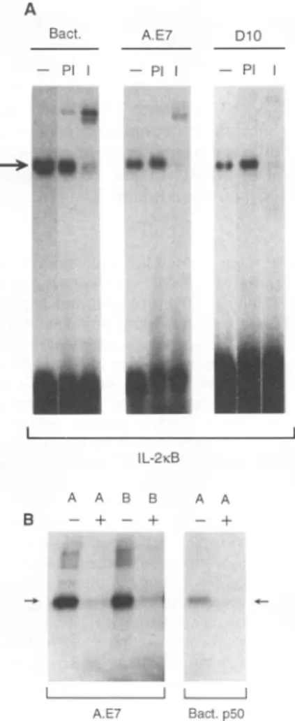
Fig.
2.The
NF-KC complex is a p50
ho-modimer.
(A) Alteration of the
NF-KC DNA
complex
by anti-p50. The extracts added to
each
binding assay
were asfollows:
affinity-purified,
bacterially
expressed p50 (100 ng)
(Bact.);
resting A.E7 nuclear extract (2
jg)
(A.E7); and resting nuclear
extract(2
jg)
from
D1O.G4.1,
anontransformed
CD4+
TH2
T
lym-phocyte clone
(D10) (25). Nuclear
extractswere
either untreated
(-), mixed with either
preimmune rabbit
serum(1
jil)
(Pl),
orimmune
serum
(1
p1)
(I)
for
10min
atambient
tempera-ture,
thenadded
to aDNA
binding assay that
contained the IL-2KB
probe.
(B)
Ultraviolet
cross-linking
of the
NF-KC
complex
from
resting
A.E7
cells and
bacterial p50. Probes
weredesigned
such that
only
half
of the IL-2KB
sequence
wassubstituted with
bromodeoxyu-ridine
(BrdU)
and[a-32P]deoxycytosine
triphos-phate
(dCTP)
asdescribed
(23, 26). The
Aprobe contains the
5'half-site
substituted with
and
the Bprobe contains
the 3' half-sitesub-stituted.
Binding reactions contained
eitherresting A.E7 nuclear
extracts(A.E7)
oraffinity-purified, bacterially
expressed
NF-KB
p50
sub-unit
(Bact.
p50),
and lanes
marked
+werefrom
binding
assays
inwhich
an-50-fold
molar excess ofunlabeled IL-2KB
oligonucleotide
was
added. The
arrowindicates the
50-kD
species.
1453 1 ~ ~ ~~ ~ ~ ~ ~ ~ ~ ~ ~on October 30, 2012
www.sciencemag.org
Downloaded from
sion
through
its
binding
tothe
IL-2KB
site.If
NF-KC
acted
as a repressor,itcould block
transactivation
of the
IL-2KB
site
by
NF-KB
until removed by
antigen
stimulation.
We
nextprepared pools of
A.E7
cells
stably transfected with multimers of
NF-KB
binding
sites
from
the IL-2
genedirecting
the
activity
of
abacterial chloramphenicol
acetyltransferase (CAT)
gene(Fig.
1B)
(23).
The
IL-2KB
binding
motif responded
well only
toantigen
and
APCs
and
wasrelatively
insensitive
toAPCs
alone.
Acti-vation
of the
IL-2KB
motif
wasalmost
completely blocked by CsA. These
effects
are
likely
tostrongly influence
IL-2
geneFig. 3. Repression
of IL-2 promoteractivity by
Acetylatlon(%)expression
ofp50.
A0.3-kbmouseIL-2promot- 0er-CAT construct
(5 ,g)
was transfected into p50EL-4
cells
withincreasing
amounts ofa con- 0.5structwith the adenovirus major latepromoter
during expression
of eitherwild-type
p50
or a Emutant form of
p50
that contained asingle
R 5amino acid deletion
in thecoding
sequence 0othat eliminates
binding activity
(p50A) (24).
c 0psoa
Corresponding
amounts of theexpression
vec-tor DNA without any
p50 coding
sequence 0.5wereaddedsothat in eachcase atotal of 10
:
2gg
of DNA was used in each transfection. AfterQ
transfection,
the EL-4 cellswereincubatedfordr
40 hours and
split
into twopools.
Onepool
wasstimulated
overnight
with concanavalin A(2
0g/ml)
and PMA(20 ng/ml)
beforeharvesting (shaded bars);
the otherpool
wasuntreated(filled
bars). Samples
were normalizedby
protein
concentration. We measured conversion ofchloram-phenicol
toitsacetylated
formusing
aphosphorimager (Molecular Dynamics, Sunnyvale,
Califor-nia).
No effectwas seen on an NFATreporter construct,
anIL-2promoter
witha mutationin theIL-2KB
site,
orthe
expression plasmid
alone(18).
Similar resultswere seeninfourexperiments
with<15%variation in replicate
samples.
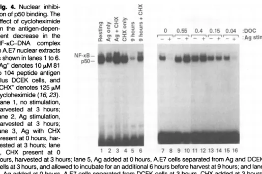
Fig.
4. Nuclear inhibi- xtion of
p50 binding.
TheX +
effect of
cycloheximide
,,
>.x
,,,
on the
antigen-depen-
so +
X 0 0.55 0.4 0.15 0.04 DOC0 0 05 . .500 0
dent decrease in the Q mm
=Zw s s
r_+_+'
r+-
+r_ +'
:Ag:stAim
NF-KC-DNA
complex
in A.E7nuclear extracts
isshownin lanes
1
to6. NF-I_-_"Ag"denotes 10pM81
to 104 peptide antigen
plus DCEK cells, and
CHX" denotes 125 gM cycloheximide(16, 23).n
Lane 1, nostimulation,
harvested at 3hours;
lane 2, Ag stimulation. harvested at 3 hours; lane 3, Ag with CHX presentat0hours,har-vestedat3
hours;
lane4, CHX present at 0 1 2 3 4 5 6 7 8 9 1011 1213141516
hours,
harvested at 3hours;
lane5, Ag
addedat0hours,
A.E7 cellsseparated
fromAg
andDCEKcells at3
hours,
and allowed to incubate foranadditional 6 hoursbefore harvest at 9hours;
and lane6, Ag
added at 0hours,
A.E7 cellsseparated
from DCEK cells at 3hours,
CHX added at 3hours,
then allowed to incubate foran additional 6 hours before harvest at 9 hours. Allextracts gave
comparable binding
toan1g
octamerprobe.
The effect of DOConNF-KCbinding activity
is showninlanes7to16. Nuclearextractswerefromunstimulated
(-;
lanes7, 9, 11, 13, 15)
and stimulatedwith10
puM
81 to 104peptide antigen plus
DCEK cells for 3 hours(+;
lanes8, 10, 12, 14, 16) (23).
Control
experiments
showed that thereactivatedp50 complex
could besupershifted
withanti-p50
serumandwas
competed
withthe IL-2KBoligonucleotide (26).
expression
because
mutations
inthe
IL-2KB
site
eliminate
-80%
of the
activityof
the
IL-2
promoter(7, 18). Stable transfectants
of
A.E7 cells with
constructscontaining
the
IgKB
site
wereactivated approximately
equally by
APC
alone,
Ag
+APC,
oreither
treatment
with CsA (18). Thus,
expression
of the IL-2KB
motif
wasspecifically
antigen-restricted and
CsA-sensitive
in
nontrans-formed T
cells,
which
correlated well with
regulation of the
NF-KC
complex.
To
identify the
molecular
natureof the
NF-KC,
weprepared
antisera
tothe
bacteri-ally expressed
murinep50
subunit
of NF-KB
(anti-p50)
and tested
for
recognition
by
EMSA (Fig. 2A). Anti-p50 and
notpreim-mune sera
shifted the complex of
bacterial
p50
with the IL-2KB
site.
The NF-KC
com-plex
in
the
nontransformed
T
cells clones
A.E7
and DlO.G4.1
(22)
comigrated with
that of bacterial
p50
and
wasalso
efficiently
shifted by anti-p50. Ultraviolet (UV)
cross-linking studies verified that the
NF-KC
com-plex
in
A.E7
cells
consisted of
two50-kD
subunits each
binding
toonehalf-site of the
IL-2KB
site
(Fig. 2B). These findings,
to-gether
with the fact
that NF-KC had the
binding
characteristics
of
purified
p50
previ-ously
described
(24),
suggestthat NF-KC
is
ahomodimer of the
NF-KB
p50
subunit.
To
directly
testwhether the NF-KB
p50
subunit could inhibit IL-2
promoteractivity
in
T
cells,
wetransfected
aplasmid
expres-sing
the
protein-coding
sequenceof
p50
with
a300-bp
IL-2
promoter-CAT
geneinto
EL-4
T
lymphoma cells (Fig. 3).
Stim-ulation of the
transfected
cells with PMA
and concanavalin A
induced IL-2
promoteractivity.
Increasing
amountsof the
p50
expression
plasmid
decreased the
responseof the IL-2
promoterin
adose-dependent
fashion. A
plasmid
expressing
amutated
p50
protein
that
wasincapable
of
binding
to
DNA
had
noeffect
(Fig. 3).
Using
the
EMSA,
wenexttested
wheth-er
protein
synthesis
wasrequired
for the
decrease
in
p50
binding
tothe IL-2KB
site
(Fig. 4).
The
protein
synthesis
inhibitor
cycloheximide
blocked
the decrease
in
p50
after
antigen
stimulation.
By
contrast,NF-KB
binding
wasaugmented
rather than
blocked
by
cycloheximide (25).
Cyclohexi-mide
in
the absence
of
antigen
stimulation
did
notaffect
p50
binding. Even when
anti-genwas
removed after
a3-hour
incubation,
the
p50
complex
continued
todecrease for
atleast
upto9
hours. The
decrease
in
the
p50
complex depended
oncontinued
protein
synthesis, because,
with addition of
cyclo-heximide after 3
hours, the
p50 binding
activity
after 9 hours
wasequivalent
tothe
amountof decrease
at3
hours.
The
requirement
for
ongoing protein
synthesis suggested
that
anewly synthesized
protein
orproteins
caused the decrease
in
p50 binding
activity. This
protein could
tably, was not increased by any of these
treatments.
Binding to a probe containing
the octamer sequence showed that the
ex-tracts were
of comparable quality for each
experiment.
Northem (RNA) blot analysis revealed
that
the IL-2 gene was strongly expressed
only after antigen and APC treatment, and
this expression was
blocked by CsA (Fig.
1A) (5, 7, 21). A
small amount of IL-2
mRNA was seen
with PMA and
ionomy-cin,
but not with either agent alone. Thus,
IL-2 mRNA
induction correlated with
re-moval of NF-KC. These results suggested
that NF-KC could inhibit
IL-2
gene
expres-1454
on October 30, 2012
www.sciencemag.org
Sl
l~
simply degrade p50 or could bind
to
p50
and inhibit
its
ability
to
bind DNA. The
cytoplasmic protein
I-KB
interacts with
NF-KB
and inhibits its DNA binding activity.
Dissociating agents,
such as the detergent
deoxycholate (DOC), release NF-KB from
IKB
in
cytosolic extracts and allow NF-KB
to
bind
DNA
(26). By contrast, treatment
of
cytosol from either resting or stimulated
A.E7
cells with DOC yielded no p50 DNA
binding activity (18). However, when
nu-clear extracts from stimulated A.E7 cells
were
treated with DOC, we detected p50
DNA
binding activity that was equivalent
to
that
in
resting A.E7
extracts (Fig. 4).
The release
of p50 required more than
0.04%
DOC and
was
maximal
at
0.4%.
The recovered binding activity
was
indis-tinguishable from p50 on the basis of
EM-SAs in
the presence
of competitor
oligonu-cleotides and anti-p50 (18).
In
addition,
the
inhibitory activity
could be separated
from p50 by DNA affinity chromatography
and
was
capable of inhibiting p50 binding
in
an
untreated resting A.E7 extract (27).
Thus,
a
newly
synthesized protein causes p50
to
be
in
a
non-DNA
binding form
in
the
nucleus of
T
cells after antigen stimulation.
In T
lymphoma
cells, expression from
the
IL-2KB site is
activated
by
inducers
of
NF-KB,
such
as
lectins
or
phorbol ester
alone, and
is
resistant
to
CsA (7, 21).
In
A.E7
cells,
we
find the
IL-2KB
site
requires
full antigen
stimulation (antigen
+
APCs)
and
is
sensitive
to
CsA.
The
regulation of
the
IL-2KB site
may
begin
to
explain the
observation
that
IL-2
can
be
produced after
partially inducing
signals
such
as PMA in
EL4
cells,
or
lectins
in
the
case
of Jurkat
cells,
without
costimulatory
signals
(7,
21
).
Such
stimuli
are
never
sufficient
for
IL-2
gene expression
or
lymphokine production
in
nontransformed
T
cells
(5, 16). Indeed,
stimulation of
nontransformed
T
cells
by
partial signals
such
as
lectins
induces
a
functionally
nonresponsive
state
(anergy)
(5).
Also, complexes
in
T
cell
tumor
lines
such
as
KBF-1
and
TCF
resemble the
p50
dimers
in
A.E7
cells,
but do
not
display
the
same
regulatory
features,
emphasizing the
importance
of
transcriptional
studies
in
nontransformed
T
lymphocytes
(8, 11,
18).
Our
findings
reveal
a
physiologic
function
for p50
in
the
T
cell response
to
antigen
in
that
p50
homodimers appear
to
inhibit
acti-vation of
gene
expression
by
NF-KB. The
p50-p50
complex
is
also found
in
lymph
node
T
cells and
thymocytes,
which
implies
a
regulatory role
in
vivo
(18).
One
of the
paradoxes of
NF-KB
as a
widely
used
signal-ing
mechanism
is
that, because
it
can
be
induced
by
so
many agents,
it
lacks the
specificity
apparently
needed
to
activate
genes
in response to
unique stimuli
(11,
12).
The
p50
(NF-KC) control
pathway
offers
one
solution
to
this
problem.
Many
agents
increased NF-KB in
A.E7 cells, but activities
of the IL-2KB
site
and the
IL-2 gene
ap-peared to be blocked unless the p50 complex
decreased. Thus, the IL-2KB site may only
be
active when the appropriate
combination
of
stimuli leads to synthesis of a p50
inhibi-tory
molecule. Inhibition of the p50
com-plex is a
regulatory paradigm that differs from
the
cytoplasmic regulation of
NF-KB
by
1KB
because the non-DNA binding form of the
p50 complex
appears to be retained entirely
in the
nucleus.
REFERENCES AND NOTES
1. C. G. Fathman, F. W. Fitch, K. A. Denis, 0. N.White, in Fundamental Immunology, W. E. Paul, Ed. (Raven, New York, 1989), pp. 803-815; C. Jamieson, P. G. McCaffrey, A. Rao, R. Sen, J. Immunol. 147, 416 (1991); U. Hazan etal., Proc. Nati. Acad.Sci.U.S.A. 147, 7861 (1990). 2. S.-M. Kang etal., Mol. Cell.Biol.,in press. 3. K. A.Brorson, B. Beverly,S.-M. Kang, M. Lenardo,
R. H.Schwartz, J.Immunol. 147, 3601 (1991). 4. M. J.Lenardo, Nature 353, 858 (1991). 5. M. K.Jenkins and R. H. Schwartz, J. Exp. Med.
165, 302 (1987); J. R. Lamb, B. J. Skidmore, N. Green, J. M. Chiller, M. Feldman, ibid. 157,1434 (1983); D.L. Mueller, M. K. Jenkins, L. Chiodetti, R. H.Schwartz, J. Immunol. 144, 3701 (1990); C. T. Weaver, C. M. Hawrylowicz, E. R. Unanue, Proc.
Natt.
Acad.Sci. U.S.A. 85, 8181 (1988). 6. T.Fujita, H. Shibuya, T. Ohashi, K. Yamanishi, T.Taniguchi, Cell 46, 401 (1986); D. B. Durand, M. R. Bush, J. G.Morgan, A. Weiss, G. Crabtree, J. Exp. Med. 165, 395 (1987); G. Crabtree, Science 243, 355 (1989); D. B. Durand et al., Mol. Cell. Biol. 8,1715 (1988); H. Shibuya and T. Taniguchi,
Nucteic
Acids Res. 17, 9173 (1989).7. J.-P. Shaw et
at.,
Science241, 202 (1988); M. J. Lenardo, A. Kuang, A. Gifford, D. Baltimore, Proc.Natt.
Acad. Sci. U.S.A. 85, 8825 (1988); M. J. Lenardo,C.-M. Fan, T. Maniatis, D. Baltimore, Cell 57, 287 (1989); H. Shibuya, M. Yoneyama, T. Taniguchi, Int. Immunol. 1, 143 (1989); B. Hoyos, D. W. Ballard, E. Bohnlein, M. Siekevitz, W. C.Greene, Science244, 457(1989).
8. A. Radler-Pohl, I. Pfeffer, M. Karin, E. Serfling,
NewBiol. 2, 566 (1990); M. Yano et al., EMBO J. 6, 3317(1987).
9. E. A.Emmel et al.,Science 246, 1617 (1989). 10. P. Baeuerle, Biochim. Biophys. Acta 1072, 63
(1991).
11. M. Lenardo and D.Baltimore,Cell 58, 227 (1989); M. Grilli,J.-S. Chiu, M. Lenardo,
tnt.
Rev.Cytol.,in press.12. V.Bours et al.,
Mot.
Cell. Biol. 12, 685 (1992); V. Bours, J. Villalobos, P. R. Burd, K. Kelly, U.Siebenlist,Nature348, 76(1990); S.Ghoshetal., Cell 62, 1019 (1990); K. Kawakami, C. Schei-dereit,R.G. Roeder, Proc.Natl.Acad.Sci.U.S.A.
85, 4700 (1988); M. Kieran etal., Cell 62, 1007 (1990); F. Logeat etal., EMBO J. 10, 1827 (1991); R. Meyer et al., Proc.Nati.Acad.Sci. U.S.A. 88, 966(1991);G.P.Nolan,S.Ghosh, H.-C. Liou,P. Tempst, D. Baltimore,Cell 64, 961 (1991); S. M. Ruben etal., Science251, 1490 (1991); R.-P. Rysecketal., Mol. Cell. Biol. 12, 675 (1992); R. M. Schmid, N. D. Perkins, C. S. Duckett, P.C.
An-drews, G. J. Nabel, Nature 352, 733 (1991); R. Sen and D. Baltimore, Cell 46, 705(1986); K. C. Wilhelmsen, K. Eggleton, H. M. Temin, J. Virol. 52, 172(1984).
13. M. L. Schmitz andP. A. Baeuerle, EMBO J. 10, 3805(1991);R. Tenetal.,ibid.11, 195(1992). 14. N. Davis et al., Science 253, 1268 (1991); S.
Haskill et al., Cell 65, 1281 (1991);L. D.Kerret
at.,
Genes Dev. 5, 1464(1991); S. E. Lux,K. M.John, V. Bennett, Nature 344, 36 (1990); C. C.
Thompson,T. A.Brown,S.L.McKnight,Science 253, 672(1991).
15. Cell culture and cell stimulations. The A.E7 cells
SCIENCE * VOL. 256 * 5JUNE1992
responded to a pigeon cytochrome c peptide (amino acids 81 to 104) in the context of MHC class 11 Ek and weregrown as described (5) [T. T. Hecht, D. L. Longo,L.A. Matis, J.Immunol. 131, 1049(1983)]. DCEK cells were grown in Dulbec-co's modified essential medium (DMEM) as de-scribed (16). Antigen stimulation and magnetic cell separation will bedescribed more fully else-where (S.Kang, A. Tran, M. Lenardo, in prepara-tion). Briefly, DCEK cells were incubated with magnetic beads (Dynabeads, Dynal, Oslo, Nor-way) for 2 days. The DCEK cells were then trypsinized, resuspended in fresh medium, and placed in a magnetic field (Advanced Magnetics, Cambridge, MA) for 5min. The supernatant was removed, and the magnetic pelleting was repeat-ed three more times to remove all nonmagnetic DCEK cells. Antigenstimulations for extracts used A.E7 cells (50 x 106) purified by Lympholyte M (Cedarlane, Hornby, ON), according to the manu-facturer's instructions mixed with 25 x 106 mag-netic bead-loaded DCEK cells and pigeon cy-tochrome c peptide (10
gM
cyanogen bromide peptide fragment 81 to 104) in 10mlof media. The cells wereharvested at various times and separat-ed by vigorouspipetting, and tubes were placed in amagnetic field. Thesupematant was removed, and the separation repeated twice. Treatments with ionomycin(Calbiochem, La Jolla, CA) (2 ,uM) and PMA(Sigma, St. Louis, MO) (20ng/ml) were similarly performed but without DCEK cells or antigen. CsA was used at 200 IM. Anisomycin (Sigma) (10 FM) orcycloheximide (Sigma) (125gg/ml)
were used with a30-minpreincubation. 16. F. Ronchese, R. H. Schwartz, R. N. Germain,Nature 329, 254 (1987).
17. Nuclear extracts were prepared as described [S. Fiering etal.,GenesDev.4, 1823 (1990)]. Protein concentrations were determined with a Bio-Rad (Richmond, CA) assay kit. The EMSAs were per-formed as described [L. Staudt etal., Science 241, 577 (1988)]with acustom-designed minigel appa-ratus [M.Lenardo, unpublished data; Owl Scientif-ic, Cambridge, MA) with the following modifica-tions: nuclear extract (2
jig),
poly[dl:dC] (0.5bg),and 32P end-labeled oligonucleotide probe (20,000 cpm) were used in lipage buffer [1 x=6.7 mM tris) Cl (pH 7.5), 3.3 mMsodium acetate, 1 mM EDTA] in a final volume of 6
pi
(7).Deoxycholate treatments were carried out as in (26) with minor modifications: 1gl
ofextract (2mg/mi)was quickly mixed with 1.2 or 0.9gi
of 4% DOC(wN)
(for 0.55% or 0.4% finalconcentration, respectively), or with 1.2 or0.6 pl of 2% DOC(wN)
(for 0.25 or 0.13% finalconcentration, respectively), then im-mediately transferred to binding buffer (7 Al) con-taining 10x lipage buffer (0.7 EI), bovine serum albumin (0.5gi)
(20 mg/mI, BoehringerMann-heim), Buffer D + 1% NP-40
(vN)
(1g])
[J. D.Dignam, R. M. Lebovitz, R. G. Roeder, Nucleic
Acids Res. 11, 1475 (1983)], 10% NP-40
(vN)
(1g1i),
andlabeled probe [20,000(cpm)]for 15min before gel loading. Polyclonal rabbit antisera were prepared against a bacterially produced and affin-ity-purifiedrecombinant mouse p50 subunit (19). Anti-p50 (1pi)
or preimmune serum (1pi)
was preincubated with nuclear extract (1gi)
(2mg/mi)for 15min at roomtemperature. UV cross-linking experiments were carried out asdescribed (24). The A probe consistedof the annealed oligonucle-otides
5'-CCAAGAGGGAMCACCTAAATCC-3'
and GGAMAGG-3', and the "B" probe5'-GGAWAGGTGAAATC-CCTCTIGG-3'
and5'-CCAAGAGGG-3' oligonucleotides. Studies of binding site specificity, comigration with affinity-purified NF-KB, and recognition by anti-p50 and anti-p65 sera confirmed that the complex labeled NF-KB was ap50-p65 heterodimer.
18. S.-M. Kang, A.-C. Tran, M. G. Grilli, M. J. Lenardo, unpublished data.
19. M. G. Grilliand M. J. Lenardo,unpublished data; S. W. Krasnow et
at.,
Cytokine 3, 372 (1991). 20. M. Kronke etat.,
Proc.Natt.
Acad.Sci. U.S.A. 81,5214
(1984);
E. A. Emmel etal., Science246,1617(1989).
21. T.
Brabletz,
I.Pietrowski,
E.Serfling,
Nucleic1455
0,19...a O R:- . ... 'O' I~ .M
on October 30, 2012
www.sciencemag.org
AcidsRes.19, 61 (1991);C. Randak,T.Brabletz,
M.Bergenrother, I.Sobotta,E.Serfling, EMBO J. 9, 2529 (1990).
22. J. Kaye et al.,J. Exp.Med. 158, 836(1983).
23. Oligonucleotidesand constructs.The IgKBsite is
describedinJ. Pierceet al.[Proc.Nati. Acad.Sci. U.S.A.85, 1482(1988)].The IL-2KBoligo nude-otide spans -212 to -195 of theIL-2enhancer. Thesequence of these oligonucleotides isinthe legend to Fig. 1, except thata5'-TCGA-3'
over-hang exists at the 5' end.For stabletransfectants,
minimal c-fos promoter-bacterial CAT reporter constructs wereprepared as described[M.
Len-ardo, J. W. Pierce, D. Baltimore, Science236,
1573 (1987)]. TheIgKBconstruct is"J16' from Pierce etal.(23). (vide supra). The IL-2KB con-structcontained sixcopiesof the IL-2KB
oligonu-cleotide in the Sal site of the A56 minimal
c-fos-CATvector.Constructswereconfirmedby DNAsequencing, and functionwascheckedby
transienttransfection into EL-4 cells. For stable
transfections,A.E7 cellswereplacedin IL-2 (30 U/mI) (intheform of MLA-144supernatant)for 3 days andtransfectedwith251g of linearizedtest
plasmid and 2.5 >g of Eco RI-linearized
pSV2Neo by electroporation [A.J. Cann et
al.,
Oncogene 3,123(1989)]with 300V,960
gF
inaBio-Rad genepulserapparatus. After
electropo-ration, the cellswereplacedintoIL-2-containing
mediumfor 2 days before the addition ofG418
(0.8mg/mI) [Genticin,BethesdaResearch Labo-ratories(BRL)]. The cells were thenpooled and grown inG418-containingmedium for more than 1 monthbefore use. Two differentpoolsof
trans-fectantsweretested foreachconstruct.For stim-ulations,weused 5x10⁵Tlymphocytesand2.5
X 105 DCEK cells, with or without 81 to 104 peptide (1 FM) in 2ml. The cells were incubated for 14 to 16 hours and harvested as described above. The CAT assays were performed with standard procedures[C. Gorman et al., Mol. Cell. Biol. 2, 1044 (1982)] and extracts were normal-ized by protein concentration. Chloramphenicol conversion was quantitated with a phosphorim-ager (Molecular Dynamics,Sunnyvale, CA). Tran-sient transfections into EL-4 cells wereperformed by the DEAE-dextran procedure as described (7).
24. M. B.Urban, R.Schreck,P.A.Baeuerle,EMBO J. g 10, 1817(1991); M.B. Urban and P. A. Baeuerle, New Biol. 3, 279 (1991).
25. R.Sen and D. Baltimore, Cell 47, 921 (1986). 26. P. Baeuerle and D. Baltimore, Science242, 540
(1988).
27. J.J.-S. ChiuandM. J. Lenardo, unpublished data. 28. We thank R. Schwartzand B.Beverlyfor advice and assistance in thegrowth of nontransformed T cellclones; R.Germainfor advice on bead sep-aration; D. D.A'Ambrosio and K. Bruhn for labo-ratoryassistance;U.Siebenlist, V. Bours,G. Fran-zoso, and P. Bressler for the p50 expression
constructs,discussions, and sharingunpublished
results; H.Shibuya and T. Taniguchi for IL-2CAT
constructs;S. Ghoshand D.Baltimorefor the p50 cDNA; and D. Levens, J. Mellentin, U.Siebenlist,
and L. Staudtfor comments on themanuscript.
S.-M. K. was supported by a Howard Hughes Medical Institute-NIH Research Scholarship and M.J.L. is supported by aCancerResearch Insti-tuteInvestigator Award.
2March 1992; accepted 8 April 1992
Nerve Growth Factor Stimulation
of the
Ras-Guanine Nucleotide Exchange
Factor
and GAP
Activities
Bao-Qun
Li,David
Kaplan, Hsiang-fu Kung, Tohru Kamata*
The biological activity of Ras proteins is thought to be controlled by the guanine nucleotide
exchange factor and the guanosine
triphosphatase activating protein (GAP). Treatment of
rat
pheochromocytoma
PC-12
cells with
nervegrowth factor (NGF) increased the amount
of
active Ras
guanosine triphosphate complex and stimulated the activities of both the
guanine nucleotide exchange factor and GAP.
In
PC-1
2
cells that
overexpressed the
tyrosine kinase encoded by the trk proto-oncogene (a component of the high-affinity
NGF
receptor), the NGF-induced activation of the regulatory proteins
waspotentiated. These
results suggest that the NGF receptor system enhances the activities of both the guanine
nucleotide
exchange factor and GAP and that the activation of Ras might be controlled by
the
balance
in
activity between these two regulatory proteins.
The
ras
proto-oncogenes
encode
membrane-bound proteins that bind guanine nucleotide
and
have low
intrinsic
guanosine
triphospha-B.-Q.
Li and T.Kamata,
Biological Carcinogenesis andDevelopment Program, Program Resources,Inc./DynCorp, National Cancer Institute, Frederick Cancer Research and Development Center, Frederick, MD 21702.
D. Kaplan, Eukaryotic Signal Transduction Group, Advanced Bioscience Laboratory- Basic Research Program, National Cancer Institute, Frederick Cancer Research and Development Center, Frederick, MD 21702.
H.-f. Kung, Laboratory of Biochemical Physiology, NCI, Frederick Cancer Research and Development
Center,Frederick, MD 21702.