Insulin-producing organoids engineered from islet
and amniotic epithelial cells to treat diabetes
Fanny Lebreton
1,7
, Vanessa Lavallard
1,7
, Kevin Bellofatto
1,7
, Romain Bonnet
1
, Charles H. Wassmer
1
,
Lisa Perez
1
, Vakhtang Kalandadze
2,3
, Antonia Follenzi
3
, Michel Boulvain
4
, Julie Kerr-Conte
5
,
David J. Goodman
6
, Domenico Bosco
1
, Thierry Berney
1
& Ekaterine Berishvili
1,2
*
Maintaining long-term euglycemia after intraportal islet transplantation is hampered by the
considerable islet loss in the peri-transplant period attributed to inflammation, ischemia and
poor angiogenesis. Here, we show that viable and functional islet organoids can be
suc-cessfully generated from dissociated islet cells (ICs) and human amniotic epithelial cells
(hAECs). Incorporation of hAECs into islet organoids markedly enhances engraftment,
via-bility and graft function in a mouse type 1 diabetes model. Our results demonstrate that the
integration of hAECs into islet cell organoids has great potential in the development of
cell-based therapies for type 1 diabetes. Engineering of functional mini-organs using this strategy
will allow the exploration of more favorable implantation sites, and can be expanded to
unlimited (stem-cell-derived or xenogeneic) sources of insulin-producing cells.
https://doi.org/10.1038/s41467-019-12472-3
OPEN
1Cell Isolation and Transplantation Center, Surgery, Geneva University Hospitals and University of Geneva, Geneva, Switzerland.2Institute of Medical
Research, Ilia State University, Tbilisi, Georgia.3Department of Health Sciences, University of Oriental Piedmont, Novara, Italy.4Department of Gynecology
and Obstetrics, Geneva University Hospitals and University of Geneva, Geneva, Switzerland.5INSERM U1190, Translational Research for Diabetes, University
of Lille, Lille, France.6Department of Nephrology, St Vincent’s Hospital, Melbourne, VIC, Australia.7These authors contributed equally: Fanny Lebreton,
Vanessa Lavallard, Kevin Bellofatto. *email:[email protected]
123456789
A
lthough intraportal islet transplantation is an established
therapy for patients with type 1 diabetes, maintaining
long-term glucose control with this approach remains
challenging, mainly due to considerable islet loss in the
peri-transplant period
1. Among the reasons for early graft loss,
inflammation at the site of implantation and impaired
revascu-larization appear as key factors
2–4. Pancreatic islets have a dense
blood supply which is inevitably disrupted by the isolation
pro-cess
5. In the
first weeks after transplantation, oxygen and
nutri-ents are delivered to avascular islets exclusively by diffusion until
they become revascularized
6. Therefore, new strategies aiming at
protecting the islets from inflammatory insults and/or promoting
graft revascularization may be effective for improving clinical islet
transplantation outcomes.
Recent studies have demonstrated the functionality of
three-dimensionally assembled
β-cell aggregates, or multicellular islet
spheroids
7,8. Modulating the cell composition by combining
different cell types of islet spheroids leads to improvement of
function and viability due to heterotypic cell–cell interactions and
reproduction of the complex natural morphology of the islet
9,10.
This strategy could be brought further, by generating
multi-cellular hybrid organoids consisting of several cell types, i.e.,
endocrine cells for regulated hormone release, and other cell types
with cytoprotective and immunomodulatory properties with the
aim to increase islet survival and function after transplantation.
Over the last decades, human amniotic epithelial cells (hAECs)
have gained interest in regenerative medicine due to their high
proliferative capacity, multilineage differentiation, ease of access,
and safety
11. hAECs express surface markers found on human
embryonic stem cells and secrete considerable amounts of
proangiogenic and anti-inflammatory growth factors, including
vascular endothelial growth factor (VEGF), basic
fibroblast
growth factor (bFGF), angiogenin (ANG), insulin-like growth
factors (IGF), and their binding proteins (IGFBPs)
12–14. In
addition, hAECs secrete high levels of hyaluronic acid, which
suppresses tumor growth factor
β (TGFβ)—a potent
profibro-genic cytokine
15. Decreased levels of TGFβ expression were
observed after AEC transplantation in mice with bleomycin- and
CCl
4-induced lung or liver injury
16,17. This growth factor
secre-tion profile and antifibrotic properties make hAECs attractive
cells for a construct designed to enhance the engraftment and
vascularization of islet cells.
In this study we successfully generated viable and functional
insulin-secreting organoids composed of hAECs and dissociated
islet cells (ICs) and have shown that incorporation of hAECs into
islet-cell constructs markedly enhances engraftment, viability and
graft function in model of cell therapy for type 1 diabetes.
Results
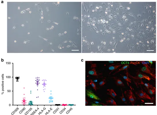
Characterization of hAECs. After initial seeding, hAECs rapidly
formed proliferating clusters and grew within 5 days into a
confluent cobblestone-shaped monolayer (Fig.
1
a). After culture,
hAECs were characterized by
flow cytometry and were positive
for epithelial (CD326), mesenchymal (CD90, CD105), embryonic
stem-cell (SSEA-4) and pluripotency (Oct-4) markers. Most
importantly, the hAECs expressed non-classical class Ib
histo-compatibility antigens HLA-G and HLA-E (Fig.
1
b, c,
Supple-mentary Fig. 8). Human amniotic epithelial cells were negative for
hematopoietic cell markers CD34, CD31, and CD45 (Fig.
1
b).
Our results are consistent with previously reported
findings
14.
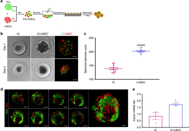
Generation and in vitro assessment of organoids. Figure
2
a
describes the process used herein to generate islet organoids by
mixing ICs and hAECs. Both IC- and IC-hAEC aggregates
formed round-shaped spheroids of uniform size with well-defined
smooth borders within 5 days (Fig.
2
b). The average diameter of
IC-hAEC organoids and IC spheroids were 139 ± 4
μm and 202 ±
2
μm (data are mean ± SEM, n = 12), respectively (Fig.
2
c).
Although ICs and hAECs formed small monocellular islands
within the organoid, most ICs were in contact with both cell types
as shown by confocal laser scanning microscopy (Fig.
2
d). No
evidence of cell loss was detected. There was no significant
dif-ference in cell viability between IC spheroids and IC-hAEC
organoids (Supplementary Fig. 1).
On day 5, harvested spheroids were assessed for insulin
expression by qPCR analysis, which demonstrated that insulin
mRNA expression was significantly upregulated in the IC-hAEC
organoids as compared with IC spheroids (Fig.
2
e).
The functionality of the spheroids was evaluated by
glucose-stimulated insulin secretion (GSIS) assay. IC-hAEC organoids
released considerably more insulin in response to high-glucose as
compared with IC spheroids and showed significantly higher SI
than controls (4.2 ± 0.4 vs 2.8 ± 0.3, data are mean ± SEM, n
= 5).
These data demonstrate that incorporation of hAECs into
islet-cell constructs enhances
β-cell function.
Organoids maintain function after hypoxic stress in vitro. Both
IC-hAEC organoids and IC spheroids were cultured under
hypoxic conditions (1% oxygen and 5% CO
2at 37 °C) for 16 h to
mimic the ischemic condition taking place in vivo in the early
phase of engraftment
18. This allowed us to examine whether
hAECs were able to confer cytoprotection and help to maintain
the functional capacity of ICs under ischemic stress. Incubation
under hypoxia rapidly caused fragmentation of IC spheroids and
increased cell death. By contrast, considerably fewer dead cells
were observed within IC-hAEC organoids (Fig.
3
a). As
antici-pated, glucose-induced insulin secretion of monocellular
spher-oids was seriously impaired. By contrast, SI of the IC-hAEC
organoids in GSIS assay was significantly higher (Fig.
3
b;
Sup-plementary Table 1). These results were further strengthened by
qPCR analysis, which showed about threefold higher insulin
mRNA expression in IC-hAEC organoids as compared with
controls (Fig.
3
c).
To assess the possible molecular mechanisms behind the
protective effect of hAECs on hypoxia-induced cell death and
dysfunction, expression of HIF-1α, a key regulator of cell
response to hypoxia was analyzed in spheroids.
Immunofluores-cence demonstrated a significant increase in the nuclear
localization of HIF-1α in IC + hAEC organoids approaching
50% compared with IC spheroids after exposure to hypoxia
(Fig.
3
d, e). The higher nuclear HIF-1α expression in IC-hAEC
was correlated with a downregulation of the apoptotic genes
Casp3, Casp8, and Casp9 and twofold upregulation of the
antiapoptotic gene Bcl2 (Fig.
3
f) compared with IC spheroids
alone. Taken together, these results suggest that the hAECs
protect islet cells from ischemia-induced apoptotic injury and
help to maintain glucose responsiveness through the upregulation
of HIF-1α expression.
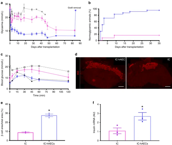
Islet organoid transplantation improves diabetes reversal. To
assess whether incorporation of hAECs into the islet organoids
could enhance engraftment and lead to better glycemic control,
diabetic SCID mice were transplanted with a marginal mass of
150 IC-hAEC organoids (IC-hAEC group, n
= 25), IC spheroids
(IC group, n
= 25), or hAECs spheroids (hAEC group, n = 5).
Mice transplanted with IC-hAEC organoids exhibited enhanced
glycemic control, compared with mice grafted with IC spheroids
(Fig.
4
a). The average nonfasting blood glucose concentrations of
mice in the IC-hAEC group were considerably lower than those
in IC group at 1 month after transplantation (7.9 ± 1.1 mmol/l for
IC-hAEC (n
= 13) vs 18.4 ± 2.1 mmol/l for IC (n = 10), data are
mean ± SEM, p < 0.0001, unpaired Student’s t test). The
cumu-lative percentage of animals reaching normoglycemia was 96% in
the IC-hAEC group vs 16% in the IC group at 1 month after the
transplantation (Fig.
4
b). In cured animals, the median time to
reach euglycemia was 5.1 ± 0.1 days in the IC-hAEC (n
= 24)
group and 30 ± 9.2 days in the IC group (n
= 8) (data are mean ±
SEM, p < 0.0001, unpaired Student’s t test). As expected, mice
transplanted with hAEC spheroids remained diabetic. Removal of
graft-bearing kidneys at different time points after transplantation
led to recurrence of hyperglycemia in all mice within 24 h,
indi-cating that the transplanted spheroids were responsible for
nor-malized glucose levels in cured animals.
To investigate the insulin secretory capacity of the graft in vivo,
IPGTT was performed at 4 weeks post transplantation. As shown
in Fig.
3
c, glucose clearance of mice in the IC-hAEC group (n
=
10) was similar to that of a nondiabetic control at all time points
after glucose loading. By contrast, the IC group (n
= 10) showed
abnormal glucose tolerance. To further support the data obtained
from the IPGTT, fasting serum insulin and C-peptide levels were
measured in the same animals. Both insulin (242 ± 32 pmol/l in
the IC-hAEC group vs 130 ± 29 pmol/l in the IC group (n
= 6),
data are mean ± SEM, p
= 0.02, unpaired Student’s t test) and
C-peptide (1140 ± 43 pmol/l in IC-hAEC group vs 732 ± 124 pmol/l
in the IC group (n
= 5), data are mean ± SEM, p = 0.01, unpaired
Student’s t test) concentrations were significantly higher in the
IC-hAEC group. These data demonstrate that incorporation of
hAECs into the islet organoids enhances functional capacity of
islet cells.
Organoid transplantation enhances graft revascularization. To
evaluate engraftment and revascularization, graft-bearing kidneys
were processed for histology. Immunohistochemical (IHC)
staining showed larger
β-cell mass, as assessed by the
insulin-positive area per
field in the IC-hAEC group compared with that
of the IC group (Fig.
4
d, e) at 120 days posttransplant. This
finding was further confirmed by qPCR analysis, which
demon-strated that insulin mRNA expression levels were significantly
higher (in the IC-hAEC group (Fig.
4
f). Similarly, more glucagon
and somatostatin-positive cells were found by IHC in the
removed grafts of IC-hAEC group compared with grafts of IC
group (Fig.
5
a–c).
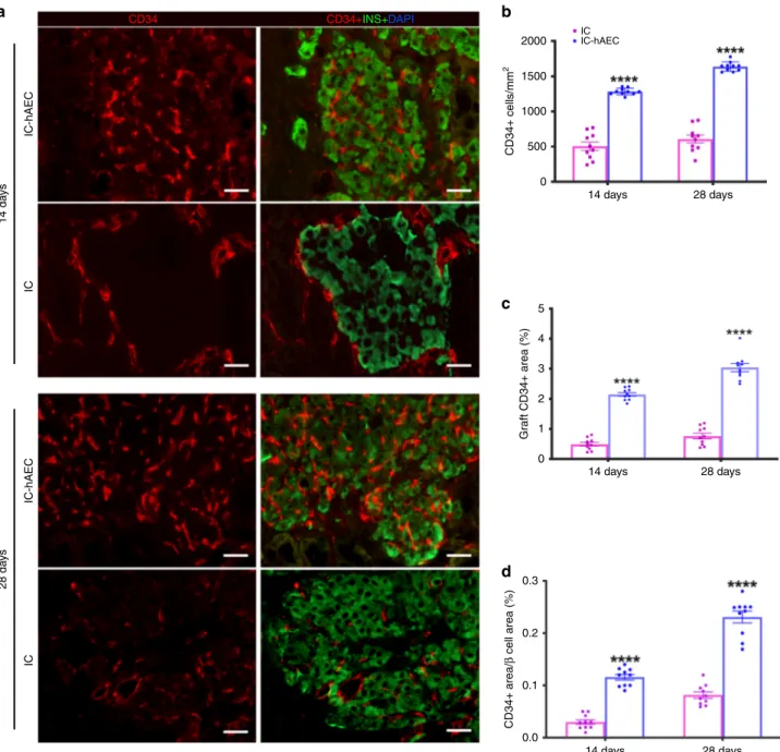
To investigate whether incorporation of hAECs into the islet
organoids promotes the process of revascularization, histological
sections of the graft-bearing kidneys, harvested at different time
points were processed for CD34 and CD31 immunostaining.
Higher CD34 and CD31 staining on histological section was
observed in IC-hAEC group compared with IC group (Fig.
6
a,
Supplementary Fig. 2). After quantification, CD34 staining was
shown to be 2–4-fold higher in IC-hAEC group compared with
IC group (Fig.
6
b–d). As expected, in both groups, CD34 staining
was higher at day 28 compared with day 14.
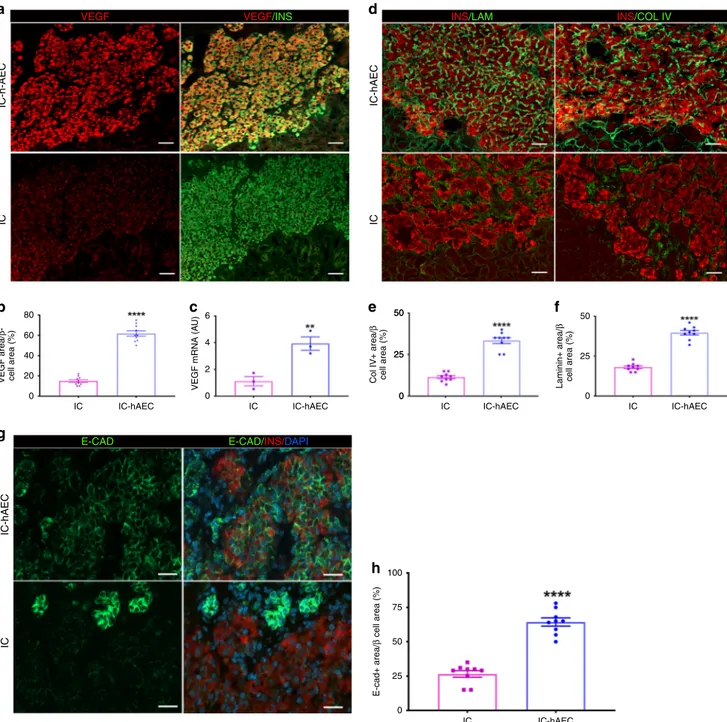
Mechanisms of improved graft function and revascularization.
To elucidate the underlying mechanisms by which hAECs
con-tribute to improved revascularization and function of the graft,
we assessed possible differences in VEGF-A production between
IC-hAEC and IC groups. Immunohistochemical staining showed
higher expression of VEGF-A in the IC-hAEC group compared
with in the IC group (Fig.
7
a). After quantification. VEGF-A
staining in the graft was three times higher in IC-hAEC compared
with IC group (Fig.
7
b). To assess whether the hAECs stimulated
production of proangiogenic factors by the islet cells, rat-specific
VEGF-A mRNA levels were measured in IC spheroids and
IC-hAEC organoids. IC-IC-hAEC organoids expressed considerably
more VEGF-A mRNA than islet IC spheroids (Fig.
7
c). Moreover,
we examined if endothelial cells in the graft tissue originated from
hAECs. To this end, histological sections were exposed to an
anti-150
b
c
a
OCT4PanCK DAPI
% positiv e cells 100 50 0 CD326 CD90CD105SSEA-4HLA-GHLA-E CD31 CD34 CD45
Fig. 1 Characterization of hAECs. a Phase-contrast microscopic images of cultured hAECs at days 2 (left panel) and 5 (right panel) after seeding. Scale
bar= 100 μm. b hAECs were characterized for various surface markers by FACS. Data are the means ± SEM, of cells obtained from sixteen different donors
labeled with specific antibodies (% positive cells). c Immunohistochemical detection of cytokeratin (red) and OCT-4 (green) in cultured hAECs; nuclei are
human specific CD31 antibody instead of the anti-rodent CD34
or CD31 antibodies used above. No CD31 staining was found at
any time point (Supplementary Fig. 3), suggesting that
endothe-lial cells are not of human origin. These results indicate that
human hAECs accelerate the revascularization process mainly by
stimulating angiogenic factors in the islet cells, but not through
differentiation into the endothelial cells.
We
finally examined whether hAEC incorporation into the
islet organoids promoted the production of extracellular matrix
proteins and adhesion molecules, which are essential to maintain
islet morphology and promoting in turn
β-cell survival and
function
19. IHC studies demonstrated that expression of collagen
IV and laminin (two major basement membrane proteins) was
higher in the IC-hAEC group (Fig.
7
d–f). Expression of
E-cadherin, an adhesion molecule involved in the maintenance of
β-cell viability and promoting insulin secretion
20, was considerably
upregulated in the IC-hAEC compared with IC group (Fig.
8
a, b).
These results suggest that incorporation of hAECs into islet-cell
constructs enhance basement membrane and production of
E-cadherin, thus ensuring proper function of islet cells.
hAECs remain within grafted organoids over 2 weeks. To
co-localize
hAECs
within
transplanted
IC-hAEC
organoids,
explanted graft-bearing kidneys were stained for anti-human
nuclear antigen antibody. Our
findings showed that while
human-derived cells were abundantly present in the
first 2 weeks
after transplantation, their number gradually declined over time,
and at the end of a 1-month period, only few HNA-positive cells
were detectable (Supplementary Fig. 4).
In vivo function and vascularization of human-derived grafts.
Islet material from two different human donors was used. Each
mouse was transplanted with 300 human IC spheroids (n
= 8) or
IC-hAEC organoids (n
= 10) into the epididymal fat pad. To
monitor graft function after transplantation, human C-peptide
levels in the blood were measured once a week. As shown in
Fig.
9
a, C-peptide levels gradually increased after transplantation
and were significantly higher in the IC-hAEC group compared
with IC spheroid controls. Glucose clearance was studied by
IPGTT 1 month after transplantation. IC-hAEC organoids
showed normal metabolic function, in contrast with the IC
spheroid group (Fig.
9
b).
Grafts were removed one month after transplantation and
processed for histological analysis to analyze engraftment and
vascularization. Histological analysis of explanted grafts from the
IC-hAEC group revealed healthy islet morphology. Grafts stained
ICsa
b
d
e
c
DIO DIL+ Mix Seeding Aggregating Spheroids
5 days ICs+hAECs hAECs IC Da y 1 Da y 5 IC-hAEC Spheroid diameter ( μ m) 250 200 150 100 IC I-hAEC IC-hAEC INS HNA 2.5 2.0 1.5 1.0 0.5 0.0 IC IC-hAEC Insulin mRNA A U
Fig. 2 In vitro characterization of islet organoids. a Schematic representation of islet organoid engineering. After labeling with Dio and DiI, ICs and hAECs
were mixed, seeded and incubated several days on 3D agarose-patterned microwells to generate islet organoids.b Phase-contrast and corresponding
fluorescence views of spheroids in one microwell on days 1 and 5. After 5 day culture (bottom), cells undergo compaction and spheroids appear to acquire a
smooth border as compared with aggregated cells at day 1. Scale bar= 50 μm. c Diameters of IC spheroids and IC-hAEC organoids (n = 12). ****p > 0.0001,
unpaired Student’s t test. d Confocal views of islet-cell construct. Cell arrangement and composition of the islet organoid on day 14. Islet-derived cells stained
for Insulin (green) and hAECs for human nuclear factor (red). Every ninth section of az-stacked and the entire 3-D reconstructed islet heterospheroid (right
panel) are shown. Scale bar= 50 μm. e Insulin mRNA expressed by IC spheroids and IC + hAEC organoids; insulin mRNA was analyzed by qPCR, arbitrary
positive for insulin, glucagon and for the presence of endothelial
cells in newly formed intra-islet micro vessels (Figs.
8
c, 9a, b). In
contrast, very little graft tissue was retrievable from mice
transplanted with IC spheroids. Explants showed extensive loss
of islet mass and poor vascularization. Consistent with these
results, explanted grafts from the human IC-hAEC group
exhibited considerably higher E-cadherin expression levels
compared with IC grafts (Supplementary Fig. 5).
These observations demonstrate that integration of hAECs into
human islet-cell organoids significantly improves their
function-ality, viability and vascularization, and confirm the findings
obtained with organoids derived from rodent islet cells.
Discussion
In this study, we have shown that incorporation of hAECs into
islet-cell constructs markedly improved secretory function and
viability in vitro, in conventional culture and in hypoxic
condi-tions, and engraftment and graft function in vivo. Combined
hAEC and islet-cell organoids hold great potential for cell-based
therapies for type 1 diabetes. Recent studies have indicated that
combining different cell types of cells into hybrid spheroids is a
tissue engineering strategy able to provide
“building blocks” for
larger tissue constructs, with enhanced cell viability, physiologic
function and proliferative ability
21. Thus, the generation of
islet-cell-based multicellular spheroids could be an interesting strategy
towards the development of novel cell-based therapies in the
treatment of diabetes. However, the generation of stable
multi-cellular islet spheroids is quite challenging due to differences in
the mode of cellular adhesion of pancreatic islet and other cell
types used so far (mostly mesenchymal stem cells)
22.
In this study, we have successfully generated viable and
func-tional islet organoids composed of hAECs and dissociated ICs.
We did not observe any segregation of islet cells and hAECs into
separate spheroidal units at any stage, as reported by other groups
attempting to generate stable hybrid spheroids enriched with
mesenchymal stem cells (MSCs)
23,24. In contrast to what has been
reported for MSCs, the even coaggregation of hAECs and
islet-derived cells clearly demonstrated by confocal microscopy can be
attributed to the epithelial origin of both cell types and the
identical mode of cellular adhesion, mediated by the cadherin
superfamily
25,26.
Significant islet loss in the early posttransplant period is one of
the reasons for suboptimal outcomes of clinical islet
transplan-tation. This occurs, at least in part, by anoikis (programmed cell
death secondary to loss of cell-to-extracellular matrix contact)
and necrosis caused by ischemia during the revascularization
process
27. Our
findings demonstrate a clear protective effect of
hAECs on islet cells in hypoxic conditions. While IC spheroids
exposed to hypoxia display extensive necrosis and impaired
function, IC-hAEC organoids preserve adequate glucose
respon-siveness and show considerable protection from cell death.
HIF-1α is a transcription factor orchestrating compensatory
responses to adapt to hypoxia through modulation of
down-stream genes involved in angiogenesis and cell survival
28–30. In
islets, HIF-1α upregulates genes involved in glucose metabolism
such as glucose transporter 2 (GLUT2) and glucokinase (GCK)
31.
Moreover, recent studies have shown that upregulation of HIF-1α
protects islets after transplantation, and thus improves islet
transplantation outcomes
32. In accordance with this, islet
bicel-lular spheroids subjected to hypoxia showed a
fivefold increase of
HIF-1α expression, which was correlated with a downregulation
of the apoptotic genes Casp3, Casp8, and Casp9, and a twofold
upregulation of the antiapoptotic gene Bcl2. These
findings
strongly suggest that hAECs protect islet cells from hypoxic
damage through HIF-1α. Moreover, we have observed that
hypoxia led to significant loss of E-cadherin expression by IC
spheroids (Supplementary Fig. 6), which was correlated with
6
b
c
a
e
f
d
6 4 2 0 3 2 1 0 Bcl2 Caps3 mRNA Casp8 Casp9 IC IC+hAEC IC IC+hAEC Stim ulation inde x HIF 1a positiv e cells (%) Insulin mRNA A U AU Liv e Dead INS HIF 1 α 5 4 3 2 1 0 60 45 30 15 0Normoxia Hypoxia IC IC-hAEC
IC IC-hAEC
Fig. 3 Organoid functionality after hypoxic stress. a Fluorescence views of IC spheroids and IC-hAEC organoids exposed to hypoxia and assessed for
viability by a FDA/PI test; green (FDI) and red (PI) signals indicate live and dead cells respectively. Scale bars= 100 μm. b Insulin secretion, expressed as
SI, of IC spheroids (magenta columns) and IC-hAEC organoids (blue columns) under normoxic and hypoxic conditions, *p < 0.03 and after hypoxic
exposure, *p < 0.02, two-way ANOVA with Sidak’s multiple comparisons test, n = 5. c Insulin mRNA expressed by IC and IC + hAEC spheroids cultured
under hypoxic conditions; insulin mRNA was analyzed by qPCR, and data presented as arbitrary units (AU) after normalization to housekeeping genes,
***p < 0.0003, unpaired Student’s t test, n = 3. d, e HIF-1α nuclear localization visualized by immunostaining and its upregulated expression in IC-hAEC
spheroids. ****p < 0.0001, unpaired Student’s t test, n = 6. Scale bar = 50 μm. f Casp3, Casp8, Casp9, and Bcl2 mRNAs expressed by IC spheroids
spheroids (magenta columns) and IC+ hAEC organoids (blue columns) cultured under hypoxic conditions; data presented as arbitrary units (AU) after
impaired insulin secretion in response to glucose stimulation. In
contrast, IC-hAEC organoids showed preserved E-cadherin
expression and adequate in vitro function.
Our in vitro
findings were corroborated with in vivo
experi-ments, which demonstrated that transplantation of islet organoids
enriched with hAECs resulted in larger
β-cell mass engraftment
and improved function. Moreover, transplantation of minimal
mass of IC-hAEC organoids but not IC spheroids normalized
blood glucose levels in STZ-induced diabetic SCID mice.
Incor-poration of hAECs into the islet-cell constructs accelerated the
rate of graft revascularization, which in turn led to a superior
engraftment.
Several studies have shown that hAECs are able to promote
endothelial cell proliferation and angiogenesis through the
secretion of trophic factors
12,13. Consistent with these data, our
findings have demonstrated that hAEC enhance vascular density
in the graft at 14 and 28 days after transplantation. Interestingly,
we did not
find human-derived endothelial cells in grafts. This
finding clearly indicates that hAECs promote revascularization
through the stimulation of angiogenic factors in the islet cells, but
not through transdifferentiation of epithelial cells. The role of
VEGF-A as a key regulator of islet vascularization and function is
well known, and a substantial loss of islet vasculature and islet
function has been observed in VEGF-A deficient islets
33. On the
other hand, manipulating islets to overproduce VEGF-A
accel-erates islet revascularization and function after transplantation
34.
Our
findings showed a fourfold upregulation of VEGF-A mRNAs
in the IC-hAEC organoids compared with IC spheroids
sug-gesting that hAECs mediate the process of neovascularization by
stimulating VEGF-A production from the
β cells. Stimulation of
VEGF-A production could also be attributed to upregulation of
HIF-1α triggered by initial hypoxia after transplantation, as
VEGF-A is known to be one of the primary target genes regulated
by HIF-1α
35. Enhanced revascularization mediated by hAECs
30
a
c
e
f
d
b
20 10 30 20 10 0 30 40 20 10 0 3 4 2 1 0 IC IC-hAECs IC IC-hAECs 0 15 30 45 60 Time (min) 75 90 105 120 IC IC-hAEC INS 0 10 20 30 40 50Days after transplantation Days after transplantation
60 70 80 90 0 5 10 15 20 25 30 35 100 80 60 40 20 0 Graft removal Glycaemia (mmol/L)
Blood glucose (mmol/L)
β
cell area/total area (%) Insulin mRNA (A
U)
Nor
moglycemic animals (%)
Fig. 4 In vivo function of islet organoids. a Blood glucose measurements. ****p < 0.0001 IC-hAEC (blue squares, n = 25) vs IC (magenta circles, n = 25),
*p IC vs, hAEC (gray triangles, n = 5), ****p < 0.0001 IC-hAEC vs hAEC, one-way ANOVA, with Tukey’s multiple comparison test. b Percentage of cured
mice after islet spheroid transplantation.c. Intraperitoneal glucose tolerance tests. **p < 0.01 IC-hAEC (blue squares, n = 10) vs IC (magenta circles, n =
10), one-way ANOVA, with Tukey’s multiple comparison test. Gray squares diabetic non-transplanted controls, gray circles non- diabetic, non-transplanted
controls.d, e Insulin-positive area of each group visualized by the immunostaining and its percentage per given area 4 months after transplantation. ****p <
0.0003, unpaired Student’s t test, n = 3. Scale bars = 500 μm. f Rat insulin mRNA levels in retrieved grafts after marginal islet spheroid transplantation.
Insulin mRNA was analyzed by qPCR, and data presented as arbitrary units (AU) after normalization to housekeeping genes. *p < 0.01 vs IC group,
contributed to superior engraftment of islet organoids as
demonstrated by adequate blood glucose control, glucose
toler-ance and serum C-peptide concentrations. Preservation of
E-cadherin expression, observed in IC-hAEC organoids but not in
IC spheroids, could also contribute to the better engraftment and
graft function in the presence of hAEC. Direct involvement of
E-cadherin in the control of
β-cell secretory function in response to
glucose was recently demonstrated by Parnaud et al.
36. Although
data on the impact of hypoxic conditions on islet cells during the
revascularization period, are lacking, hypoxia has been shown to
downregulate E-cadherin expression in other cell types, mostly in
the oncology
field
37,38. The mechanisms by which E-cadherin
expression was preserved in the presence of hAECs have yet to be
studied.
Human amniotic epithelial cells abundantly produce
extra-cellular matrix proteins
39, which have been shown to be essential
in promoting
β-cell survival and function
19,40,41. Consistent with
reported data, superior engraftment in the IC-hAEC group
was accompanied with a substantial increase of Col IV and
laminin.
Taken together, these results suggest that the integration of
hAECs into islet-cell constructs significantly improves their
functionality, viability and engraftment capacity through a variety
of mechanisms, including better resistance to ischemia,
acceler-ated revascularization and restoration of cell-to-matrix contacts.
Other authors have demonstrated the immunomodulatory
properties conferred by amniotic cells
42,43, an observation that we
have also made with our hAECs in preliminary experiments
(Supplementary Fig. 7). We will next investigate whether these
immunomodulatory properties could allow maintenance of the
grafts with minimal or no immunosuppression.
This strategy has great potential in the development of
cell-based therapies for type 1 diabetes, since the engineering of
spheroids into functional mini-organs would allow the
explora-tion of more favorable implantaexplora-tion sites and could be expanded
to unlimited sources of insulin-producing cells, such as stem cells
or xenogeneic sources.
Methods
Antibodies and reagents. References and catalog numbers of all antibodies and
reagents used are listed in Supplementary Tables 2–5.
Animals. Male, 6–8-week-old SCID mice and male 8-week-old Sprague-Dawley (SD) rats (250–300 g) were purchased from Janvier Labs (Le Genest St-Isle, France) and kept in the animal facilities at the University of Geneva with free access to food and water. All animal procedures were approved by the University of Geneva Institutional Animal Care and Use Committee.
Human samples. Studies involving human tissues were approved by the Com-mission Cantonale d’Ethique de la Recherche (CCER), in compliance with the Swiss Human Research Act (810.30). Amniotic membranes were obtained from term healthy placentas of women undergoing elective cesarean section, under CCER protocol 2017-00101. Informed, written consent was obtained from each placenta donor prior to amniotic tissue collection. Human islets isolated from brain-dead multiorgan donors were obtained from the Lille University Hospital. The use of human islets for research was approved by CCER protocol 2016-01979. Rat pancreatic islet isolation and dissociation. Rat pancreatic islets were isolated
by enzymatic digestion (collagenase V, Sigma-Aldrich) and purified by
cen-trifugation on a Ficoll density gradient44. Purified islets were incubated (37 °C, 5%
CO2) 24 h in DMEM medium (ThermoFisher Scientific) supplemented with 10%
(v/v) fetal bovine serum (FBS; Merk Millipore), 1 mmol/l sodium pyruvate (Sigma-Aldrich), 11 mmol/l glucose (Bichsel, Interlaken, Switzerland), 0.05 mmol/l 2-mercaptoethanol (Bio-Rad), 2 mmol/l L-Glutamin, 100 U/ml Penicillin and 0.1 mg/ ml Streptomycin (1% (v/v) of a L-Glutamin-Penicillin-Streptomycin stock solution
from Sigma-Aldrich). Islets were then incubated in 0.05% (w/v) trypsin-EDTA45
and dispersed into single cells.
Human pancreatic islet dissociation. Islets from two separate human islet pre-parations were used. Prior to dissociation, human islets were cultured in CMRL 1066-medium containing 5.6 mmol/L glucose and supplemented with antibiotics,
HEPES, and 10% FBS. Islets were dissociated into single cells20. Briefly, islets were
rinsed twice with PBS, resuspended in 1 mL Accutase (Innovative Cell Technolo-gies) and incubated at 37 °C with gentle pipetting every 30 s. When dissociation was considered to be complete, cells were resuspended in cold complete CMRL and incubated at 37 °C in nonadherent Petri dishes.
Isolation of human amniotic epithelial cells (hAECs). hAECs were isolated
according to the method of Miki et al.46. Briefly, the amnion was mechanically
peeled from the underlying chorion and washed four times with cold HBSS sup-plemented with 100 U/ml penicillin, 100 mg/ml streptomycin, and 0.25 mg/ml
40
b
a
c
Gluc Somato 15 10 5 0 20 Glucagon+ cells (field of vie w 200×)Somatostatin+ cells (field of vie
w 200×)
0
IC IC-hAEC IC IC-hAECs
IC-hAEC IC
Fig. 5 Immunohistochemical analysis of hormone production in the grafts. a–c Glucagon and somatostatin-positive cells quantified in each group in the field
amphotericin B. To isolate hAECs, the amnion was incubated with 0.05% trypsin/ EDTA for 40 min at 37 °C with gentle shaking. Dispersed cells were collected by
centrifugation at 500 g and after four washes were plated at a density 20 × 104cells/
cm2in DMEM/F-12 medium (ThermoFisher Scientific) supplemented 2 mmol/l
Glutamin, 100 U/ml Penicillin, and 0.1 mg/ml Streptomycin (1% (v/v) of a L-Glutamin-Penicillin-Streptomycin stock solution from Sigma-Aldrich), 1 mmol/l sodium pyruvate (Sigma-Aldrich), 1% (v/v) MEM NEAA 100X (ThermoFisher Scientific), 0.1% fungin (InvivoGen, San Diego, CA), 10% FBS, 0.05 mmol/l 2-mercaptoethanol (ThermoFisher Scientific), 10 ng/ml human recombinant
epi-dermal growth factor (EGF; Sigma-Aldrich) and cultured at 37 °C, 5% CO2in a
humidified atmosphere for 48–72 h to form a confluent monolayer. The culture medium was changed three times a week. hAECs were harvested at 80% confluence by mild trypsinization and were either used fresh or cryopreserved in 90% FBS and 10% DMSO for later use. To generate spheroids hAECs were thawed and cultured for 5 days.
Characterization of hAECs. Cultured hAECs were harvested as described above, then rinsed three times with phosphate-buffered saline (PBS)−0.1% BSA
supplemented with 0.01% sodium azide (hereafter referred as PBS–BSA-N3), and
aliquots of 106hAECs in 100μl of PBS–BSA-N3were incubated for 30 min at 4 °C
with the followingfluorescent-conjugated antibodies or matched-isotype control
IgGs: FITC-conjugated anti-CD 105, BV421-conjugated anti-CD326, PerCP-Cy5.5-conjugated anti-SSEA-4 (1:50 dilution; all from BD Biosciences), PE-conjugated anti-CD 90 (1:100 dilution; BD Biosciences) and anti-APC-PE-conjugated HLA-G (1:50 dilution, Biolegend), HLA-E (1:50 dilution; Biolegend, London, UK), CD31 (1:30 dilution; Biolegend), CD34 (1:25 dilution; Abcam), and CD45 (1:25
dilution; Biolegend) antibodies. DRAQ7 (1:10 dilution; Biostatus) far-red
fluores-cing viability staining was used to exclude dead cells. Cells were analyzed byflow
cytometry on a Gallios cytometer (Beckman Coulter, Indianapolis, Indiana, USA) using Kaluza Analysis software from Beckman Coulter (Version 1.5.20365.16139).
hAECs cultured on collagen-coated coverslips werefixed in 4%
paraformaldehyde (PFA) at 4 °C for 10 min, rinsed twice with PBS and permeabilized with PBS containing 0.5% Triton X-100 for 15 min. After two washes in PBS, slides were incubated in 1% BSA in PBS for 1 h. Samples were incubated overnight at 4 °C with anti-pancytokeratin (1:75 dilution), and anti-Oct-4 (1:200 dilution) antibodies (all from Abcam) in PBS containing 0.2% Triton X-100. Cells were rinsed twice with PBS and incubated 2 h at room temperature with
2000
b
c
d
a
IC-hAEC 14 da ys IC CD34+INS+DAPI CD34 IC IC-hAEC CD34+ cells/mm 2 Gr aft CD34+ area (%) CD34+ area/ β cell area (%) 1500 1000 500 0 5 4 3 2 1 0 0.3 0.2 0.1 0.0 14 days 28 days 14 days 28 days 14 days 28 days IC-hAEC 28 da ys ICFig. 6 Enhanced revascularization of the grafts in IC-hAEC group. a The blood vessels of the graft site were detected at day 14 and 28 using CD34
immunostaining. Scale bars= 20 μm. b–d The total number of vessels was calculated as the number of endothelial cells per mm2of the graft. The vessel/
graft and vessel/β-cell ratios were calculated as a percent of the graft and insulin-positive area respectively. ****p < 0.0001 vs IC group, two-way ANOVA,
FITC- or Cy3-conjugated secondary antibodies. Finally, cells were rinsed with PBS
and mounted with aqueous mounting solution containing DAPI (4
′,6-diamidino-2-phenylindole; ProTaqs MountFluor Anti-Fading, Quartett Biochemicals, Berlin, Germany) for nuclear counterstaining.
Generation of spheroids. Spheroids were generated on 3D agarose-patterned
microwells by distributing 500μl of warm, sterile 2.5% agarose (Promega,
Dübendorf, Switzerland) solution into 256-well micromolds, with a well diameter
400μm (3D Petri Dish; Microtissues Inc., Sigma-Aldrich). After solidification,
agarose casts were removed from the molds, transferred into 12-well culture plates, washed three times with PBS and stored at 4 °C until used. Before cell seeding, the agarose casts were equilibrated in culture medium for at least 1 h at 37 °C. To form
monocellular spheroids (IC- and hAEC spheroids) dispersed islet cells (ICs) and hAECs (128,000 cells/cast and 500 cells/spheroid) were seeded alone. Islet orga-noids were formed by mixing ICs and hAECs at a ratio of 1:1 (128,000 cells/cast and 500 cells/organoid). The number of islet cells was chosen in order to obtain IC
spheroids of ~150μm diameter, i.e., the size of an “islet equivalent” (IEQ).
After cell seeding, agarose casts were centrifuged at 500 g for 5 min to trap the cells into the microwells and cultured for 5 days. Culture medium was changed every other day. Cell aggregation and spheroid formation were observed daily under the microscope. Spheroids were removed from the casts by upside down centrifugation at 300 g for 2 min. The average diameters of spheroids were measured by analyzing light microscopy images using ImageJ software (NIH, Bethesda, MD). 50 25 0 IC-h-AEC VEGF VEGF/INS E-CAD E-CAD/INS/DAPI INS/LAM INS/COL IV IC IC-hAEC IC 80
b
g
c
e
h
f
a
d
60 40 20 VEGF area/ β-cell area (%) Col IV+ area/
β
cell area (%)
VEGF mRNA (A
U)
0
IC IC-hAEC IC IC-hAEC IC IC-hAEC IC IC-hAEC
IC IC-hAEC 6 50 100 75 50 25 0 25 0 Laminin+ area/ β cell area (%) E-cad+ area/ β cell area (%) 50 25 0 4 2 0 IC-hAEC IC
Fig. 7 The mechanisms of the enhanced vascularization and improved function of the IC-hAEC grafts. a The graft-bearing kidneys stained for VEGF-A and
insulin at day 14 after transplantation. Scale bars= 100 μm. b The mean VEGF-A expression per insulin-positive area, ****p < 0.0001 vs IC group, unpaired
Student’s t test, n = 10. c VEGF-A mRNA expressed by IC and IC + hAEC spheroids as analyzed by qPCR, data presented as arbitrary units (AU) after
normalization to housekeeping genes, with the IC group set to, **p < 0.001 vs IC group, unpaired Student’s t test, n = 3. d Representative images of the
grafts labeled for collagen IV, laminin and insulin. Scale bars= 100 μm. e, f The Col IV-positive and the laminin-positive areas were calculated as a
percentage of the graft area, ****p < 0.0001 vs IC group, unpaired Student’s t test, n = 10. g, h Expression of E-cadherin as analyzed by
immunohistochemistry in the graft site was considerably upregulated in the IC-hAEC group. Scale bars= 100 μm. ****p < 0.0001 vs IC group, unpaired
In order to visualize cell distribution in islet organoids, ICs and hAECs were
prelabeled withfluorescent carbocyanine dyes CM-DiI (red) or CM-DiO (green)
(ThermoFisher Scientific), according to the manufacturer’s protocol. Labeled cells were visualized using a spectral confocal microscope (Nikon A1R; Nikon Imaging, Egg, Switzerland).
Cell viability and functional assessment of IC-hAEC organoids. To examine whether hAECs are able to express cytoprotective abilities and help to maintain functional capacity of islet cells under ischemic stress, rat IC spheroids and
IC-hAEC organoids were cultured under hypoxic conditions (1% oxygen and 5% CO2
at 37 °C) for 16 h. 600
a
c
b
IC IC+hAEC 300 C peptide (pmol/L) 0 600 300 C peptide (pmol/L) 0 0 7 14 21 Days post-Tx Insulin Glucagon Time (min) 28 35 42 0 30 60 IC-hAEC ICFig. 8 Function of a human-derived organoids transplanted in the epididymal fat pad. a Human C-peptide measurements of mice transplanted with 300
islet organoids (IC-hAEC group, blue squares,n = 10), or with 300 islet-cell spheroids (IC group; magneta circles, n = 8) ***p < 0.0007, unpaired Student’s
t test. b Human C-peptide levels after intraperitoneal glucose challenge 4 weeks after transplantation. Magneta circles: IC, blue squares: IC-hAEC. **p <
0.008, unpaired Student’s t test, n = 5. c Representative images of the graft stained for insulin (red) and glucagon (green). Scale bar, upper panel 500 μm,
lower panel 50μm CD31 Insulin 0.2
b
a
0.1 CD31+ area/ β cell area (%) 0.0 IC IC-hAEC IC-hAEC ICFig. 9 Revascularization of the grafts transplanted in the epididymal fat pad. a Fluorescent images of the grafts stained for blood vessels (CD31, red) and
insulin (green). Scale bar= 20 μm. b Percentage of vessels per insulin-positive area. ***p < 0.0002 vs IC group, unpaired Student’s t test, n = 5. All data
Cell viability was assessed byfluorescein diacetate (FDA) and propidium iodide
(PI) staining. To assess viability, spheroids were incubated influorescent dyes for
5 min in the dark and then imaged under an epifluorescent microscope (Leica
Microsystems, Heerbrugg, Switzerland). Viable cells were stained in green (FDA), while DNA of dead cells was stained in red (PI). Cells were photographed and using the ImageJ software at least 100 organoids per condition were analyzed to determine the percentage of FDA positive cells (viable cells).
To assess insulin secretory capacity in response to glucose stimulation, 50 IC-and IC-hAEC organoids were hIC-andpicked in triplicate, transferred to an ultra-low
attachment plate and preincubated for 1 h in Krebs–Ringer buffered HEPES (pH
7.4) with 0.1% (wt/vol) BSA containing 2.8 mmol/l glucose. Organoids were then incubated at 37 °C for 1 h in low-glucose (2.8 mmol/l) and high-glucose (16.7
mmol/l) solutions, subsequently. Supernatants were collected and stored at−20 °C.
Insulin concentration in supernatants (secreted) was measured using a rat insulin ELISA kit (Mercodia, Uppsala, Sweden) and normalized to the total insulin content of the corresponding organoid lysates. The ability of organoids to secrete insulin in response to glucose was define as the ratio of the insulin secreted in high-glucose to the insulin secreted in low-glucose condition, hereafter referred as the stimulation index (SI).
Immunohistological analyses of cultured spheroids. At day 5 after culture,
recovered spheroids werefixed in 4% (w/v) PFA for 30 min at room temperature.
Followingfixation, spheroids were washed three times in PBS, permeabilized in
0.5% Triton X-100/PBS for 4 h and incubated in 0.5% BSA/0.1% Triton X-100/PBS at 4 °C for 1 h to block unspecific sites. Blocking was followed by incubation with primary antibodies against insulin (1:100 dilution, DakoCytomation, Baar, Swit-zerland), glucagon (1:4000, Sigma-Aldrich), E-cadherin (1:50; Cell Signalling Technology, Danvers, MA), human nuclear antigen (1:200; Lifespan Biosciences, Seattle, WA), and HIF1A (1:100, Abcam) used in combination as indicated in the Results section. The secondary antibodies used were goat anti-mouse, anti-rabbit,
anti-guinea pig (1:300; ThermoFisher Scientific), or goat anti-guinea pig (1:200;
Jackson ImmunoResearch Laboratories, Rheinfelden, Switzerland). Both primary and secondary antibodies were diluted in 1% BSA/0.1% Triton X-100/PBS and the incubations were carried out overnight at 4 °C. Spheroids were then transferred to Ibidi microscopy culture chambers (Ibidi, Planegg, Germany) and subjected to optical sectioning 1-μm increments in axial (z) dimension using a spectral confocal microscope (Nikon Imaging).
Diabetes induction and xenogeneic islet transplantation. One week before transplantation mice were rendered diabetic by single intraperitoneal dose of 250 mg/kg streptozotocin (Sigma-Aldrich). Nonfasting blood glucose was mea-sured daily after streptozotocin injection from blood samples taken from the tail of the animal using a portable glucometer (Freestyle Precision, Abbott Diabetes Care, Baar, Switzerland). Only mice with blood glucose levels > 20 mmol/l for at least 3 consecutive days were used as recipients.
For experiments using rat islet cells, 150 IC spheroids and IC-hAEC organoids were handpicked, packed in PE50 tubing (PhyMep, Paris, France) and transplanted under the kidney capsule of diabetic mice using a screw-drive syringe (Hamilton, Reno, NV). Nonfasting blood glucose was measured daily
during thefirst week and twice weekly thereafter. Graft function was defined as
blood glucose levels below 11.1 mmol/l. To exclude residual or recovery function of the native pancreas and to ascertain rapid loss of euglycemia, graft-bearing kidneys were removed 5, 14, 30, and 120 days after transplant and blood glucose levels were monitored.
In another set of experiments, 300 human IC spheroids and IC-hAEC organoids were transplanted into the epididymal fat pad (EFP) of SCID mice for assessment of survival, function and vascularization of human islet-cell-derived organoids. The human omentum was recently shown to be a relevant extrahepatic
site for clinical islet transplantation47, and the epididymal fat pad is considered the
murine equivalent of the human omentum for islet transplantation48.
In vivo functional assessment of transplanted islet organoids. To assess glu-cose responsiveness, intraperitoneal gluglu-cose tolerance test (IPGTT) was performed one month after islet transplantation. Briefly, after a 6-h fast, mice were injected intraperitoneally with 2.0 g/kg glucose and blood glucose measured at 0, 15, 30, 45, 60, and 120 min.
Immunohistological analyses of transplanted islet organoids. Retrieved grafts
werefixed in 4% (g/vol) PFA and embedded in paraffin or cryopreserved at −80 °C
in optimum cutting temperature (OCT) compound (Tissue-Tek, Sakura Finetek, Tokyo, Japan). Tissue samples were serially sectioned and 10 randomly picked 5 µm sections per graft were used for further analysis. Sections were processed for hematoxylin-eosin staining or immunofluorescence. Tissue samples were per-meabilized in 0.5% Triton X-100/PBS for 10 min, incubated in 0.1% BSA in PBS, pH 7.4 (PBS) for 30 min at room temperature to block nonspecific sites and incubated with primary antibodies against insulin (1:100; DakoCytomation), glu-cagon (1:4000; Sigma-Aldrich), somatostatin (1:100; DakoCytomation), human
specific CD31 (1:50; DakoCytomation), CD31 (1:50; Abcam, crossreacting with
human, pig and mouse), CD34 (1:2500; Abcam), E-cadherin (1:50; Cell Signalling
Technology), HIF1α (1:100; Abcam), human nuclear antigen (1:200; Lifespan Biosciences), VEGF (1:200; Abcam), Laminin (1:30; Sigma-Aldrich), and Collagen
IV (1:30; Bio-Rad, Basel, Switzerland) in PBS with 0.1% BSA serum for 16–18 h at
4 °C. Afterward, samples were washed twice in PBS and incubated in goat mouse, rabbit, guinea pig (1:300; ThermoFisher Scientific), or goat anti-guinea pig (1:200; Jackson ImmunoResearch Laboratories) secondary antibodies for 1 h. Histological slides were mounted with aqueous mounting medium con-taining DAPI (ProTaqs MountFluor Anti-Fading, Quartett Biochemicals, Berlin, Germany) for nuclear counterstaining
The entire images of 20 serial sections were captured using a Zeiss Axioscan. Z1 slide scanner (Zeiss, Feldbach, Germany) for automated imaging. Morphometry
andfluorescent analysis were performed using ImageJ software.
Real-time quantitative PCR analysis. Transplanted spheroids were removed from
the retrieved grafted kidneys using microscissors7.
Briefly, RNA was extracted from spheroids or microdissected grafts using the RNeasy minikit (Qiagen, Courtaboeuf, France) and reverse transcribed using the High Capacity cDNA Reverse transcription kit (ThermoFisher Scientific). Gene
amplification was achieved by RT-PCR using either the TaqMan Fast Advance
Master Mix (ThermoFisher Scientific), or the Takyon No-Rox SYBR Core Kit blue dTTP (Liège, Belgium). Primers used for amplification were rat INS2
(Rn01774648-g1), rat BCL2 (Rn99999125-m1), rat CASP3 (Rn00563902-m1), rat CASP8 (Rn00574069-m1), rat CASP9 (Rn00581212-m1), rat VEGF (5′-AAC GCG AGT CTG TGT TTT TGC-3′), rat ACTB (Rn00667869-m1), and rat RPLP1
(5′-TCT CTG AGC TTG CCT GCA (5′-TCT ACT-3′). Gene expressions values were
normalized to the housekeeping genes (Actb and Rplp1), and calculated based on
the comparative cycle threshold Ct method (2−ΔCtmethod).
Statistical analysis. All experiments in this study were reproduced with similar results for at least three times. Continuous and categorical variables are presented as mean ± SEM. Comparisons between two groups were performed with unpaired Student’s t-test or one-way ANOVA when appropriate. All statistical analyses were performed with Prism software 7.02 (GraphPad, La Jolla, CA, USA), and p < 0.05
was considered statistically significant.
Reporting summary. Further information on research design is available in the Nature Research Reporting Summary linked to this article.
Data availability
The source data underlying the main Figs. 1b, 2c, e, 3b, c, e, f, 4a, c, e, f, 5b-c, 6b-d, 7b-c, e-f, h, 8a-b and 9b are provided as a Source Datafile.
Received: 25 July 2018; Accepted: 12 September 2019;
References
1. Aghazadeh, Y. & Nostro, M. C. Cell THerapy For Type 1 Diabetes: Current
And Future Strategies. Curr. Diab Rep. 17, 37 (2017).
2. Robertson, R. P. Islet transplantation a decade later and strategies forfilling a
half-full glass. Diabetes 59, 1285–1291 (2010).
3. Jansson, L. & Carlsson, P. O. Graft vascular function after transplantation of
pancreatic islets. Diabetologia 45, 749–763 (2002).
4. Barshes, N. R., Wyllie, S. & Goss, J. A. Inflammation-mediated dysfunction
and apoptosis in pancreatic islet transplantation: implications for intrahepatic grafts. J. Leukoc. Biol. 77, 587–597 (2005).
5. Carlsson, P. O. et al. Markedly decreased oxygen tension in transplanted rat
pancreatic islets irrespective of the implantation site. Diabetes 50, 489–495
(2001).
6. Brissova, M. & Powers, A. C. Revascularization of transplanted islets: can it be
improved? Diabetes 57, 2269–2271 (2008).
7. Lavallard, V. et al. Cell rearrangement in transplanted human islets. FASEB J.
30, 748–760 (2016).
8. Hilderink, J. et al. Controlled aggregation of primary human pancreatic islet
cells leads to glucose-responsive pseudoislets comparable to native islets. J. Cell Mol. Med. 19, 1836–1846 (2015).
9. Jo, Y. H. et al. Artificial islets from hybrid spheroids of three pancreatic cell
lines. Transpl. Proc. 46, 1156–1160 (2014).
10. Penko, D. et al. Incorporation of endothelial progenitor cells into mosaic pseudoislets. Islets 3, 73–79 (2011).
11. Miki, T. Amnion-derived stem cells: in quest of clinical applications. Stem Cell Res. Ther. 2, 25 (2011).
12. Song, Y. S. et al. Transplanted human amniotic epithelial cells secrete paracrine proangiogenic cytokines in rat model of myocardial infarction. Cell Transplant. 24, 2055–2064 (2015).
13. Wu, Q. et al. Comparison of the proliferation, migration and angiogenic properties of human amniotic epithelial and mesenchymal stem cells and their effects on endothelial cells. Int J. Mol. Med. 39, 918–926 (2017).
14. Miki, T. et al. Stem cell characteristics of amniotic epithelial cells. Stem Cells
23, 1549–1559 (2005).
15. Cargnoni, A. et al. Effect of human amniotic epithelial cells on pro-fibrogenic
resident hepatic cells in a rat model of liverfibrosis. J. Cell Mol. Med. 22,
1202–1213 (2018).
16. Murphy, S. et al. Human amnion epithelial cells prevent bleomycin-induced lung injury and preserve lung function. Cell Transplant. 20, 909–923 (2011). 17. Manuelpillai, U. et al. Transplantation of human amnion epithelial cells
reduces hepaticfibrosis in immunocompetent CCl4-treated mice. Cell
Transplant. 19, 1157–1168 (2010).
18. Cantley, J. et al. The hypoxia response pathway andβ-cell function. Diabetes
Obes. Metab. 12(Suppl 2), 159–167 (2010).
19. Bosco, D. et al. Importance of cell-matrix interactions in rat islet beta-cell
secretion in vitro: role of alpha6beta1 integrin. Diabetes 49, 233–243 (2000).
20. Parnaud, G. et al. Cadherin engagement improves insulin secretion of single
humanβ-cells. Diabetes 64, 887–896 (2015).
21. Laschke, M. W. & Menger, M. D. Life is 3D: boosting spheroid function for
tissue engineering. Trends Biotechnol. 35, 133–144 (2017).
22. Hoffecker, I. T. & Iwata, H. Manipulation of cell sorting within mesenchymal stromal cell-islet cell multicellular spheroids. Tissue Eng. Part A. 20, 1643–1653 (2014).
23. Jun, Y. et al. Microchip-based engineering of super-pancreatic islets supported by adipose-derived stem cells. Biomaterials 35, 4815–4826 (2014). 24. Shin, J. Y. et al. Transplantation of bicellular spheroids of islet cells and
mesenchymal stem cells for effective angiogenesis and antiapoptosis. Tissue Eng. Part A. 21, 1024–1035 (2015).
25. Parnaud, G. et al. Cadherin engagement protects human beta-cells from
apoptosis. Endocrinology 152, 4601–4609 (2011).
26. Roy, R. et al. Epithelial-to-mesenchymal transition enhances the
cardioprotective capacity of human amniotic epithelial cells. Cell Transplant.
24, 985–1002 (2015).
27. Gibly, R. F. et al. Advancing islet transplantation: from engraftment to the immune response. Diabetologia 54, 2494–2505 (2011).
28. Semenza, G. L. Regulation of mammalian O2 homeostasis by hypoxia-inducible factor 1. Annu Rev. Cell Dev. Biol. 15, 551–578 (1999). 29. Hofer, T., Wenger, H. & Gassmann, M. Oxygen sensing, HIF-1α
stabilization and potential therapeutic strategies. Pflug. Arch. 443, 503–507 (2002).
30. Kotch, L. E. et al. Defective vascularization of HIF-1α-null embryos is not
associated with VEGF deficiency but with mesenchymal cell death. Dev. Biol.
209, 254–267 (1999).
31. Cheng, K. et al. Hypoxia-inducible factor-1alpha regulates beta cell function in
mouse and human islets. J. Clin. Invest. 120, 2171–2183 (2010).
32. Stokes, R. A. et al. Hypoxia-inducible factor-1α (HIF-1α) potentiates β-cell
survival after islet transplantation of human and mouse islets. Cell Transplant. 22, 253–266 (2013).
33. Brissova, M. et al. Pancreatic islet production of vascular endothelial growth factor—a is essential for islet vascularization, revascularization, and function. Diabetes 55, 2974–2985 (2006).
34. Mathe, Z. et al. Tetracycline-regulated expression of VEGF-A in beta cells induces angiogenesis: improvement of engraftment following transplantation. Cell Transplant. 15, 621–636 (2006).
35. Ramakrishnan, S., Anand, V. & Roy, S. Vascular endothelial growth factor
signaling in hypoxia and inflammation. J. Neuroimmune Pharmacol. 9,
142–160 (2014).
36. Parnaud, G. et al. Blockade of beta1 integrin-laminin-5 interaction affects spreading and insulin secretion of rat beta-cells attached on extracellular
matrix. Diabetes 55, 1413–1420 (2006).
37. Chen, S. et al. Conversion of epithelial-to-mesenchymal transition to mesenchymal-to-epithelial transition is mediated by oxygen concentration in pancreatic cancer cells. Oncol. Lett. 15, 7144–7152 (2018).
38. Cai, Y. et al. KGF inhibits hypoxia-induced intestinal epithelial cell apoptosis by upregulating AKT/ERK pathway-dependent E-cadherin expression. Biomed. Pharmacother. 105, 1318–1324 (2018).
39. Alitalo, K. et al. Extracellular matrix components synthesized by human amniotic epithelial cells in culture. Cell 19, 1053–1062 (1980).
40. Ris, F. et al. Impact of integrin-matrix matching and inhibition of apoptosis on
the survival of purified human beta-cells in vitro. Diabetologia 45, 841–850
(2002).
41. Hammar, E. et al. Extracellular matrix protects pancreatic beta-cells against apoptosis: role of short- and long-term signaling pathways. Diabetes 53, 2034–2041 (2004).
42. Miki, T. Stem cell characteristics and the therapeutic potential of amniotic epithelial cells. Am. J. Reprod. Immunol. 80, e13003 (2018).
43. Insausti, C. L. et al. Amniotic membrane-derived stem cells:
immunomodulatory properties and potential clinical application. Stem Cells
Cloning 7, 53–63 (2014).
44. Borot, S. et al. Quantification of islet loss and graft functionality during immune rejection by 3-tesla MRI in a rat model. Transplantation 96, 438–444 (2013).
45. Phelps, E. A. et al. Advances in pancreatic islet monolayer culture on glass surfaces enable super-resolution microscopy and insights into beta cell ciliogenesis and proliferation. Sci. Rep. 7, 45961 (2017).
46. Miki, T. et al. Isolation of amniotic epithelial stem cells. Curr. Protoc. Stem Cell Biol. Chapter 1, Unit 1E.3 (2007).
47. Baidal, D. A. et al. Bioengineering of an intraabdominal endocrine pancreas.
N. Engl. J. Med. 376, 1887–1889 (2017).
48. Weaver, J. D. et al. Vasculogenic hydrogel emhances islet survival. Engraftment, and function in leading extrahepatic sites. Sci. Adv. 3, e1700184 (2017).
Acknowledgements
This work was supported by grants from the Swiss National Science Foundation (Grant #310030_173138, to T.B., E.B., and D.B.), from the Fondation Privée des HUG ("CONFIRM Project" to T.B.), from the Fondation Romande de Recherche sur le Diabète (to E.B. and T.B.) and from the European Foundation for the Study of Diabetes (to E.B.). Human islets were provided thanks to a grant from the Juvenile Diabetes Research Foundation (JDRF grant #31-2012-783).
Author contributions
E.B. conceived the idea, conceptualized and supervised the study. E.B. and T.B. initiated and designed the experiments. F.L., V.L., R.B., V.K., L.P., K.B., D.G. and C.H.W. per-formed the experiments. J.K.C. isolated and provided human islets. M.B. provided human placentas. V.L., F.L., A.F., D.B., T.B. and E.B. analyzed the data. All authors provided input for the paper writing and discussion.
Competing interests
The authors declare no competing interests.
Additional information
Supplementary informationis available for this paper at
https://doi.org/10.1038/s41467-019-12472-3.
Correspondenceand requests for materials should be addressed to E.B.
Peer review informationNature Communications thanks Camillo Ricordi and the other, anonymous, reviewer(s) for their contribution to the peer review of this work. Peer reviewer reports are available.
Reprints and permission informationis available athttp://www.nature.com/reprints
Publisher’s note Springer Nature remains neutral with regard to jurisdictional claims in published maps and institutional affiliations.
Open Access This article is licensed under a Creative Commons Attribution 4.0 International License, which permits use, sharing, adaptation, distribution and reproduction in any medium or format, as long as you give appropriate credit to the original author(s) and the source, provide a link to the Creative Commons license, and indicate if changes were made. The images or other third party material in this article are included in the article’s Creative Commons license, unless indicated otherwise in a credit line to the material. If material is not included in the article’s Creative Commons license and your intended use is not permitted by statutory regulation or exceeds the permitted use, you will need to obtain permission directly from the copyright holder. To view a copy of this license, visithttp://creativecommons.org/
licenses/by/4.0/.