Received: July 3, 2020. Accepted: October 29, 2020. Pre-published: January 7, 2021.
©2021 Ferrata Storti Foundation
Material published in Haematologica is covered by copyright. All rights are reserved to the Ferrata Storti Foundation. Use of published material is allowed under the following terms and conditions:
https://creativecommons.org/licenses/by-nc/4.0/legalcode. Copies of published material are allowed for personal or inter-nal use. Sharing published material for non-commercial pur-poses is subject to the following conditions:
https://creativecommons.org/licenses/by-nc/4.0/legalcode, sect. 3. Reproducing and sharing published material for com-mercial purposes is not allowed without permission in writing from the publisher.
Correspondence:
FRANCESCO BERNARDI [email protected]Haematologica
2021
Volume 106(2):351-362
https://doi.org/10.3324/haematol.2020.248542Ferrata Storti Foundation
A
ctivated factor VII (FVIIa), the first protease of clotting, expresses
its physiological procoagulant potential only after complexing
with tissue factor (TF) exposed to blood. Deep knowledge of the
FVIIa-TF complex and F7 gene helps to understand the Janus-faced
clini-cal findings associated to low or elevated FVII activity (FVIIc). Congenital
FVII deficiency, the most frequent among the recessively inherited
bleed-ing disorders, is caused by heterogeneous mutations in the F7 gene.
Complete FVII deficiency causes perinatal lethality. A wide range of
bleeding symptoms, from life-threatening intracranial hemorrhage to
mild mucosal bleeding, is observed in patients with apparently modest
differences in FVIIc levels. Though clinically relevant FVIIc threshold
lev-els are still uncertain, effective management, including prophylaxis, has
been devised, substantially improving the quality of life of patients. The
exposure of TF in diseased arteries fostered investigation on the role of
FVII in cardiovascular disease. FVIIc levels were found to be predictors of
cardiovascular death and to be markedly associated to F7 gene variation.
These genotype-phenotype relationships are among the most
extensive-ly investigated in humans. Genome-wide anaextensive-lyses extended association
to numerous loci that, together with F7, explain >50% of FVII level
plas-ma variance. However, the ability of F7 variation to predict thrombosis
was not consistently evidenced in the numerous population studies.
Main aims of this review are to highlight i) the biological and clinical
information that distinguishes FVII deficiency from the other clotting
dis-orders and ii) the impact exerted by genetically predicted FVII level
vari-ation on bleeding as well as on the thrombotic states.
Biochemical, molecular and clinical aspects
of coagulation factor VII and its role in
hemostasis and thrombosis
Francesco Bernardi1and Guglielmo Mariani2
1Department of Life Science and Biotechnology, University of Ferrara, Ferrara, Italy and 2Department of Science and Technology, University of Westminster, London, UK
ABSTRACT
Introduction
Blood coagulation is initiated by the formation of a complex between tissue fac-tor (TF), a single-pass transmembrane glycoprotein, and activated facfac-tor VII (FVIIa), a serine protease highly dependent for its procoagulant activity on TF.1-4 The
absence of either of these components is incompatible with life.5However, small
amounts of these proteins, interacting in the FVIIa-TF complex, are sufficient to
ini-tiate a number of reactions6on membrane surfaces.4The FVIIa-TF complex might
also possess non-hemostatic, signaling properties.7
Detailed knowledge of the physiological and biochemical properties of FVIIa has enabled its pharmacological application as recombinant FVIIa (rFVIIa), a landmark in the management of bleeding disorders.8 Genetic9 and clinical studies have defined the heterogeneous molecular basis10-12of FVII deficiency13and could lay the foundations for gene therapy.
Extensive plasma studies and genetic investigations14 have defined the impor-tance of the F7 gene variation in determining the large FVII variance in plasma con-centration,15with implications in predisposition to thrombosis16-18in both individu-als and the population as a whole.
A comprehensive review on FVII is complex because of the wealth of informa-tion available both in the field of hemostasis and thrombosis (Figure 1). The main aim of this review is to provide an integrated and balanced perspective of the
bio-logical and clinical information currently available. That is, the relationship between the reduced levels of FVII/bleeding tendency and high levels of FVII/ risk of cardiovascular disease. We will particularly focus on dis-tinctive features of the FVII protein and F7 gene (Table 1) as well as on open issues (Table 2).
Genetics and biochemical aspects
Expression of the F7 gene and FVII protein is shown in Figure 2. Key biochemical and genetic findings are summa-rized on a historical time line in Figure 1A, and some of the key unanswered questions are summarized in Table 2. The
F7 gene9(12.8 KB) is located on chromosome 13q34, 2 KB apart from the homologous F10 gene, which suggests evo-lution by duplication. In addition to F10, the F7 gene struc-ture and coding sequence19displays a noticeable homology also with F9 (factor IX, FIX) and PROC (protein C).
Mutations that disrupt promoter activity in severe FVII deficiency20,21highlight the importance of the transcription factors SP1 and HNF4α, but do not sustain the
androgen-dependent rescue observed for mutations in the F9 pro-moter in hemophilia B. The evolutionary history of the F7 promoter region, which contains frequent polymor-phisms22,23modulating FVII expression (see dedicated para-graph), may differ in human populations.
The F7 gene gives rise to three mRNA transcripts12 through FVII mRNA alternative splicing. Whereas two transcripts (NM_019616.4 and NM_000131.4) encode an identical mature and circulating FVII protein, the third (NM_001267554.1) lacks the amino terminal domains and is of unknown physiological significance.
The FVII protein and FVIIa-TF complex
A scheme of FVII activation, activity and inhibition is shown in Figure 3. Whereas the mature circulating FVII
protein (50 KDa) sequence19is composed of 406 residues,
the mRNA (NM_019616.4) encodes 60 additional aminoacids, the pre-propeptide sequences which drive FVII biosynthesis/secretion and are removed by intracellu-lar proteolysis (Figure 2). The first numbering (1-406 residues) is currently used in protein studies, and the sec-ond one (1-466 codons) in molecular genetics.
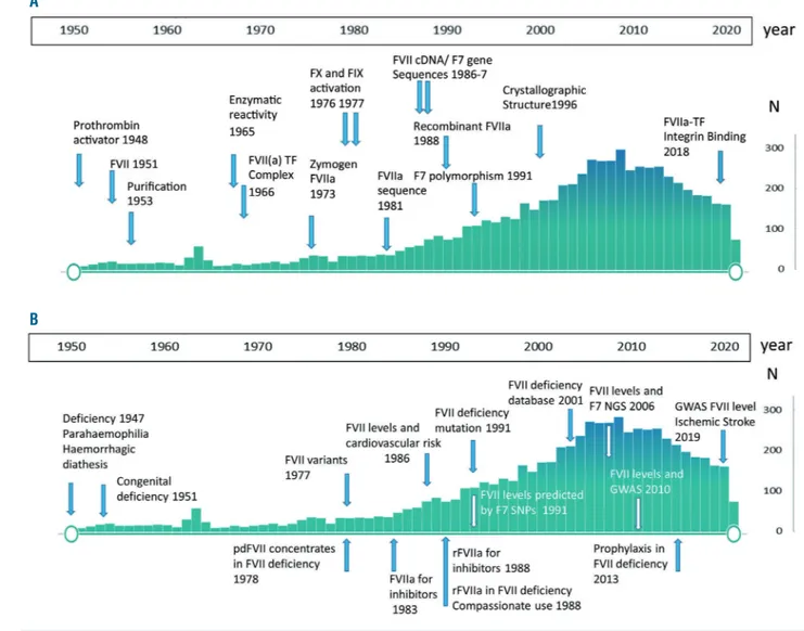
Figure 1. Publications over 70 years and some key achievements related to coagulation factor VII. (A) Coagulation factor VII (FVII) biochemistry and F7 genetics. (B) FVII deficiency, FVII level associated cardiovascular disease, FVII levels and F7 gene: N: number of publications reported in Pubmed. Only some of the discoveries are referenced in the text due to space constraints. A complete list is available on request. GWAS: Genome Wide Analysis Study; NGS: next-generation sequencing.
A
FVII zymogen circulates in plasma at low concentration (500 ng/mL, 5nM) and the extracellular proteolytic cleav-age1of a minute portion (about 1%, 0.1 nM) of FVII giving rise to FVIIa, occurs between residues Arg152-Ile153, (Figure 2)2producing the light and heavy chains. The origin of FVIIa in plasma is still debated. Substantially decreased levels of plasma FVIIa in individuals with congenital FIX deficiency suggest that the generation of FVIIa is depend-ent on an activation product of FIX. Recdepend-ently, it has been proposed that different forms of activated FIX (FIXα and FIXb) participate in a reciprocal activation mechanisms of FVII(a) and FIX(a) (white and red-curved arrows, Figure 3, left panel) that would not require a protein cofactor.24
The FVIIa heavy chain contains the domain character-ized by the (chymotrypsin) serine protease family catalytic triad. The light chain contains the calcium binding, vitamin K-dependent domain (gamma-carboxyl-glutamic, GLA) and two epidermal growth factor-like domains, essential for the interaction with membranes and other proteins2of the coagulation cascade.
Considerable variation in FVIIa plasma concentrations between individuals has been reported (Figure 4),25 exceed-ing one order of magnitude. Differently from other serine protease of the coagulation cascade, FVIIa displays a plas-ma half-life (2-3 hours) remarkably close to that of the FVII zymogen, which might be explained by the “zymogen-like” properties of FVIIa that have been weakened by mutagenesis, thus increasing its activity.26
FVIIa interacts with TF, a membrane receptor exposed following vascular lesion (extrinsic pathway). The
FVIIa-TF complex,1 essentially conserved in all jawed
vertebrates,27activates both FIX and factor X (FX)28(Figure
3) on the platelet surface in the presence of Ca2+, leading to the generation of thrombin and the subsequent deposition of fibrin.
The crystal structure of the complex between the active-site-inhibited FVIIa and the cleaved, soluble extracellular domain of TF (sTF) revealed the details of the contoured embrace of FVIIa and sTF domains and the extensive inter-molecular contacts.29Neutron and X-ray scattering experi-ments30 suggested that FVIIa in solution has an elongated domain structure with significant flexibility, which allows rapid interaction with sTF over a large surface area to form the high-affinity complex (dissociation constant, KD 2−5 nM).
The FVIIa-TF complex is highly dependent on specific lipids for physiological activity.2 Activated phospholipid membranes host both TF, an integral membrane protein, and FVIIa, which binds membranes through its GLA domain. Externalization of phosphatidylserine to the outer membrane leaflet and allosteric TF disulphide bond
exchanges (decryption on membranes)4make TF the
FVIIa-activating cofactor. The FVIIa-TF complex formation shapes the FVIIa catalytic site by allosteric interactions and increases its activity up to 106–fold. As a matter of fact, cir-culating FVIIa is the active portion of the total FVII mass only after high-affinity binding with TF. These FVIIa-TF complex-specific molecular events that provide FVIIa with its physiological activity are believed to represent the true initiation of the extrinsic pathway (Table 1).
The physiological negative control of the FVIIa-TF com-plex (Figure 3) occurs through the reversible inhibition by tissue factor pathway inhibitor (TFPI),31 mediated by the TFPI Kunitz domain 1 and with protein S as cofactor.32An
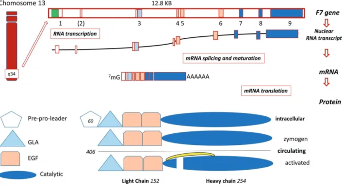
Figure 2. Schematic diagram of the F7 gene and factor VII protein expression.Upper part: Exons are numbered (1-9) and colored in accordance with the encoded protein domains (lower part). Exon 2 is in parenthesis because it is not included in the most abundant mature mRNA. FVII: factor VII; F7: factor VII gene; KB: kilobase. In addition to the complete nuclear transcript the most represented FVII mRNA is indicated. Lower part: Protein domains are indicated with different colors. GLA, tri-angle, γ-carboxyglutamic acid-containing domain. EGF, trapezium, epidermal growth factor-like domain. Green box, promoter. The complete intracellular protein, and the circulating forms are depicted. The numbers of residues in the pre-pro-leader sequence (n=60), in the circulating forms (n=406) and in the light (n=152) and heavy (n=254) chains are indicated. The interchain disulphide bridge is also depicted (yellow bracket).
apparently redundant and irreversible inhibition of FVIIa is also exerted by the serpin antithrombin (AT),33and a sub-stantial portion of FVIIa may be cleared through this rela-tively stable complex (FVIIa-AT),34which circulates in plas-ma at a concentration similar to that of FVIIa. FVII and FVIIa also bind the protein C receptor on endothelial cells (EPCR) with relevant affinity, but at present the patho-physiological significance of this interaction is unclear.
Congenital factor VII deficiency
Definition, prevalence and epidemiology
FVII deficiency was first described as a bleeding disorder
by Alexander and colleagues13and represents a model dis-ease to understand the pathophysiology of recessively inherited coagulation deficiencies. Relevant findings on FVII deficiency are summarized on a historical time scale in Figure 1B, and some of the open issues are summarized in Table 2. Congenital FVII deficiency (OMIM 227500, ORPHA 327) is defined as a bleeding disorder associated with FVII coagulant activity below 50% of normal. However, this threshold includes asymptomatic heterozy-gotes for causative mutations and may comprise individu-als homozygous for frequent FVII lowering single
nucleotide polymorphisms (SNP).35These subgroups
sub-stantially contribute to the belief that FVII deficiency is a mild bleeding disorder.36
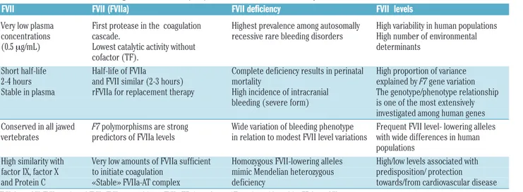
Figure 4. F7 genotype driven dif-ferences in FVIIa, FVIIc and FVIIAg in the European popula-tion. Mean values of FVIIa (mU/mL), FVIIc (% of PNP), and FVIIAg (% of PNP) in genotype groups determined by the promot-er (5′F7 ins del) and missense 353(413)Arg/Gln polymorphisms. Standard deviation is reported above each column. The number of subjects is reported in paren-theses. Significant differences, groups 1–6 (P<0.001): FVIIc: 1 vs. 2, 4, 6; 4 vs. 6; FVIIa: 1 vs. 4, 6; 2 vs. 6; 4 vs. 6; and FVIIag 1 vs. 4, 6. Adapted from “Contribution of factor VII genotype to activated FVII levels. Differences in geno-type frequencies between Northern and Southern European Populations”.107 FVII: factor VII;
FVIIc: FVII activity; FVIIAg: FVII antigen; PNP: pooled normal plas-ma; ins: insertion; del: deletion
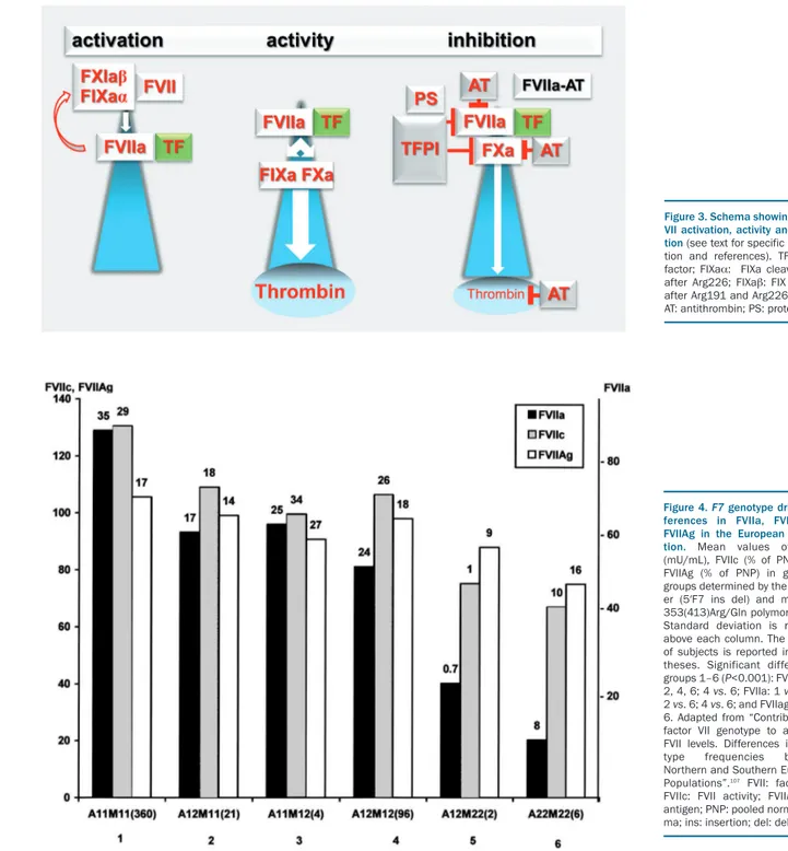
Figure 3. Schema showing factor VII activation, activity and inhibi-tion (see text for specific informa-tion and references). TF: tissue factor; FIXaα: FIXa cleaved only after Arg226; FIXab: FIX cleaved after Arg191 and Arg226 (ref. 24);
FVII deficiency is believed to be very rare (one case in
500,000 individuals).36,37 The World Federation of
Hemophilia38 estimates around 5,000 FVII-deficient indi-viduals in the countries covered by this organization. However, the Italian Registry of Congenital Rare Bleeding Disorders39(RBD) reported 1,023 cases with FVII deficien-cy among the 2,178 patients registered. This figure, similar to that reported by the UK registry (n=1.373),38suggests a prevalence only slightly lower than that of hemophilia B, at least in Italy (FVII deficiency, 1:59,000; haemophilia B, 1:67,000 individuals). Accordingly, FVII deficiency accounts for the most frequent RBD in most registries (36-49%).37,39 These data support the concept (Table 1) that FVII deficiency, including mild, moderate and severe forms, might be much more prevalent than previously reported. Large differences in prevalence, with variation reaching one order of magnitude, have been reported in those geographical areas where consanguineous marriages are frequent.
Clinical picture
The bleeding tendency in FVII deficiency varies from forms more severe than those seen in the hemophilias40to very mild forms or asymptomatic cases. A simple classifi-cation of the clinical severity was proposed:10 i) severe forms (central nervous system [CNS] and/or gastrointesti-nal [GI] bleeding and/or hemarthrosis; ii) moderate forms
(three or more symptoms other than CNS and/or GI bleeding and/or hemarthrosis); iii) mild forms (one or two symptoms, mostly muco-cutaneous, and other than CNS and/or GI and/or hemarthrosis).
In the International F7 Registry and the Seven Treatment Evaluation Registry (STER)41that include 787 cases, 12% of individuals had a severe to very severe phe-notype and three fourth of them bled within the first year of age (Table 4). About 20% of individuals with FVII defi-ciency suffer from a bleeding disorder that can be life-threatening and/or be a major handicap. On the other hand, the most frequently observed patient cohort (>50%) is characterized by muco-cutaneous types of bleeding: epistaxis (two thirds), gum bleeding (one third), easy bruising (one third), and menorrhagia (70% of the females). In general, the bleeding tendency remains the same during the lifetime of the patient with the first bleeding event being a strong and independent predictor
of the risk for the subsequent bleeding phenotype.41
Co-inheritance of factor V Leiden enhances thrombin forma-tion and is associated with a mild bleeding phenotype.42
Levels of FVIIc higher than 26%11,41,43may have a nil or minimal clinical relevance in terms of spontaneous bleed-ing and subjects with FVIIc levels <10% who remain asymptomatic most of their lives are not rare in being diagnosed during family studies or hemostatic screenings.10,11,35
Table 1. Distinctive features of coagulation factor VII (FVII), activated FVII , FVII deficiency, FVII levels and cardiovascular disease.
FVII FVII (FVIIa) FVII deficiency FVII levels
Very low plasma First protease in the coagulation Highest prevalence among autosomally High variability in human populations concentrations cascade. recessive rare bleeding disorders High number of environmental (0.5 mg/mL) Lowest catalytic activity without determinants
cofactor (TF).
Short half-life Half-life of FVIIa Complete deficiency results in perinatal High proportion of variance 2-4 hours and FVII similar (2-3 hours) mortality explained by F7 gene variation Stable in plasma rFVIIa for replacement therapy High incidence of intracranial The genotype/phenotype relationship bleeding (severe form) is one of the most extensively investigated among human genes
Conserved in all jawed F7 polymorphisms are strong Wide variation of bleeding phenotype Frequent FVII level- lowering alleles vertebrates predictors of FVIIa levels in relation to modest FVII level variations with wide differences in human populations
High similarity with Very low amounts of FVIIa sufficient Homozygous FVII-lowering alleles High/low levels associated with factor IX, factor X to initiate coagulation mimic Mendelian heterozygous predisposition/ protection and Protein C «Stable» FVIIa-AT complex deficiency towards/from cardiovascular disease
FVII: factor VII; FVIIa: activated FVII; rFVIIa: recombinant FVIIa; TF: tissue factor; AT: serpin antithrombin; F7: factor VII gene.
Table 2. Some open issues in factor VII (FVII) protein and F7 genetics, FVII deficiency, FVII levels and cardiovascular disease.
FVII / F7 FVII deficiency F7 gene variation cardiovascular disease
Mechanisms underlying Residual FVII concentration predicting the most Population features causing large differences in the FVIIa levels severe symptoms is still puzzling. prediction of cardiovascular disease risk by F7 genotypes Residual FVII concentration preventing spontaneous
bleeding is difficult to define.
Decryption of TF for FVIIa Short plasma half life of FVII/rFVIIa for therapeutic Impact of FVII-lowering alleles in the protection binding and catalytic activity purposes. from cardiovascular disease
Discrepancy between rFVIIa pharmacokinetic and pharmacodynamic data
Physiological balance between Impact of FVII-lowering alleles in increasing the Impact of FVII-increasing alleles in the predisposition FVIIa inhibitors clinical penetrance of pathological F7 gene lesions toward specific cardiovascular disease forms
However, the therapeutic thresholds conferring protec-tion from traumatic or post-operative bleeds are still a matter of debate.44,45Surgical bleeding has been reported in 15-24% of cases42,45with FVIIc <7%.42Bleeds in the first post-operative day46are particularly frequent if the bleed-ing disorder was not diagnosed before, and are mostly reported during orthopedic procedures,46 a major hemo-static challenge. Presenting symptoms as post-circumci-sion bleeds and hemorrhage from the umbilical stump frequently herald a severe disease form.
Diagnosis
The spectrum of bleeding symptoms is wide, involves both males and females, can start after birth and may occur at any age. The most important predictors of bleed-ing risk, familial and personal histories, can be silent as observed in diseases with recessive inheritance. The rele-vance of the gynecological and obstetric histories are dis-cussed in a dedicated paragraph.
The laboratory workup47 performed after a bleeding episode or during a family screening includes routine screening assays (prothrombin time [PT], activated partial thromboplastin time [aPTT]), followed by FVIIc measure-ment, which is necessary to confirm the diagnosis. Evaluation of FVIIc is determined with a one-stage assay adding diluted plasma samples to FVII-deficient plasma and thromboplastin as the source of TF. The accuracy of FVIIc assays is related to the sensitivity of the thrombo-plastin reagent and to the quality of the FVII-deficient plasma and of the calibrators. The most suitable activa-tors are the human placenta-derived and the recombinant
preparations,47that are also recommended to monitor the
treatment with rFVIIa.25New chromogenic or fluorogenic
assays for FVIIa are available for research purposes and to monitor treatment with rFVIIa.48
The sensitivity of the FVIIc assay to levels below 2% may be poor, with negative implications for the accurate definition of the relationship between FVII levels and bleeding phenotypes (Table 1), as well as between geno-type and phenogeno-type. A low assay sensitivity may explain some inconsistencies such as the paradoxical observation of asymptomatic cases with low FVII levels. Furthermore, severe bleeding is sporadically observed in individuals with moderately reduced levels of FVII. The combination of the low assay sensitivity with the minimal amount of FVIIa-TF complex needed to trigger coagulation does not permit one to efficiently differentiate clinical phenotypes. As a consequence, in FVII deficiency a variety of bleeding phenotypes are observed in the presence of apparently modest differences in FVIIc levels (Table 1). In summary, the clotting tests do not always predict the degree of bleeding tendency in the single individual.
The presence of the FVII protein (FVII antigen [FVIIag]) can be determined by enzyme-linked immunosorbent assays or immune-turbidimetric assays, using monoclon-al epitope-specific antibodies.47 The FVIIag assay47 does not predict the bleeding tendency, but does allow one to distinguish between type I (quantitative defects, with decrease of both FVIIc and FVIIAg), and type II defects (qualitative defects, with low FVIIc and normal or reduced levels of FVIIAg), and may help to clarify the dif-ferent mutational mechanisms.
Molecular genetics
The key findings are summarized on a historical time
scale in Figure 1B, and the main findings in individuals with FVII deficiency are shown in Table 3. The function-al characterization of protein variants in plasma was car-ried out in seminal studies49well before the genetic char-acterization of FVII deficiencies. The first F7 gene muta-tions in FVII deficiency were reported in the UK50 and Italian populations, and more than 1,000 genetic diag-noses in several countries are reported in the International Registry on FVII deficiency (IRF7)10and the Greifswald Registry.11Mutations and residual FVII levels have been presented in databases.51 The recent EHAD
database12 (http://www.umd.be/F7/W_F7/index.html)
includes 221 unique variants identified in 728 individu-als, of great help for the evaluation of F7 mutations found during genetic counseling and diagnosis, including prenatal diagnosis.5
F7 mutations have been detected by sequencing coding,
splicing and in promoter regions in the vast majority of patients. High throughput genomic sequencing will help in the case no mutations are found.52 Cases with severe symptoms are virtually all homozygous or doubly het-erozygous for F7 mutations. Symptomatic mutations impairing vitamin K metabolism, and thus posttransla-tional modification of GLA residues, have been found to cause combined vitamin K-dependent clotting factor defi-ciency that includes an abnormal FVII biosynthesis and low FVIIc levels.
As reported in registries11 and databases,12 single nucleotide variants are the most common type of gene defect (around 90%), and include a large proportion (between two thirds and three quarters) of missense mutations.
Numerous missense mutations have been found in the homozygous condition, related to geographical, ethnic and mutational pattern features. Clusters of homozygotes often indicate genetic isolates or ethnic customs favoring consanguinity (see below).
Since they have been well presented in databases,12we
shall briefly comment on a few of them.
The severe Gln100(160)Arg53,54 and the moderate/mild Ala294(354)Val55,56are prevalent in Northern Europe. The frameshift mutation in the last carboxyl terminal codons (Pro404(464)Hfs) is very prevalent in cis with the Ala294(354)Val missense change and causes a 28 residues-elongated FVII molecule. This moderate to severe and combined variant is frequent in Central Europe and espe-cially in Slovakia.10The mild Gly331(391)Ser mutation is prevalent in the gulf of Naples, Italy, as well as in other
countries.57 Other mild/asymptomatic changes, the
Arg304(364)Gln50, Cys310(370)Phe/Ser and
Thr359(419)Met substitutions, have been found in several populations, suggesting recurrent nucleotide changes at a CpG site. The frequent Arg304(364)Gln is also character-ized by discrepant FVII activity findings depending on the thromboplastin reagent.49
Splicing mutations are not rare,52and the homozygous
splicing mutation IVS7+5 g>a, relatively frequent in Italy, has been used for cellular58,59and animal60models of gene therapy.
F7 deletions are also not rare and together with
duplica-tions, insertions and indel rearrangements represent about 10% of all genetic lesions.12However, large and extended deletions have not been described in homozygous individ-uals. Differently from hemophilia A and B, individuals with complete FVII deficiency might die perinatally or
shortly after birth due to severe hemorrhage.5Complete FVII deficiency causes perinatal mortality in “knockout” mice, in which the presence of a trace amount of FVII (0.7%) seems sufficient for survival with symptoms.61 Several cases with heterozygous combined FVII and FX deficiency, due to large deletions within the terminal end
of chromosome 13, have been reported.62
Recombinant expression after site-directed mutagenesis has been performed for a number of mutations detected in patients, which permits one to investigate the molecular bases of the deficiency. Recombinant studies of the rare and potentially null homozygous nonsense mutations support the notion that gain-of-function and translational readthrough over premature stop codons may prevent truly null conditions and may contribute to the unexpect-edly variable bleeding phenotype in individuals
homozy-gous for F7 nonsense changes.63Whereas life-threatening
symptoms have been associated with the p.Ser52(112)X, moderate bleeding was observed in p.Cys82 (132)X homozygotes. The Arg402(462)X change permits the secretion of a small amount of protein with gain-of-func-tion features, causing a mild phenotype paradoxically associated with a “null” mutation.64
Chimeric fluorescent FVII molecules have also been constructed to investigate intracellular processing of vari-ants, and chemical chaperones have been used to
stimu-late their secretion.54 The FVII protein has been purified and biochemically characterized for very few variants,
among these the frequent Arg304(364)Gln50 and
Ala294(354)Val55 changes, as well as the severe
Gln160Arg33and the activation sequence Val154(214)Gly65 changes. These laborious experiments provided strong support for the understanding of the mild or severe coag-ulation phenotypes in vivo and for FVII altered expression and properties.
The homozygous conditions for polymorphisms pre-dicting lower FVII levels might not be pathogenic but mimic Mendelian heterozygous FVII deficiency35(Table 1), which frequently leads to the request for clotting defect diagnosis in pre-surgical screenings, and generates confu-sion in estimating the incidence of inherited FVII deficien-cy (see dedicated section). In light of their effects on low-ering FVII plasma levels, these variants might contribute to pathogenicity of co-inherited variants.52As a matter of fact the frequencies of the FVII-lowering alleles were signifi-cantly higher51in the FVII-deficient individuals tested than in controls, suggesting that the presence of these alleles may have increased, through increased clinical signifi-cance (Table 2), the likelihood of identifying “causative”
F7 gene lesions. Genotyping of these frequent variants
would be especially useful for the interpretation of mild and moderate FVII deficiency.
Table 3. Overview of molecular genetics findings in factor VII deficiency.
Molecular genetics of FVII deficiency
Genetic diagnosis Sanger sequencing of exons and splicing junctions in >1,000 individuals with FVII deficiency Next-generation sequencing in a few individuals
Phenotypic assays do not define specific genetic defects* No highly frequent gene lesions
Large heterogeneity of genetic causes (n>300 different mutations) Geographical/ethnic clustering of identical-by-descent mutations (n>15) Several recurrent mutations in different populations
Mutation zygosity and Severe deficiencies caused by compound heterozygous/homozygous mutations disease severity Increased prevalence of homozygotes in genetic isolates/consanguineous marriages
Otherwise asymptomatic heterozygous variants associated with frequent FVII-lowering single nucleotide polymorphism in mild deficiencies
Mutation type frequency Missense>>Small deletions>Splicing>Nonsense>Large deletions (not reported in homozygosity) Recombinant expression Numerous variants expressed after site directed mutagenesis
A few purified protein variants
Translational read through over premature termination codons Gain-of-function premature termination codon
*The asymptomatic Arg364Gln is detectable by using different thromboplastins59. Small deletions include also insertions and indels. FVII: factor VII.
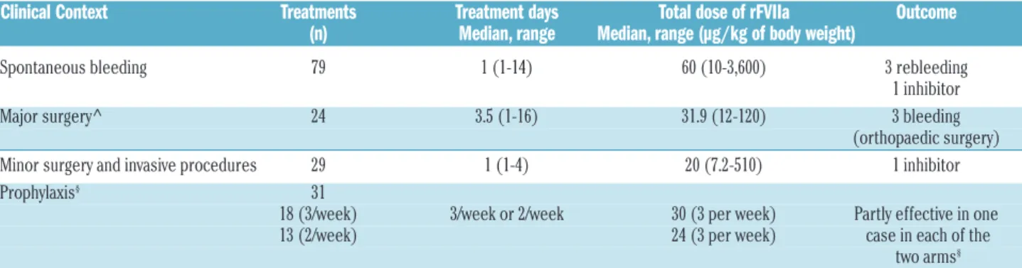
Table 4. Management of factor VII deficiency using recombinant factor VII (synopsis of the STER study data).
Clinical Context Treatments Treatment days Total dose of rFVIIa Outcome (n) Median, range Median, range (µg/kg of body weight) Spontaneous bleeding 79 1 (1-14) 60 (10-3,600) 3 rebleeding 1 inhibitor Major surgery^ 24 3.5 (1-16) 31.9 (12-120) 3 bleeding (orthopaedic surgery) Minor surgery and invasive procedures 29 1 (1-4) 20 (7.2-510) 1 inhibitor Prophylaxis§ 31
18 (3/week) 3/week or 2/week 30 (3 per week) Partly effective in one 13 (2/week) 24 (3 per week) case in each of the two arms§
The data express data analysis, not recommendations; ^ Mariani et al. 2011; §Napolitano et al. 2013 (no statistical difference between arms). FVII: factor VII; rFVII: recombinant
Replacement therapy and prophylaxis
Replacement therapy (RT) options are determined by: the rarity of the disorder; the availability and supply of products and the economic and geographical factors. These include: i) recombinant FVIIa (rFVIIa), ii) plasma-derived FVII (pdFVII), iii) fresh frozen plasma (FFP) and prothrombin complex concentrates (PCC). In the STER prospective trial (comprising 312 RT) most therapies were carried out with rFVIIa (78%), the remaining with FFP (10%), pdFVII concentrates (10%%) and PCC (2%). Based on the modest catalytic activity of FVIIa in the absence of TF, the therapeutic/prophylactic use of FVIIa and rFVIIa (initially proposed for bleeding diathesis other than FVII deficiency) represents a therapeutic milestone8 and distinguishes replacement therapy in FVII deficiency from that in the other coagulation defects (Table 1). Although in FVII deficiency rFVIIa is employed (Figure 1A) at doses (Table 3) much lower than those needed for patients with FVIII inhibitors, supraphysiological FVIIa concentrations are also produced in plasma of FVII defi-cient subjects.
It is still a matter of debate whether rFVIIa acts through the binding with TF, provided by microparticles shed into the circulation following diverse stimuli, or binds at high concentration66 to anionic phospholipids exposed on activated platelets, thus directly activating FX to FXa. FXa would in turn generate thrombin, bypass-ing the tenase complex. It is temptbypass-ing to speculate that the physiological FVIIa-TF function (Figure 3) prevails at the rFVIIa doses used in FVII deficiency and in the pres-ence of normal FVIII levels. rFVIIa easily diffuses into the extravascular spaces where it could be retained for extended time periods. As supported by pharmacokinet-ic studies,67rFVIIa prolongs its pharmacological effects at low concentration, in accordance with the hypothesis of physiological binding to TF.
rFVIIa has a very good safety-to-efficacy ratio.46 One-day therapy with ‘intermediate’ doses of rFVIIa can be effective and safe for the treatment of most of sponta-neous bleeds as well as for minor surgery and invasive procedures (Table 3). Replacement with rFVIIa is also effective to prevent bleeding in major surgical proce-dures.46
The large volume of the infusions and the limited availability make pd-FVII concentrates less appealing. Currently, the average pd-FVII dosages used are 15–20 IU/kg for mucosal bleeding, and 30–40 IU/kg for severe or life-threatening hemorrhages.
Although FFP is easily available in developing coun-tries, its effectiveness is limited owing to the high risk of fluid overload and the consequent need for slow infu-sions.
In the case of mild mucosal bleeds (epistaxis, mild menorrhagia) tranexamic acid and hormones are current-ly considered.
Anti-fibrinolytic agents are contraindicated in hema-turia, and may trigger thrombosis in association to PCC.
Prophylaxis is warranted for patients with the most severe bleeding picture and should be prescribed from childhood or soon after the first bleeding event.45,68 Effective prophylaxis schedules have been reported for rFVIIa68,69and pdFVII concentrates.70
Treatment complications
Inhibitors to FVII are a rare (1-2%)71 complication that
occurs mainly in severely deficient patients, particularly in children younger than 1 year on prophylaxis. We observed only high responders (>5 BU Bethesda Unit)71, and in the presence of a high-titer inhibitor to FVII treatment becomes a problem.71
At variance with the homologous deficiency of FIX (F9, HB), complete homozygous gene deletions that predis-pose to FIX inhibitors have never been detected in FVII deficiency (Table 1),5,12and FVII inhibitors have never been shown to be complicated by allergic reactions.
Paradoxically, a dozen of cases with FVII deficiency and thrombosis have been reported. Surgical interventions and/or replacement therapies had a close temporal rela-tionship with thrombotic episodes, but apparently spon-taneous events were also reported.68,72 Different replace-ment therapies were associated with the thrombotic events: PCC (three cases), rFVIIa (three cases), pdFVII (two cases), FFP (one case), no replacement (three cases).72This suggests that FVII deficiency does not seem to offer pro-tection from strong thrombosis risk factors such as sur-gery/high dose radiotherapy.
Women with factor VII deficiency
Autosomally transmitted RBD occur as frequently in women as in men, but women may experience more bleeding than men because gynecological and obstetric challenges to hemostasis add an important burden to the background bleeding related to the hemostatic defect. Menstruation and ovulation are associated with an increased risk of bleeding, as are pregnancy and delivery.73 While in normal and heterozygous women FVII plasma levels rise during pregnancy, no such increase is observed in women with a severe homozygous deficiency. As a consequence, symptomatic women with FVII deficiency may continue to bleed during pregnancy and postpartum. In a large study (234 women with FVII deficiency),74 men-orrhagia during the reproductive age occurred in half of the cases, and in 12% represented the first bleed. Further, frequent gynecological problems, such as uterine fibroids, were diagnosed earlier because of the hemostatic defect. Although FVIIc was an important predictor of gynecolog-ical bleeding, other determinants including endocrine pathologies may also play a role.
For severe menorrhagia, management has been moved from hemostatic agents like tranexamic acid, oral contra-ceptives and intra-uterine devices, to replacement therapy and prophylaxis with effective single- or multiple-dose schedules.68,73,74This will hopefully change clinical practice that, until recently, included surgical approaches such as endometrial ablation or hysterectomy.
Therapeutics in development
The first attempts to improve rFVIIa activity through mutagenesis, or its half-life in plasma by glycoPEGylation, have increased FVII antigenicity. Longer-acting FVIIa has been tested in different recombinant preparations, which are currently in clinical trials (reviewed in Menegatti et
al.)75: i) addition of a c-terminal peptide76potentially suit-able for subcutaneous administration that led to pro-longed pharmacodynamics effects, ii) an Fc receptor-fused rFVIIa that displayed a 5-fold longer plasma half-life and iii) an albumin-fused rFVIIa molecules that showed improved features compared to rFVIIa (2- to 3-fold longer half-life and 4- to 8-fold lower clearance) in patients with congenital FVII deficiency.77Recently, an engineered
albu-min-fused rFVIIa molecule showed enhanced transcellular transport upon intranasal delivery and extended plasma half-life in transgenic mice.78
The development of non-substitutive therapy for the hemophilias, such as the use of anti-antithrombin RNA interference or of aptamer and monoclonal antibodies directed against TFPI, have raised expectations for improvement of the quality of life in individuals with FVII deficiency.75
Based on adeno-associated viral-mediated expression of FVII79, gene therapy has produced sustained correction of severe FVII deficiency in dogs. In addition, gene therapy using FVIIa, inserted into the same viral vector, could also be used to treat congenital FVII deficiency with lower vec-tor doses, which increases the chance of efficient and long term expression of the transgene. Small engineered RNA have also been used as an approach of personalized gene therapy to correct a human FVII splicing mutation in mice.59
Factor VII levels,
F7 genotypes and
cardiovascu-lar disease
Several findings have created interest and still lead to an intense investigation on the role of FVII in cardiovascular disease (Figure 1B). Exposure of TF to blood in (coronary) artery disease, and especially plaque rupture, may favor FVIIa-TF complex formation. Further, lipids are important components of atherosclerosis and determinants of FVII activation and activity as well.79,80 Additional links between high FVII levels and risk for thrombosis have
been suggested81by finding that the TF-FVIIa-Xa complex
activates FVIII before coagulation amplification and that heparanase increases generation of FXa by the
FVIIa-TF-complex.82Furthermore, the FVIIa-TF complex is believed
to mediate non-hemostatic functions in diverse biological processes, such as angiogenesis, inflammation, atheroscle-rosis and vascular and cardiac remodeling, by activating signaling pathways (reviewed in D'Alessandro et al.)84 through FVIIa-integrin binding and PAR2 cleavage.85
The F7 promoter may respond to a number of metabolic components, and FVIIc levels are associated with several environmental factors linked to atherosclerosis i.e., body mass index, dietary fat intake, plasma lipids and particu-larly triglyceride concentration.
Age- and sex-related variations in FVII levels add com-plexity to the investigation of clinical correlates. FVII lev-els were found to increase with age and to be significantly lower in women than men at younger ages. Postmenopausal women displayed the highest levels,
except when undergoing hormone replacement therapy.86
This conundrum of environmental factors interacts with the F7 gene variation.
Factor VII levels and F7/genome wide genotypes
FVII levels show ample variation in the normal popula-tion and have a substantial heritable component, also in relation to polymorphisms15 (Figure 4). Missense,87repeat number variation,58 insertion/deletion22 and SNP23 were found to be associated with FVIIc, FVIIa and FVIIag levels (Figure 4) through epigenetic, transcription,
biosynthesis-or stability-mediated mechanisms. Whereas twin studies88
estimated about 60% of genetic-associated level of vari-ance in plasma, the F7 locus variation accounted for up to
40% of variance. Genotype effects, perhaps stronger on FVIIa than on FVIIag (Figure 4),15 might modulate the response to environmental stimuli and the sex-dependent regulation.89Serum phospholipids were found to be strong and F7 genotype-associated FVIIa determinants.90
The number of F7 single SNP was substantially increased by highthroughput F7 gene sequencing, which
enabled very informative F7 population studies.91
Genome-wide association studies (GWAS) confirmed14,92 the strong association between FVII levels and F7 gene
variation.93 Importantly, GWAS have detected a number
of genomic regions associated with FVII levels (GCKR,
ADH4, MS4A6A, PROCR, APOA5, HNF4A, REEP3-JMJD1C, JAZF1-AS1, MLXIPL and XXYLT1),14,92that could explain an additional one fifth of the FVII variance in plas-ma levels and are also, in part, associated with lipids which in turn modulate FVII activity. These gene-based association scan initiatives have been established in sever-al populations.94 Overall, this picture defines one of the most extensively investigated relationship between geno-types and multiple quantitative phenogeno-types (FVIIc, FVIIag and FVIIa) (Table 1).
Levels, genotypes and cardiovascular disease
The Northwick Park Heart Study investigators were the first to report that high FVII levels were predictors of
death due to coronary disease,16and a number of studies
confirmed this observation in different populations by also evaluating FVIIa levels.95
Recent investigations on the level of the FVIIa-AT com-plex, which may reflect levels of FVIIa as well its interac-tion with TF, indicated that higher complex concentra-tions were associated with increased mortality in the Cardiovascular Health Study.96 In patients with stable coronary artery disease (Verona Heart Study) higher FVIIa-AT complex levels were associated with increased cardio-vascular mortality and increased thrombin and FXa gener-ation,97 particularly in the coagulation initiation phase.98 However, in small groups of patients with unstable angi-na, acute myocardial infarction99or post-infarction80FVIIa levels were not higher than in controls. High plasma FVIIag levels were associated with failure of thrombolytic therapy in patients with myocardial infarction.100Elevated levels of FVII have not been consistently associated with
venous thromboembolism.101
The influence of F7 genotypes on the hemostatic bal-ance and on the susceptibility to cardiovascular disease has been extensively investigated in several large cohorts of patients in relation to both myocardial infarction and stroke. F7 polymorphisms with an opposite effect on FVIIa levels may positively or negatively modulate the risk of MI in males with advanced coronary artery disease,18 and some FVII genotypes may protect against myocardial infarction17 by affecting transcription levels and reducing protein functional activity. The modulation of stroke risk in atrial fibrillation by F7 genotypes may follow a similar scheme102 and recently, in a meta-analysis of several GWAS14 variations in FVII-related genes and FVIIc levels were associated with a risk of the incidence of ischemic stroke in the general population. The physiological effects of FVII lowering alleles may represent a natural model for anticoagulation; in fact F7 polymorphisms were shown to play a role in determining the initial response to war-farin103and influencing the risk of thrombosis in patients with essential thrombocythemia104.
Large haplotype studies confirmed that the F7 gene strongly influences FVII levels, but associations with coro-nary artery disease and FVII level were inconsistent.105 Variable genetic, environmental and atherogenic risk fac-tors in different populations may explain this discrepancy (Table 2). The frequencies of F7 genotypes predicting lower FVII levels are higher in Caucasians and much lower in Chinese and Malays populations. On the other hand, the distribution of these F7 genotypes covaries across pop-ulations in Europe with the rate of myocardial infarction mortality, being higher in countries at low risk.106
The most recent meta-analysis of large studies14appears to be consistent with positive and potentially clinically important causal effects of FVIIc levels, both on coronary artery disease and venous thromboembolism. However, several open issues remain (Table 2) and the usefulness of FVIIc and FVIIa evaluation in patients with cardiovascular risks is still unclear.
Conclusive remarks
In summary, in the 70 years since FVII investigation started, research has been particularly intense (Figure 1), and provided excellent examples at the basic science and
translational levels. The multidisciplinary approaches have spanned structural biology, biochemistry, recombi-nant protein biotechnology, molecular and population genetics, pharmacology, clinics and epidemiology, and encompassed various areas of hematology, hemostasis and thrombosis.
The large number of studies on FVII have not only served to substantially improve our knowledge on FVII biology and related phenotypes and to improve the quali-ty of life of the individuals with FVII deficiency, but also to train three generations of scientists in different fields.
Disclosures
Pfizer Research Grant
Contributions
FB and GM wrote the Manuscript
Acknowledgments
The authors express their gratitude to Prof. Peter Lydyard (emeritus at UCL London, UK and associate at Georgia University, Tbilisi Georgia) for his very helpful text revision and Dr Barbara Lunghi for her help in the selection of references.
References
1. Nemerson Y, Esnouf MP. Activation of a proteolytic system by a membrane lipopro-tein: mechanism of action of tissue factor. Proc Natl Acad Sci U S A. 1973;70(2):310-314.
2. Gajsiewicz JM, Morrissey JH. Structure-function relationship of the interaction between tissue factor and factor VIIa. Semin Thromb Hemost. 2015;41(7):682-690.
3. McVey JH. The role of the tissue factor pathway in haemostasis and beyond. Curr Opin Hematol. 2016;23(5):453-461. 4. Ansari SA, Pendurthi UR, Rao LVM. Role of
cell surface lipids and thiol-disulphide exchange pathways in regulating the encryption and decryption of Tissue Factor. Thromb Haemost. 2019;119(6):860-870. 5. McVey JH, Boswell EJ, Takamiya O, et al.
Exclusion of the first EGF domain of factor VII by a splice site mutation causes lethal factor VII deficiency. Blood 1998;92(3):920-926.
6. Lawson JH, Kalafatis M, Stram S, Mann KG. A model for the tissue factor pathway to thrombin. I. An empirical study. Biol Chem. 1994;269(37):23357-23366. 7. Zelaya H, Rothmeier AS and Ruf W. Tissue
factor at the crossroad of coagulation and cell signaling. J Thromb Haemost. 2018;16(10):1941-1952.
8. Hedner U, Glazer S, Pingel K, et al. Successful use of recombinant factor VIIa in a patient with severe hemophilia A during synovectomy. Lancet. 1988;2(8621):1193. 9. O'Hara PJ, Grant FJ, Haldeman BA, et al.
Nucleotide sequence of the gene coding for human factor VII, a vitamin K-dependent protein participating in blood coagulation. Proc Natl Acad Sci U S A. 1987;84(15):5158-5162.
10. Mariani G, Herrmann FH, Dolce A, et al. International Factor VII Deficiency Study Group. Clinical phenotypes and factor VII genotype in congenital factor VII deficien-cy. Thromb Haemost. 2005;93(3):481-487.
11. Herrmann FH, Wulff K, Auerswald G, et al. Greifswald Factor FVII Deficiency Study Group. Factor VII deficiency: clinical mani-festation of 717 subjects from Europe and Latin America with mutations in the factor 7 gene. Haemophilia. 2009;15(1):267-280. 12. Giansily-Blaizot M, Rallapalli PM, Perkins
SJ, et al. The EAHAD blood coagulation factor VII variant database. Hum Mutat. 2020;41(7):1209-1219.
13. Alexander B, Goldstein R, Landwehr G, Cook CD. Congenital SPCA deficiency: a hitherto unrecognized coagulation defect with hemorrhage rectified by serum and serum fractions. J Clin Invest. 1951;30(6): 596-608.
14. de Vries PS, Sabater-Lleal M, Huffman JE, et al. A genome-wide association study iden-tifies new loci for factor VII and implicates factor VII in ischemic stroke etiology. Blood. 2019;133(9):967-977.
15. Bernardi F, Marchetti G, Pinotti M, et al. Factor VII gene polymorphisms contribute about one third of the factor VII level vari-ation in plasma. Arterioscler Thromb Vasc Biol. 1996;16(1):72-76.
16. Meade TW, Mellows S, Brozovic M, et al. Haemostatic function and ischaemic heart disease: principal results of the Northwick Park Heart Study. Lancet. 1986;2(8506): 533-537.
17. Iacoviello L, Di Castelnuovo A, De Knijff P, et al. Polymorphisms in the coagulation factor VII gene and the risk of myocardial infarction. N Engl J Med. 1998;338(2):79-85.
18. Girelli D, Russo C, Ferraresi P, et al. Polymorphisms in the factor VII gene and the risk of myocardial infarction in patients with coronary artery disease. N Engl J Med. 2000;343(11):774-780.
19. Hagen FS, Gray CL, O'Hara P, et al. Characterization of a cDNA coding for human factor VII. Proc Natl Acad Sci U S A. 1986;83(8):2412-2416.
20. Arbini AA, Pollak ES, Bayleran JK, High KA, Bauer KA. Severe factor VII deficiency due to a mutation disrupting a hepatocyte nuclear factor 4 binding site in the factor
VII promoter. Blood. 1997;89(1):176-82. 21. Barbon E, Pignani S, Branchini A, Bernardi
F, Pinotti M, Bovolenta M. An engineered tale-transcription factor rescues transcrip-tion of factor VII impaired by promoter mutations and enhances its endogenous expression in hepatocytes. Sci Rep. 2016;6:28304.
22. Marchetti G, Patracchini P, Papacchini M, Ferrati M, Bernardi F. A polymorphism in the 5' region of coagulation factor VII gene (F7) caused by an inserted decanucleotide. Hum Genet. 1993;90(5):575-576.
23. van't Hooft FM, Silveira A, Tornvall P, et al. Two common functional polymorphisms in the promoter region of the coagulation factor VII gene determining plasma factor VII activity and mass concentration. Blood. 1999;93(10):3432-3441.
24. Misenheimer TM, Kumfer KT, Bates BE, Nettesheim ER, Schwartz BS. A candidate activation pathway for coagulation factor VII. Biochem J. 2019;476(19):2909-2926. 25. Morrisey JH, Macik BG, Neuenschwander
PF, Comp PC. Qantitation of activated fac-tor VII levels in plasma using a tissue facfac-tor mutant selectively deficient in promoting factor VII activation. Blood. 1993;81(3):734-744.
26. Sorensen AB, Tuneew I, Anders Svensson L, et al. Beating tissue factor at its own game: Design and properties of a soluble tissue factor-independent coagulation fac-tor VIIa. J Biol Chem. 2020;295(2):517-528. 27. Davidson CJ, Hirt RP, Lal K, et al. Molecular evolution of the vertebrate blood coagula-tion network. Thromb Haemost. 2003;89 (3):420-428.
28. Osterud B, Rapaport SI. Activation of factor IX by the reaction product of tissue factor and factor VII: additional pathway for initi-ating blood coagulation. Proc Natl Acad Sci U S A. 1977;74(12):5260-5264.
29. Banner DW, D'Arcy A, Chene C, et al. The crystal structure of the complex of blood coagulation factor VIIa with soluble tissue factor. Nature 1996;380(6569):41-46. 30. Rode-Mosbaek C, Nolan D, Persson E,
Extensive small-angle X-ray scattering stud-ies of blood coagulation factor VIIa reveal interdomain flexibility Biochemistry. 2010;49(45):9739-9745.
31. Girard TJ, Warren LA, Novotny WF, Bejcek BE, Miletich JP, Broze GJ Jr. Identification of the 1.4 kb and 4.0 kb messages for the lipoprotein associated coagulation inhibitor and expression of the encoded protein. Thromb Res. 1989;55(1):37-50.
32. Hackeng TM, Rosing J. Protein S as cofac-tor for TFPI. Arterioscler Thromb Vasc Biol. 2009;29(12):2015-2020.
33. Broze GJ Jr, Majerus PW. Purification and properties of human coagulation factor VII. J Biol Chem. 1980;255(4):1242-1247. 34. Agersø H, Brophy DF, Pelzer H, et al.
Recombinant human factor VIIa (rFVIIa) cleared principally by antithrombin follow-ing intravenous administration in hemo-philia patients. J Thromb Haemost. 2011;9(2):333-338.
35. Bernardi F, Castaman G, Pinotti M, et al. Mutation pattern in clinically asympto-matic coagulation factor VII deficiency. Hum Mutat. 1996;8(2):108-115.
36. Gallani D, Wheeler AP, Neff AT. Rare coag-ulation factor deficiencies. In Hoffman et al. Hematology, Basic Principles and Practice. Chapter 137:2034-50. Elsevier, Philadelphia (PA) USA, 2018.
37. Palla R, Peyvandi F, Shapiro A. Rare bleed-ing disorders: diagnosis and treatment. Blood. 2015;125(13):2052-2061.
38. World Federation of Hemophilia. Report on the Annual Global Survey 2017.
39. Abbonizio F, Hassan JH, Riccioni R, Arcieri R, Giampaolo A. National Registry of con-genital bleeding disorders (AICE). Report 2017. Istituto Superiore di Sanità, Rapporti Istisan. 2019,III:54.
40. Bernardi F, Dolce A, Pinotti M, et al. International Factor VII Deficiency Study Group. Major differences in bleeding symptoms between factor VII deficiency and hemophilia B. J Thromb Haemost. 2009;7(5):774-779.
41. Di Minno MND, Dolce A, Mariani G on behalf of the STER Study Group. Bleeding symptoms at disease presentation and pre-diction of ensuing bleeding in inherited FVII deficiency. Thromb Haemost. 2013;109(6):1051-1059.
42. Castoldi E, Govers-Riemslag JW, Pinotti M, et al. Coinheritance of factor V (FV) Leiden enhances thrombin formation and is associ-ated with a mild bleeding phenotype in patients homozygous for the FVII 9726þ5G>A (FVII Lazio) mutation. Blood. 2003;102(12):4014-4020.
43. Benlakhl F, Mura T, Schved JK, Giansily-Blaizot M on behalf of the French Study Group of FVII Deficiency. A retrospective analysis of 157 surgical procedures per-formed without replacement therapy in 83 unrelated factor VII-deficient patients. J Thromb Haemost. 2011;152(6):340-346. 44. Giansily-Blaizot M, Verdier R,
Biron-Adréani C, et al. Analysis of biological phe-notypes from 42 patients with inherited factor VII deficiency: can biological tests predict the bleeding risk? Haematologica. 2004;89(6):704-709.
45. Siboni SM, Biguzzi E, Mistretta C, Garagiola L, Peyvandi F. Long-term prophy-laxis in severe FVII deficiency. Haemophilia. 2015;21(6):812-819. 46. Mariani G, Dolce A, Batorova A et al.
Recombinant, activated factor VII for sur-gery in factor VII deficiency: a prospective evaluation – the surgical STER. Br J Haematol. 2011;152(3):340-346.
47. Sevenet PO, Kaczor DA, Depasse F. Factor VII deficiency: from basics to clinical labo-ratory diagnosis and patient management. Clin Appl Thromb Hemost. 2017;23 (7):703-710.
48. Cid AR, Lorenzo JI, Haya S, Montoro JM, Casana P, Aznar JA. A comparison of FVII:c and FVIIa assays for the monitoring of recombinant factor VIIa treatment. Haemophilia. 2001,7(1):39-41.
49. Girolami A, Fabris F, Dal Bo Zanon R, Ghiotto G, Burul A. Factor VII Padua: a congenital coagulation disorder due to an abnormal factor VII with a peculiar activa-tion pattern. J Lab Clin Med. 1978;91(3): 387-395.
50. O'Brien DP, Gale KM, Anderson JS, et al. Purification and characterization of factor VII 304-Gln: a variant molecule with reduced activity isolated from a clinically unaffected male. Blood. 1991;78(1):132-140.
51. McVey JH, Boswell E, Mumford AD, Kemball-Cook G, Tuddenham EG. Factor VII deficiency and the FVII mutation data-base. Hum Mutat. 2001;17(1):3-17. 52. Ferraresi P, Balestra D, Guittard C, et al.
Next-generation sequencing and recombi-nant expression characterized aberrant splicing mechanisms and provided correc-tion strategies in factor VII deficiency. Haematologica. 2020;105(3):829-837. 53. Kemball-Cook G, Johnson DJ, Takamiya O,
Banner DW, McVey JH, Tuddenham EG. Coagulation factor VII Gln100 --> Arg. Amino acid substitution at the epidermal growth factor 2-protease domain interface results in severely reduced tissue factor binding and procoagulant function. J Biol Chem. 1998;273(14):8516-8521.
54. Andersen E, Chollet ME, Baroni M, et al. The effect of the chemical chaperone 4-phenylbutyrate on secretion and activity of the p.Q160R missense variant of coagula-tion factor FVII. Cell Biosci. 2019;9:69. 55. Toso R, Pinotti M, High KA, Pollak ES,
Bernardi F. A frequent human coagulation Factor VII mutation (A294V, c152) in loop 140s affects the interaction with activators, tissue factor and substrates. Biochem J. 2002;363(Pt 2):411-416.
56. Herrmann FH, Wulff K, Strey R, Siegemund A, Astermark J, Schulman S, International Greifswald Registry of FVII deficiency. Variability of clinical manifestation of fac-tor VII-deficiency in homozygous and het-erozygous subjects of the european F7 gene mutation A294V. Haematologica. 2008;93 (8):1273-1275.
57. Etro D, Pinotti M, Wulff K, et al. The Gly331Ser mutation in factor VII in Europe and the Middle East. Haematologica. 2003;88(12):1434-1436.
58. Pinotti M, Toso R, Redaelli R, Berrettini M, Marchetti G, Bernardi F. Molecular mecha-nisms of FVII deficiency: expression of mutations clustered in the IVS7 donor splice site of factor VII gene. Blood. 1998;92(5):1646-1651.
59. Pinotti M, Rizzotto L, Balestra D, et al. U1-snRNA-mediated rescue of mRNA process-ing in severe factor VII deficiency. Blood. 2008;111(5):2681-2684.
60. Balestra D, Faella A, Margaritis P, et al. An engineered U1 small nuclear RNA rescues splicing defective coagulation F7 gene expression in mice. J Thromb Haemost. 2014;12(2):177-185.
61. Rosen ED, Xu H, Liang Z, Martin JA, Suckow M, Castellino FJ. Generation of genetically-altered mice producing very low levels of coagulation factor VII.
Thromb Haemost. 2005;94(3):493-497. 62. Pavlova A, Preisler B, Driesen J, et al.
Congenital combined deficiency of coagu-lation factors VII and X--different genetic mechanisms. Haemophilia. 2015;21(3):386-391.
63. Branchini A, Ferrarese M, Lombardi S, Mari R, Bernardi F, Pinotti M. Differential func-tional readthrough over homozygous non-sense mutations contributes to the bleeding phenotype in coagulation factor VII defi-ciency. J Thromb Haemost. 2016;14(10): 1994-2000.
64. Branchini A, Rizzotto L, Mariani G, et al. Natural and engineered carboxy-terminal variants: decreased secretion and gain-of-function result in asymptomatic coagula-tion factor VII deficiency. Haematologica. 2012;97(5):705-709.
65. Toso R, Bernardi F, Tidd T, et al. Factor VII mutant V154G models a zymogen-like form of factor VIIa. Biochem J. 2003;369(Pt 3):563-571.
66. Augustsson C, Persson E. In vitro evidence of a tissue factor-independent mode of action of recombinant factor VIIa in hemo-philia. Blood. 2104;124(20):3172-3174. 67. Morfini M, Batorova A, Mariani G, et al.
Pharmacokinetic properties of recombinant FVIIa in inherited FVII deficiency account for a large distribution at steady state and a prolonged pharmakodinamic effect. Thromb Haemost. 2014;112(2):424-425. 68. Napolitano M, Giansily-Blaizot M, Dolce
A, et al. Prophylaxis in congenital factor VII deficiency: indications, efficacy and safety. Results from the Seven Treatment Evaluation Registry (STER). Haematologica. 2013;98(4):538-544. 69. Kuperman AA, Barg AA, Fructhman Y, et
al. Primary prophylaxis for children with severe congenital FVII deficiency. Clinical and laboratory assessment. Blood Cells Mol Dis. 2017;67:86-90.
70. Mariani G, Mannucci PM, Mazzucconi MG, Capitanio A. Treatment of congenital factor VII deficiency with a new concen-trate. Thromb Haemost. 1978;39(3):675-682.
71. Mariani G, Konkle AB, Kessler CM. Inhibitors in hemophilias. In Hoffman et al. Hematology, Basic Principles and Practice. 5th edition. Chapter 136:2023-33. Elsevier, Philadelphia (PA) USA, 2018.
72. Mariani G, Herrmann FH, Shulman S, et al. Thrombosis in inherited FVII deficiency. J Thromb Haemost. 2003;1(10):2153-2158. 73. Peyvandi F, Garagiola I, Menegatti M.
Gynecological and obstetrical manifesta-tions of inherited bleeding disorders in women. J Thromb Haemost. 2011;9(Suppl 1):S236-245.
74. Napolitano M, Di Minno MN, Batorova A, et al. Women with congenital factor VII deficiency: clinical phenotype and treat-ment options from two international stud-ies. Haemophilia. 2016;22(5):752-759. 75. Menegatti M, Peyvandi F. Treatment of rare
factor deficiencies other than hemophilia. Blood. 2019;133(5):415-424.
76. Bar-Ilan A, Livnat T, Hoffmann M, et al. In vitro characterization of MOD-5014, a novel longacting carboxy-terminal peptide (CTP)- modified activated FVII. Haemophilia. 2018;24(3):477-486. 77. Laros-van Gorkom B, André Holme P,
Joch C, et al. Pharmacokinetics and phar-macodynamics of a recombinant fusion protein linking activated coagulation factor VII with human albumin (rVIIa-FP) in patients with congenital FVII deficiency. Hematology. 2020;25(1):17-25.
engineered human albumin enhances half-life and transmucosal delivery when fused to protein-based biologics. Sci Transl Med. 2020;12(565):eabb0580.
79. Marcos-Contreras OA, Smith SM, Bellinger DA, et al. Sustained correction of FVII defi-ciency in dogs using AAV-mediated expres-sion of zymogen FVII. Blood. 2016;127(5): 565-571.
80. Miller GJ. Dietary fatty acids and the haemostatic system. Atherosclerosis. 2005; 179(2):213-227.
81. Moor E, Silveira A, van't Hooft F, et al. Coagulation factor VII mass and activity in young men with myocardial infarction at a young age. Role of plasma lipoproteins and factor VII genotype. Arterioscler Thromb Vasc Biol. 1995;15(5):655-664.
82. Kamikubo Y, Mendolicchio GL, Zampolli A, et al. Selective factor VIII activation by the tissue factor–factor VIIa–factor Xa com-plex Blood. 2017; 130(14):1661-1670. 83. Nadir Y, Brenner B, Fux L, Shafat I, Attias J,
Vlodavsky I. Heparanase enhances the gen-eration of activated factor X in the presence of tissue factor and activated factor VII. Haematologica. 2010;95(11):1927-1934. 84. D'Alessandro E, Posma JJN, Spronk HMH,
Ten Cate H. Tissue Factor (:Factor VIIa) in the heart and vasculature: more than an envelope. Thromb Res. 2018;168:130-137. 85. Rothmeier AS, Liu E, Chakrabarty S, et al.
Identification of the integrin-binding site on coagulation factor VIIa required for proangiogenic PAR2 signaling. Blood. 2018;131(6):674-685.
86. Scarabin PY, Vissac AM, Kirzin JM, et al. Population correlates of coagulation factor VII. Importance of age, sex, and menopausal status as determinants of acti-vated factor VII. Arterioscler Thromb Vasc Biol. 1996;16(9):1170-1176.
87. Green F, Kelleher C, Wilkes H, Temple A, Meade T, Humphries S. A common genetic polymorphism associated with lower coag-ulation factor VII levels in healthy individu-als. Arterioscler Thromb. 1991;11(3):540-546.
88. de Lange M, Snieder H, Ariens RA, Spector TD, Grant PJ. The genetics of haemostasis: a twin study. Lancet. 2001;357(9250):101-105.
89. Eriksson-Berg M, Deguchi H, Hawe E, et al. Influence of factor VII gene polymorphisms
ulation factor VII concentrations in middle-aged women with and without manifest coronary heart disease. Thromb Haemost. 2005;93(2):351-358.
90. Mariani G, Bernardi F, Bertina R, et al. Serum phospholipids are the main environ-mental determinants of activated factor VII in the most common FVII genotype. European Union Concerted Action "Clotart". Haematologica. 1999;84(7):620-626.
91. Sabater-Lleal M, Almasy L, Martínez-Marchán E, et al. Genetic architecture of the F7 gene in a Spanish population: impli-cation for mapping complex diseases and for functional assays. Clin Genet. 2006;69(5):420-428.
92. Smith NL, Chen MH, Dehghan A, et al. Novel associations of multiple genetic loci with plasma levels of factor VII, factor VIII, and von Willebrand Factor: The CHARGE (Cohorts for Heart and Aging Research in Genome Epidemiology) Consortium. Circulation. 2010;121(12):1382-1392. 93. Tang W, Schwienbacher C, Lopez LM, et
al. Genetic associations for activated partial thromboplastin time and prothrombin time, their gene expression profiles, and risk of coronary artery disease meta-analy-sis. Am J Hum Genet. 2012;91(1):152-162. 94. Taylor KC, Lange LA, Zabaneh D, et al. A gene-centric association scan for coagula-tion factor VII levels in european and African Americans: the candidate gene association resource (CARe) consortium. Hum Mol Genet. 2011;20(17):3525-3534. 95. Kario K, Miyata T, Sakata T, Matsuo T,
Kato H. Fluorogenic assay of activated fac-tor VII. Plasma facfac-tor VIIa levels in relation to arterial cardiovascular diseases in Japanese. Arterioscler Thromb. 1994;14(2): 265-274.
96. Olson NC, Raffield LM, Lange LA, et al. Associations of activated coagulation factor VII and factor VIIa-antithrombin levels with genome-wide polymorphisms and cardiovascular disease risk. J Thromb Haemost. 2018;16(1):19-30.
97. Martinelli N, Girelli D, Baroni M, et al. Activated factor VII-antithrombin complex predicts mortality in patients with stable coronary artery disease: a cohort study. J Thromb Haemost. 2016;14(4):655-666. 98. Baroni M, Martinelli N, Lunghi B, et al.
to investigate hypercoagulability in plasma from patients with ischemic heart disease. Thromb Res. 2020;189:140-146.
99. Merlini PA, Ardissino D, Oltrona L, Broccolino M, Coppola R, Mannucci PM. Heightened thrombin formation but nor-mal plasma levels of activated factor VII in patients with acute coronary syndromes. Arterioscler Thromb Vasc Biol. 1995;15(10): 1675-1679.
100.Holm J, Tödt T, Berntorp E, Erhardt L. Failure of thrombolytic therapy in patients with myocardial infarction is associated with high plasma levels of factor VII anti-gen. Thromb Haemost. 1998;79(5):928-931.
101.Folsom AR, Cushman M, Heckbert SR, Ohira T, Rasmussen-Torvik L, Tsai MY. Factor VII coagulant activity, factor VII -670A/C and -402G/A polymorphisms, and risk of venous thromboembolism. J Thromb Haemost. 2007;5(8):1674-1678. 102.Roldán V, Marín F, González-Conejero R, et
al. Factor VII -323 decanucleotide D/I poly-morphism in atrial fibrillation: implications for the prothrombotic state and stroke risk. Ann Med. 2008;40(7):553-559.
103.D'Ambrosio RL, D'Andrea G, Cappucci F, et al. Polymorphisms in factor II and factor VII genes modulate oral anticoagulation with warfarin. Haematologica. 2004;89 (12):1510-1516.
104.Buxhofer-Ausch V, Olcaydu D, Gisslinger B, et al. Decanucleotide insertion polymor-phism of F7 significantly influences the risk of thrombosis in patients with essential thrombocythemia. Eur J Haematol. 2014;93 (2):103-111.
105.Ken-Dror G, Drenos F, Humphries SE, et al. Haplotype and genotype effects of the F7 gene on circulating factor VII, coagulation activation markers and incident coronary heart disease in UK men. J Thromb Haemost. 2010;8(11):2394-2403. 106.Donati MB, Zito F, Castelnuovo AD,
Iacoviello L. Genes, coagulation and cardio-vascular risk. J Hum Hypertens. 2000;14(6): 369-372.
107.Bernardi F, Arcieri P, Bertina RM, et al. Contribution of factor VII genotype to acti-vated FVII levels. Differences in genotype frequencies between northern and south-ern European populations. Arterioscler Thromb Vasc Biol. 1997;17(11):2548-2553.