doi: 10.3389/fnagi.2018.00371
Edited by: J. Arturo García-Horsman, University of Helsinki, Finland Reviewed by: Christian Windischberger, Medizinische Universität Wien, Austria Julia Jade Harris, Imperial College London, United Kingdom *Correspondence: Richard G. Wise [email protected] Received: 19 July 2018 Accepted: 25 October 2018 Published: 13 November 2018 Citation: Wright ME and Wise RG (2018) Can Blood Oxygenation Level Dependent Functional Magnetic Resonance Imaging Be Used Accurately to Compare Older and Younger Populations? A Mini Literature Review. Front. Aging Neurosci. 10:371. doi: 10.3389/fnagi.2018.00371
Can Blood Oxygenation Level
Dependent Functional Magnetic
Resonance Imaging Be Used
Accurately to Compare Older and
Younger Populations? A Mini
Literature Review
Melissa E. Wright
1,2and Richard G. Wise
1*
1Cardiff University Brain Imaging Research Center, School of Psychology, Cardiff University, Cardiff, United Kingdom, 2School of Optometry and Vision Sciences, Cardiff University, Cardiff, United Kingdom
A wealth of research has investigated the aging brain using blood oxygenation level
dependent functional MRI [Blood oxygen level dependent (BOLD) functional magnetic
resonance imaging (fMRI)]. However, many studies do not consider the aging of the
cerebrovascular system, which can influence the BOLD signal independently from
neural activity, limiting what can be inferred when comparing age groups. Here, we
discuss the ways in which the aging neurovascular system can impact BOLD fMRI, the
consequences for age-group comparisons and possible strategies for mitigation. While
BOLD fMRI is a valuable tool in this context, this review highlights the importance of
consideration of vascular confounds.
Keywords: aging, brain imaging, cerebral blood flow, cerebral hemodynamics, fMRI, neurovascular coupling
INTRODUCTION
With an expanding older population, research into the aging brain has become increasingly
important. Blood oxygen level dependent (BOLD) an functional magnetic resonance imaging
(fMRI) has often been applied to investigate how age affects neural function; common findings
include increased task-induced activation in frontal areas (
Park et al., 2003
;
Langenecker et al.,
2004
;
Gutchess et al., 2005
), decreased activation in occipital (
Madden, 2004
) and temporal areas
(
Park et al., 2003
;
Gutchess et al., 2005
), and altered functional connectivity (
Dørum et al., 2016
)
with increasing age.
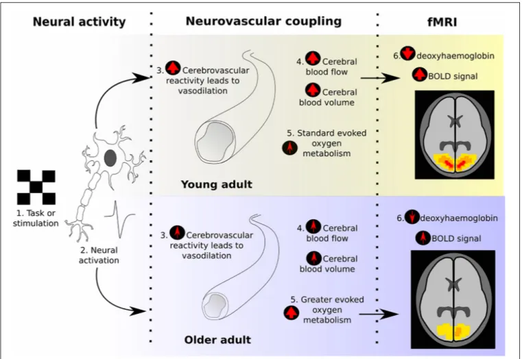
However, the BOLD signal does not directly index changes in neural activity (as
summarized in Figure 1). The BOLD signal reflects the balance between changes in cerebral
blood flow (CBF), determined by the characteristics of neurovascular coupling (NVC), and
changes in tissue oxygen consumption. Following increased neural activation, the signaling
processes that underlie NVC lead to vasodilation and increased blood flow, tending to
increase BOLD signal. Simultaneously, the increased oxygen consumption, needed to fuel
the increased neural activity, will decrease the BOLD signal. The picture is complicated
further by the additional dependence of BOLD signal changes on
the volume of the relevant venous blood compartment (cerebral
blood volume, CBV) and the local vascular architecture.
This complexity leaves BOLD fMRI open to confounds when
comparing age groups. If there are age-related differences in
NVC or oxygen consumption, then a difference in BOLD signal
would be produced independently of a difference in neural
activation. Therefore, the interpretability of any BOLD fMRI
studies that compare age groups without neurovascular controls
must be questioned. Here, we review how aging impacts the
neurovascular system and brain metabolism and how age may
confound BOLD fMRI studies, as well as discuss strategies for
addressing this issue.
AGING NEUROVASCULAR SYSTEM
Vasculature
Firstly, the structural integrity of the cerebral vasculature is
compromised during aging (
Gazzaley and D’Esposito, 2004
). This
includes thickening of the capillary basement membrane (
Alba
et al., 2004
) and decreases in capillary density, though the latter
has more mixed evidence (
Meiger-Ruge et al., 1980
;
Kalaria,
1996
). While the differing results for capillary density may
reflect different methodologies and variability in neurovascular
health (especially as reported changes tend not to be large;
Riddle et al., 2003
), they could also be due to the age ranges
included. Specifically, decreases in capillary density may only
begin in later senescence;
Brown et al. (2007)
found a stronger
negative correlation with age and density when only considering
participants over 60 years old. Therefore, this trend may not be
visible in younger age ranges. Structural changes (which impact
vessel elasticity and functionality) would influence CBF and CBV
dynamic changes, which then impact BOLD signal magnitude
and timing (
Brown et al., 2003
). For example, it has been
suggested that age-related arterial stiffness, which is especially
seen centrally (
O’Rourke and Hashimoto, 2007
), may lead to
CBF pulsatility increases despite overall CBF decreases (
Tarumi
et al., 2014
). A loss of vessels is also especially important when
comparing across aging studies, as different magnet strengths and
pulse sequences bias the BOLD signal to different vessel sizes
(
Mueller-Bierl et al., 2007
).
Cerebral Blood Flow (CBF)
Lower resting-CBF has been demonstrated in older adults using
arterial spin labelling (ASL) fMRI (
Restom et al., 2007
), even
when potential confounds such as partial volume effects and
age-related cortical thinning have been corrected for (
Asllani
et al., 2009
;
Chen et al., 2011
). This has been replicated with
different scanning techniques, though the magnitude differs
across regions (
Martin et al., 1991
;
Bertsch et al., 2009
;
Chen
et al., 2011
). It is suggested that this CBF decrease is due to
age-related differences in CO
2end-tidal partial pressure, which
reflects CO
2arterial tension, rather than an independent effect
of age (
De Vis et al., 2015
). CBF is an important parameter
in determining the BOLD response, with studies finding an
inverse correlation between basal CBF and BOLD signal change,
(
Cohen et al., 2002
;
Brown et al., 2003
;
Stefanovic et al., 2006
)
in line with the deoxy-hemoglobin dilution model (
Hoge et al.,
1999
). Indeed, age-related BOLD differences have been shown
to significantly decrease when lower CBF is accounted for,
suggesting a substantial vascular contribution to the observed
BOLD age-differences (
Zebrowitz et al., 2016
).
Cerebrovascular Reactivity (CVR)
Age also influences Cerebrovascular Reactivity (CVR), which
refers to the increase in CBF and CBV following exposure to a
vasodilatory stimulus, such as after neural activity in order to
meet the increased energy demand. Neurovascular coupling relies
on CVR being preserved. A decrease in vasodilatory capacity is
reported in aged rats (
Tamaki et al., 1995
).
Flück et al. (2014)
used transcranial Doppler ultrasound (TCD) to investigate this
in humans and concluded that older adults had reduced CVR,
which may be due to vascular stiffening. However, in TCD
the blood velocities through the posterior and middle cerebral
arteries are measured rather than absolute CBF, so there may be
unobserved regional dependences, and the lack of direct tissue
measurement of CBF using TCD means that results should be
interpreted with caution (
Coverdale et al., 2014
;
Reinstrup et al.,
2014
). Other studies have used a CO
2-inhalation task (
Liu et al.,
2013a
) or breath-holding challenge, (
Riecker et al., 2003
) in
which BOLD fMRI has been used monitor evoked-CBF changes.
Despite breath-holding challenges having potential confounds
(e.g., metabolic rate or lung function can influence the quantity
of blood CO
2) and not showing a linear relationship between
holding time and arterial partial pressure of CO
2, (
Fierstra
et al., 2013
) similar results were found between breath-hold
and CO
2inhalation. For both methods, older adults showed
lower CVR. Importantly, while uncorrected BOLD fMRI in
Liu
et al. (2013a)
implied decreases in task-related V1 and medial
temporal lobe activity with age, this effect disappeared when CVR
was corrected for. Additionally, a stronger increase in bilateral
frontal gyrus activation in older adults was found after
CVR-correction, which may reflect compensatory mechanisms as there
was no difference in memory scores (
Liu et al., 2013a
). This
provides strong evidence for an age-related decrease in CVR; this
would lead to a smaller amount of vasodilation and subsequent
evoked CBF, deoxyhaemoglobin, and BOLD signal, causing an
under-representation of neural responses in older groups when
comparing across ages.
Cerebral Metabolic Rate of Oxygen
(CMRO
2
)
Another key factor regulating BOLD signal is the rate at which
oxygen is extracted from the blood and used for energy release
(rate of cerebral metabolic oxygen consumption; CMRO
2).
An increase in CMRO
2would lead to an increase in
deoxy-hemoglobin in the venous vessels, and thus a decrease in BOLD
signal due to the paramagnetic properties of deoxy-hemoglobin
(
Schwarzbauer and Heinke, 1999
). Although age-related changes
in resting CMRO
2have been reported, there is conflicting
evidence as to their direction.
Peng et al. (2014)
investigated
this using CBF, blood oxygen saturation percentage, and total
FIGURE 1 | Illustration of the neurovascular coupling processes that give rise to the BOLD response, and how these typically change between young and old adults, according to previous literature. The final “fMRI” section demonstrates what effect would be expected on the BOLD response due to these age-related changes, assuming equal neural activity.
blood oxygen capacity to estimate resting CMRO
2, which is
suggested to be a reliable method (
Liu et al., 2013b
). They
found a significant increase in CMRO
2in older adults, which
could act as a compensatory mechanism for declining cognitive
functions or weakening CVR and CBF. Conversely,
De Vis
et al. (2015)
reported a decrease in baseline CMRO
2, which was
present in all regions except for the occipital cortex (although the
parietal cortex lost significance once end-tidal partial pressure
of CO
2was controlled for). This conflicting result is reflected
in other studies, using similar and alternative techniques (
Lu
et al., 2011
;
Aanerud et al., 2012
). It is possible that the method
by which CMRO
2was derived influenced results, as different
methods were used (e.g., ASL vs. phase-contrast flow velocity).
It may also simply reflect the greater within- and
between-subject variability that has been reported in older participants’
BOLD signals (
Kannurpatti et al., 2010
;
Garrett et al., 2013
;
Baum and Beauchamp, 2014
but see ). Of particular relevance for
interpreting task-related BOLD signal changes is the difference
in evoked CMRO
2, of which several studies report an
age-related increase (
Restom et al., 2007
;
Hutchison et al., 2013
).
In the presence of an equal blood flow response across age
groups this would have the effect of reducing the BOLD signal
response.
Interactions
between
changes
in
NVC
and
oxygen
consumption may have the greatest confounding effect;
for example, alterations in CMRO
2and CBF may explain
lower BOLD responses in older adults, as higher demand
but diminished supply would lead to higher venous
deoxy-hemoglobin concentrations (
Lu et al., 2011
;
Hutchison et al.,
2013
). Furthermore, these effects are clinically silent and thus
not excluded by controlling for health status (e.g., cardiac or
neurological illness). Several studies have linked age-related
changes in BOLD signal to neurovascular factors, such as signal
timing (
Taoka et al., 1998
) and increased voxel-wise noise
(
D’Esposito et al., 1999
). Another finding that confounds the
comparison of age groups using BOLD fMRI is that of increased
within- and between-subject variability that has been reported in
older adults (e.g.,
Kannurpatti et al., 2010
;
Baum and Beauchamp,
2014
). It may be that variations in NVC and levels of CMRO2
contribute to this greater variability (though this cannot be the
only contributor, as increased neural variation is also found
electro-physiologically;
McIntosh et al., 2014
). However, the
extent of this increased variability with age was found to vary
greatly based on the motion correction pipeline, despite the
pipelines having similar goals, introducing extra difficulty in age
group comparisons (
Turner et al., 2015
). Therefore, to better
interpret age-related changes in BOLD signal, these factors must
be considered and examined.
DRUG-RELATED FACTORS
Other age-related factors may also indirectly impact the
neurovascular system. For example, older adults tend to use more
prescription and over-the-counter drugs (
Francis et al., 2005
).
Although studies often exclude certain medications, such as
beta-blockers or anti-depressants, they rarely account for
over-the-counter products such as non-steroidal anti-inflammatory drugs
(NSAIDs). A survey of aspirin use (a common NSAID) in adults
over 45 years old found that 52% reported current use, many did
so for primary prevention, and that regular use was associated
with markers of a healthy lifestyle (
Williams et al., 2015
). This
indicates that over-the-counter NSAID use is higher in older
adults, even if they are relatively healthy. If these medications
alter NVC, it may confound the BOLD signal. There has been
little research into this, though aspirin was found to reduce
resting-CBF in an
in vivo rabbit model, which was suggested
to be due to inhibiting prostacyclin and/or nitric oxide (
Bednar
and Gross, 1999
). A human study investigated the effects of two
other NSAIDs, naproxen (available over-the-counter in lower
doses) and indomethacin (prescription only). Using transcranial
Doppler sonography, it found a significant decrease in resting
and visually evoked blood velocity, possibly due to inhibiting
vasodilatory processes (
Szabo et al., 2014
). These results may also
be more reflective of average older adult NSAID use than other
work as the medication was administered orally in usual doses
over 2 days, rather than an injection (
Jensen et al., 1993
) or a
single high dose (
Markus et al., 1994
). However, they used young
adults so it is inconclusive whether the same effect would be
evoked in the aged neurovascular system. Other types of NSAIDs
may also have differing effects; for example, CBV and CBF didn’t
change after short-term exposure to ibuprofen in piglets (
Pellicer
et al., 1999
). While under-researched in older adults, increased
use of NSAIDs may contribute to an inaccurate estimation of
neural activity differences in older adults. Further studies should
examine the influence of over-the-counter medications in older
adults and on the BOLD response.
POTENTIAL CONTROLS
As non-neuronal factors independently alter the BOLD response,
such factors should be understood and controlled for as far
as possible to maximize the interpretability of fMRI data. One
approach involves normalizing BOLD fMRI data with measure
of the effectiveness of NVC, CVR being commonly used under
the hypothesis that it explains a lot of variability in NVC. CVR
can be estimated by altering arterial blood CO
2(a vasodilator),
and measuring the CBF/BOLD increase. One approach involves
a CO
2-inhalation task with simultaneous fMRI recording and
using the change in BOLD to normalize the task-related response
(see
Liu et al., 2013a
). However, an inhalation task may prove
strenuous for older participants. Breath-holding paradigms,
which involve the participant naturally increasing blood CO
2by
holding their breath, may be more tolerable and have shown
comparable results (
Kastrup et al., 2001
;
Handwerker et al.,
2007
). Breath-holding is suggested to be reliable even in those
with poor breath-hold performance, such as older adults (
Bright
and Murphy, 2013
). However, breath-holding can cause severe
motion artifacts, particularly in high-field strengths, such as 7T.
Confounds could also be caused by patient anxiety over a
breath-hold or inhalation task, such as increased movement or increased
heart rate, which may occur if an older participant finds the task
difficult to perform.
Another possibility is resting state fluctuation amplitude
(RSFA), which examines the signal amplitude variation when
the participant is ‘task free’, thus reducing confounds such as
head movement which may be seen with a breath-holding or a
hypercapnia challenge (e.g.,
Hall et al., 2011
). The RSFA has been
suggested as an index of vascular contributions to variability in
the BOLD response as it will depend on local CVR and CBV.
Tsvetanov et al. (2015)
used RSFA to scale BOLD data and
found that the magnitude of age-related differences significantly
decreased. Furthermore, though RSFA has been criticized for
confounding together neural and neurovascular properties (
Lipp
et al., 2015
), these contributions have been separated using
magnetoencephalography and neurovascular function measures
(
Tsvetanov et al., 2015
). Only vascular factors mediated
age-effects on RSFA, which suggests that RSFA is largely driven by
neurovascular contributions (
Wolpe et al., 2016
). However, RSFA
has also been reported to have poorer repeatability and model
fit than breath holding tasks (
Lipp et al., 2015
). Although these
methods of estimating CVR have major limitations when applied
to an older population, they offer an empirical way of reducing
non-neural variability.
An alternative to trying to ‘correct’ the BOLD response
is to measure a process that is likely to more closely
reflect levels of neural activity, such as CMRO
2. By
calibrating fMRI to quantitatively examine the amount
of
oxygen
metabolism,
reflective
of
oxidative
energy
release, we avoid the influence of factors such as
resting-CBF
with
established
age
differences.
Methods
have
been proposed to map CMRO
2reliably, such as using
combined hyperoxia and hypercapnia (
Wise et al., 2013
;
Germuska et al., 2016
). However, this again involves gas
inhalation, which may be too strenuous or uncomfortable
for very old participants. A method involving diffuse
optical tomography with BOLD and ASL fMRI may
therefore be better as it does not involve gas inhalation
but was found to be highly correlated with previous
methods (
Yücel et al., 2014
). This also does not assume
a constant CMRO
2, which is not always found (
Xu et al.,
2011
).
Another option is implementing a control task that is assumed
to not show neural age-related changes (e.g., a simple motor
task;
D’Esposito et al., 1999
). A difference between groups would
therefore suggest a global NVC confound. The signal and noise
characteristics (i.e., the haemodynamic response function) within
each group could then be characterized to create a global
normalization factor (
Samanez-Larkin and D’Esposito, 2008
),
assuming that it can be extended across the brain. However, this
approach assumes equivalent neural activity across age groups,
which may not be valid even with simple motor tasks(
Sailer et al.,
2000
;
Inuggi et al., 2011
). It also does not account for
region-dependant variation in neurovascular function (
Ances et al.,
2008
;
Devonshire et al., 2012
). Additionally, the neurovascular
system has different influences on BOLD signal depending
on the task (
Kannurpatti et al., 2010
), possibly because they
utilize different functional and structural networks. Signals
characterized in control tasks therefore may not be generalizable.
The above controls may be best when used in tandem
with designs intended to reduce neurovascular confounds,
which also have the advantage of being easier to implement.
Investigating relative change within groups or varying the
conditions parametrically may be useful as they provide an
internal control (
Gazzaley and D’Esposito, 2004
;
Ankudowich
et al., 2016
). However, these designs will still be confounded
by resting levels of CBF influencing the magnitude of
evoked BOLD signal (
Cohen et al., 2002
). Another possible
method is event-related fMRI, which allows the separation
of cognitive processes; if age-related differences are found
in one stage of a cognitive task but not another, which
both use similar brain areas (e.g., memory encoding vs.
retrieval), observed differences are more likely to be neuronal
in origin (
Rypma and D’Esposito, 2000
;
Gazzaley and
D’Esposito,
2004
).
Although
the
methodological
design
depends heavily on the research question, designs such as the
above may be useful for comparing age groups with BOLD
fMRI.
CONCLUSION
The BOLD signal reflects the complex interaction of many
factors, such as CVR, CBF, and CMRO
2. When using BOLD fMRI
to compare across age groups, researchers must be aware that age
can impact the majority of these non-neural factors, so differences
across age groups cannot be solely contributed to neuronal
changes. Neuronal changes, if present, may be exaggerated or
masked by age-related alterations in factors such as
resting-CBF. This requires careful consideration during study design and
interpretation, so as not to falsely highlight neuronal changes that
are not present or miss out on identifying neuronal changes that
are present.
Finally, it should be noted that most of the neurovascular
changes noted are part of the normal aging process and
may have an independent effect on cognition (
Moorhouse
and Rockwood, 2008
;
Bangen et al., 2014
). For example,
neurovascular uncoupling, without CBF changes, leads to
significant cognitive impairment in mice (
Tarantini et al.,
2015
). Monitoring these factors using fMRI also has
interesting applications in clinically assessing neurovascular
health. While BOLD fMRI requires careful consideration if
interpreting signals as neuronal in origin, more broadly it
demonstrates great potential as a tool, in aging research,
that
is
sensitive
to
neurovascular
and
neurometabolic
changes.
AUTHOR CONTRIBUTIONS
MW conceived the review, literature search, and wrote
the manuscript. RW conceived the review and revised the
manuscript.
REFERENCES
Aanerud, J., Borghammer, P., Chakravarty, M. M., Vang, K., Rodell, A. B., Jónsdottir, K. Y., et al. (2012). Brain energy metabolism and blood flow
differences in healthy ageing.J. Cerebral Blood Flow Metab. 32, 1177–1187.
doi: 10.1038/jcbfm.2012.18
Alba, C., Vidal, L., Díaz, F., Villena, A., and de Vargas, I. P. (2004). Ultrastructural and quantitative age-related changes in capillaries of the dorsal lateral geniculate nucleus.Brain Res. Bull. 64, 145–153. doi: 10.1016/j.brainresbull. 2004.06.006
Ances, B. M., Leontiev, O., Perthen, J. E., Liang, C., Lansing, A. E., and Buxton, R. B. (2008). Regional differences in the coupling of cerebral blood flow and oxygen metabolism changes in response to activation: implications
for BOLD-fMRI.Neuroimage 39, 1510–1521. doi: 10.1016/j.neuroimage.2007.
11.015
Ankudowich, E., Pasvanis, S., and Rajah, M. N. (2016). Changes in the modulation of brain activity during context encoding vs. context retrieval across the
adult lifespan. Neuroimage 139, 103–113. doi: 10.1016/j.neuroimage.2016.
06.022
Asllani, I., Habeck, C., Borogovac, A., Brown, T. R., Brickman, A. M., and Stern, Y. (2009). Separating function from structure in perfusion imaging of the aging
brain.Hum. Brain Mapp. 30, 2927–2935. doi: 10.1002/hbm.20719
Bangen, K. J., Nation, D. A., Clark, L. R., Harmell, A. L., Wierenga, C. E., Dev, S. I., et al. (2014). Interactive effects of vascular risk burden and advanced age on cerebral blood flow.Front. Ageing Neurosci. 6:159. doi: 10.3389/fnagi.2014. 00159
Baum, S. H., and Beauchamp, M. S. (2014). Greater BOLD variability in older
compared with younger adults during audiovisual speech perception.PLoS One
9:e111121. doi: 10.1371/journal.pone.0111121
Bednar, M. M., and Gross, C. E. (1999). Aspirin reduces experimental cerebral
blood flow in vivo. Neurol. Res. 21, 488–490. doi: 10.1080/01616412.1999.
11740963
Bertsch, K., Hagemann, D., Hermes, M., Walter, C., Khan, R., and Naumann, E.
(2009). Resting cerebral blood flow, attention, and ageing.Brain Res. 1267,
77–88. doi: 10.1016/j.brainres.2009.02.053
Bright, M. G., and Murphy, K. (2013). Reliable quantification of BOLD fMRI
cerebrovascular reactivity despite poor breath-hold performance.Neuroimage
83, 559–568. doi: 10.1016/j.neuroimage.2013.07.007
Brown, G. G., Zorrilla, L. T. E., Georgy, B., Kindermann, S. S., Wong, E. C., and Buxton, R. B. (2003). BOLD and perfusion response to finger-thumb apposition after acetazolamide administration: differential relationship to global perfusion. J. Cerebral Blood Flow Metab. 23, 829–837. doi: 10.1097/01.WCB.0000071887. 63724.B2
Brown, W. R., Moody, D. M., Thore, C. R., Challa, V. R., and Anstrom, J. A. (2007). Vascular dementia in leukoaraiosis may be a consequence of capillary loss not only in the lesions, but in normal-appearing white matter and cortex as well. J. Neurol. Sci. 257, 62–66. doi: 10.1016/j.jns.2007.01.015
Chen, J. J., Rosas, H. D., and Salat, D. H. (2011). Age-associated reductions in
cerebral blood flow are independent from regional atrophy.Neuroimage 55,
468–478. doi: 10.1016/j.neuroimage.2010.12.032
Cohen, E. R., Ugurbil, K., and Kim, S.-G. (2002). Effect of basal conditions on the magnitude and dynamics of the blood oxygenation level-dependent fMRI
response.J. Cereb. Blood Flow Metab. 22, 1042–1053. doi: 10.1097/00004647-200209000-00002
Coverdale, N. S., Gati, J. S., Opalevych, O., Perrotta, A., and Shoemaker, J. K. (2014). Cerebral blood flow velocity underestimates cerebral blood flow during
modest hypercapnia and hypocapnia.J. Appl. Physiol. 117, 1090–1096. doi:
10.1152/japplphysiol.00285.2014
De Vis, J. B., Hendrikse, J., Bhogal, A., Adams, A., Kappelle, L. J., and Petersen, E. T. (2015). Age-related changes in brain hemodynamics; a calibrated MRI study:
age-related changes in brain hemodynamics.Hum. Brain Mapp. 36, 3973–3987.
doi: 10.1002/hbm.22891
D’Esposito, M., Zarahn, E., Aguirre, G. K., and Rypma, B. (1999). The effect of normal ageing on the coupling of neural activity to the bold hemodynamic
response.Neuroimage 10, 6–14. doi: 10.1006/nimg.1999.0444
Devonshire, I. M., Papadakis, N. G., Port, M., Berwick, J., Kennerley, A. J., Mayhew, J. E. W., et al. (2012). Neurovascular coupling is brain region-dependent. Neuroimage 59, 1997–2006. doi: 10.1016/j.neuroimage.2011.09.050
Dørum, E. S., Alnaes, D., Kaufmann, T., Richard, G., Lund, M. J., Tønnesen, S., et al. (2016). Age-related differences in brain network activation and co-activation during multiple object tracking.Brain Behav. 6:e00533. doi: 10.1002/brb3.533 Fierstra, J., Sobczyk, O., Battisti-Charbonney, A., Mandell, D. M., Poublanc, J.,
Crawley, A. P., et al. (2013). Measuring cerebrovascular reactivity: what stimulus to use? Measuring cerebrovascular reactivity.J. Physiol. 591, 5809– 5821. doi: 10.1113/jphysiol.2013.259150
Flück, D., Beaudin, A. E., Steinback, C. D., Kumarpillai, G., Shobha, N., McCreary, C. R., et al. (2014). Effects of ageing on the association between cerebrovascular
responses to visual stimulation, hypercapnia and arterial stiffness. Front.
Physiol. 5:49. doi: 10.3389/fphys.2014.00049
Francis, S.-A., Barnett, N., and Denham, M. (2005). Switching of prescription drugs to over-the-counter status: is it a good thing for the elderly?Drugs Ageing 22, 361–370. doi: 10.2165/00002512-200522050-00001
Garrett, D. D., Kovacevic, N., McIntosh, A. R., and Grady, C. L. (2013). The modulation of BOLD variability between cognitive states varies by age and processing speed.Cereb. Cortex 23, 684–693. doi: 10.1093/cercor/bhs055 Gazzaley, A. H., and D’Esposito, M. (2004). “BOLD functional MRI and cognitive
ageing,” inCognitive Neuroscience of Ageing: Linking Cognitive and Cerebral
Ageing, eds R. Cabeza, L. Nyberg, and D. Park (New York, NY: Oxford University Press).
Germuska, M., Merola, A., Murphy, K., Babic, A., Richmond, L., Khot, S., et al. (2016). A forward modelling approach for the estimation of oxygen extraction fraction by calibrated fMRI.Neuroimage 139, 313–323. doi: 10.1016/ j.neuroimage.2016.06.004
Gutchess, A. H., Welsh, R. C., Hedden, T., Bangert, A., Minear, M., Liu, L. L., et al. (2005). Ageing and the neural correlates of successful picture encoding: frontal activations compensate for decreased medial-temporal activity.J. Cogn. Neurosci. 17, 84–96. doi: 10.1162/0898929052880048
Hall, E. L., Driver, I. D., Croal, P. L., Francis, S. T., Gowland, P. A., Morris, P. G., et al. (2011). The effect of hypercapnia on resting and stimulus induced MEG
signals.Neuroimage 58, 1034–1043. doi: 10.1016/j.neuroimage.2011.06.073
Handwerker, D. A., Gazzaley, A., Inglis, B. A., and D’Esposito, M. (2007). Reducing vascular variability of fMRI data across ageing populations using a
breathholding task.Hum. Brain Mapp. 28, 846–859. doi: 10.1002/hbm.20307
Hoge, R. D., Atkinson, J., Gill, B., Crelier, G. R., Marrett, S., and Pike, G. B. (1999). Investigation of BOLD signal dependence on cerebral blood flow and oxygen
consumption: The deoxyhemoglobin dilution model. Magn. Reson. Med.
42, 849–863. doi: 10.1002/(SICI)1522-2594(199911)42:5<849::AID-MRM4>3. 0.CO;2-Z
Hutchison, J. L., Lu, H., and Rypma, B. (2013). Neural mechanisms of age-related slowing: the CBF/ CMRO2 ratio mediates age-differences in BOLD signal
and human performance.Cereb. Cortex 23, 2337–2346. doi: 10.1093/cercor/
bhs233
Inuggi, A., Amato, N., Magnani, G., González-Rosa, J. J., Chieffo, R., Comi, G., et al. (2011). Cortical control of unilateral simple movement in healthy
ageing. Neurobiol. Ageing 32, 524–538. doi: 10.1016/j.neurobiolaging.2009.
02.020
Jensen, K., Freundlich, M., Bünemann, L., Therkelsen, K., Hansen, H., and Cold, G. E. (1993). The effect of indomethacin upon cerebral blood flow in healthy
volunteers. The influence of moderate hypoxia and hypercapnia.Acta Neuroch.
124, 114–119. doi: 10.1007/BF01401132
Kalaria, R. N. (1996). Cerebral vessels in ageing and Alzheimer’s disease. Pharmacol. Therap. 72, 193–214. doi: 10.1016/S0163-7258(96)00116-7 Kannurpatti, S. S., Motes, M. A., Rypma, B., and Biswal, B. B. (2010). Neural and
vascular variability and the fMRI-BOLD response in normal ageing.Magn.
Reson. Imaging 28, 466–476. doi: 10.1016/j.mri.2009.12.007
Kastrup, A., Krüger, G., Neumann-Haefelin, T., and Moseley, M. E. (2001). Assessment of cerebrovascular reactivity with functional magnetic resonance
imaging: comparison of CO2 and breath holding.Magn. Reson. Imaging 19,
13–20. doi: 10.1016/S0730-725X(01)00227-2
Langenecker, S. A., Nielson, K. A., and Rao, S. M. (2004). fMRI of healthy older
adults during Stroop interference.Neuroimage 21, 192–200. doi: 10.1016/j.
neuroimage.2003.08.027
Lipp, I., Murphy, K., Caseras, X., and Wise, R. G. (2015). Agreement and repeatability of vascular reactivity estimates based on a breath-hold task and a resting state scan.Neuroimage 113, 387–396. doi: 10.1016/j.neuroimage.2015. 03.004
Liu, P., Hebrank, A., Rodrigue, K., Kennedy, K., Section, J., Park, D., et al. (2013a). Age-related differences in memory-encoding fMRI responses after accounting
for decline in vascular reactivity. Neuroimage 78, 415–425. doi: 10.1016/j.
neuroimage.2013.04.053
Liu, P., Xu, F., and Lu, H. (2013b). Test-retest reproducibility of a rapid method
to measure brain oxygen metabolism.Magn. Reson. Med. 69, 675–681. doi:
10.1002/mrm.24295
Lu, H., Xu, F., Rodrigue, K. M., Kennedy, K. M., Cheng, Y., Flicker, B., et al. (2011). Alterations in cerebral metabolic rate and blood supply across the adult lifespan. Cereb. Cortex 21, 1426–1434. doi: 10.1093/cercor/bhq224
Madden, D. J. (2004). Age-related changes in neural activity during visual target
detection measured by fMRI.Cereb. Cortex 14, 143–155. doi: 10.1093/cercor/
bhg113
Markus, H. S., Vallance, P., and Brown, M. M. (1994). Differential effect of three cyclooxygenase inhibitors on human cerebral blood flow velocity and carbon dioxide reactivity.Stroke 25, 1760–1764. doi: 10.1161/01.STR.25.9.1760 Martin, A. J., Friston, K. J., Colebatch, J. G., and Frackowiak, R. S. J. (1991).
Decreases in regional cerebral blood flow with normal ageing.J. Cereb. Blood Flow Metab. 11, 684–689. doi: 10.1038/jcbfm.1991.121
McIntosh, A. R., Vakorin, V., Kovacevic, N., Wang, H., Diaconescu, A., and Protzner, A. B. (2014). Spatiotemporal dependency of age-related changes in brain signal variability.Cereb. Cortex 24, 1806–1817. doi: 10.1093/cercor/ bht030
Meiger-Ruge, W., Hunziker, O., Schulz, U., Tobler, H., and Schweizer, A. (1980). Stereological changes in the capillary network and nerve cells of the ageing
human brain.Mech. Ageing Dev. 14, 684–689.
Moorhouse, P., and Rockwood, K. (2008). Vascular cognitive impairment: current
concepts and clinical developments.Lancet Neurol. 7, 246–255. doi: 10.1016/
S1474-4422(08)70040-1
Mueller-Bierl, B. M., Uludag, K., Pereira, P. L., and Schick, F. (2007). Magnetic field distribution and signal decay in functional MRI in very high fields (up to 9.4
T) using monte carlo diffusion modeling.Int. J. Biomed. Imaging 2007:70309.
doi: 10.1155/2007/70309
O’Rourke, M. F., and Hashimoto, J. (2007). Mechanical factors in arterial ageing. J. Am. Coll. Cardiol. 50, 1–13. doi: 10.1016/j.jacc.2006.12.050
Park, D. C., Welsh, R. C., Marshuetz, C., Gutchess, A. H., Mikels, J., Polk, T. A., et al. (2003). Working memory for complex scenes: age differences in frontal
and hippocampal activations.J. Cogn. Neurosci. 15, 1122–1134. doi: 10.1162/
089892903322598094
Pellicer, M., Aparicio, F., and Caba`nas, E. A. (1999). Effect of the cyclo-oxygenase blocker ibuprofen on cerebral blood volume and cerebral blood flow during
normocarbia and hypercarbia in newborn piglets.Acta Paediatr. 88, 82–88.
doi: 10.1080/08035259950170664
Peng, S.-L., Dumas, J. A., Park, D. C., Liu, P., Filbey, F. M., McAdams, C. J., et al. (2014). Age-related increase of resting metabolic rate in the human brain. Neuroimage 98, 176–183. doi: 10.1016/j.neuroimage.2014.04.078
Reinstrup, P., Ryding, E., Asgeirsson, B., Hesselgard, K., Unden, J., and Romner, B. (2014). Cerebral blood flow and transcranial doppler sonography measurements of CO2-Reactivity in acute traumatic brain injured patients. Neurocrit. Care 20, 54–59. doi: 10.1007/s12028-012-9727-8
Restom, K., Bangen, K. J., Bondi, M. W., Perthen, J. E., and Liu, T. T. (2007). Cerebral blood flow and BOLD responses to a memory encoding task: A
comparison between healthy young and elderly adults.Neuroimage 37, 430– 439. doi: 10.1016/j.neuroimage.2007.05.024
Riddle, D. R., Sonntag, W. E., and Lichtenwalner, R. J. (2003). Microvascular plasticity in ageing.Ageing Res. Rev. 2, 149–168. doi: 10.1016/S1568-1637(02) 00064-8
Riecker, A., Grodd, W., Klose, U., Schulz, J. B., Groschel, K., Erb, M., et al. (2003). Relation between regional functional MRI activation and vascular reactivity to carbon dioxide during normal ageing.J. Cereb. Blood Flow Metab. 23, 565–573. doi: 10.1097/01.WCB.0000056063.25434.04
Rypma, B., and D’Esposito, M. (2000). Isolating the neural mechanisms of
age-related changes in human working memory.Nat. Neurosci. 3, 509–515. doi:
10.1038/74889
Sailer, A., Dichgans, J., and Gerloff, C. (2000). The influence of normal ageing
on the cortical processing of a simple motor task.Neurology 55, 979–985.
doi: 10.1212/WNL.55.7.979
Samanez-Larkin, G. R., and D’Esposito, M. (2008). Group comparisons: imaging the ageing brain.Soc. Cogn. Affect. Neurosci. 3, 290–297. doi: 10.1093/scan/ nsn029
Schwarzbauer, C., and Heinke, W. (1999). Investigating the dependence of
BOLD contrast on oxidative metabolism.Magn. Reson. Med. 41, 537–543.
doi: 10.1002/(SICI)1522-2594(199903)41:3<537::AID-MRM16>3.0.CO;2-V Stefanovic, B., Warnking, J. M., Rylander, K. M., and Pike, G. B. (2006). The effect
of global cerebral vasodilation on focal activation hemodynamics.Neuroimage
30, 726–734. doi: 10.1016/j.neuroimage.2005.10.038
Szabo, K., Rosengarten, B., Juhasz, T., Lako, E., Csiba, L., and Olah, L. (2014). Effect of non-steroid anti-inflammatory drugs on neurovascular coupling in humans. J. Neurol. Sci. 336, 227–231. doi: 10.1016/j.jns.2013.10.048
Tamaki, K., Nakai, M., Yokota, T., and Ogata, J. (1995). Effects of ageing and chronic hypertension on cerebral blood flow and cerebrovascular CO2 reactivity in the rat.Gerontology 41, 11–17. doi: 10.1159/000213657
Taoka, T., Iwasaki, S., Uchida, H., Fukusumi, A., Nakagawa, H., Kichikawa, K., et al. (1998). Age correlation of the time lag in signal change on EPI-fMRI.J. Comput. Assist. Tomogr. 22, 514–517. doi: 10.1097/00004728-199807000-00002 Tarantini, S., Hertelendy, P., Tucsek, Z., Valcarcel-Ares, M. N., Smith, N.,
Menyhart, A., et al. (2015). Pharmacologically-Induced Neurovascular
uncoupling is associated with cognitive impairment in mice.J. Cereb. Blood
Flow Metab. 35, 1871–1881.doi: 10.1038/jcbfm.2015.162
Tarumi, T., Khan, M. A., Liu, J., Tseng, B. M., Parker, R., Riley, J., et al. (2014). Cerebral hemodynamics in normal ageing: central artery stiffness, wave reflection, and pressure pulsatility.J. Cereb. Blood Flow Metab. 34, 971–978. doi: 10.1038/jcbfm.2014.44
Tsvetanov, K. A., Henson, R. N. A., Tyler, L. K., Davis, S. W., Shafto, M. A., Taylor, J. R., et al. (2015). The effect of ageing on fMRI: Correction for the confounding effects of vascular reactivity evaluated by joint fMRI and MEG in
335 adults: vascular influences on BOLD signal with ageing.Hum. Brain Mapp.
36, 2248–2269. doi: 10.1002/hbm.22768
Turner, B. O., Lopez, B., Santander, T., and Miller, M. B. (2015). One dataset, many conclusions: BOLD variability’s complicated relationships with age and motion artifacts.Brain Imag. Behav. 9, 115–127. doi: 10.1007/s11682-014-9351-7 Williams, C. D., Chan, A. T., Elman, M. R., Kristensen, A. H., Miser, W. F., Pignone,
M. P., et al. (2015). Aspirin use among adults in the U.S.Am. J. Prevent. Med. 48, 501–508. doi: 10.1016/j.amepre.2014.11.005
Wise, R. G., Harris, A. D., Stone, A. J., and Murphy, K. (2013). Measurement of OEF and absolute CMRO2: MRI-based methods using interleaved and
combined hypercapnia and hyperoxia.Neuroimage 83, 135–147. doi: 10.1016/j.
neuroimage.2013.06.008
Wolpe, N., Ingram, J. N., Tsvetanov, K. A., Geerligs, L., Kievit, R. A., Henson, R. N., et al. (2016). Ageing increases reliance on sensorimotor prediction through structural and functional differences in frontostriatal circuits.Nat. Commun. 7:13034. doi: 10.1038/ncomms13034
Xu, F., Uh, J., Brier, M. R., Hart, J., Yezhuvath, U. S., Gu, H., et al. (2011). The influence of carbon dioxide on brain activity and metabolism in conscious
humans.J. Cereb. Blood Flow Metab. 31, 58–67. doi: 10.1038/jcbfm.2010.153
Yücel, M. A., Evans, K. C., Selb, J., Huppert, T. J., Boas, D. A., and Gagnon, L. (2014). Validation of the hypercapnic calibrated fMRI method using DOT–
fMRI fusion imaging.Neuroimage 102, 729–735. doi: 10.1016/j.neuroimage.
2014.08.052
Zebrowitz, L., Ward, N., Boshyan, J., Gutchess, A., and Hadjikhani, N. (2016). Dedifferentiated face processing in older adults is linked to lower resting state metabolic activity in fusiform face area.Brain Res. 1644, 22–31. doi: 10.1016/j. brainres.2016.05.007
Conflict of Interest Statement: The authors declare that the research was conducted in the absence of any commercial or financial relationships that could be construed as a potential conflict of interest.
Copyright © 2018 Wright and Wise. This is an open-access article distributed under the terms of the Creative Commons Attribution License (CC BY). The use, distribution or reproduction in other forums is permitted, provided the original author(s) and the copyright owner(s) are credited and that the original publication in this journal is cited, in accordance with accepted academic practice. No use, distribution or reproduction is permitted which does not comply with these terms.