of December 6, 2011
This information is current as
2004;173;3838-3843
J Immunol
Cafaro, Paola Rimessi, Antonella Caputo and Barbara Ensoli
Bonaccorsi, Mauro Marastoni, Fabiola Micheletti, Aurelio
Fabris, Arianna Bottoni, Alessandro Canella, Angela
Riccardo Gavioli, Eleonora Gallerani, Cinzia Fortini, Marina
Proteasome Composition and Enzymatic Activity
Cytotoxic T Cell Epitopes by Modifying
HIV-1 Tat Protein Modulates the Generation of
References
http://www.jimmunol.org/content/173/6/3838.full.html#related-urls
Article cited in:
http://www.jimmunol.org/content/173/6/3838.full.html#ref-list-1
, 20 of which can be accessed free at:
cites 39 articles
This article
Subscriptions
http://www.jimmunol.org/subscriptions
is online at
The Journal of Immunology
Information about subscribing to
Permissions
http://www.aai.org/ji/copyright.html
Submit copyright permission requests at
Email Alerts
http://www.jimmunol.org/etoc/subscriptions.shtml/
Receive free email-alerts when new articles cite this article. Sign up at
Print ISSN: 0022-1767 Online ISSN: 1550-6606.
Immunologists, Inc. All rights reserved.
by The American Association of
Copyright ©2004
9650 Rockville Pike, Bethesda, MD 20814-3994.
The American Association of Immunologists, Inc.,
is published twice each month by
The Journal of Immunology
on December 6, 2011
www.jimmunol.org
HIV-1 Tat Protein Modulates the Generation of Cytotoxic T
Cell Epitopes by Modifying Proteasome Composition and
Enzymatic Activity
1
Riccardo Gavioli,
2* Eleonora Gallerani,* Cinzia Fortini,* Marina Fabris,
†Arianna Bottoni,*
Alessandro Canella,* Angela Bonaccorsi,
†Mauro Marastoni,
‡Fabiola Micheletti,
§Aurelio Cafaro,
§Paola Rimessi,
†Antonella Caputo,
†and Barbara Ensoli
§Tat, the trans activation protein of HIV, is produced early upon infection to promote and expand HIV replication and transmis-sion. However, Tat appears to also have effects on target cells, which may affect Ag recognition both during infection and after vaccination. In particular, Tat targets dendritic cells and induces their maturation and Ag-presenting functions, increasing Th1 T cell responses. We show in this work that Tat modifies the catalytic subunit composition of immunoproteasomes in B and T cells either expressing Tat or treated with exogenous biological active Tat protein. In particular, Tat up-regulates latent membrane protein 7 and multicatalytic endopeptidase complex like-1 subunits and down-modulates the latent membrane protein 2 subunit. These changes correlate with the increase of all three major proteolytic activities of the proteasome and result in a more efficient generation and presentation of subdominant MHC-I-binding CTL epitopes of heterologous Ags. Thus, Tat modifies the Ag processing and modulates the generation of CTL epitopes. This may have an impact on both the control of virally infected cells during HIV-1 infection and the use of Tat for vaccination strategies. The Journal of Immunology, 2004, 173: 3838 –3843.
T
at is a regulatory protein of HIV-1 produced very early after infection and essential for HIV-1 gene expression, replication, and infectivity (1). During acute infection of T cells by HIV-1, Tat is also released in the extracellular milieu in the absence of cell death or permeability changes (2– 4). Extracel-lular Tat is taken up by neighbor cells, where it modulates viral and/or cellular functions depending on the protein concentration, conformational state, and cell type (2– 6). In addition, native Tat protein targets and is very efficiently taken up by monocyte-de-rived dendritic cells (DC),3and, after internalization, it induces DC maturation, augmenting allogeneic and Ag-specific presentation by DC and increasing Th1 responses against recall Ags (6). We have shown recently that vaccination with the Tat protein or tat DNA protects monkeys against challenge with pathogenic simian HIV, and that this protection correlates with Th1 responses and CTL activity (7, 8).
CTLs recognize peptide epitopes expressed at the surface of target cells in association with MHC class I molecules. The major enzymatic activity responsible for the generation of class I-associated peptides is
that of the proteasome, a large multicatalytic protease that is essential for the degradation of intracellular proteins (9). Proteasomes have three distinct catalytic subunits called 1, 2, and 5, which exhibit postacidic, tryptic-like, and chymotryptic-like activity, respectively (10). When cells are exposed to IFN-␥, these three catalytic subunits are substituted with new components termed latent membrane protein 2 (LMP2), LMP7, and multicatalytic endopeptidase complex like-1 (MECL1), which are incorporated into a modified proteasome form known as immunoproteasome (11). The proteolytic activity of immu-noproteasomes is characterized by a reduced cleavage after acidic amino acids and by an increased cleavage after hydrophobic and basic residues, which are the most frequent residues found at the COOH terminus of MHC class I-binding peptides (12, 13).
It has recently been shown that the Tat protein interacts with both␣ subunits and LMP7 and MECL1 catalytic subunits of the proteasome (14), and that Tat inhibits the proteolytic activity of 20S proteasome by competing with PA28 regulator for binding to 20S (15, 16). In this study, we analyzed the subunit composition and activity of immunoproteasomes expressed in cells endog-enously expressing Tat or treated with a fully active exogenous Tat protein. We showed that Tat affects the subunit composition of immunoproteasomes and increases all three major proteolytic ac-tivities. This results in more efficient generation and presentation of subdominant MHC-I-binding CTL epitopes derived from heterologous Ags.
Materials and Methods
CellsLymphoblastoid cell lines (LCL) were established by in vitro infection of B lymphocytes from healthy donors (MIN and MON) with the B95.8 strain of EBV. Transduction of HIV tat (BH10 clone) into MIN and MON LCLs was performed by infection with the retroviral vector pBabeP carrying the puromycin resistance gene (17). Puromycin-selected cells were tested for the presence and expression of tat by DNA-PCR, RT-PCR, and Northern blot analysis. Jurkat T cells expressing Tat have been previously described
Departments of *Biochemistry and Molecular Biology,†Experimental and Diagnostic Medicine, and‡Pharmaceutical Sciences, University of Ferrara, Ferrara, Italy; and §Laboratory of Virology, Istituto Superiore di Sanita`, Rome, Italy
Received for publication April 13, 2004. Accepted for publication July 12, 2004. The costs of publication of this article were defrayed in part by the payment of page charges. This article must therefore be hereby marked advertisement in accordance with 18 U.S.C. Section 1734 solely to indicate this fact.
1This work was supported by grants from Istituto Superiore di Sanita` (AIDS Project), Italian Concerted Action on HIV-AIDS Vaccine Development, Italian Association for Cancer Research, and Ministero dell’Universita` e della Ricerca Scientifica e Tech-nologica. E.G. and C.F. were supported by a PhD fellowship awarded by the Uni-versity of Ferrara, and M.F. by an AIDS fellowship by Istituto Superiore di Sanita`. 2Address correspondence and reprint requests to Dr. Riccardo Gavioli, Department of Biochemistry and Molecular Biology, Universita` di Ferrara, Via Luigi Borsari 46, 44100 Ferrara, Italy. E-mail address: [email protected]
3Abbreviations used in this paper: DC, dendritic cell; EBNA, EBV-encoded nuclear Ag; IRF, IFN regulatory factor; LCL, lymphoblastoid cell line; LMP, latent mem-brane protein; MECL, multicatalytic endopeptidase complex like-1.
Copyright © 2004 by The American Association of Immunologists, Inc. 0022-1767/04/$02.00
on December 6, 2011
www.jimmunol.org
(18). Cells were cultured in medium supplemented with 800g/ml neo-mycin (Sigma-Aldrich, St. Louis, MO). Control cells and cells expressing Tat presented similar amounts of HLA class I molecules and adhesion molecules as judged by immunofluorescence analysis using mAbs specific for HLA-A,-B,-C, HLA-A2, HLA-DR, CD40, CD11a, CD80, and CD86 molecules (BD Pharmingen, San Diego, CA).
HIV-1 Tat protein
HIV-1 Tat from the human T lymphotropic virus type IIIB isolate (BH10 clone) was expressed in Escherichia coli and purified by heparin-affinity chromatography and HPLC, as described previously (6). The lyophilized Tat protein was stored at⫺80°C to prevent oxidation, reconstituted in degassed buffer before use, and handled, as described (3). Different lots of Tat were used with reproducible results, and, in all cases, endotoxin con-centration was undetectable (detection threshold: 0.05 EU/g).
Purification of proteasomes
Cells were lysed with glass beads, as previously described (19). Superna-tants were ultracentrifuged for 1 h at 100,000⫻ g and loaded into an affinity column containing a matrix derivatized with the MCP21 mAb spe-cific for the␣2 subunit of the proteasome (Affinity Technology, Exeter, U.K.). Proteasomes were eluted with 25 mM Tris-HCl, pH 7.5, containing 2 M NaCl, and 0.5-ml fractions were collected. Homogeneity of the eluted material was confirmed by analysis of an aliquot by 12% SDS-PAGE and Coomassie blue staining of the gel. Fractions containing proteasomes were combined and dialyzed against 25 mM Tris-HCl, pH 7.5. Protein concen-tration was determined using the bicinchoninic acid method (Pierce, Rockford, IL).
Western blot assay
Equal amounts of proteins or equal amounts of purified proteasomes were loaded on a 12% SDS-PAGE and electroblotted onto Protran nitrocellulose membranes (Schleicher & Schuell Microscience, Keene, NH). Blots were probed with Abs specific for␣2, LMP2, LMP7, MECL1, and PA28␣ sub-units (Affinity) and developed by ECL (Amersham Biosciences, Uppsala, Sweden).
Enzymatic assays
The chymotrypsin-like, trypsin-like, and postacidic activities of purified proteasomes were tested using the fluorogenic substrates Suc-LLVY-AMC, Boc-LRR-Suc-LLVY-AMC, and Ac-YVAD-Suc-LLVY-AMC, respectively, as previously described (19). Fluorescence was determined by a fluorometer (Spec-trafluor plus; Tecan, Salzburg, Austria). Proteasome activity is expressed as arbitrary fluorescence units. The in vitro degradation of CLGGLLTMVA-GAVW (CLG⫹ 5) was performed using 500g of peptide and 127 g of purified proteasomes in 300l of activity buffer at 37°C. At different time points, 60l of sample was collected, and the reaction was stopped by adding 2 vol of ethanol at 0°C. Digestion mixtures were centrifuged, and 80l of supernatants was collected and separated by HPLC. Fractionation was simultaneously monitored at 210 and 280 nm. Fractions were collected and tested by IFN-␥ ELISPOT assays.
Synthetic peptides
Peptides were synthesized by solid phase method and purified by HPLC to
⬎98% purity, as previously described (20). Structure verification was
per-formed by elemental and amino acid analysis and mass spectrometry. Pep-tides were dissolved in DMSO at 10⫺2M, kept at⫺20°C, and diluted in PBS before use.
Generation of CTL cultures
HLA A11-restricted specific CTL cultures reacting against the EBV-encoded nuclear Ag 4 (EBNA4)-derived IVTDFSVIK (IVT, aa 416 – 424) and AVFSRKSDAK (AVF, aa 399 – 408) epitopes (21) were obtained by stimulation of lymphocytes from the HLA-A11-positive EBV-seropositive donor MC with the autologous LCL. HLA-A2-restricted EBV-specific CTL cultures reacting against the LMP2-derived CLGGLLTMV (CLG, aa 426 – 434) epitope (22) and the LMP1-derived YLQQNWWTL (YLQ, aa 159 –167) epitope (23) were obtained by stimulation of lymphocytes from the HLA-A2-positive EBV-seropositive donor RG with peptide-pulsed T2 cells, as previously described (24). CTL cultures were maintained in me-dium supplemented with 10 U/ml rIL-2 (Chiron, Milan, Italy).
Cytotoxicity assay
Target cells were labeled with Na251CrO4for 90 min at 37°C. Cyto-toxicity tests were routinely run at different E:T ratios in triplicate.
Percentage of specific lysis was calculated as 100⫻ (cpm sample ⫺ cpm medium)/(cpm Triton X-100⫺ cpm medium) (21). Spontaneous release was always⬍20%.
ELISPOT assay
CTLs (4⫻ 104) were seeded in triplicate on microplate 96-well unifilter (Whatman, Maidstone, Kent, U.K.) coated with an anti IFN-␥ mAb (Pierce). CTLs were stimulated with 20l of each HPLC fraction derived from the in vitro digestion of CLG⫹ 5 peptide. CTLs incubated with medium alone were used as negative control, whereas CTL stimulated with PHA (Wellcome Diagnostics, Dartford, U.K.) represented the positive con-trol. Plates were incubated for 24 h and washed, and then a biotinylated anti-IFN-␥ mAb (1 g/ml; Pierce) was added to the wells. After 60 min, the plates were washed again and HRP-conjugated streptavidin (Pierce) was added at room temperature for 45 min. Individual IFN-␥-producing cells were detected using 3-amino-9-ethylcarbazole cromogen kit (Sigma-Aldrich) and counted by ELISPOT reader (AELVIS, Hannover, Germany). The number of specific IFN-␥-secreting T cells, expressed as spot-forming cells per 106cells, was calculated by subtracting the negative control values.
FIGURE 1. Expression of proteasomes in LCL expressing the HIV-1 tat gene. A, Equal amounts of proteins from total cell lysates from MIN and MON LCLs transduced with pBabeP (MIN-0 and MON-0, respectively) or with pBabeP-Tat (MIN-Tat and MON-Tat, respectively) were fractionated by SDS-PAGE, transferred onto nitrocellulose filters, and probed with mAbs or polyclonal antisera specific for the␣2 subunit, PA28␣, LMP2, LMP7, and MECL1. B, The intensity of specific bands was measured by densitometry. Data are expressed as percentage of increase in ODs of spe-cific bands detected in Tat-expressing cells relative to control cells. Mean ⫾ SEM of four independent experiments is shown. C, Equal amounts of purified proteasomes (1g) were fractionated by SDS-PAGE, transferred onto nitrocellulose filters, and probed with mAbs or polyclonal antisera␣2 subunit, LMP2, LMP7, and MECL1. One representative ex-periment of four performed is shown. D, The intensity of specific bands was measured by densitometry. Data are expressed as percentage of in-crease in ODs of specific bands detected in proteasomes purified from Tat-expressing cells relative to proteasomes from control cells. Mean⫾ SEM of three independent experiments is shown.
3839 The Journal of Immunology
on December 6, 2011
www.jimmunol.org
Results
Endogenously expressed Tat or exogenous native Tat protein modulates proteasome composition and activity
To evaluate the level of proteasome expression in the presence of endogenous Tat, LCL were permanently transduced with a Tat-expressing vector (MIN-Tat and MON-Tat) or the vector alone (MIN-0 and MON-0), and total cell lysates were examined by Western blot analysis with Abs specific for the constitutive ␣2 subunit of proteasomes. As shown in Fig. 1, all cells expressed similar amounts of proteasomes. Because LCLs constitutively ex-press immunoproteasomes (25), we then evaluated the exex-pression of the IFN-␥-inducible PA28␣ regulator and that of the catalytic  subunits LMP2, LMP7, and MECL1. Both LCL-Tat lines showed no difference in the level of expression of PA28␣ as compared with control cells. In contrast, Tat-expressing cells showed a marked down-regulation of LMP2 and up-regulation of LMP7 and MECL1 subunits, respectively (Fig. 1, A and B).
We then evaluated the expression of the catalytic subunits in-corporated in proteasomes purified from cell lysates. Proteasomes purified from Tat-expressing cells presented a down-regulation of LMP2 and up-regulation of LMP7 and MECL1 subunits as com-pared with proteasomes purified from vector-transduced cells (Fig. 1, C and D), respectively. Down-regulation of LMP2 and up-reg-ulation of MECL1 and LMP7 were also observed at transcriptional level (data not shown).
Increase of LMP7 and MECL1 and decrease of LMP2 were detected in proteasome purified from a Jurkat cell line stably trans-fected with Tat (JSL3-Tat) as compared with control JSL3-0 cells (Fig. 2).
These findings indicate that the endogenous expression of Tat protein modifies the catalytic subunit composition of immunoproteasomes.
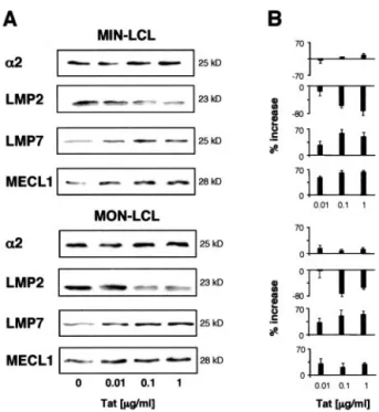
Because the HIV Tat protein is efficiently taken up by cells (2– 6), we tested the effect of exogenous Tat protein on protea-somes in MIN and MON LCLs cultured in the absence or presence
of increasing concentrations (0.01–1g/ml) of the native Tat pro-tein. After treatment, proteasomes purified from Tat-treated cells showed a down-regulation of LMP2 and an up-regulation of LMP7 and MECL1, as compared with proteasomes purified from un-treated cells (Fig. 3). These results demonstrate that both endog-enously expressed Tat and exogenous native Tat protein modify the subunit composition of immunoproteasomes.
To investigate whether the differences in subunit composition correlated with differences in enzymatic activity, we analyzed the cleavage specificity of equal amounts of proteasomes isolated from MIN-Tat, MON-Tat cells, or control cells. Chymotryptic-like, tryptic-like, and postacidic activities were all augmented in pro-teasomes purified from cells expressing Tat, as compared with control cells (Fig. 4). This is in agreement with the pattern of expression of the three catalytic subunits in Tat-expressing cells, because activation of LMP7 and MECL1 expression is associated with increased chymotryptic and tryptic activities, whereas a re-duction of LMP2 expression is associated with an increased post-acidic activity, respectively (10, 26, 27).
Tat modifies the generation and recognition of CTL peptide epitopes derived from EBV latent Ags
Because proteasomes play a key role in the generation of CTL epitopes, we investigated the effect of Tat on the presentation of dominant or subdominant CTL epitopes using as models LCLs endogenously expressing Tat or treated with the Tat protein.
LCLs were chosen because they express all EBV latent Ags, including EBNA1, 2, 3, 4, 5, and 6, and LMP1 and 2. These Ags, except for the nuclear Ag 1, are all targets of CTL and contain a large number of well-characterized CTL epitopes (28). Therefore, we evaluated the killing of LCLs expressing the immunodominant
FIGURE 2. Expression of proteasomes in Jurkat cells expressing the HIV-1 tat gene. A, Equal amounts of purified proteasomes (1g) from Jurkat cells transfected with the vector alone (JSL3-0) or with the tat gene (JSL3-Tat) were fractionated by SDS-PAGE, transferred onto nitrocellu-lose filters, and probed with mAbs or polyclonal antisera specific for␣2 subunit, LMP2, LMP7, and MECL1. One representative experiment of four performed is shown. B, The intensity of specific bands was measured by densitometry. Data are expressed as percentage of increase in ODs of specific bands detected in proteasomes purified from Tat-expressing cells relative to proteasomes from control cells. Mean⫾ SEM of three inde-pendent experiments is shown.
FIGURE 3. Expression of proteasome subunits in cells treated with the HIV-1 Tat protein. A, MIN and MON LCLs were treated for 24 h at 37°C with 0.01, 0.1, or 1g/ml native Tat protein. Equal amounts of protea-somes (1g) were fractionated by SDS-PAGE, transferred onto nitrocel-lulose filters, and probed with mAbs or polyclonal antisera specific for the
␣2 subunit, LMP2, LMP7, and MECL1. One representative experiment of
three performed is shown. B, The intensity of specific bands was measured by densitometry. Data are expressed as percentage of increase in ODs of specific bands detected in Tat-treated cells relative to control cells. Mean⫾ SEM of three independent experiments is shown.
on December 6, 2011
www.jimmunol.org
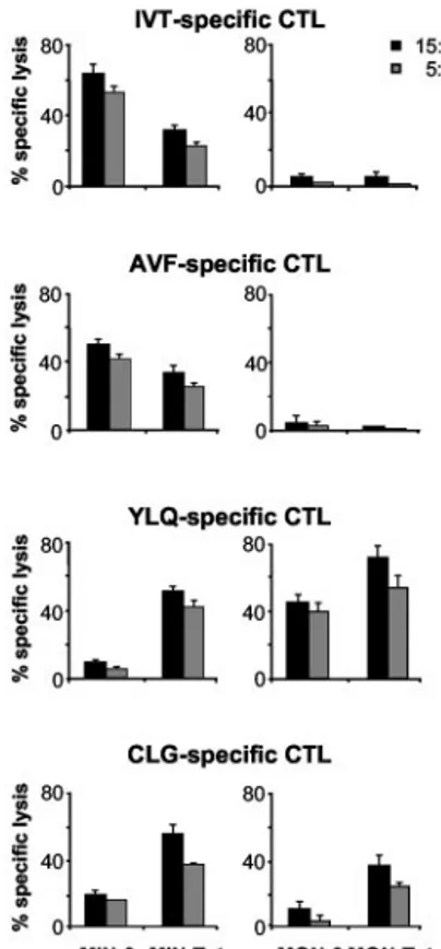
IVT and AVF HLA-A11-presented epitopes (21), and the sub-dominant YLQ and CLG HLA-A2-presented epitopes, respec-tively (22, 23). To this end, the HLA-2- and HLA-A11-positive MIN LCL and the HLA-A2-positive MON LCL, transduced or not with Tat, were used as targets in cytotoxic assays using CTL cul-tures specific for IVT, AVF, CLG, and YLQ epitopes (Fig. 5). As expected, IVT- and AVF-specific CTLs efficiently lysed A11-matched LCL (21). In contrast, YLQ- and CLG-specific CTLs rec-ognized the target cells less efficiently (22–24). This is due to the poor expression of these two epitopes on the surface of EBV-infected B cells (28). However, a decrease in IVT- and AVF-spe-cific CTL killing and an increase in YLQ- and CLG-speAVF-spe-cific kill-ing were observed in Tat-expresskill-ing cells as compared with control cells. No killing of the HLA-A11-negative MON LCL, either expressing Tat or the empty vector, was observed by using IVT- and AVF-specific CTL cultures (Fig. 5).
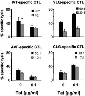
In a second set of experiments, we evaluated the CTL killing of MIN LCL untreated or treated with 0.1 g/ml Tat protein. As expected from the results of the previous experiments, LCLs treated with Tat were less sensitive to IVT- and AVF-specific CTL killing, but were lysed more efficiently by YLQ- and CLG-specific CTLs (Fig. 6).
These findings suggested that the effect of Tat on proteasome composition and enzymatic activity results in changes in epitope presentation, leading to increased CTL recognition of subdominant epitopes.
Efficient in vitro generation of the subdominant CLG epitope by proteasomes purified from Tat-expressing cells
To confirm whether proteasomes from Tat-expressing cells are able to generate subdominant epitopes more efficiently, we ana-lyzed the in vitro degradation of a CLG peptide precursor contain-ing 5 aa at the C terminus (CLG⫹ 5), corresponding to the wild-type sequence of the LMP2 Ag. Proteasomes were purified from MIN-Tat or MIN-0, and the in vitro CLG precursor degradation was evaluated by HPLC analysis. We observed that after 2 h, pro-teasomes isolated from control cells degraded 20% of the CLG⫹ 5 peptide precursor, while proteasomes purified from
Tat-express-ing cells degraded 80% of the CLG⫹ 5 peptide (Fig. 7A). This level of degradation was only reached by proteasomes from con-trol cells after 24 h (data not shown).
We then determined the presence of CLG peptides/precursors by testing the HPLC-fractionated digestion products for their ca-pacity to activate CLG-specific CTLs by IFN-␥ ELISPOT assays. Fractions purified from digests obtained after 30, 60, and 90 min of incubation, respectively, did not activate CTL responses (data not shown), suggesting that they did not contain any active peptide. In contrast, fractions 4 and 8, which were obtained after 120 min of degradation by proteasomes purified from MIN-Tat, stimulated CLG-specific CTL responses (Fig. 7B). This indicated that such fractions contained the CLG epitope or a longer antigenic epitope. In contrast, only a weak CLG-specific CTL response was observed with HPLC fraction 8, obtained after 120 min of degradation by proteasomes isolated from MIN-0 control cells. No HPLC fraction able to stimulate CLG-specific CTL responses after 24 h of in vitro degradation by proteasomes purified from control cells was de-tected (data not shown).
These results indicate that proteasomes purified from Tat-ex-pressing cells exhibit different proteolytic activity, capable of ef-ficiently generating subdominant peptide epitopes.
Discussion
We have shown in this study that the HIV-1 Tat protein, an early product of HIV-infected cells, modifies the subunit composition
FIGURE 4. Enzymatic activity of proteasomes in LCL expressing the HIV-1 tat gene. Proteasomes (2.5g) purified from cell lysates of the indicated cell lines were incubated for 30 min at 37°C with Suc-LLVY-AMC, Boc-LRR-Suc-LLVY-AMC, and Ac-YVAD-AMC to evaluate chymotryptic-like, tryptic-chymotryptic-like, and postacidic activities, respectively. Data are expressed as arbitrary fluorescence units. Mean⫾ SEM of three independent exper-iments is shown.
FIGURE 5. CTL killing of cells expressing the HIV-1 tat gene. The HLA-A2-, positive MIN and the HLA-A2-positive, HLA-A11-negative MON LCLs transduced with pBabeP (MIN-0 and MON-0, re-spectively) or with pBabeP-Tat (MIN-Tat and MON-Tat, rere-spectively) were used as targets in cytotoxic assays of CTLs specific for the HLA-A11-presented, EBNA4-derived IVT and AVF epitopes, the HLA-A2-pre-sented LMP1-derived YLQ epitope, and the HLA-A2-preHLA-A2-pre-sented LMP2-derived CLG epitope, respectively. Results are expressed as percentage of specific lysis. The mean⫾ SEM of the results from three independent experiments is shown.
3841 The Journal of Immunology
on December 6, 2011
www.jimmunol.org
and activity of proteasomes. In particular, we demonstrated that proteasomes in cells of B and T cell origin, expressing an endog-enous Tat or treated with a biologically active Tat protein, present an up-regulation of the LMP7 and MECL1 subunits and a down-modulation of the LMP2 subunit. Changes in proteasome subunit composition induced by Tat result in the increase of all three major proteolytic activities of the proteasomes.
Selective down-regulation of LMP2 by viral gene products has already been observed (29, 40). Of note, the LMP2 gene is under the control of the STAT1-IFN regulatory factor-1 (IRF-1) complex that, after binding with LMP2 promoter, induces transcription. It is therefore of interest that the Tat protein associates with IRF-1 (30), and it is tempting to speculate that this may result in IRF-1 se-questration and down-regulation of LMP2 transcription and expression.
Of note, Tat-dependent LMP2 down-regulation, both at protein and RNA levels (data not shown), did not affect the incorporation of MECL1 into proteasomes. LMP2 incorporation into immuno-proteasomes is an early event during assembly, and it has been shown that subsequent incorporation of MECL1 normally requires LMP2 (31, 32). This is due to a cooperative assembly process (33) mediated by not yet defined MECL1 and LMP2 propeptide-protein interactions. We speculate that Tat may also play a role in the assembly of proteasomal subunits by interacting with MECL1 propeptide, resulting in MECL1 incorporation without the need of LMP2. Indeed, it has recently been shown that Tat interacts with both␣ subunits and LMP7 and MECL1 subunits of the proteasome (14). Tat has also been shown to inhibit the proteolytic activity of 20S proteasome by competing with PA28 regulator for binding to 20S (15, 16). In contrast with these data, no effect on the expres-sion of PA28 in Tat-expressing cells nor on the activity of immu-noproteasomes treated in vitro with Tat protein was detected (data not shown). However, we used a monomeric fully active Tat de-rived from the human T cell leukemia virus-IIIB isolate (BH10 clone, subtype B), which lacks Asp67
, shown by Huang et al. (16) to be required for the interaction with the 20S core particle.
We also showed that changes of the proteolytic activities of proteasomes in Tat-expressing LCLs or in LCLs treated with the Tat protein correlate with a different presentation of EBV-derived epitopes. In particular, Tat decreases the presentation of two im-munodominant CTL epitopes (IVT and AVF) presented by HLA-A11 molecules, and increases the presentation of two subdominant epitopes (YLQ and CLG) presented by HLA-A2. In fact, protea-somes from Tat-expressing cells are more efficient both in the deg-radation of a CLG peptide precursor and in the generation of im-munogenic CLG peptide fragments. A similar phenomenon has been observed for an HLA-A2-presented epitope expressed in mel-anoma cells (34), suggesting that the presence of LMP2 may spe-cifically affect the range of peptides presented by some HLA class I alleles (i.e., HLA-A2). Further confirming the critical role of the LMP2 subunit in the generation of CTL epitopes, it has been dem-onstrated that influenza-specific CTL responses to the two most dominant determinants decrease in LMP2 knockout mice, whereas responses to two subdominant epitopes are greatly enhanced (35). Changes in immunodominance may be particularly relevant in vaccination strategies aimed at the control of viral infections and tumors. In fact, a decrease in the presentation of immunodominant epitopes concomitant with an increase of subdominant and cryptic epitopes may be beneficial for the elimination of virally infected or tumor cells. Indeed, it is well established that immunodominant epitopes are very prone to mutations and to CTL escape (36, 37), while subdominant epitopes are more conserved and may induce protective immune responses (38, 39).
In conclusion, our results demonstrate that the HIV-1 Tat pro-tein modulates proteasome composition and activity, and that this affects the generation of peptide Ags recognized by CTLs. These
FIGURE 6. CTL killing of cells treated with exogenous HIV-1 Tat pro-tein. The HLA-A2-, HLA-A11-positive MIN LCLs, untreated or treated with 0.1g/ml Tat for 24 h, were used as target in cytotoxic assays of CTLs specific for the HLA-A11-presented EBNA4-derived IVT and AVF epitopes, the HLA-A2-presented LMP1-derived YLQ epitope, and the HLA-A2-presented LMP2-derived CLG epitope, respectively. Results are expressed as percentage of specific lysis. Results from one representative experiment of three performed are shown.
FIGURE 7. In vitro degradation of a CLG epitope precursor by protea-somes purified from Tat-expressing cells. A, The CLG⫹ 5 peptide was incubated with proteasomes purified from MIN-Tat or from MIN-0 LCLs. The precursor degradation was followed at different time points, and the degradation of CLG⫹ 5 was evaluated by HPLC analysis. Data are ex-pressed as percentage of degradation. The mean of the results from three independent experiments is shown. B, The digestion products obtained af-ter 120 min of degradation were purified by HLPC; the indicated fractions were collected and tested by IFN-␥ ELISPOT for their capacity to activate CLG-specific CTLs. Data are expressed as spot-forming cells (SFC) per 106cells. The mean of the results from three independent experiments, performed in triplicates, is shown.
on December 6, 2011
www.jimmunol.org
data may have important implication in the immune recognition of HIV-infected cells, the CTL control of HIV-associated viral infec-tions and malignancies, and the immunogenicity of Tat itself. We have shown recently that vaccination with Tat protein or tat DNA protected monkeys against challenge with pathogenic simian HIV, and that protection correlated with Th1 responses and CTL activity (7, 8). Furthermore, we have shown that the Tat protein induces maturation of DC and increases both allogeneic and recall Ag pre-sentation by DC (6). These observations, together with the findings presented in this work, suggest that native Tat is not only an Ag, but also a novel adjuvant capable of modifying CTL epitope hi-erarchy and responses against heterologous Ags favoring the gen-eration of subdominant CTL epitopes. Therefore, the Tat protein may represent an important tool for broadening the spectrum of the epitopes recognized by CTLs, and for increasing the chances to prevent the appearance of CTL escape in vaccine strategies against HIV, and more in general against intracellular pathogens and tumors.
Acknowledgments
We thank C. Sgadari, E. Fanales-Belasio, F. Nappi, S. Moretti, and V. Fiorelli for the testing of Tat lots.
References
1. Wu, Y., and J. W. Marsh. 2003. Gene transcription in HIV infection. Microbes Infect. 5:1023.
2. Ensoli, B., G. Barillari, S. Z. Salahuddin, R. C. Gallo, and F. Wong-Staal. 1990. Tat protein of HIV-1 stimulates growth of cells derived from Kaposi’s sarcoma lesions of AIDS patients. Nature 345:84.
3. Ensoli, B., L. Buonaguro, G. Barillari, V. Fiorelli, R. Gendelman, R. A. Morgan, P. Wingfield, and R. C. Gallo. 1993. Release, uptake, and effect of extracellular human immunodeficiency virus type 1 Tat protein on cell growth and viral trans-activation. J. Virol. 67:277.
4. Chang, H. C., F. Samaniego, B. C. Nair, L. Bonauguro, and B. Ensoli. 1997. Tat protein exits from cells via a leaderless secretory pathway and binds to extracel-lular matrix-associated heparan sulfate proteoglycans through its basic region. AIDS 11:1421.
5. Frankel, A. D., and C. O. Pabo. 1988. Cellular uptake of the Tat protein from human immunodeficiency virus. Cell 55:1189.
6. Fanales-Belasio, E., S. Moretti, F. Nappi, G. Barillari, F. Micheletti, A. Cafaro, and B. Ensoli. 2002. Native HIV-1 Tat protein is selectively taken up by mono-cyte-derived dendritic cells and induces their maturation, Th-1 cytokine produc-tion and antigen presenting funcproduc-tion. J. Immunol. 168:197.
7. Cafaro, A., A. Caputo, C. Fracasso, M. T. Maggiorella, D. Goletti, S. Baroncelli, M. Pace, L. Sernicola, M. L. Koanga-Mogtomo, M. Betti, et al. 1999. Control of SHIV-89.6P infection of cynomolgus monkeys by HIV-1 Tat protein vaccine. Nat. Med. 5:643.
8. Cafaro, A., F. Titti, C. Fracasso, M. T. Maggiorella, S. Baroncelli, A. Caputo, D. Goletti, A. Borsetti, M. Pace, E. Fanales-Belasio, et al. 2001. Vaccination with DNA containing tat coding sequences and unmethylated CpG motifs protects cynomolgus monkeys upon infection with simian/human immunodeficiency virus (SHIV89.6P). Vaccine 19:2862.
9. Rock, K. L., and A. L. Goldberg. 1999. Degradation of cell proteins and the generation of MHC class I-presented peptides. Annu. Rev. Immunol. 17:739. 10. Dick, T. P., A. K. Nussbaum, M. Deeg, W. Heinemeyer, M. Groll, M. Schirle,
W. Keilholz, S. Stevanovic, D. H. Wolf, R. Huber, et al. 1998. Contribution of proteasomal-subunits to the cleavage of peptide substrates analyzed with yeast mutants. J. Biol. Chem. 273:25637.
11. Tanaka, K., and M. Kasahara. 1998. The MHC class I ligand-generating system: roles of immunoproteasomes and the interferon-␥-inducible proteasome activator PA28. Immunol. Rev. 163:161.
12. Gaczynska, M., K. L. Rock, and A. L. Goldberg. 1993.␥-Interferon and expres-sion of MHC genes regulate peptide hydrolysis by proteosomes. Nature 365:264. 13. Sijts, A. J. A. M., T. Ruppert, B. Rehermann, M. Schmidt, U. Koszinowski, and P.-M. Kloetzel. 2000. Efficient generation of a hepatitis B virus cytotoxic T lym-phocyte epitope requires the structural features of immunoproteasomes. J. Exp. Med. 191:503.
14. Apcher, G. S., S. Heink, D. Zantopf, P. M. Kloetzel, H.-P. Schmid, R. J. Mayer, and E. Kruger. 2003. Human immunodeficiency virus-1 Tat protein interacts with distinct proteasomal␣ and  subunits. FEBS Lett. 553:200.
15. Seeger, M., K. Ferrell, R. Frank, and W. Dubiel. 1997. HIV-1 Tat inhibits the 20 S proteasome and its 11 S regulator-mediated activation. J. Biol. Chem. 272:8145.
16. Huang, X., U. Seifert, U. Salzmann, P. Henklein, R. Preissner, W. Henke, A. J. Sijts, P. M. Kloetzel, and W. Dubiel. 2002. The RTP site shared by the HIV-1 Tat protein and the 11S regulator subunit␣ is crucial for their effects on proteasome function including antigen processing. J. Mol. Biol. 323:771.
17. Morgenstern, J. P., and H. Land. 1990. Advanced mammalian gene transfer: high titre retroviral vectors with multiple drug selection markers and a complementary helper-free packaging cell line. Nucleic Acids Res. 18:3587.
18. Caputo, A., J. G. Sodroski, and W. A. Haseltine. 1990. Constitutive expression of HIV-1 tat protein in human Jurkat T cells using a BK virus vector. J. Acquired Immune Defic. Syndr. 3:372.
19. Gavioli, R., T. Frisan, S. Vertuani, G. W. Bornkamm, and M. G. Masucci. 2001. c-Myc overexpression activates alternative pathways for intracellular proteolysis in lymphoma cells. Nat. Cell Biol. 3:283.
20. Micheletti, F., A. Canella, S. Vertuani, M. Marastoni, L. Tosi, S. Volinia, S. Traniello, and R. Gavioli. 2000. Supra-agonist peptides enhance the reactiva-tion of memory cytotoxic T lymphocyte responses. J. Immunol. 165:4264. 21. Gavioli, R., M. G. Kurilla, P. O. de Campos-Lima, L. E. Wallace, R. Dolcetti,
R. J. Murray, A. B. Rickinson, and M. G. Masucci. 1993. Multiple HLA A11-restricted cytotoxic T-lymphocyte epitopes of different immunogenicities in the Epstein-Barr virus-encoded nuclear antigen 4. J. Virol. 67:1572.
22. Lee, S. P., W. A. Thomas, R. J. Murray, F. Khanim, S. Kaur, L. S. Young, M. Rowe, M. Kurilla, and A. B. Rickinson. 1993. HLA A2.1-restricted cytotoxic T cells recognizing a range of Epstein-Barr virus isolates through a defined epitope in latent membrane protein LMP2. J. Virol. 67:7428.
23. Khanna, R., S. R. Burrows, J. Nicholls, and L. M. Poulsen. 1998. Identification of cytotoxic T cell epitopes within Epstein-Barr virus (EBV) oncogene latent membrane protein 1 (LMP1): evidence for HLA A2 supertype-restricted immune recognition of EBV-infected cells by LMP1-specific cytotoxic T lymphocytes. Eur. J. Immunol. 28:451.
24. Micheletti, F., R. Guerrini, A. Formentin, A. Canella, M. Marastoni, M. Bazzaro, R. Tomatis, S. Traniello, and R. Gavioli. 1999. Selective amino acid substitutions of a subdominant Epstein-Barr virus LMP2-derived epitope increase HLA/pep-tide complex stability and immunogenicity: implications for immunotherapy of Epstein-Barr virus-associated malignancies. Eur. J. Immunol. 29:2579. 25. Frisan, T., V. Levitsky, A. Polack, and M. Masucci. 1998. Phenotype-dependent
differences in proteasome subunit composition and cleavage specificity in B cell lines. J. Immunol. 160:3281.
26. Gaczynska, K., K. L. Rock, T. Spies, and A. L. Goldberg. 1994. Peptidase ac-tivities of proteasomes are differentially regulated by the major histocompatibility complex-encoded genes for LMP2 and LMP7. Proc. Natl. Acad. Sci. USA 91:9213.
27. Gaczynska, K., A. L. Goldberg, K. Tanaka, K. B. Hendil, and K. L. Rock. 1996. Proteasome subunits X and Y alter peptidase activities in opposite ways to the interferon-␥-induced subunits LMP2 and LMP7. J. Biol. Chem. 271:17275. 28. Rickinson, A. B., and D. J. Moss. 1997. Human cytotoxic T lymphocyte
re-sponses to Epstein-Barr virus infection. Annu. Rev. Immunol. 15:405. 29. Chatterjee-Kishore, M., F. van den Akker, and G. R. Stark. 2000. Adenovirus
E1A down-regulates LMP2 transcription by interfering with the binding of Stat1 to IRF1. J. Biol. Chem. 275:20406.
30. Sgarbanti, M., A. Borsetti, N. Moscufo, M. C. Bellocchi, B. Ridolfi, F. Nappi, G. Marsili, G. Marziali, E. M. Coccia, B. Ensoli, and A. Battistini. 2002. Mod-ulation of human immunodeficiency virus 1 replication by interferon regulatory factors. J. Exp. Med. 195:1359.
31. Nandi, D., E. Woodward, D. B. Ginsburg, and J. J. Monaco. 1997. Intermediates in the formation of mouse 20S proteasomes: implications for the assembly of precursor subunits. EMBO J. 16:5363.
32. Griffin, T. A., D. Nandi, M. Cruz, H. J. Fehling, L. V. Kaer, J. J. Monaco, and R. A. Colbert. 1998. Immunoproteasome assembly: cooperative incorporation of interferon␥ (IFN-␥)-inducible subunits. J. Exp. Med. 187:97.
33. De, M., K. Jayarapu, L. Elenich, J. J. Monaco, R. A. Colbert, and T. A. Griffin. 2003.2 subunit propeptides influence cooperative proteasome assembly. J. Biol. Chem. 278:6153.
34. Morel, S., F. Le´vy, O. Burlet-Schiltz, F. Brasseur, M. Probst-Kepper, A.-L. Pei-trequin, B. Monsarrat, R. Van Velthoven, J.-C. Cerottini, T. Boon, et al. 2000. Processing of some antigens by the standard proteasome but not immunoprotea-some results in poor presentation by dendritic cells. Immunity 12:107. 35. Chen, W., C. C. Norbury, Y. Cho, J. W. Yewdell, and J. R. Bennink. 2001.
Immunoproteasomes shape immunodominance hierarchies of antiviral CD8⫹T cells at the levels of T cell repertoire and presentation of viral antigens. J. Exp. Med. 193:1319.
36. Phillips, R. E., S. Rowland-Jones, D. F. Nixon, F. M. Gotch, J. P. Edwards, A. O. Ogunlesi, J. G. Elvin, J. A. Rothbard, C. R. M. Bangham, C. R. Rizza, and A. J. McMichael. 1991. Human immunodeficiency virus genetic variation that can escape cytotoxic T cell recognition. Nature 354:453.
37. De Campos-Lima, P. O., R. Gavioli, Q.-J. Zhang, L. Wallace, R. Dolcetti, M. Rowe, A. B. Rickinson, and M. G. Masucci. 1993. HLA-A11 epitope-loss isolates of Esptein-Barr virus from a highly HLA A11⫹population. Science 260:98.
38. Feltkamp, M. C. W., G. R. Vreugdenhil, M. P. M. Vierboom, E. Ras, S. H. van der Burg, J. ter Schegget, C. J. M. Melief, and W. M. Kast. 1995. Cytotoxic T lymphocytes raised against a subdominant epitope offered as a syn-thetic peptide eradicate human papilloma virus type 16-induced tumors. Eur. J. Immunol. 25:2638.
39. Rodriguez, F., M. K. Slifka, S. Harkins, and J. I. Whitton. 2001. Two overlapping subdominant epitopes identified by DNA immunization induce protective CD8⫹ T-cell populations with differing cytolytic activities. J. Virol. 75:7399. 40. Zeidler, R., G. Eissner, P. Meissner, S. Uebel, R. Tampe´, S. Lazis, and W.
Ham-merschmidt. 1997. Downregulation of TAP1 in B lymphocytes by cellular and Epstein-Barr virus-encoded interleukin-10. Blood 90:2390.
3843 The Journal of Immunology