ANAL CANCER IN PEOPLE
LIVING WITH HIV:
THE IMPORTANCE OF THE SCREENING
AND OF EARLY DIAGNOSIS
F. D’ALEO
1
, M. CECCARELLI
1,2
, E. VENANZI RULLO
1,2
, A. FACCIOLÀ
1
, F. D’ANDREA
1
,
C. MICALI
1
, M. COCO
1
, M. R. PINZONE
2,3
, E. FOCÀ
4
, F. CONDORELLI
5
, I. PICERNO
6
,
G. VISALLI
6
, B. CACOPARDO
3
, G. NUNNARI
1
, G. F. PELLICANÒ
7
Abstract – Objective: HIV-positive patients suffer from higher cancer-related mortality
com-pared to the general population. Anal cancer (AC) is considered as a rare form of neoplasm,
ac-counting for 4% of all cancers of the lower gastrointestinal tract in the general population.
Ap-proximately 88% of AC cases are associated with human papillomavirus (HPV) infection. This paper
purpose is the diagnostic and therapeutic management of AC in HIV infect people.
KEYWORDS: Anal Cancer, PLWH, HIV, ART, HPV, Screening.
1
Department of Clinical and Experimental Medicine, University of Messina, Messina, Italy
2
Department of Pathology and Laboratory Medicine, School of Medicine, University of Pennsylvania,
Philadelphia, PA, USA
3
Department of Clinical and Experimental Medicine, Unit of Infectious Diseases, University of Catania, Catania, Italy
4Department of Clinical and Experimental Sciences, Division of Infectious Diseases, University of Brescia, Brescia, Italy
5Department of Pharmacological Sciences, Università del Piemonte Orientale “A. Avogadro”, Novara, Italy
6Department of Biomedical and Dental Sciences and Morpho Functional Imaging, University of Messina,
Messina, Italy
7
Department of Human Pathology of the Adult and the Developmental Age “G. Barresi”, University of Messina,
Messina, Italy
INTRODUCTION
Over the last decades, because of the broad use of
combination antiretroviral therapy (cART), the life
expectancy of patients living with human
immuno-deficiency virus (HIV) and acquired immune
defi-ciency syndrome (AIDS) improved. This brought to
a continuous increase in the total number of people
living with HIV (PLWH) worldwide, a population
who bears the burden of associated health
condi-tions that complicate long-term HIV infection
1-23.
Among them, a constantly growing incidence
of non-AIDS defining cancers (NADCs) such as
hepatocellular carcinoma (HCC), lung cancer and
anal carcinoma accompanied the decrease of AIDS
defining cancers (ADCs) such as Kaposi’s sarcoma
and Hodgkin’s lymphoma
1,24-26.
HIV-positive patients suffer from a higher
can-cer-related mortality than the general population.
Studies have shown that this higher mortality is
caused by the degree of immunosuppression,
irre-spectively of whether the tumor is
infection-associ-ated or not. NADCs are burdened by a poorer
out-come compared to AIDS-associated malignancies,
which might be positively influenced by the
applica-tion of cART
27,28before the onset of acquired
immu-nodeficiency syndrome (AIDS.
The aim of this paper is to comprehensively review
the literature about the diagnostic and therapeutic
man-agement of anal cancer (AC) in HIV-infected people.
DEFINITION AND CLASSIFICATION
OF AC
AC is defined as cancer arising from the squamous
epithelium of the anus, making it distinct from
colorectal cancer. The anal canal consists of
strati-fied squamous epithelium originating outside the
body and extending into the anus up to the dentate
line, the point where it intersects the columnar
epi-thelium of the rectum. AC, as cervical cancer, may
be HPV-associated and arise from precursor lesions
defined dysplasia or intraepithelial neoplasia (anal
intraepithelial neoplasia, AIN). Due to the
con-cordant histopathological characteristics, in 2001
Bethesda classification revised the nomenclature for
anal dysplasia, making it similar to the one utilized
for cervical lesions. Cytology plays an important
role in the diagnosis of anal lesions
49-51.
The cytological examination follows to
distin-guished from low- and high-grade dysplasia and
lesions with undetermined significance. Low-grade
squamous intraepithelial lesion (LSIL) cytology
corresponds to the histological diagnosis of anal
intraepithelial neoplasia grade 1 (AIN1) while the
high-grade squamous intraepithelial lesions (HSIL)
correspond to intraepithelial neoplasia grade 2/3 or
anal carcinoma in situ (AIN2/3). The role of
low-grade AIN for tumor progression is controversial,
seen that these may sporadically demonstrate
spon-taneous regression, while AIN2 and AIN3 are
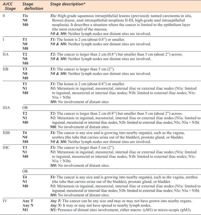
po-tential precursor lesions of AC. The most recent
ver-sion of TNM staging of AC is showed in Table 1.
S
creeningSimilar to cervical intraepithelial neoplasia (CIN)
and cervical cancer, these cancers may be
prevent-able by an early diagnosis, which could be reached
thanks to mass screening
25.
However, despite the high-incidence of AC, the
screening is not currently routinely effected, not
even in HIV-infected MSM, who are burdened by
the highest risk and could actually benefit from an
early diagnosis and early therapy.
Similarly to what happens for cervical
neopla-sia, cytology has been proposed to screen for AC in
high-risk population
52,53. This kind of screening has
not been studied and is not currently recommended
in the general population
37,41,53-58.
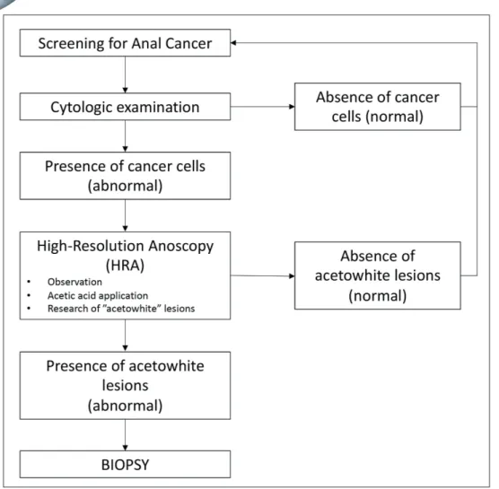
DIAGNOSIS (FIG. 1)
Incidence of AC has dramatically risen in several parts
of the world, including Europe and the United States,
in the general population. It is even more significant
among MSM, for whom where the incidence rates up
to 37 per 100,000, to rise up to 135 per 100,000, in
EPIDEMIOLOGY
AC is considered a rare form of neoplasm in the
gen-eral population, accounting for 4% of all cancers of
the lower gastrointestinal tract. Approximately 88%
of AC cases are associated with human
papilloma-virus (HPV) infection, with HPV 16 being the most
commonly detected type, followed by HPV 18, 32
and 34. In AC, two major morphologic kinds are
highlighted: squamous cell carcinoma (SCC),
ac-counting for 70% of the cases, associated to HPV in
80% of the cases, and adenocarcinoma (ADC), less
frequently related to HPV
29-32.
PLWH have elevated rates of AC, because of a
higher rate of sexually transmitted infections,
espe-cially infection by high-risk type of papillomavirus.
The incidence of AC is elevated in PLWH when
com-pared to the general population and it is especially
high in the HIV-positive males who have sex with
males (MSM) population, as it has been
demonstrat-ed in several studies
33-36. Some authors reported that
the rates of AC are in HIV+ MSM 135 per 100,000,
in HIV+ non-MSM of 45 per 100,000, and in HIV
negative non-MSM of 2 per 100,000. The same study
also found AC rates of 30 per 100,000 in HIV positive
women and the authors highlighted how there were
no cases of AC in the HIV-negative women
37.
PATHOGENESIS
Recent data shows that the majority of the squamous
cell ACs are apparently linked to HPV. It is often
a persistent high-risk HPV (HR-HPV) genotype,
most commonly 16 and 18, infecting the squamous
epithelium and causing a neoplastic transformation
in the rectal mucosa. As we can observe in the case
of other HPV associated tumors, it exists a sequence
from persistent infection to invasive cancer. An
im-portant role in this process is played by the
dysregu-lation of autophagy
30,38-46.
An explanation of the higher risk for AC among
PLWH might be that HIV is associated with a higher
incidence of HR-HPV infection, which can promote
the development of AC. Moreover, both HIV and AC
are associated with inability to clear HPV infection and
simultaneous infection with multiple strains of HPV
43.
The mechanism of tumorigenesis has been found
to be the inactivation of tumor suppression genes.
Mu-tations in the p53, DCC, and/or APC tumor suppressor
genes have been identified as antecedent events. Much
like the development of colorectal adenocarcinoma,
a pattern of chromosomal instability is evident in the
genesis of the ACs. Some author proposed the
micro-satellite instability rather than chromosomal
instabil-ity to be the possible pathway for rapid progression
to-wards invasive carcinoma in HIV positive cases
24,47,48.
the rectum until encountering the rectal wall, then
re-moving the swab with a twisting motion while
apply-ing lateral pressure. This technique allows gatherapply-ing
samples of the transitional zone and anal canal. The
swab is then processed using a liquid cytology
tech-nique older than Papanicolaou staining. The sample
obtained is then analyzed by a pathologist
61.
Some authors reported that, in HIV negative
MSM, anal cytology sensitivity was between
47-70% for the detection of intraepithelial neoplasia of
any grade. Performing a HPV molecular test, such
as polymerase chain reaction (PCR), on the same
specimens may help improving the diagnostic
sen-sitivity.
HIV+ MSM
29,54,55,59. The burden of this cancer
contin-ues to rise, with only 10% of patients with metastatic
disease surviving more than 2 years
60.
Moreover, 95% of HIV+ MSM are seropositive
for the related viral pathogen, HPV (subtypes 16, 18,
32 and 34).
The most important diagnostic methods for AC
are anal cytology with histology staging and
radio-logical techniques.
A
nAlc
ytologyAnal cytology is currently applied to screen for
dys-plasia or intraepithelial neodys-plasia. It is performed
in-serting a water-moistened polyester fiber swab into
TABLE 1 . Clinical Staging of Anal Cancer, according to AJCC Cancer Staging Manual, 8th edition.
AJCC Stage Stage description*
Stage definition
0 Tis Tis: High-grade squamous intraepithelial lesions (previously named carcinoma in situ,
N0 Bowen diseas, anal intraepithelial neoplasia II-III, high-grade anal intraepithelial M0 neoplasia). It describes a situation where the cancer is limited to the epithelium layer
(the most external) of the mucosa.
N0 & M0: Neither lymph nodes nor distant sites are involved.
I T1 T1: The lesion is 2 cm (about 0.8”) or smaller.
N0 N0 & M0: Neither lymph nodes nor distant sites are involved. M0
IIA T2 T2: The cancer is larger than 2 cm (0.8”) but smaller than 5 cm (about 2”) across.
N0 N0 & M0: Neither lymph nodes nor distant sites are involved.
M0
IIB T3 T3: The cancer is larger than 5 cm (2”).
N0 N0 & M0: Neither lymph nodes nor distant sites are involved.
M0
T1 T1: The lesion is 2 cm (about 0.8”) or smaller.
N1 N1: Metastasis in inguinal, mesorectal, internal iliac or external iliac nodes (N1a: limited
M0 to inguinal, mesorectal or internal iliac nodes; N1b: limited to external iliac nodes; N1c: N1a + N1b)
M0: No involvement of distant sites
IIIA OR
T2 T2: The cancer is larger than 2 cm (0.8”) but smaller than 5 cm (about 2”) across.
N1 N1: Metastasis in inguinal, mesorectal, internal iliac or external iliac nodes (N1a: limited to
M0 inguinal, mesorectal or internal iliac nodes; N1b: limited to external iliac nodes; N1c: N1a + N1b) M0: No involvement of distant sites.
IIIB T4 T4: The cancer is any size and is growing into nearby organ(s), such as the vagina,
N0 urethra (the tube that carries urine out of the bladder), prostate gland, or bladder. M0 N0 & M0: Neither lymph nodes nor distant sites are involved.
IIIC T3 T3: The cancer is larger than 5 cm (2”).
N1 N1: Metastasis in inguinal, mesorectal, internal iliac or external iliac nodes (N1a: limited
M0 to inguinal, mesorectal or internal iliac nodes; N1b: limited to external iliac nodes; N1c: N1a + N1b)
M0: No involvement of distant sites.
OR
T4 T4: The cancer is any size and is growing into nearby organ(s), such as the vagina, urethra
N1 (the tube that carries urine out of the bladder), prostate gland, or bladder.
M0 N1: Metastasis in inguinal, mesorectal, internal iliac or external iliac nodes (N1a: limited to
inguinal, mesorectal or internal iliac nodes; N1b: limited to external iliac nodes; N1c: N1a + N1b)
M0: No involvement of distant sites.
IV Any T Any T: The cancer can be any size and may or may not have grown into nearby organs.
Any N Any N: It may or may not have spread to nearby lymph nodes.
currently the gold-standard to assess loco-regional
dis-ease, but EUS is more specific for small lesions. PET/
CT has been recommended because of high sensitivity
in identifying involved lymph nodes
65-67.
TREATMENTS
Several therapies are nowadays possible for
intraep-ithelial lesion or an invasive neoplasia. Despite its
growing incidence, AC remains a rare condition and
requires a high level of expertise to correctly
diag-nose and treat; all individuals found to have positive
anal cytology should be referred to expert centers
for treatment and therapy
68.
t
opicAltherApyTopical therapy consists in the direct application of
a medication on the specific lesion or the entire anal
canal. Available medications include trichloroacetic
acid (TCA), 5-flurouracil, and the immune
modula-tor imiquimod
69, 70.
However, several studies showed that the
efficien-cy of local eradication of HPV-associated anogenital
lesions with electrocautery was superior to that of
lo-cal chemotherapy in HIV-positive MSM
69, 70.
If the cytologic examination comes out positive,
the next step consists in localizing the source of
those atypical cells with High Resolution Anoscopy
(HRA). HRA consists of a direct examination of the
squamo-columnar junction between the anus and the
rectum, of the anal canal and of the perianal skin
un-der magnification using a colposcope. During a first
phase examination when the anoscope is placed into
the anus with lidocaine lubrication, a direct
observa-tion is performed. The anoscope is then removed, and
a swab soaked in 3-5% acetic acid solution is placed
into the anal canal for two minutes. The acetic acid
ap-plication helps distinguishing the epithelium infected
by HPV from the healthy one, creating “acetowhite”
areas, which can be observed at the anoscope.
The latest and final step is the biopsy of the
sus-pected zone
56, 61-64.
r
AdiologicAlStAgingA number of imaging techniques can be used for the
diagnosis and the staging of the AC. Computerized
to-mography (CT), Magnetic Resonance Imaging (MRI),
Endo-Anal Ultrasound (EUS) and Positron Emission
Tomography (PET) scanning are combined to allow
an assessment of the local and distant spreading,
in-cluding involvement of other organs and nodes. MRI is
Fig. 1. Screening dia-gram for anal cancer.
general population, the incidence of this cancer is
constantly increasing, especially in PLWH, who
represenst an optimal target population for mass
screening.
It is essential to acquire new data about the
ef-fects of the vaccination campaign on the incidence
of AC. Further studies are needed to achieve these
outcomes.
c
onflictofi
ntereStThe Authors declare that they have no conflict of interests
REFERENCES
1) Visalli G, Facciolà A, D’Aleo F, Pinzone MR, Condorelli F, Picerno I, Nunnari G, Pellicanò GF, Ceccarelli M, Venanzi Rullo E. HPV and urinary bladder carcinoma: a review of the literature. World Cancer Res J 2018; 5: e1038. 2) D’Aleo F, Cama BAV, Paolucci IA, Venanzi Rullo E,
Con-dorelli F, Facciolà A, Di Francia R, Savasta A, Pinzone MR, Picerno I, Visalli G, Nunnari G, Pellicanò GF, Ceccarelli M. New and old assumptions on lung cancer in people living with HIV. World Cancer Res J 2018; 5: e1036. 3) D’Aleo F, Ceccarelli M, Venanzi Rullo E, Facciolà A, Di
Rosa M, Pinzone MR, Condorelli F, Visalli G, Picerno I, Berretta M, Pellicanò GF, Nunnari G. Hepatitis C-related hepatocellular carcinoma: diagnostic and therapeutic management in HIV-patients. Eur Rev Med Pharmacol Sci 2017; 21: 5859-5867.
4) Pomerantz RJ, Nunnari G. HIV and GB virus C--can two viruses be better than one? N Engl J Med 2004; 350: 963-965.
5) Pinzone MR, Berretta M, Cacopardo B, Nunnari G. Epstein-barr virus- and Kaposi sarcoma-associated her-pesvirus-related malignancies in the setting of human immunodeficiency virus infection. Semin Oncol 2015; 42: 258-271.
6) Pinzone MR, Di Rosa M, Celesia BM, Condorelli F, Malaguarnera M, Madeddu G, Martellotta F, Castro-nuovo D, Gussio M, Coco C, Palermo F, Cosentino S, Cacopardo B, Nunnari G. LPS and HIV gp120 modu-late monocyte/macrophage CYP27B1 and CYP24A1 expression leading to vitamin D consumption and hypovitaminosis D in HIV-infected individuals. Eur Rev Med Pharmacol Sci 2013; 17: 1938-1950.
7) Pinzone MR, Cacopardo B, Condorelli F, Rosa MD, Nunnari G. Sirtuin-1 and HIV-1: an overview. Curr Drug Targets 2013; 14: 648-652.
8) Visalli G, Bertuccio MP, Currò M, Pellicanò G, Sturniolo G, Carnevali A, Spataro P, Ientile R, Picerno I, Cavallari V, Piedimonte G. Bioenergetics of T cell activation and death in HIV type 1 infection. AIDS Res Hum Retrovi-ruses 2012; 28:1110-1118.
9) Trovato M, Ruggeri RM, Sciacchitano S, Vicchio TM, Picerno I, Pellicanò G, Valenti A, Visalli G. Serum inter-leukin-6 levels are increased in HIV-infected patients that develop autoimmune disease during long-term follow-up. Immunobiology 2018; 223: 264-268. 10) Squillace N, Ricci E, Quirino T, Gori A, Bandera A,
Carenzi L, De Socio GV, Orofino G, Martinelli C, Madeddu G, Rusconi S, Maggi P, Celesia BM, Cordier L, Vichi F, Calza L, Falasca K, Di Biagio A, Pellicanò GF, Bonfanti P, CISAI Study Group. Safety and tolerability
Electrocautery constitutes a first-line treatment
for intraepithelial neoplasia. A possible advantage
of surgical excision is the chance to perform a
his-topathological examination of the tissue removed.
Despite being ineffective on a high percentage of
the patients, topical therapy appears to be generally
well tolerated
69,70.
S
urgicAltherApySurgery for AC is associated with significant
mor-bidity, often requiring a large excision of healthy
tissue around the cancerous lesion, and yet it is
as-sociated with a variable rate of recurrence. A
lo-cal resection, which removes only the tumor plus
a small margin (edge) of the normal tissue around
the tumor, is mostly used to treat cancers of the
anal margin when the tumor is small, and it has not
spread to nearby tissues or lymph nodes.
Abdomi-noperineal resection is a major operation, which
consists in the complete removal of the anus and the
creation of a colostomy. Surgical therapy is a
com-mon treatment for rectal cancer when the cancer
is spread well above the anus. This technique
con-sists in the resection of the entire rectal cancer with
the adjacent normal rectal tissue and surrounding
lymph nodes through an incision made in the lower
abdomen. Local resection may be an option for early
stage AC that has not spread to the lymph nodes or
surrounding tissue
71-75.
PREVENTION
HPV infection can be currently prevented thanks to
the introduction of a vaccination against the virus.
Three kinds of vaccine against HPV are available: the
quadrivalent Gardasil, against HPV-serotypes 6, 11,
16 and 18; the nine-valent Gardasil-9, against
HPV-serotypes 6, 11, 16, 18, 31, 33, 45, 52 and 58; and the
bivalent against HR-HPV-serotypes 16 and 18.
The quadrivalent HPV (qHPV) vaccine has been
demonstrated to prevent persistent anal HPV
infec-tions as well as anal intraepithelial neoplasia grades
2-3 in young MSM not previously infected;
howev-er, some recent studies showed that the quadrivalent
HPV vaccine was not effective in preventing new
anal infections or improving high-grade squamous
intraepithelial lesions in adults aged older 27 years
with HIV
40,51,76-78.
CONCLUSIONS
AC is often associated with HPV, arising from the
squamous epithelium of the anus. In some groups,
such as PLWH and especially MSM, it shows a high
progression risk. Despite its low frequency in the
Pellicanò GF. Circulating angiopoietin-like protein 2 levels are associated with decreased renal function in HIV+ subjects on cART: a potential marker of kidney disease. Biomed Rep 2019; 10: 140-144.
24) D’Aleo F, Venanzi Rullo E, Ceccarelli M, Facciolà A, Condorelli F, Pinzone MR, Cacopardo B, Di Rosa M, Nunnari G, Pellicanò GF. HIV and colorectal cancer. New insights and review of the literature. World Cancer Res J 2018; 5: e1122.
25) Ceccarelli M, Venanzi Rullo E, Facciolà A, Madeddu G, Cacopardo B, Taibi R, D’Aleo F, Pinzone MR, Picerno I, Di Rosa M, Visalli G, Condorelli F, Nunnari G, Pellicanò GF. Head and neck squamous cell carcinoma and its correlation with human papillomavirus in people living with HIV: a systematic review. Oncotarget 2018; 9: 17171-17180.
26) Facciolà A, Venanzi Rullo E, Ceccarelli M, D’Aleo F, Di Rosa M, Pinzone MR, Condorelli F, Visalli G, Picerno I, Fisichella R, Nunnari G, Pellicanò GF. Kaposi’s sarcoma in HIV-infected patients in the era of new antiretrovi-rals. Eur Rev Med Pharmacol Sci 2017; 21: 5868-5879. 27) Engels EA, Biggar RJ, Hall HI, Cross H, Crutchfield A, Finch JL, Grigg R, Hylton T, Pawlish KS, McNeel TS, Goedert JJ. Cancer risk in people infected with human immunodeficiency virus in the United States. Int J Can-cer 2008; 123: 187-194.
28) Castilho JL, Luz PM, Shepherd BE, Turner M, Ribeiro SR, Bebawy SS, Netto JS, McGowan CC, Veloso VG, Engels EA, Sterling TR, Grinsztejn B. HIV and cancer: a comparative retrospective study of Brazilian and U.S. clinical cohorts. Infect Agents Cancer 2015; 10: 4. 29) Carneiro Pereira AC, de Lacerda HR, do Rego Barros
RC. Diagnostic methods for prevention of anal cancer and characteristics of anal lesions caused by HPV in men with HIV/AIDS. Braz J Infect Dis 2008; 12: 293-299.
30) Casper C, Crane H, Menon M, Money D. HIV/AIDS comorbidities: impact on cancer, noncommunicable diseases, and reproductive health. In: Holmes KK, Ber-tozzi S, Bloom BR, Jha P (eds) Disease Control Priorities, Third Edition (Volume 6): major infectious diseases. The World Bank, 2017.; pp. 45-66.
31) Robbins HA, Pfeiffer RM, Shiels MS, Li J, Hall HI, En-gels EA. Excess cancers among HIV-infected people in the United States. J Natl Cancer Inst 2015; 107: dju503.
32) Chiu CG, Smith D, Salters KA, Zhang W, Kanters S, Milan D, Montaner JSG, Coldman A, Hogg RS, Wise-man SM. Overview of cancer incidence and mortality among people living with HIV/AIDS in British Columbia, Canada: implications for HAART use and NADM devel-opment. BMC Cancer 2017; 17: 270.
33) Hessol NA, Katz MH, Liu JY, Buchbinder SP, Rubino CJ, Holmberg SD. Increased incidence of Hodgkin disease in homosexual men with Hiv-infection. Ann Intern Med 1992; 117: 309-311.
34) Aldersley J, Lorenz DR, Misra V, Uno H, Gabuzda D. Increased risk of anal squamous cell carcinoma in HIV-positive men with prior hepatitis B virus infection. AIDS 2019; 33: 145-152.
35) Machalek DA, Poynten M, Jin F, Fairley CK, Farnsworth A, Garland SM, Hillman RJ, Petoumenos K, Roberts J, Tabrizi SN, Templeton DJ, Grulich AE. Anal human pap-illomavirus infection and associated neoplastic lesions in men who have sex with men: a systematic review and meta-analysis. Lancet Oncol 2012; 13: 487-500. 36) Palefsky JM, Rubin M. The epidemiology of anal human
papillomavirus and related neoplasia. Obstet Gynecol Clin N Am 2009; 36: 187-200.
of elvitegravir/cobicistat/emtricitabine/tenofovir diso-proxil fumarate in a real life setting: data from surveil-lance cohort long-term toxicity antiretrovirals/antivirals (SCOLTA) project. PLoS One 2017; 12: e0179254. 11) Bellissimo F, Pinzone MR, Cacopardo B, Nunnari G.
Diagnostic and therapeutic management of hepato-cellular carcinoma. World J Gastroenterol 2015; 21: 12003.
12) Nunnari G, Sullivan J, Xu Y, Nyirjesy P, Kulkosky J, Cavert W, Frank I, Pomerantz RJ. HIV type 1 cervico-vaginal reservoirs in the era of HAART. AIDS Res Hum Retroviruses 2005; 21: 714-718.
13) Nunnari G, Leto D, Sullivan J, Xu Y, Mehlman KE, Kulkosky J, Pomerantz RJ. Seminal reservoirs during an HIV type 1 eradication trial. AIDS Res Hum Retroviruses 2005; 21: 768-775.
14) Martellotta F, Berratta M, Cacopardo B, Fisichella R, Schioppa O, Zanghì A, Spartà D, Cappellani A, Talamini R, Izzi I, Ridolfo A, Torresin A, Fiorica F, Tirelli U. Clinical presentation and outcome of squamous cell carcinoma of the anus in HIV-infected patients in the HAART-era: a GICAT experience. Eur Rev Med Pharmacol Sci 2012; 16: 1283-1291.
15) Celesia BM, Nigro L, Pinzone MR, Coco C, La Rosa R, Bisicchia F, Mavilla S, Gussio M, Pellicanò G, Milioni V, Palermo F, Russo R, Mughini MT, Martellotta F, Taibi R, Cacopardo B, Nunnari G. High prevalence of undiagnosed anxiety symptoms among HIV-positive individuals on cART: a cross-sectional study. Eur Rev Med Pharmacol Sci 2013; 17: 2040-2046.
16) Celesia BM, Castronuovo D, Pinzone MR, Bellissimo F, Mughini MT, Lupo G, Scarpino MR, Gussio M, Palermo F, Cosentino S, Cacopardo B, Nunnari G. Late presen-tation of HIV infection: predictors of delayed diagnosis and survival in Eastern Sicily. Eur Rev Med Pharmacol Sci 2013; 17: 2218-2224.
17) Bearz A, Vaccher E, Martellotta F, Spina M, Talamini R, Lleshi A, Cacopardo B, Nunnari G, Berretta M, Tirelli U. Lung cancer in HIV positive patients: the GICAT experi-ence. Eur Rev Med Pharmacol Sci 2014; 18: 500-508. 18) Nuvoli S, Caruana G, Babudieri S, Solinas P, Pellicanò
G, Piras B, Fiore V, Bagella P, Calia GM, Yue M, Spanu A, Madeddu G. Body fat changes in HIV patients on highly active antiretroviral therapy (HAART): a longitu-dinal DEXA study. Eur Rev Med Pharmacol Sci 2018; 22: 1852-1859.
19) Facciolà A, Ceccarelli M, Venanzi Rullo E, D’Aleo F, Condorelli F, Visalli G, Cacopardo B, Pinzone MR, Di Rosa M, Nunnari G, Pellicanò GF. Prostate cancer in HIV-positive patients- a review of the literature. World Cancer Res J 2018; 5: e1136.
20) D’Andrea F, Ceccarelli M, Facciolà A, Nunnari G, Pel-licanò GF, Venanzi Rullo E. Breast cancer in women living with HIV. Eur Rev Med Pharmacol Sci 2019; 23: 1158-1164.
21) Ceccarelli M, Venanzi Rullo E, Vaccaro M, Facciolà A, D’Aleo F, Paolucci IA, Cannavò SP, Cacopardo B, Pinzone MR, Pellicanò GF, Condorelli F, Nunnari G, Guarneri C. HIV-associated psoriasis: epidemiology, pathogenesis, and management. Dermatol Ther 2019; 75: e12806
22) Venanzi Rullo E, Ceccarelli M, Condorelli F, Facciolà A, Visalli G, D’Aleo F, Paolucci I, Cacopardo B, Pinzone MR, Di Rosa M, Nunnari G, Pellicanò GF. Investigational drugs in HIV: pros and cons of entry and fusion inhib-itors (Review). Mol Med Rep 2019; 19: 1987-1995. 23) Pinzone MR, Ceccarelli M, Venanzi Rullo E, Maresca M,
Bruno R, Condorelli F, Di Rosa M, Madeddu G, Focà E, Calcagno A, Celesia BM, Cacopardo B, Nunnari G,
56) Goncalves PH, Montezuma-Rusca JM, Yarchoan R, Uldrick TS. Cancer prevention in HIV-infected popula-tions. Semin Oncol 2016; 43: 173-188.
57) Robison K, Cronin B, Bregar A, Luis C, DiSilvestro P, Schechter S, Pisharodi L, Raker C, Clark M. Anal cytol-ogy and human papillomavirus genotyping in women with a history of lower genital tract neoplasia com-pared with low-risk women. Obstet Gynecol 2015; 126: 1294-1300.
58) Phillips AA, Justman JE. Screening HIV-infected pa-tients for non-AIDS-defining malignancies. Curr HIV/ AIDS Rep 2009; 6: 83-92.
59) Tanaka LF, Latorre MDRDO, Gutierrez EB, Curado MP, Dal Maso L, Herbinger K-H, Froeschl G, Heumann C. Cancer survival in people with AIDS: a popula-tion-based study from São Paulo, Brazil. Int J Cancer 2017; 142: 524-533.
60) Chin-Hong PV, Vittinghoff E, Cranston RD, Browne L, Buchbinder S, Colfax G, Da Costa M, Darragh T, Benet DJ, Judson F, Koblin B, Mayer KH, Palefsky JM. Age-related prevalence of anal cancer precursors in homosexual men: the EXPLORE study. J Natl Cancer Inst 2005; 97: 896-905.
61) Tong WWY, Jin F, McHugh LC, Maher T, Sinclair B, Grulich AE, Hillman RJ, Carr A. Progression to and spontaneous regression of high-grade anal squamous intraepithelial lesions in HIV-infected and uninfected men. AIDS 2013; 27: 2233-2243.
62) Palefsky JM, Holly EA, Ralston ML, Jay N, Berry JM, Darragh TM. High incidence of anal high-grade squa-mous intra-epithelial lesions among HIV-positive and HIV-negative homosexual and bisexual men. AIDS 1998; 12: 495-503.
63) Palefsky JM, Holly EA, Hogeboom CJ, Berry JM, Jay N, Darragh TM. Anal cytology as a screening tool for anal squamous intraepithelial lesions. J Acquir Immune Defic Syndr Hum Retrovirol 1997; 14: 415-422. 64) Sendagorta E, Herranz P, Guadalajara H, Bernardino
JI, Viguer JM, Beato MJ, Garcia-Olmo D, Pena JM. Prevalence of abnormal anal cytology and high-grade squamous intraepithelial lesions among a cohort of HIV-infected men who have sex with men. Dis Colon Rectum 2014; 57: 475-481.
65) Hutchings M. FDG-PET after two cycles of chemother-apy predicts treatment failure and progression-free sur-vival in Hodgkin lymphoma. Blood 2006; 107: 52-59. 66) Counts SJ, Kim AW. Diagnostic imaging and newer
modalities for thoracic diseases PET/computed tomo-graphic imaging and endobronchial ultrasound for staging and its implication for lung cancer. PET Clin 2018, 13: 113-126.
67) Collettini F, Lutter A, Schnapauff D, Hildebrandt B, Puhl G, Denecke T, Wust P, Gebauer B. Unresectable col-orectal liver metastases: percutaneous ablation using CT-Guided High-Dose-Rate Brachytherapy (CT-HDBRT). Fortschr Röntgenstr 2014; 186: 606-612.
68) Macaya A, Muñoz-Santos C, Balaguer A, Barberà MJ. Interventions for anal canal intraepithelial neoplasia. Cochrane Database Syst Rev 2012; 12: CD009244 69) Cranston RD, Baker JR, Liu Y, Wang L, Elishaev E, Ho
KS. Topical application of trichloroacetic acid is effi-cacious for the treatment of internal anal high-grade squamous intraepithelial lesions in HIV-positive men. Sex Transm Dis 2014; 41: 420-426.
70) Singh JC, Kuohung V, Palefsky JM. Efficacy of trichlo-roacetic acid in the treatment of anal intraepithelial neoplasia in HIV-positive and HIV-negative men who have sex with men. J Acquir Immune Defic Syndr 2009, 52: 474-479.
37) Sigel K, Dubrow R, Silverberg M, Crothers K, Braithwaite S, Justice A. Cancer screening in patients infected with HIV. Curr HIV/AIDS Rep 2011; 8: 142-152.
38) Pinzone MR, Fiorica F, Di Rosa M, Malaguarnera G, Malaguarnera L, Cacopardo B, Zanghi G, Nunnari G. Non-AIDS-defining cancers among HIV-infected peo-ple. Eur Rev Med Pharmacol Sci 2012; 16: 1377-1388. 39) Lin CC, Hsieh MC, Hung HC, Tsao SM, Chen SC,
Yang HJ, Lee YT. Human papillomavirus prevalence and behavioral risk factors among HIV-infected and HIV-uninfected men who have sex with men in Taiwan. Medicine 2018; 97: e13201.
40) Taylor S, Bunge E, Bakker M, Castellsagué X. The incidence, clearance and persistence of non-cervical human papillomavirus infections: a systematic review of the literature. BMC Infect Dis 2016; 16: 293. 41) Palefsky J. Human papillomavirus infection in
HIV-in-fected persons. Top HIV Med 2007; 15: 130-133. 42) Shebl FM, Engels EA, Goedert JJ. Opportunistic
intes-tinal infections and risk of colorectal cancer among people with AIDS. AIDS Res Hum Retroviruses 2012; 28: 994-999.
43) Roberts JR, Siekas LL, Kaz AM. Anal intraepithelial neo-plasia: a review of diagnosis and management. World J Gastrointest Oncol 2017; 9: 50-53.
44) Gautier M, Brochard C, Lion A, Henno S, Mallet AL, Bodere A, Bouguen G, Lièvre A, Siproudhis L. High-grade anal intraepithelial neoplasia: progression to invasive can-cer is not a can-certainty. Dig Liver Dis 2018; 48: 806-811. 45) Gandra S, Azar A, Wessolossky M. Anal high-risk
human papillomavirus infection and high-grade anal intraepithelial neoplasia detected in women and het-erosexual men infected with human immunodeficiency virus. HIV AIDS (Auckl) 2015; 7: 29-34.
46) Slama J, Sehnal B, Duesek L, Zima T, Cibula D. Impact of risk factors on prevalence of anal HPV infection in women with simultaneous cervical lesion. Neoplasma 2015; 62: 308-314.
47) Brickman C, Palefsky JM. Cancer in the HIV-infected host: epidemiology and pathogenesis in the antiretro-viral era. Curr HIV/AIDS Rep 2015; 12: 388-396. 48) Riedel DJ, Tang LS, Rositch AF. The role of viral
co-infec-tion in HIV-associated non-AIDS-related cancers. Curr HIV/AIDS Rep 2015; 12: 362-372.
49) Müller MF, Ibrahim AEK, Arends MJ. Molecular patho-logical classification of colorectal cancer. Virchows Arch 2016; 469: 125-134.
50) Shah N, Schechter S, Garcia-Henriquez N. Adjuvant chemotherapy after preoperative chemoradiation im-proves survival in patients with locally advanced rectal cancer. Dis Colon Rectum 2018; 61: e35.
51) Hoots BE, Palefsky JM, Pimenta JM, Smith JS. Human papillomavirus type distribution in anal cancer and anal intraepithelial lesions. Int J Cancer 2009; 124: 2375-2383. 52) Kan M, Wong PHP, Press N, Wiseman SM. Colorectal
and anal cancer in HIV/AIDS patients: a comprehensive review. Expert Rev Anticancer Ther 2014; 14: 395-405. 53) Long KC, Menon R, Bastawrous A, Billingham R.
Screen-ing, surveillance, and treatment of anal intraepithelial neoplasia. Clin Colon Rectal Surg 2016; 29: 57-64. 54) D’Andrea F, Ceccarelli M, Venanzi Rullo E, Facciolà
A, D’Aleo F, Cacopardo B, Iacobello C, Costa A, Al-tavilla G, Pellicanò GF, Nunnari G. Cancer screening in HIV-infected patients: early diagnosis in a high-risk population. World Cancer Res J 2018; 5: e1130. 55) Ceccarelli M, Condorelli F, Venanzi Rullo E, Pellicanò GF.
Editorial - Improving access and adherence to screening tests for cancers: a new, though old, challenge in the HIV epidemics. World Cancer Res J 2019; 5: e1030.
76) Palefsky JM, Giuliano AR, Goldstone S, Moreira ED, Aranda C, Jessen H, Hillman R, Ferris D, Coutlée F, Stoler MH, Marshall JB, Radley D, Vuocolo S, Haupt RM, Guris D, Garner EIO. HPV vaccine against anal HPV infection and anal intraepithelial neoplasia. N Engl J Med 2011; 365: 1576-1585.
77) Joura EA, Giuliano AR, Iversen O-E, Bouchard C, Mao C, Mehlsen J, Moreira ED Jr., Ngan Y, Petersen LK, Lazca-no-Ponce E, Pitisuttithum P, Restrepo JA, Stuart G, Woel-ber L, Yang YC, Cuzick J, Garland SM, Huh W, Kjaer SK, Bautista OM, Chan ISF, Chen J, Gesser R, Moeller E, Ritter M, Vuocolo S, Luxembourg A. A 9-Valent HPV vaccine against infection and intraepithelial neoplasia in women. New Engl J Med 2015, 372: 711-723. 78) Petrosky E, Bocchini JAJ, Hariri S, Chesson H, Curtis CR,
Saraiya M, Unger ER, Markowitz LE. Use of 9-Valent Human Papillomavirus (HPV) vaccine: updated HPV vac-cination recommendations of the Advisory Committee on Immunization Practices. MMWR Morb Mortal Wkly Rep 2015; 64: 300-304.
. 71) Chang GJ, Berry JM, Jay N, Palefsky JM, Welton ML.
Surgical treatment of high-grade anal squamous in-traepithelial lesions: a prospective study. Dis Colon Rectum 2002; 45: 453-458.
72) Panther LA, Wagner K, Proper J, Fugelso DK, Chatis PA, Weeden W, Nasser IA, Doweiko JP, Dezube BJ. High resolution anoscopy findings for men who have sex with men: inaccuracy of anal cytology as a predictor of histologic high-grade anal intraepithelial neoplasia and the impact of HIV serostatus. Clin Infect Dis 2004; 38: 1490-1492.
73) Siekas LL, Aboulafia DM. Establishing an anal dysplasia clinic for HIV-infected men: initial experience. AIDS Read 2009; 19: 178-186.
74) Kreuter A, Brockmeyer NH, Altmeyer P, Wieland U. Anal intraepithelial neoplasia in HIV infection. J Dtsch Dermatol Ges 2008, 6: 925-934.
75) Marchesa P, Fazio VW, Oliart S, Goldblum JR, Lavery IC. Perianal Bowen’s disease: a clinicopathologic study of 47 patients. Dis Colon Rectum 1997; 40: 1286-1293.