Pro
filing of Flavonol Derivatives for the Development of
Antitrypanosomatidic Drugs
Chiara Borsari,
†,+Rosaria Luciani,
†,+Cecilia Pozzi,
‡,+Ina Poehner,
§,+Stefan Henrich,
§Matteo Trande,
†Anabela Cordeiro-da-Silva,
∥Nuno Santarem,
∥Catarina Baptista,
∥Annalisa Tait,
†Flavio Di Pisa,
‡Lucia Dello Iacono,
‡Giacomo Landi,
‡Sheraz Gul,
⊥Markus Wolf,
⊥Maria Kuzikov,
⊥Bernhard Ellinger,
⊥Jeanette Reinshagen,
⊥Gesa Witt,
⊥Philip Gribbon,
⊥Manfred Kohler,
⊥Oliver Keminer,
⊥Birte Behrens,
⊥Luca Costantino,
†Paloma Tejera Nevado,
◆Eugenia Bifeld,
◆Julia Eick,
◆Joachim Clos,
◆Juan Torrado,
#María D. Jiménez-Antón,
#,¶1María J. Corral,
#,¶1Jose
́ M
aAlunda,
#,¶1Federica Pellati,
†Rebecca C. Wade,
§,∇,○Stefania Ferrari,*
,†Stefano Mangani,*
,‡and Maria Paola Costi*
,††
Department of Life Sciences, University of Modena and Reggio Emilia, Via G. Campi 103, 41125 Modena, Italy
‡Department of Biotechnology, Chemistry and Pharmacy, University of Siena, Via Aldo Moro 2, 53100 Siena, Italy
§Molecular and Cellular Modeling Group, Heidelberg Institute for Theoretical Studies, 69118 Heidelberg, Germany
∥
Instituto de Investigac
̧ão e Inovação em Saúde, Universidade do Porto and Institute for Molecular and Cell Biology, 4150-180 Porto,
Portugal
⊥
Fraunhofer Institute for Molecular Biology and Applied Ecology-ScreeningPort, Schnackenburgallee 114 D-22525, Hamburg,
Germany
#
Complutense University of Madrid, 28040 Madrid, Spain
∇
Center for Molecular Biology (ZMBH), DKFZ-ZMBH Alliance, Heidelberg University, 69120 Heidelberg, Germany
○Interdisciplinary Center for Scienti
fic Computing (IWR), Heidelberg University,69120 Heidelberg, Germany
◆Bernhard Nocht Institute for Tropical Medicine, D-20359 Hamburg, Germany
¶1
Instituto de Investigacio
́n Hospital 12 de Octubre, 28041 Madrid, Spain
*
S Supporting InformationABSTRACT:
Flavonoids represent a potential source of new antitrypanosomatidic leads. Starting from a library of natural
products, we combined target-based screening on pteridine reductase 1 with phenotypic screening on Trypanosoma brucei for hit
identification. Flavonols were identified as hits, and a library of 16 derivatives was synthesized. Twelve compounds showed EC
50values against T. brucei below 10
μM. Four X-ray crystal structures and docking studies explained the observed structure−activity
relationships. Compound 2 (3,6-dihydroxy-2-(3-hydroxyphenyl)-4H-chromen-4-one) was selected for pharmacokinetic studies.
Encapsulation of compound 2 in PLGA nanoparticles or cyclodextrins resulted in lower in vitro toxicity when compared to the
free compound. Combination studies with methotrexate revealed that compound 13
(3-hydroxy-6-methoxy-2-(4-methoxyphenyl)-4H-chromen-4-one) has the highest synergistic e
ffect at concentration of 1.3 μM, 11.7-fold dose reduction
index and no toxicity toward host cells. Our results provide the basis for further chemical modifications aimed at identifying novel
antitrypanosomatidic agents showing higher potency toward PTR1 and increased metabolic stability.
■
INTRODUCTION
Protozoan parasites of the Trypanosomatidae family are the
etiological agents of several signi
ficant neglected tropical
diseases including human African trypanosomiasis (HAT),
Received: May 9, 2016
Published: July 14, 2016
Article
pubs.acs.org/jmc
Chagas
’ disease, and leishmaniasis, which collectively affect
nearly 10 million people worldwide. HAT is caused by the
bloodstream form of Trypanosoma brucei.
1Leishmania spp.
infect macrophages and cause a wide spectrum of symptoms
ranging from cutaneous lesions to potentially fatal visceral
infections.
2Trypanosoma cruzi, the etiological agent of Chagas
’
disease, is an obligate intracellular parasite that invades di
fferent
internal organs. Since e
ffective vaccines are lacking, the control
of these diseases is based on vector control and chemotherapy.
However, the drugs currently available are characterized by high
toxicity, limited e
fficacy, and require a long period of treatment.
The
first line drugs have been used for over half a century, and
their e
fficacy is compromised by widespread resistance.
3−5Therefore, there is an urgent requirement for new, safe, and
e
ffective drugs. The drug discovery process against these
parasites is hampered by the intrinsic biology of these
organisms that can include intracellular stages or central
nervous system involvement.
6Although there are very few validated drug targets for
leishmaniasis and HAT, a target-based approach is extensively
used in the drug discovery process for neglected tropical
diseases.
7The folate pathway is an established target for the
treatment of bacterial infections and some parasitic diseases
such as malaria.
8Drugs targeting folate-dependent enzymes
represent useful candidates against trypanosomatidic infections
and key enzymes of the folate metabolism are thymidylate
synthase (TS) and dihydrofolate reductase (DHFR), which is
the target of the antifolate drugs methotrexate (MTX),
pyrimethamine (PYR), and trimethoprim (TMP). However,
the activity of classical inhibitors of DHFR against Leishmania
and Trypanosoma is reduced because of pteridine reductase 1
(PTR1).
9PTR1 is responsible for the salvage of pterins in
parasitic trypanosomatids and has overlapping activity with
DHFR, providing a metabolic bypass to alleviate DHFR
inhibition.
10−12Under physiological conditions, PTR1 is
responsible for only 10% of the reduction of folic acid required
by the cell, but when classic antifolate drugs inhibit DHFR, the
gene encoding the NADPH-dependent PTR1 is upregulated
and PTR1 provides the reduced folates necessary for parasite
survival.
13Therefore, PTR1 is considered a promising target for
the development of improved therapies. This protein is
considered a validated target in T. brucei, even though a
correlation between inhibition of T. brucei PTR1 (TbPTR1)
and inhibition of parasite growth has not been demonstrated
yet. The combination with MTX, a DHFR inhibitor, has been
shown to improve the efficacy and the potency of PTR1
inhibitors.
14,15In the drug discovery process, natural compounds represent a
potential source of new leads.
16−19Di
fferent natural
com-pounds from plants including
flavonoids have shown
antitrypanosomatidic activity.
20,21Although
flavonoids have
been found to inhibit polyamine biosynthesis, the mechanism
of action has not been fully elucidated.
21−24Flavonoids exert
pleiotropic effects, and different targets have been suggested to
explain at least part of their antiparasitic activity.
25−27Flavonoids have been proposed as PTR1 inhibitors only in
silico.
28Although some
flavonoids are very effective in vitro,
they lack notable in vivo activity due to their low
bioavailability.
22However, an efficient drug delivery strategy
together with a clear de
finition of their molecular targets could
lead to the exploitation of the antiparasitic potential of these
natural products.
22The purpose of the present study was to identify new PTR1
inhibitors showing antitrypanosomal (T. brucei, T. cruzi) and
antileishmanial (Leishmania infantum) activity by screening a
library of natural products and then by using a medicinal
chemistry approach to design, synthesize, and biologically
evaluate a new library. Assessment of early ADME and toxicity
properties provided selection criteria for further studies. MTX
has previously been combined with PTR1 inhibitors to achieve
synergistic/additive e
ffects on T. brucei.
14In our study, we
evaluated the antiparasitic activity of the compounds both alone
and in combination with MTX. Pharmacokinetic studies using
BALB/c mice showed that one of our compounds (compound
2) was characterized by a short half-life, and therefore it was
loaded in poly(lactic-co-glycolic acid) (PLGA) nanoparticles
and solubilized with cyclodextrins, aiming to increase its
stability and bioavailability.
■
RESULTS AND DISCUSSION
On-Target Screening of a Natural Product Library and
Hit Identi
fication. To identify potential new drugs for treating
parasitic diseases, we investigated a library of natural products.
Starting from a library of 98 compounds, a computational
docking analysis against TbPTR1 together with consideration
of structural diversity allowed the restriction of the library to 38
compounds that were tested for PTR1 inhibition. These 38
phytochemicals (NP-1
−NP-38), consisting of twelve phenolic
acids,
five flavanones, nine flavones, six flavonols, two catechins,
two triterpenes, and two anthraquinones, were investigated for
in vitro inhibition of TbPTR1 and for growth inhibition of T.
brucei. Due to their ability to bind similar folate-related
metabolites, PTR1 inhibitors can also interact with other
folate-dependent enzymes such as TS and DHFR both from
parasite and human cells. Both human enzymes can be
considered potential o
ff-targets for PTR1 inhibitors, therefore
all the compounds were assessed for their selectivity toward the
human TS and human DHFR. The data obtained are depicted
Figure 1.Activity profile of the 38 phytochemicals screened against TbPTR1, hTS, and hDHFR and against the T. brucei parasite. The IC50 is
indicated by the color: dark-green, 0−30 μM; green, 31−90 μM; light-green, 90−150 μM; yellow, 151−250 μM; red, >250 μM; gray, not tested. *, Catechins;**, triterpenes; ***, anthraquinones.
in
Figure 1
. All chemical structures and IC
50values are reported
in Supporting Information,
Table S1
.
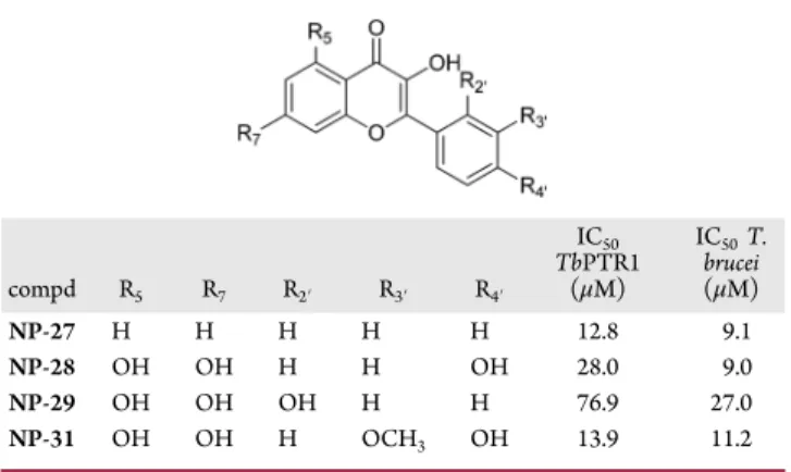
With the exception of quercetin dehydrate (NP-30), all
flavon-3-ol type aglycones showed a significant TbPTR1
inhibition, with 3-hydroxy
flavone (NP-27, IC
50= 12.8
μM)
and isorhamnetin (NP-31, IC
50= 13.9
μM) being the most
potent, whereas the glycosylated
flavonoid (NP-32) did not
inhibit TbPTR1. Most phenolic acids showed no inhibitory
activity toward TbPTR1, highlighting the importance of a
condensed ring (chromen-4-one) for protein inhibition. The
presence of two rings, one aromatic and the other one aliphatic
(NP-8) or both aromatic (NP-10), slightly increased the
inhibitory activity with respect to the other phenolic acid
derivatives.
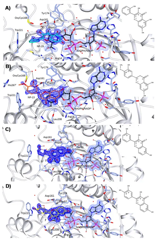
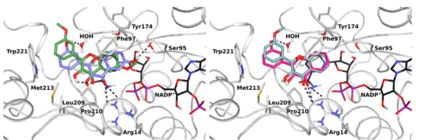
Figure 2.Crystal structures of TbPTR1 (gray cartoon, interacting residues in sticks) in complex with NADPH/NADP+(in sticks, black carbon atoms) and four inhibitors (in sticks) (A) NP-29 (cyan), (B) NP-13 (orange), (C) compound 2 (lilac), and (D) compound 7 (yellow). Hydrogen bond interactions (dashed lines) in the active site are shown. The 2Fo− Fcelectron density maps corresponding to the inhibitors (dark-blue wire)
and NADPH/NADP+(light-blue wire), contoured at the 1σ level are shown. The chemical structures and atom names of the ligands are specified in the insets.
The simplest
flavonol (NP-27, IC
50= 12.8
μM) was 10 times
more potent than the corresponding
flavone (NP-18, IC
50=
120.8
μM). This implies the importance of OH at position R3.
Moreover, isorhamnetin (NP-31, IC
50= 13.9
μM) was over 35
times more active against TbPTR1 than the corresponding
flavone (chrysoeriol, NP-25, IC
50> 490
μM). The insertion of
a hydroxyl group at position R5 led to a decrease in the
inhibitory activity. Indeed, the simplest
flavone (NP-18, IC
50=
120.8
μM) is more than 20-fold more active than primuletin
(NP-19, IC
50> 2450
μM), which has a hydroxyl group at
position 5. On target screening of the NP series indicates that
flavonols are the most promising compounds, with NP-31
being the most active and selective hit (selectivity index: SI
hTS/TbPTR1 > 35; SI hDHFR/TbPTR1 = 36). Nevertheless,
a clear structure
−activity relationship (SAR) could not be
established from this data set, and we could not reliably
determine how the number and the pattern of hydroxyl/
methoxy substituents in
fluenced the activity.
The 38 compounds were also assessed for their inhibitory
activity against T. brucei parasite (
Figure 1
, Supporting
Information,
Table S1
). With the exception of NP-9, NP-10,
NP-22, and NP-35, all the natural
flavonoids screened showed
antitrypanosomal activity with IC
50lower than 30
μM.
Figure 1
shows that there is no direct correlation between the
antitrypanosomal activity of the NP compounds and their
PTR1 inhibition e
ffect.
X-ray Crystallographic Studies of the Natural
Prod-ucts. To understand the
flavonoid interactions into the protein
binding site, the 18 compounds showing IC
50against TbPTR1
lower than 150
μM (compounds NP-8, NP-10, NP-12, NP-13,
NP-18, NP-21, NP-22, NP-23, NP-24, NP-26, NP-27, NP-28,
NP-29, NP-31, NP-35, NP-36, NP-37, and NP-38;
Figure 1
)
were selected for structural analysis. We obtained crystal
structures of the TbPTR1
−NADPH/NADP
+complexes for
two moderately active natural products (NP-29 (datiscetin)
IC
50TbPTR1= 76.9
μM and NP-13 (hesperetin) IC
50= 104.2
μM) only. These compounds show inhibitory activity against
TbPTR1 and selectivity against at least one of the human
proteins (TS and DHFR;
Figure 1
). Statistics for data
collection and re
finement are reported in the Supporting
Information,
Tables S2 and S3
. In both cases, the crystal
asymmetric unit contains the functional unit of the enzyme, the
TbPTR1 tetramer. The tertiary structure of the TbPTR1
subunits is typical of the short-chain dehydrogenases/
reductases (SDR) superfamily and shows a single
α/β domain
consisting of a seven-stranded parallel
β-sheet sandwiched
between two sets of
α-helices.
29The TbPTR1 active site is
mainly formed by a single chain, with one end blocked by the
C-terminus of the partner subunit. In this L-shaped depression,
the cofactor binds in an extended conformation entrapped by a
network of highly conserved hydrogen bonds.
29The
substrate-binding loop (residues 207−215) interacts with both the
cofactor and the substrate. Two surface-exposed parts of the
sequence, residues 104
−112 and 143−151, are usually poorly
visible in TbPTR1 crystal structures and they were not included
in our models. The overall structure of each subunit of the
TbPTR1 complexes is the same as indicated by the small
root-mean-square deviations (RMSDs) after C
α superimposition of
the four subunits in each tetramer (0.16−0.26 Å for NP-13 and
0.11
−0.41 Å for NP-29). The binding of the cofactor is
essential to create both the catalytic site and the
substrate-binding pocket, indicating an ordered sequential reaction
mechanism with NADPH binding before the substrate.
30The
pterin moiety of substrates and of pterin-like inhibitors binds in
a
π-sandwich between the nicotinamide ring of NADPH/
NADP
+and the aromatic side chain of Phe97.
31Pterin also
interacts with the NADPH/NADP
+β-phosphate, and this
interaction is strongly conserved in pterin-like inhibitors.
31TbPTR1-NADPH/NADP
+-
NP-29. The chromen-4-one core of
NP-29
binds in the biopterin binding pocket as described
above. The ligand is supported by a network of H-bonds
involving the cofactor and the surrounding residues in the
active site cavity (see
Figure 2
A). The hydroxyl group at
position 7 on the chromen-4-one of NP-29 (the NP-29 atom
numbering is given in the inset in
Figure 2
A) donates a H-bond
to the NADPH/NADP
+β-phosphate and accepts a H-bond
from an amino group on Arg14. The hydroxyl group at position
5 accepts a H-bond from NADPH/NADP
+ribose OH
2′
and
donates a H-bond to the hydroxyl group of Tyr174, which
donates a H-bond to the inhibitor carbonyl oxygen at position
4. Furthermore, the carbonyl oxygen and the hydroxyl group at
position 3 of NP-29 are both H-bonded to a water molecule
linked by a further H-bond to the side chain of Asp161. An
additional water molecule, which is highly conserved
throughout all TbPTR1 structures, mediates the interaction
between the position 3 hydroxyl group of NP-29 and the
backbone carbonyl oxygen of Gly205 (not shown). The
o-phenol moiety of NP-29 is located in a hydrophobic pocket of
the TbPTR1 active site cavity, surrounded by the side chains of
Val206, Leu209, Pro210, Met213, and Trp221. The 2
′ hydroxyl
group on this ring is close (
∼4 Å) to the carbonyl oxygen of
Gly205 and forms an intramolecular H-bond (2.4 Å) with the
hydroxyl group at position 3 of the chromen-4-one moiety.
TbPTR1-NADPH/NADP
+-
NP-13. The chromen-4-one core of
NP-13
also binds in the biopterin binding pocket, but it is
rotated by about 180
° with respect to its orientation in the
TbPTR1
−NP-29 complex (
Figure 2
B). The ether oxygen at
position 1 on the chromen-4-one moiety of NP-13 points
toward the side chains of Asp161 and Tyr174. The hydroxyl
group at position 7 of the NP-13 chromen-4-one is within
H-bonding distance from the NADPH/NADP
+ribose OH
2′and
the side chain of Ser95. The NP-13 hydroxyl group at position
5 accepts a H-bond from an amino group of Arg14 and donates
a H-bond to the backbone carbonyl oxygen of Leu208. The
o-methoxy-phenol (o-guaiacol) ring in position 2 on the
chromen-4-one makes a T-shaped stacking interaction with
the aromatic side chain of Trp221. Furthermore, the 3
′
phenolic moiety is within H-bonding distance from the Asp161
side chain and forms two water-mediated interactions with the
backbone nitrogen of Cys168 and the amide nitrogen of
Asn175 (not shown). The methoxy oxygen at position 4
′
interacts with the sulfur of the modi
fied Cys168
(S-oxy-cysteine), whereas the terminal methyl makes van der Waals
contacts with the side chains of Trp221 and His267 of the
partner subunit.
In summary, structural comparison of the two TbPTR1
complexes reveals two opposite orientations of the natural
flavonoid inhibitors NP-13 and NP-29 in the biopterin binding
pocket in which the chromen-4-one substituents interact with
di
fferent residues surrounding the cavity. A double binding
mode was previously observed for pteridine-like inhibitors and
MTX.
15Flavonoid Library Design and Synthesis. The crystal
structures showed that
flavonoids occupy the biopterin binding
site of TbPTR1, suggesting that the
flavonol moiety could
provide a new sca
ffold for the development of PTR1 inhibitors.
The
flavonoids selected as starting points for the development
of compounds to treat trypanosomatidic diseases are depicted
in
Table 1
. Even though the two crystal structures showed that
the hydroxyl group at position 5 could establish H-bonds with
the protein and the cofactor, the inhibitory data on natural
flavonoids (
Figure 1
) suggested that a hydroxyl group in this
position leads to a decrease in the inhibitory activity against
TbPTR1. Therefore, we decided to synthesize compounds with
hydroxyl substituents at positions 6 or 7 of the chromenone
A-ring because, according to the crystal structures, these should
be able to establish H-bonds with the NADPH/NADP
+ribose
and the side chain of Ser95 or with the NADPH/NADP
+β-phosphate and Arg14 (
Figure 3
). Moreover, we performed
modi
fications at positions 3′, 4′, and 5′ on ring B to explore the
SAR. As shown in
Figure 3
, hydroxyl groups in positions 3
′, 4′,
and 5
′ could form a H-bonding interaction with the Asp161
side chain and two water-mediated interactions with the
backbone nitrogen of Cys168 and the amide nitrogen of
Asn175. Many natural products have both hydroxyl and
methoxy groups, thus we synthesized and biologically evaluated
eight hydroxylated (1
−8) and eight methoxylated (9−16)
compounds aiming to investigate how the number and the
pattern of hydroxyl/methoxy substituents in
fluenced the
activity.
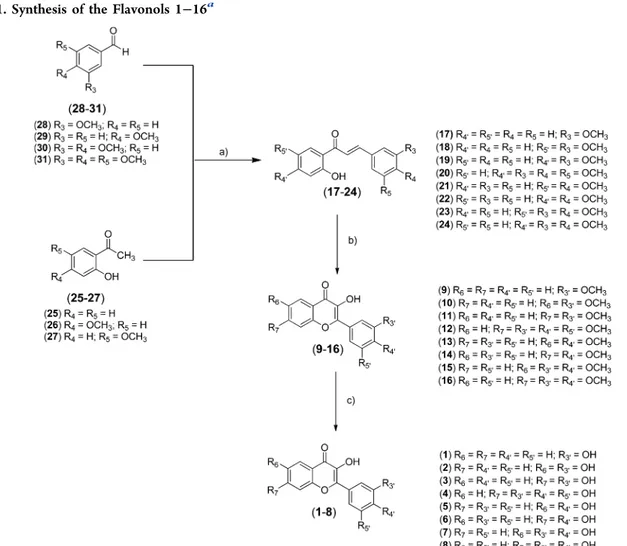
The synthesis of compounds 1
−16 is depicted in
Scheme 1
.
All the compounds have been already reported in literature, but
as far as we know, they have never been proposed as PTR1
inhibitors. The intermediate chalcones (17
−24) were
synthe-sized by Claisen
−Schmidt condensation using substituted
acetophenones and benzaldehydes in the presence of NaOH
as the base in ethanol. The reaction was carried out as
previously reported in literature for similar compounds.
26Afterward, the chalcones (17
−24) were converted into the
corresponding methoxylated
flavonols (9−16), using the
Flynn
−Algar−Oyamada method for epoxidation and
subse-quent intramolecular cyclization of the open-chain structure.
25The reaction was performed with hydrogen peroxide in
aqueous base (1 M NaOH). Cleavage of methoxy protecting
groups with boron tribromide gave the hydroxylated
flavonols
1
−8 in high yield. All synthesized compounds were
characterized by
1H NMR,
13C NMR, and mass analysis. The
obtained NMR and mass data are reported in the
Supporting
Information
(pp 33
−37) and were compared with those
available in literature.
Target Compound Pro
file. In our study, a panel of assays
was performed and activity/toxicity data were considered
during the hit selection/prioritization.
32Together with the
potency data (measured as IC
50, i.e., the compound
concentration producing 50% reduction of targeted enzyme
activity and parasite cell growth), we considered the selectivity
index (IC
50toward the parasite compared with compound
cytotoxicity IC
50on host cells) and early toxicity properties. We
have assessed the entire pro
file of the 16 synthesized
compounds, and the whole data panel has been evaluated
before selecting compounds for pharmacokinetic studies.
Testing of Synthetic Flavonols against PTR1 Enzymes.
All
flavonols were investigated for their activity toward T. brucei
(TbPTR1) and L. major PTR1 (LmPTR1). The results are
shown in
Figure 4
and Supporting Information,
Table S4
. All
hydroxylated compounds except compound 5 showed
sig-ni
ficant inhibitory activity against LmPTR1 and TbPTR1 at 50
μM concentration, with compound 2 being the most potent.
Table 1. Chemical Structures and IC
50Values for Selected
Compounds from the Screened Natural Products Library
compd R5 R7 R2′ R3′ R4′ IC50 TbPTR1 (μM) IC50T. brucei (μM) NP-27 H H H H H 12.8 9.1 NP-28 OH OH H H OH 28.0 9.0 NP-29 OH OH OH H H 76.9 27.0 NP-31 OH OH H OCH3 OH 13.9 11.2
Figure 3.Design of the synthetic library (left). NP-13 (in yellow) into TbPTR1 (in gray). The amino acids involved in the interactions of the designed compounds are shown in different colors. For clarity reasons, two residues involved in the interactions are not shown (Cys168 and Asn175). Superimposition of the four crystal structures of TbPTR1 (right) (chain A gray ribbon; chain D pink ribbon; relevant active site residues as light-blue sticks) in complex with NADPH/NADP+ (sticks, black carbon atoms) and the inhibitors (stick) NP-13 (orange), NP-29 (cyan),
compound 2 (lilac), and compound 7 (yellow). The three different binding modes adopted by the ligands can be appreciated as well as the movement of Trp221 (yellow sticks) upon binding of compound 7.
Methylation resulted in almost complete loss of inhibitory
activity. The most noteworthy examples are represented by two
pairs: compounds 10 and 2 (4% and 96% inhibition of TbPTR1
at 50
μM, respectively) and compounds 12 and 4 (no
inhibition of TbPTR1 at 50
μM and 85% inhibition at 50 μM,
respectively). The introduction of a hydroxyl group on ring A
in compound 2 led to an increase of the inhibitory potency
against both enzymes of almost 3-fold with respect to
compound 1, which bears an unsubstituted ring (34%
inhibition of TbPTR1 at 50
μM). To explain a complete
SAR, X-ray crystallographic studies and docking analyses were
carried out. The compounds were evaluated also toward
TcPTR2, and none of the compounds signi
ficantly inhibited
TcPTR2 activity (data not shown).
Crystal Structures of Synthetic Flavonoid
−TbPTR1
Complexes. Compounds 2 and 7, which show inhibitory
activity against TbPTR1, were submitted to X-ray
crystallo-graphic characterization aimed at comparing their binding
poses in the PTR1 active site with those of the natural
flavonoids and thus gaining further information for compound
optimization.
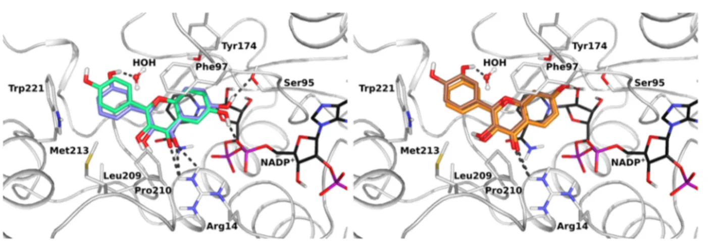
TbPTR1-NADPH/NADP
+−2. The structure of TbPTR1 in
complex with NADPH/NADP
+and compound 2 was
determined at 1.38 Å resolution. Ligand placement and key
interactions within the active site cavity are reported in
Figure
2
C. Compound 2, like NP-13 and NP-29, binds in the
substrate binding pocket, with the chromen-4-one in a
π-sandwich interaction between the nicotinamide ring of
NADPH/NADP
+and the aromatic side chain of Phe97. The
chromen-4-one core of compound 2 adopts the binding mode
observed for NP-13, with the oxygen atom at position 1
directed toward the side chain of Asp161. The R6 hydroxyl
group on the chromen-4-one is within H-bonding distance
from the hydroxyl group of Ser95 and the NADPH/NADP
+β-phosphate oxygen. The carbonyl oxygen at position 4
completes the interactions made by the chromen-4-one by
receiving two H-bonds from an amino group of Arg14 and a
water molecule that is H-bonded to the NADPH/NADP
+β-phosphate O2. These interactions, together with the stacking of
the chromen-4-one described above, bring its 3 position
hydroxyl group within H-bonding distance from the backbone
Scheme 1. Synthesis of the Flavonols 1
−16
aaReaction conditions: (a) NaOH (3 M), EtOH, rt; (b) H
2O2, NaOH (1 M), EtOH, rt; (c) BBr3(1 M in dry DMC), dry DMC, 0°C → rt.
Figure 4.Inhibitory activity against TbPTR1 (in gray) and LmPTR1 (in black). The control compound was pyrimethamine, a PTR1 inhibitor (100% inhibition at 50μM against both PTR1 enzymes).
carbonyl oxygen of Leu208. The phenyl ring in position 2 on
the chromen-4-one establishes a T-shaped stacking interaction
with the aromatic side chain of Trp221 and forms van der
Waals interactions with the side chains of Val206 and Leu209
(not shown). The meta-hydroxyl group at the 3
′ position on
the phenyl moiety of 2 is only involved in a water mediated
H-bond network linking it to the backbone carbonyl of Gly205
and the side chain of Asp161 (not shown).
TbPTR1-NADPH/NADP
+−7. The structure of TbPTR1 in
complex with NADPH/NADP
+and compound 7 has been
determined at 1.76 Å resolution. Ligand placement and key
interactions within the active site cavity are shown in
Figure
2
D, where it can be observed that compound 7 adopts exactly
the same pose as compound 2, making the same interactions
with the enzyme and cofactor. The only di
fference consists in
the steric repulsion of the additional hydroxyl group at position
4
′ that forces the side chain of Trp221 to move away by about
1.4 Å. The 4′ hydroxyl group is not involved in H-bonds but is
solvent exposed and presumably can form a H-bond with water.
The superposition of the four crystal structures is shown in
Figure 3
. The orientation of compounds 2 and 7 in the
TbPTR1 cavity is the same as for NP-13, with the inhibitor
bicyclic core directed toward the opposite side of the cavity
with respect to NP-29. Due to the larger steric hindrance of the
substituents on the 2-o-methoxy-phenolring of NP-13, a little
rearrangement of the whole molecule occurs with a rotation of
about 35
° of the bicyclic core with respect to 2 and 7 that
allows the side chain of Trp221 to remain in the position
observed in the TbPTR1
−2 complex (
Figure 2
C). The rotation
of NP-13 allows direct H-bonding between the 3
′ hydroxyl
group of NP-13 and Asp161, while the interaction of the
corresponding group of 2 and 7 is mediated by a water
molecule. The displacement of the Trp221 side chain occurring
upon binding of compound 7 can be appreciated (Trp221 as
yellow sticks in
Figure 3
, right).
A puzzling aspect of all four crystal structures is that the
di
fferent inhibitors have been systematically observed bound to
three of the four TbPTR1 subunits, whereas the NADPH/
NADP
+cofactor is found bound to all subunits in three
instances out of four, although with lower occupancy in those
subunits where the inhibitor is absent. The absence of the
inhibitor is correlated with larger disorder in the substrate
binding loop that brings Trp221 into the active site cavity.
However, this loop always shows temperature factors higher
than average, even in the subunits where both cofactor and
inhibitor are bound. The disorder of this region is independent
of the crystal packing because, when the loop is disordered
(e.g., in subunit C in TbPTR1
−NP-29), it approaches the
symmetry-related subunits in the same way as the ordered loop
(e.g., in subunit A in TbPTR1
−NP-29) located on the opposite
face of the TbPTR1 tetramer. Con
firming this observation, the
other two ordered loops (e.g., in subunits B and D in
TbPTR1
−NP-29) do not make close intermolecular contacts.
We can conclude that stabilization due to the
π-stacking
between the R2-aromatic ring of the inhibitor and the side
chain of Trp221 is critical for the ordering of the substrate
binding loop.
Computational Docking of Synthetic Compounds to
TbPTR1 and LmPTR1 and SAR Analysis. Docking studies
were conducted to further expand the SAR to all the
hydroxylation/methoxylation patterns in the synthesized
compounds. In addition, computational docking allowed the
evaluation of possible binding modes and a corresponding SAR
for LmPTR1, for which no crystal structure with these
compounds could be solved.
Like the crystallographic studies, computational docking
showed multiple possible binding modes for the
chromen-4-one system, which roughly group into three classes: (1) a
typical substrate-like binding mode involving H-bonding with
the NADPH/NADP
+cofactor and Ser95 of TbPTR1 (or
Ser111 of LmPTR1), as observed in the complexes of
compounds 2, 7, and NP-13; (2) an alternative binding
mode with chromen-4-one
flipped by roughly 180° as observed
in the complex with NP-29; and (3) an inverse binding mode
solely observed in docking studies, with ring B rather than the
chromen-4-one in a stacking orientation between the NADPH/
NADP
+nicotinamide and Phe97 of TbPTR1 (or Phe113 of
LmPTR1). While three of the four crystal structures feature
binding mode (1), a general dependence of the binding mode
on the substituent pattern is notable both from crystallographic
and docking studies. In docking, many of the observed binding
modes were found to have highly similar contacts and
H-bonding partners, thus creating a favorable entropic
contribu-tion to the binding of this compound class (see also Supporting
Information,
Tables S5 and S6
). However, due to their large
number of H-bond donors/acceptors,
flavonoids can adopt
Figure 5.Comparison of the effects of hydroxyl substituents at the R6 (left) and the R7 (right) positions of ring A on the binding of the synthetic flavonoids to TbPTR1. Superimposition of constraint docking poses for compound 2 (in sticks, lilac carbons) and compound 5 (in sticks, pale-green carbons) (left) and compound 3 (in sticks, orange carbons) and compound 6 (in sticks, brown carbons) (right) in TbPTR1 (in cartoon with interacting residues in sticks with gray carbons) in complex with NADPH/NADP+(in sticks, black carbon atoms). A conserved water molecule is shown in ball-and-stick representation. Hydrogen bonds are indicated by dark-gray dotted lines.
additional binding modes with different, often water mediated,
interactions. We therefore used a ligand constraint docking
approach to favor arrangements close to the crystal complexes
to assess the potential of other synthetic derivatives to bind in
similar orientations.
The crystal structures and docking indicate that
hydrox-ylation patterns on ring A influence the orientation of
chromen-4-one in the pocket. Precisely, compounds hydroxylated at
position R6 show di
fferent H-bonding partners (mostly
NADPH/NADP
+phosphate and Ser95/Ser111 of TbPTR1/
LmPTR1, respectively) compared with those hydroxylated at
R7 (mostly NADPH/NADP
+ribose and Tyr174/Tyr194 or
TbPTR1/LmPTR1, respectively), see
Figure 5
. This di
fference
also a
ffected the compound activity (compare R6-hydroxylated
compound 2 (96% TbPTR1 inhibition at 50
μM) and the
corresponding R7-hydroxylated compound 3 (47% TbPTR1
inhibition at 50
μM)). While the geometry of compound 2 fits
ideally in the TbPTR1 binding pocket, the slight reorientation
observed for compound 3 leads to a loss of H-bonds and
stabilizing hydrophobic contacts, halving the compound
activity. Thus, hydroxylation of ring A can
fine-tune the
arrangement of the chromen-4-one system within the pocket
and critically in
fluence the interaction pattern of rings B and C.
Methoxylations, in contrast to hydroxylations, were found to
lead to activity loss. For example, the inhibitory activity against
TbPTR1 and LmPTR1 drops substantially when comparing
compound 2 with the corresponding methoxylated compound
10
(96% and 4% TbPTR1 inhibition at 50
μM; 86% and no
LmPTR1 inhibition, respectively). Methoxylations on the
A-ring cause a displacement of the chromen-4-one from the
primary stacking geometry to accommodate the bulkier
methoxy group in close proximity to the NADPH/NADP
+cofactor and Ser95 of TbPTR1 (Ser111 of LmPTR1), see
Figure 6
, left. Notably, this adverse e
ffect was also apparent in
the comparison of compounds 1 and 9, bearing no substitution
on ring A but a hydroxylation or methoxylation on R3
′ of the
B-ring. Whereas the activity of compound 9 is negligible,
compound 1 shows about 35% inhibition against both TbPTR1
and LmPTR1 (at 50
μM). A H-bond donor functionality on
R3
′ has an important stabilizing effect, as it can interact with a
water molecule bridging Asp161 and Gly205 of TbPTR1 (see
Figure 6
, right) or Asp181 and Gly225 of LmPTR1. As this
water, according to our WatCH analysis, is almost 100%
conserved in both TbPTR1 and LmPTR1, it provides a clear
advantage for R3
′ hydroxylated compounds. This is further
supported by the crystal structures of TbPTR1 with
compounds 2 and 7, where the R3
′ hydroxylation is in
H-bonding distance to this water site. In agreement with this
observation, compound 2 with the m-hydroxyphenyl was over
30 times more active than compound 5 with a p-hydroxyphenyl
ring (96% and 3% TbPTR1 inhibition at 50
μM, respectively).
In contrast to a meta-hydroxylation, a para-hydroxylation
cannot interact with the structural water, see
Figure 5
. Again,
this e
ffect was sensitive to the hydroxylation pattern of ring A,
as can be observed by comparing compounds 3 (R3
′ and R7
hydroxylation, 47% TbPTR1 inhibition at 50
μM) and
compound 6 (R4
′ and R7 hydroxylation, 33% TbPTR1
inhibition at 50
μM), see
Figure 5
. While p-substituted
compounds generally show a drop in inhibitory activity
compared to their respective m-substituted counterparts, the
e
ffect is weaker for the second pair with the R7 hydroxylation
on ring A, possibly because R7 substitutions lead to a slight
rotation of the stacking chromen-4-one, directing the
p-hydroxyl toward a more solvent-exposed region of the pocket.
In conclusion, the e
ffects of individual hydroxyl substituents are
not purely local and do not show a completely additive e
ffect;
rather, the combinations of ring A and B hydroxylations will
determine the
final binding mode.
On the basis of the binding modes observed for TbPTR1, we
evaluated possible binding orientations to LmPTR1 by docking.
Notably, all compounds with R4
′ hydroxylation (compounds 5
and 6), compounds with R3
′ and R4′ hydroxylations
(compounds 7 and 8) and one compound with R3
′
hydroxylation combined with an R7 substitution on ring A
(compound 3) were found to be slightly more active toward
LmPTR1 than TbPTR1. While the depth of the biopterin
binding pocket is well conserved and the interactions for
chromen-4-one are practically identical in both proteins, there
are notable di
fferences in the opening of the pocket that can
explain the observed activity di
fferences (see sequence
alignment of interaction partners in Supporting Information,
Figure S2
). While in TbPTR1, R3
′ hydroxylations on the B-ring
mainly come into contact with the conserved water molecule
and Cys168 (see
Figure 5
) and R4
′ hydroxylated compounds
can hardly form direct H-bonds with the receptor, LmPTR1
generally has a more polar opening part of the pocket. Three
Figure 6.Comparison of the effects of hydroxyl and methoxy substituents at the R6 (left) and the R3′ (left and right) positions on the binding of the syntheticflavonoids to TbPTR1. Constraint docking poses are shown for compound 2 (in sticks, lilac carbons), compound 10 (in sticks, dark-green carbons) (left), compound 9 (in sticks, pale-cyan carbons), and compound 1 (in sticks, magenta carbons) (right) in TbPTR1 (in cartoon with interacting residues in sticks with gray carbons) in complex with NADPH/NADP+(in sticks, black carbon atoms). A conserved water molecule is
major di
fferences are crucially important: His241, Tyr283, and
Arg287 from the neighboring subunit in LmPTR1, correspond
to Trp221, Leu263, and His267 from the neighboring subunit
in TbPTR1, thereby providing several additional polar contact
points. As the side chain of Arg287 in LmPTR1 is much bigger
than that of His267 in TbPTR1, arginine is much more
accessible to the ligands than histidine. Furthermore, as
observed in crystal structures of TbPTR1, Trp221 is rather
motile and di
fferent rotamers lead to an open or a rather closed
form of the pocket, thereby potentially blocking ligand access to
His267. While Trp221 of TbPTR1 did not provide a
H-bonding contact to any of the studied compounds, induced
fit
docking studies of the compounds to LmPTR1 showed
movement of His241 to allow additional H-bonding
interactions in the most stable structure, as also shown in
Figure 7
(see also Supporting Information,
Table S5
). Overall,
π−π stacking and hydrophobic contacts are clearly the main
stabilizing factors for
flavonoids in TbPTR1, whereas a
tendency toward a higher number of H-bond donor/acceptor
contacts may stabilize hydroxylations in both the meta- and
para-positions of ring B slightly better in LmPTR1.
While addition of a para-hydroxylation for the R7 substituted
ring A did not lead to a signi
ficant change in activity against
TbPTR1 (compound 3, 47%; compound 8, 53% TbPTR1
inhibition at 50
μM), compound 4 with hydroxylations at R3′,
R4
′, and R5′ yielded a clear activity increase (85% TbPTR1
inhibition at 50
μM). Considering the lack of direct polar
contact points in TbPTR1, this is surprising and there may be
two possible explanations: (1) bridging water molecules may
enable additional contacts for the second meta-hydroxylation,
e.g., with Trp221, or (2) the compound may adopt an inverted
binding mode, placing the pyrogallol moiety in a stacking
orientation with Phe97 and the cofactor, where it could make
strong H-bonds and thereby keep the more hydrophobic
chromen-4-one in the hydrophobic TbPTR1 subpocket, where
it is able to form additional hydrophobic contacts.
Testing of Synthetic Flavonols against
TbDHFR,
LmDHFR, hDHFR, and hTS. The compounds showing
inhibitory activity against PTR1 (1
−8) were evaluated for
their activity against TbDHFR, LmDHFR, and hDHFR. The
results are reported in the Supporting Information,
Table S7
.
With the exception of compounds 1 and 2, all the tested
compounds had inhibitory activities against TbDHFR and
LmDHFR below 25% at 50
μM. Compounds 1 and 2 slightly
inhibit, respectively, TbDHFR and LmDHFR (42
−43% at 50
μM). Therefore, we can conclude that compounds 1−8 have
low inhibitory activity against the parasitic DHFRs. With the
exception of compound 2, all the compounds had inhibitory
activities against hDHFR below 30% at 50
μM. The IC
50of
compound 2 against hDHFR is 50
μM and against TbPTR1 is
4.3
μM, thus compound 2 has a selectivity index higher than 10.
None of the tested compounds inhibited hTS at 50
μM (data
not shown).
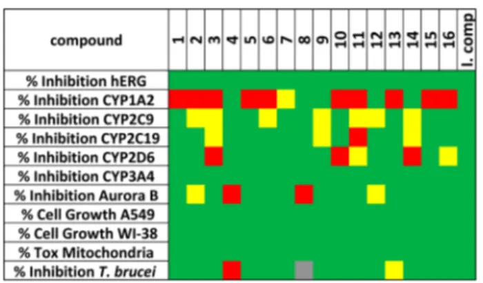
Early Toxicity Studies. To characterize the compounds
properties and to de
fine their toxicological profile, we used an
assay panel typical of those employed in drug discovery projects
for hit/lead selection.
32Our panel includes hERG,
five
cytochrome P450s, (CYP1A2, CYP2C9, CYP2C19, CYP2D6,
and CYP3A4), Aurora B kinase, A549 (human lung
adenocarcinoma epithelial) and WI-38 (fetal lung
fibroblasts)
cell lines and mitochondrial toxicity. The results of the
screening are reported in
Figure 8
as a tra
ffic light code
(from red through yellow to green for increasing biological
e
ffect and decreasing toxicity of the compounds) and in
Supporting Information,
Table S8
. An ideal lead compound
should have all parameters colored in green. In detail, the
percentage of mitochondrial toxicity and of inhibition toward
hERG, CYP isoforms, and Aurora B kinase should be below
30%, while the percentage of A549/WI-38 cells growth,
measuring cytotoxicity, should be above 70%. A few
compounds showed a percentage of inhibition toward hERG
> 10% at 10
μM. Apart from compounds 4 and 8, all the
compounds inhibited at least one isoform of cytochrome P450
(mostly CYP1A2), while only two compounds (4 and 8)
exhibited a signi
ficant inhibitory activity against Aurora B kinase
Figure 7.Superimposition of the crystal structure of LmPTR1 (PDB ID 1E92) in cartoon representation and interacting residues in sticks representation. Chains A and D (containing Arg287) are colored in pale-pink and magenta, respectively) and the best predicted receptor conformation obtained in the induced-fit docking study starting from this crystal structure (His241 in H-bonding contact to compound 7) in complex with NADPH/NADP+ (in sticks, black carbons) and
compound 7 (in sticks, yellow carbons). A conserved water molecule is shown in ball-and-stick representation. Hydrogen bonds are
indicated by dark-gray dotted lines. Figure 8.Early toxicity properties combined with inhibitory activity against T. brucei. The data are reported as a traffic light system. An ideal compound (I. comp.) should have all the parameters green. The cells are colored in green when the percentage of inhibition of T. brucei and the percentage of A549 and W1−38 cell growth is between 60 and 100, while the percentage of inhibition of CYP isoforms, hERG, Aurora B kinase and mitochondrial toxicity is between 0 and 30. Cells are colored in red when data indicates toxicity or inactivity. Yellow stands for a borderline value (30−60%): moderately active or slightly toxic compound. Gray: not tested.
at 10
μM. None of the compounds was either cytotoxic or
showed mitochondrial toxicity.
Testing of Synthetic Compounds on Cultured
Para-sites. With the exception of compound 8, all the molecules
were tested at 10
μM against T. brucei bloodstream form,
intracellular T. cruzi, and L. infantum intracellular amastigotes
(
Figure 9
). Almost all tested compounds were active against T.
brucei, while only two compounds, 4 and 12, were slightly
active against L. infantum, showing inhibitory activities of 10
and 35%, respectively. Twelve compounds (1, 2, 5
−12, 14−
16) presented EC
50values against T. brucei between 1 and 8
μM (
Table 2
), while compounds 3, 4, and 13 showed EC
50values of 12.29, 18.04, and 20.12
μM, respectively. Only the
methoxylated compounds 10, 11, 14, and 16 showed
antiparasitic activity toward T. cruzi trypomastigotes similar to
that of nifurtimox (NFX), the reference drug for the treatment
of Chagas
’ disease. Compound 10 is the most active (EC
50=
4.5
μM, NFX EC
50= 1.6
μM).
The series was assessed for cytoxicity on THP1
macrophage-like cells to evaluate the NOAEL (no observed adverse e
ffect
level). The reference compound was pentamidine with a
NOAEL of 10
μM and a selectivity index of 6440. The
selectivity indexes of our compounds, given by the ratio
between the EC
50toward T. brucei and CC
50toward THP1,
were lower than 10 with the exception of compound 12 (SI =
17) (
Table 2
). The selectivity index acceptable for a compound
to be considered non toxic
33is at least 10, thus our compounds
showed slight toxicity.
Combination Studies and Evaluation of Synergy. All
compounds were evaluated in combination with MTX
independently of their PTR1 inhibitory activity against the
recombinant protein. We aimed to assess the potential gain in
potency and the reduction in toxicity through the combination
of our compounds with MTX, a well-known DHFR inhibitor.
In trypanosomatids, MTX is expected to exert a synergistic
behavior in combination with PTR1 inhibitors. Moreover,
MTX can show a synergistic e
ffect in combination with
non-PTR1 inhibitors through a multitarget inhibition. Preliminary
combination experiments in T. brucei were conducted by
fixing
MTX at 4
μM (MTX average EC
30against T. brucei) and
varying the concentration of the selected compounds between
1.25 and 20
μM (Supporting Information,
Table S10
). The
synergy coe
fficients of all the compounds tested at the different
concentrations (1.25, 2.5, 5, 10, 20
μM) are reported in
Supporting Information,
Table S11
. Eight compounds
presented a synergistic e
ffect (synergy coefficient >1) when
combined with MTX. The antiparasitic activity against T. brucei
of the eight compounds both alone and in combination with
MTX is shown in
Figure 10
. Compounds 5, 6, and 13
consistently presented the highest synergy coe
fficients
(max-imum synergy coe
fficient >1.5). Compound 13 was the most
synergic with a calculated maximum synergy coe
fficient of 3.1
at 1.25
μM (Supporting Information,
Table S11
). Since it
showed the highest synergy coe
fficient together with a safe
toxicological pro
file (
Figure 8
, the only poor (red) parameter is
for CYP1A2 (72% inhibition at 10
μM)), it was selected for
further synergistic combination studies.
To better evaluate the gain in potency of the combination
MTX
−13, we employed the software Compusyn and a
constant ratio between the concentrations of the two
compounds chosen from their EC
50values (EC
5013
= 20
μM; EC
50MTX = 15; constant ratio = 1.3).
34This enabled us
to quantify synergism between the two compounds
(combina-tion index, CI) and to determine the dose reduc(combina-tion (dose
reduction index, DRI) needed to observe the studied e
ffect in
the combination compared with the dose of each drug alone.
The combination of MTX and compound 13 at the EC
50concentration had a CI of 0.174
± 0.047. When CI is lower
than 1, the combination can be considered synergistic,
therefore the observed CI shows strong synergism and con
firms
the initial data (Supporting Information,
Table S10
). At higher
EC values, CI becomes lower, indicating very strong synergism
(
Table 3
). The EC
50determined for the combination showing
CI of 0.147 was 3.15
± 0.33 μM. This enabled an average DRI
at the EC
50of 14.6
± 3.7 for MTX and of 11.7 ± 2.7 for
compound 13 (IC
50MTX = 1.3
μM and IC
5013
= 1.7
μM)
(Supporting Information,
Figure S3C
). At the EC
90of the
mixture, the predicted DRI is over 100-fold for both
compounds when compared with the EC
50of the compounds
alone. The isobologram is reported in Supporting Information,
Figure S3
. The toxicity of the MTX
−13 mixture on THP1 was
determined at three different concentrations (12.5, 25, 50 μM),
and no synergistic toxicity on human cells was observed
(Supporting Information,
Figure S4 and Table S12
). Therefore,
the identi
fied combination MTX−13 is selective for the
inhibition of the parasite growth, while there is no synergy in
Figure 9.Antiparasitic activity of the synthesized compounds against Trypanosoma brucei (in gray), Trypanosoma cruzi (in green), and Leishmania infantum (in black) at 10μM. The reference compounds were pentamidine (IC50= 1.55± 0.24 nM) for T. brucei, miltefosine
(IC50= 2.65± 0.4 μM) for L. infantum, and nifurtimox (IC50= 2.2±
0.4μM) for T. cruzi.
Table 2. EC
50against
T. brucei, NOAEL, and Selectivity
Index of the Synthesized Compounds
a compd EC50T. brucei (μM) ± SD CCNOAEL50± SD or selectivity index (CC50/EC50) 1 5.18± 1.10 20 4 2 7.56± 0.51 53± 2 7 3 12.29± 2.82 80± 2 6 4 18.04± 0.50 100 5 5 2.32± 0.42 20 7 6 4.29± 0.71 20 4 7 1.36± 0.57 10 6 9 2.32± 1.01 10 4 10 1.43± 0.42 10 7 11 1.14± 0.24 10 8 12 1.17± 1.07 20 17 13 20.12± 2.27 20 1 14 1.10± 0.47 10 9 15 2.02± 0.51 10 5 16 2.99± 1.86 25 8 pentamidine 0.00155± 0.00024 10 6440 aEC50 and NOAEL represent the arithmetic average of at least two
the toxicity toward the host cells. These data together with the
predicted DRI indicate that the combination improves the
selectivity indexes for both MTX and compound 13.
Pharmacokinetic Studies, in Vivo Assays, and
Com-pound Delivery. ComCom-pound 2 was selected for
pharmacoki-netic studies because it was the most active compound against
TbPTR1 (IC
50= 4.3
μM) that showed activity against the
parasite T. brucei (EC
50= 7.6
μM) and it presented a safe
profile, see
Figure 8
. The hERG IC
50(99.3
μM) was more than
20-fold higher than the target IC
50. With the exception of the
CYP1A2 IC
50(6.1
μM), the IC
50against the cytochrome
isoforms was higher than 10
μM. The solubility of compound 2
was evaluated using UV−Vis spectroscopy. In vivo
bioavail-ability and half-life of compound 2 were evaluated in BALB/c
mice treated IV with 1 mg/kg based on its low solubility. The
molecule showed a short half-life (t
1/2= 7.6 min), and it was
not detected in blood after 30 min (the plasma levels are shown
in Supporting Information,
Figure S4
). With the aim of
improving the plasma levels of compound 2, we used two
di
fferent drug delivery systems: the molecule was encapsulated
in PLGA nanoparticles and solubilized with hydroxypropyl-
β-cyclodextrins. The encapsulation did not change the physical
properties of the PLGA nanoparticles as reported in Supporting
Information,
Table S13
. The unloaded and loaded NPs were
characterized by dynamic light scattering (DLS) in terms of
size, polydispersity index, and
ζ potential. We evaluated the in
vivo bioavailability of compound 2 encapsulated in
nano-particles in BALB/c mice (IV and per os administration) and
formulated with cyclodextrins after per os administration to
NMRI mice. From preliminary data, both formulations did not
signi
ficantly increase the levels of compound 2 in plasma
samples from mice (data not shown). However, the in vitro
activity of compound 2 both in PLGA and in cyclodextrins
solution was preserved when compared to the free compound
while the toxicity on THP1 was diminished (
Table 4
).
■
CONCLUSION
This is the
first study in which we have confirmed the potential
of
flavonols as PTR1 inhibitors. Apart from compounds 4 and
8, none of the 16 synthesized
flavonols have previously been
reported in literature for their antiparasitic activity.
22Twelve of
these compounds show EC
50values against T. brucei below 10
μM. We performed combination studies with MTX and
compound 13, a non-PTR1 inhibitor, which was the
best-performing compound. Although the reason for the synergy is
unknown, compound 13 is an interesting compound for drug
development due to the synergistic behavior with MTX and the
low toxicity on host cells. Target identification studies could be
carried out aiming to explain the mechanism of action. The
synthesized compounds were also evaluated for the activity
against T. cruzi trypomastigotes, and compound 10 turned out
to have a potency comparable to that of nifurtimox, the drug
currently used to treat Chagas
’ disease (EC
50compound 10 =
4.5
μM, NFX EC
50= 1.6
μM). We carried out early in vitro
toxicity assays, and overall the library showed a satisfactory
profile. Combining the activity/toxicity data, we selected
compound 2 for pharmacokinetic studies and we observed a
very quick turnover of the molecule in BALB/c mice. Neither
encapsulation in PLGA nanoparticles nor solubilization with
cyclodextrins increased the plasma level of compound 2;
however, both formulations allowed to maintain the in vitro
activity of the compound and to reduce the toxicity. Our crystal
structures and SAR analysis provide a basis for structure-based
drug design aimed at identifying novel
flavonol-like compounds
with increased potency and selectivity toward PTR1 and able to
overcome the problems of classical
flavonols.
■
EXPERIMENTAL SECTION
Preparation of the Natural Products Library. A library of 98 natural compounds, consisting of 8 flavanones, 19 flavones, 20 flavonols, 2 dihydroflavonols, 13 anthocyanins, 2 catechins, 2 isoflavones, 4 chalcones, 18 phenolic acids and derivatives, 1 aurone, 5 anthraquinones, 3 triterpenes, and 1 phloroglucinol was purchased from Extrasynthese (Genay, France) and Sigma-Aldrich-Fluka (Milan, Italy) for drug screening. The degree of purity of all these molecules was checked by HPLC. The HPLC analyses were performed on an Agilent Technologies (Waldbronn, Germany) modular model 1100 system, consisting of a vacuum degasser, a quaternary pump, an autosampler, a thermostated column compartment, and a diode array detector (UV/DAD). The chromatograms were recorded using an Agilent Chemstation for LC and LC-MS systems (Rev. B.01.03). An Ascentis C18 column (250 mm × 4.6 mm I.D., 5 μm, Supelco,
Bellefonte, PA, USA) was used, with a mobile phase composed of (A) water (H2O) and (B) acetonitrile (ACN). The gradient elution was
modified as follows: 0−3 min 25% B, 3−10 min from 25 to 30% B, from 10−40 min from 30 to 40% B, which was held for 5 min. The postrunning time was 5 min. Theflow rate was 1.0 mL/min. The column temperature was set at 30°C. The sample injection volume was 5μL. The UV/DAD acquisitions were carried out in the range 190−600 nm, and chromatograms were acquired at 310, 330, 370, and 520 nm. All the compounds tested were found to be stable, and degradation products were not detected in the HPLC chromatograms. Supporting Information,Table S1shows the purity (>95%) of the 38 natural compounds screened. The natural compounds were stored at low temperature (−80 °C), protected from light and humidity.
Computational Studies. Virtual Screening of the Natural Product Library. The library of 98 natural products was subjected to Figure 10.Trypanocidal activity of eight synthesized compounds alone and in combination with MTX. The compounds were tested at three different concentrations: 5 μM (in green), 2.5 μM (in light-pink), 1.25 μM (in light-blue). Antiparasitic activity of MTX is shown in dark-red.
virtual screening using the GOLD software (version 5.1)35,36following the protocol validated in previous studies on PTR1.13The compounds
were docked into the binding sites of the targets, LmPTR1 (PDB ID 1E92) and TbPTR1 (PDB ID 3JQ9), and the off-targets, hDHFR (PDB ID 1U72) and hTS (PDB ID 1HVY). Ten docking poses were generated for each ligand in each target, and these were visually inspected and manually ranked, considering the Goldfitness score, hydrogen bonding, and π−π aromatic interactions. A search of BioAssay data in PubChem was done to filter out promiscuous compounds. The set of 38 compounds given in Supporting Information,Table S1was selected for inhibition assays considering the above properties and structural diversity.
Docking of Synthetic Flavonoid Derivatives. The 3D structures of the compounds were created from SMILES strings and optimized with the OPLS_2005 force field using Maestro.37 Ionization states and
tautomers were generated at pH 7.0± 0.5 using Epik.37Up to eight stereoisomers and one low-energy ring conformation were generated per compound. Important structural water sites were identified using the WatCH clustering approach.38All available chains of the LmPTR1 crystal structures and 99 chains from TbPTR1 crystal structures (see Supporting Information,Tables S14 and S15for a list of structures) were superimposed and their water sites were clustered with a distance criterion of 2.4 Å. Water sites with at least 50% conservation were considered conserved.
The PTR1 structures for docking consisted of chain A of the relevant crystal structure and a C-terminal tripeptide from the neighboring subunit pointing into the chain A active site (Val266 to Ala268 in TbPTR1 and Thr286 to Ala288 in LmPTR1). The side -chain oxygen atom on the modified Cys 168 in the crystal structure of TbPTR1 was removed. Crystallographic solvent molecules were removed and replaced by the conserved water molecules identified by WatCH clustering. The LmPTR1 structure (PDB ID 1E92) was aligned to the TbPTR1 template (PDB ID 5JCJ). PrepWizard was used to assign bond orders and add hydrogen atoms.37The N- and C-termini of chain A were capped with N-acetyl and N-methyl amide groups, respectively. The protonation state of the NADPH/NADP+
cofactor was computed at pH 7.0± 0.5. Protein protonation states were assigned by PROPKA43 at pH 7.0. The H-bond network was optimized and all hydrogens were subjected to a restrained minimization. A 20 Å× 20 Å × 20 Å docking grid was centered on Phe97 of TbPTR1 or Phe113 of LmPTR1. The following hydroxyl groups were set rotatable: Ser95 and Tyr174 in TbPTR1 and Ser111, Thr184, Tyr191, Tyr194, Thr195, and Tyr283 in LmPTR1, as well as those of the NADPH/NADP+ribose.
Docking was performed with and without ligand-based constraints using the Glide software.39−42 For the ligand constraints, protein preparation was performed including compound 7 (substrate-like binding mode, PDB ID 5JCJ) or compound NP-29 (inhibitor-like binding mode, PDB ID 5JCX) or the highest scoring docking solution from unconstrained docking of compound 4 with ring B in a stacking orientation (alternate binding mode). The ligands were then separated from the structure to provide reference geometries. The van der Waals radii of the ligand atoms were scaled by 0.80, and a partial charge cutoff of 0.15 was used. XP (extra precision) docking and flexible ligand sampling were set. Nitrogen inversions and ring conformations were sampled, and biased sampling of torsions was performed for amides only and set to penalize a nonplanar conformation. Epik state penalties were added to the docking score, intramolecular H-bonds were rewarded, and the planarity of conjugated π groups was
Table
3.
Summary
Report
of
Computer-Simulated
CI
and
DRI
Values
for
MTX
and
Compound
13
Combinations
at
50%,
75%,
90%,
and
95%
Inhibition
of
T.
brucei
Growth
a CI values at the reported EC DRI values at the reported EC drug combination combination ratio EC 50 EC 75 EC 90 EC 95 EC 50 EC 75 EC 90 EC 95 MTX + 13 (2:1.5) 0.174 ± 0.047 0.075 ± 0.036 0.034 ± 0.023 0.020 ± 0.016 14.58 ± 3.71 b 41.95 ± 10.52 b 127.36 ± 60.20 b 278.17 ± 172.13 b 11.72 ± 2.79 b 27.45 ± 6.90 b 65.56 ± 16.42 b 119.72 ± 29.15 b a Data reported are the average of three independent experiments. Data analysis was carried out using the CompuSyn software. 34 . b Values on top are for the MTX and on the bottom are for compound13
Table 4. in Vitro Activity against
T. brucei of Compound 2 in
Three Di
fferent Formulations
apreparation T. brucei EC50(μM) THP1 CC50± SD or NOAEL
2 7.56± 0.51 53± 2
PLGA-2 5.70± 0.23 >100
cyclodextrin-2 3.27± 0.16 >100
aEC
50 and NOAEL are the arithmetic mean of at least two