The authors’ full names, academic de-grees, and affiliations are listed in the Appendix. Address reprint requests to Dr. De Bruyne at the Cardiovascular Center Aalst, Onze-Lieve-Vrouw Clinic, Moorsel-baan 164, B-9300 Aalst, Belgium, or at bernard . de . bruyne@ olvz-aalst . be. * A list of the FAME 2 Investigators is
provided in the Supplementary Appen-dix, available at NEJM.org.
Drs. Xaplanteris and Fournier and Drs. Jüni and De Bruyne contributed equally to this article.
This article was published on May 22, 2018, at NEJM.org.
N Engl J Med 2018;379:250-9. DOI: 10.1056/NEJMoa1803538 Copyright © 2018 Massachusetts Medical Society.
BACKGROUND
We hypothesized that fractional flow reserve (FFR)–guided percutaneous coronary intervention (PCI) would be superior to medical therapy as initial treatment in patients with stable coronary artery disease.
METHODS
Among 1220 patients with angiographically significant stenoses, those in whom at least one stenosis was hemodynamically significant (FFR, ≤0.80) were random ly assigned to FFRguided PCI plus medical therapy or to medical therapy alone. Patients in whom all stenoses had an FFR of more than 0.80 received medical therapy and were entered into a registry. The primary end point was a composite of death, myocardial infarction, or urgent revascularization.
RESULTS
A total of 888 patients underwent randomization (447 patients in the PCI group and 441 in the medicaltherapy group). At 5 years, the rate of the primary end point was lower in the PCI group than in the medicaltherapy group (13.9% vs. 27.0%; hazard ratio, 0.46; 95% confidence interval [CI], 0.34 to 0.63; P<0.001). The difference was driven by urgent revascularizations, which occurred in 6.3% of the patients in the PCI group as compared with 21.1% of those in the medicaltherapy group (hazard ratio, 0.27; 95% CI, 0.18 to 0.41). There were no significant differ ences between the PCI group and the medicaltherapy group in the rates of death (5.1% and 5.2%, respectively; hazard ratio, 0.98; 95% CI, 0.55 to 1.75) or myocar dial infarction (8.1% and 12.0%; hazard ratio, 0.66; 95% CI, 0.43 to 1.00). There was no significant difference in the rate of the primary end point between the PCI group and the registry cohort (13.9% and 15.7%, respectively; hazard ratio, 0.88; 95% CI, 0.55 to 1.39). Relief from angina was more pronounced after PCI than after medical therapy.
CONCLUSIONS
In patients with stable coronary artery disease, an initial FFRguided PCI strategy was associated with a significantly lower rate of the primary composite end point of death, myocardial infarction, or urgent revascularization at 5 years than medi cal therapy alone. Patients without hemodynamically significant stenoses had a favorable longterm outcome with medical therapy alone. (Funded by St. Jude Medical and others; FAME 2 ClinicalTrials.gov number, NCT01132495.)
ABS TR ACT
FiveYear Outcomes with PCI Guided
by Fractional Flow Reserve
P. Xaplanteris, S. Fournier, N.H.J. Pijls, W.F. Fearon, E. Barbato, P.A.L. Tonino, T. Engstrøm, S. Kääb, J.-H. Dambrink, G. Rioufol, G.G. Toth, Z. Piroth, N. Witt, O. Fröbert, P. Kala, A. Linke, N. Jagic, M. Mates, K. Mavromatis, H. Samady, A. Irimpen, K. Oldroyd, G. Campo, M. Rothenbühler, P. Jüni, and B. De Bruyne,
for the FAME 2 Investigators*
Original Article
A
mong patients with acute coro-nary syndromes, early percutaneous cor onary intervention (PCI) increases the survival rate and decreases the rate of recurrent myocardial infarction.15 In contrast, in patientswith stable coronary artery disease, there is per sistent controversy about the role and timing of PCI to improve clinical outcomes and provide symptomatic relief.6,7 Since the potential benefit
of revascularization depends on the extent and severity of ischemia, careful identification of ste noses capable of inducing ischemia is essential.8,9
Current guidelines recommend the measurement of the coronary fractional flow reserve (FFR) for this purpose.1012
The Fractional Flow Reserve versus Angiogra phy for Multivessel Evaluation (FAME) 2 trial was designed to target stenoses capable of in ducing ischemia (FFR, ≤0.80) in a large myocar dial territory and to refrain from PCI in patients with hemodynamically nonsignificant stenoses (FFR, >0.80). We hypothesized that an initial strategy of FFRguided PCI plus medical therapy would provide better longterm outcomes than an initial strategy of medical therapy alone. Here, we describe the prespecified 5year follow up of the trial.
Methods Trial Design
We conducted this randomized, multicenter trial to compare FFRguided PCI plus medical therapy with medical therapy alone in patients with stable coronary artery disease. The shortterm outcomes (mean followup, 7 months) have been reported previously.13 The trial was sponsored by
St. Jude Medical; the sponsor did not provide support for the current analysis. The academic members of the steering committee designed the trial protocol (available with the full text of this article at NEJM.org), which was approved by all the relevant local review boards. An indepen dent data and safety monitoring board oversaw the trial.
The sponsor was involved in the collection of the data during the first 3 years of the trial but not in the trial design or conduct, the subse quent data collection, the writing and review of the manuscript, or the decision to submit it for publication. The two first authors and two last authors had full access to all the data in the
trial and vouch for the accuracy and complete ness of the data and analyses and for the fidel ity of the trial to the protocol.
Participants and Randomization
Patients with stable coronary artery disease were enrolled at 28 sites in Europe and North America.13
Patients with stable angina or documented silent ischemia who had at least one stenosis with a 50% diameter in a large epicardial artery that was suitable for PCI were eligible. The full list of the inclusion and exclusion criteria is provid ed in the Supplementary Appendix, available at NEJM.org.
Measurements of FFR were made for all angio graphically significant lesions. Each patient with at least one hemodynamically significant steno sis (FFR, ≤0.80) was randomly assigned in a 1:1 ratio to receive either FFRguided PCI plus medical therapy (PCI group) or medical therapy alone (medicaltherapy group). The randomiza tion schedule was computergenerated, stratified according to site, blocked (with randomly varied block sizes), and concealed with the use of cen tral randomization. Patients in whom all angio graphically significant stenoses were hemody namically nonsignificant (FFR, >0.80) did not undergo randomization but received medical therapy and were included in a registry. Written informed consent was obtained from all the patients.
Treatment
Patients who were assigned to the PCI group received a loading dose of clopidogrel (at a dose of 600 mg) and aspirin immediately before the procedure if they were not already taking these medications. All stenoses with an FFR of 0.80 or less were treated with second or thirdgeneration drugeluting stents. All the patients who under went PCI received clopidogrel at a dose of 75 mg daily for at least 12 months.
Trial End Points and Follow-up
The primary end point was a composite of death from any cause, myocardial infarction, or urgent revascularization. Urgent revascularization was defined as any unplanned hospital admission that was due to symptoms that led to revascular ization during the same hospitalization. Second ary end points included the components of the primary end point as well as death from cardiac
causes, any revascularization, stroke, and stent thrombosis. Endpoint definitions are provided in the Supplementary Appendix. Angina was classified according to the Canadian Cardiovas cular Society (CCS) functional classification, in which classes range from I to IV, with higher classes indicating greater limitations on physical activity owing to angina.
Followup was originally scheduled at 1 month, 6 months, and 1, 2, 3, 4, and 5 years. A total of 50% of the patients in the registry cohort were randomly selected and followed in the same manner as the trial patients. In November 2014, the sponsor decided to close out the trial once all the included patients had completed their 3year visit. The reason indicated by the sponsor was that results were unlikely to change sub stantially with longer patient followup, particu larly in view of the high rate of crossover of pa tients who had been assigned to medical therapy alone. The academic steering committee subse quently invited all 28 sites to participate in an additional 5year followup, and 19 sites partici pated (Table S1 in the Supplementary Appendix). Throughout the trial, detailed narratives were obtained for each potential event. Events that were ascertained before the original trial close out were adjudicated by an independent clinical events committee whose members were unaware of the trial group assignments. Events that were ascertained after the closeout were adjudicated by two cardiologists who were not involved in the trial and who were unaware of the trial group assignments.
Statistical Analysis
The trial was powered to determine the superior ity of FFRguided PCI over medical therapy alone with respect to the primary end point at 2 years. However, recruitment of the patients was stopped prematurely after the randomization of 888 of the originally intended 1632 patients. Recruit ment was discontinued at the recommendation of the data and safety monitoring board because of a significant difference in the rate of the pri mary end point in favor of the PCI group.13 De
tails of the original samplesize calculation are provided in the Supplementary Appendix.
Betweengroup comparisons of the end points were performed with the Mantel–Cox method for the calculation of hazard ratios and 95% confi
dence intervals and with the logrank test for corresponding P values. Kaplan–Meier curves were constructed. Landmark analyses were per formed according to landmark time points at 7 days and 3 years, with hazard ratios calculated separately for events that occurred before and after these time points. Landmark analyses were accompanied by tests for interaction between treatment and time.
There was no prespecified adjustment for mul tiple testing of secondary end points. However, because it was considered to be of importance to formally examine the components of the pri mary end point separately in this followup analysis, we informally adopted a post hoc Bon ferroni correction, which allowed for the three components of the primary end point to be tested at an alpha level of 0.0167 (0.05 ÷ 3). Since the widths of 95% confidence intervals were not adjusted for multiple comparisons, these inter vals should not be used for inference about treat ment effects. All the analyses were performed according to the intentiontotreat principle by an author who is a statistician in an academic clinical trials unit (Clinical Trials Unit Bern, University of Bern, Switzerland).
R esults Participants and Follow-up
Between May 15, 2010, and January 15, 2012, a total of 1220 patients were enrolled, including 888 in the randomized trial. Of these, 447 pa tients were assigned to PCI plus medical therapy and 441 to medical therapy alone. The remain ing 332 patients, who had an FFR more than 0.80 in all lesions, were enrolled in the registry, and half these patients (166 patients) were ran domly selected for followup. The characteristics of the patients at baseline were similar in the PCI group and the medicaltherapy group (Table 1). Tables S2 through S5 in the Supplementary Ap pendix present comparisons of the baseline characteristics between patients in the random ized trial and those in the registry cohort and according to site participation in the 5year follow up (yes or no).
Figure S1 and Table S6 in the Supplementary Appendix present the flow of patients through the different phases of the trial. In the PCI group, 435 of 447 patients underwent the planned pro
cedure; the remaining 12 patients were treated with balloon angioplasty, coronaryartery bypass grafting, or medical therapy alone (Fig. S1 in the Supplementary Appendix). In the medicaltherapy group, 439 of 441 patients received the planned treatment; the remaining 2 patients erroneously underwent PCI. In the registry, 165 of 166 pa tients received medical therapy, and 1 underwent PCI. Details of the medical therapy in each group are provided in Table S7 in the Supple mentary Appendix.
In the 19 sites that participated in the 5year followup, the median length of followup was 60.5 months (interquartile range [IQR], 59.8 to 61.7) in the PCI group, 60.5 months (IQR, 59.8 to 61.7) in the medicaltherapy group, and 60.6 months (IQR, 59.9 to 62.5) in the registry co hort, with complete followup information avail able through 5 years for 371 of 395 patients (93.9%) in the PCI group, 362 of 389 (93.1%) in the medicaltherapy group, and 133 of 147 (90.5%) in the registry. In the 9 sites that did not
Characteristic PCI Group (N = 447) Medical-Therapy Group (N = 441) Age — yr 63.5±9.4 63.9±9.6 Age >60 yr — no. (%) 282 (63.1) 279 (63.3) Male sex — no. (%) 356 (79.6) 338 (76.6) Body-mass index† 28.3±4.3 28.4±4.5 Family history of coronary artery disease — no./total no. (%) 216/446 (48.4) 207/441 (46.9) Current smoking — no. (%) 89 (19.9) 90 (20.4) Hypertension — no. (%) 347 (77.6) 343 (77.8) Hypercholesterolemia — no. (%) 330 (73.8) 348 (78.9) Diabetes mellitus — no. (%)
Any 123 (27.5) 117 (26.5)
Insulin-dependent 39 (8.7) 39 (8.8) Renal insufficiency — no. (%)‡ 8 (1.8) 12 (2.7) Peripheral vascular disease — no. (%) 43 (9.6) 47 (10.7) History of stroke or TIA — no. (%) 33 (7.4) 28 (6.3) History of myocardial infarction — no. (%) 164 (36.7) 165 (37.4) History of PCI in target vessel — no. (%) 80 (17.9) 76 (17.2) Angina — no./total no. (%)§
No angina or asymptomatic 53/447 (11.9) 46/440 (10.5) CCS class I 82/447 (18.3) 98/440 (22.3) CCS class II 204/447 (45.6) 197/440 (44.8) CCS class III 80/447 (17.9) 65/440 (14.8) CCS class IV 28/447 (6.3) 34/440 (7.7) Silent ischemia — no. (%) 73 (16.3) 73 (16.6) Left ventricular ejection fraction <50% — no. (%) 83 (18.6) 56 (12.7) * Plus–minus values are means ±SD. There were no significant differences between the two randomly assigned groups,
with the exception of left ventricular ejection fraction of less than 50% (P = 0.02). PCI denotes percutaneous coronary intervention, and TIA transient ischemic attack.
† The body-mass index is the weight in kilograms divided by the square of the height in meters.
‡ Renal insufficiency was defined as a creatinine level of more than 2.0 mg per deciliter (177 μmol per liter).
§ Angina was classified according to the Canadian Cardiovascular Society (CCS) functional classification, in which classes range from I to IV, with higher classes indicating greater limitations on physical activity owing to angina.
participate in the 5year followup, the median length of followup was 35.7 months (IQR, 34.9 to 36.3) in the PCI group, 35.6 months (IQR, 35.0 to 36.0) in the medicaltherapy group, and 35.3 months (IQR, 34.9 to 36.0) in the registry, with complete followup information available through 3 years for 46 of 52 patients (88%), 44 of 52 patients (85%), and 15 of 19 patients (79%), respectively. Details are provided in Table S6 in the Supplementary Appendix.
End Points
At least one primary endpoint event (death, myocardial infarction, or urgent revasculariza tion) occurred in 62 patients (13.9%) in the PCI group, as compared with 119 (27.0%) in the medicaltherapy group (hazard ratio, 0.46; 95% confidence interval [CI], 0.34 to 0.63; P<0.001) (Table 2). The Kaplan–Meier curves for the pri mary end point are shown in Figure 1, and in Figure S2 in the Supplementary Appendix. In the registry cohort, 26 patients (15.7%) had at least one primary endpoint event; the rates in the PCI group and the registry cohort did not differ sig
End Points PCI Group (N = 447) Medical-Therapy Group
(N = 441) Hazard Ratio (95% CI) Registry Cohort (N = 166)
no. of patients (%) no. of patients (%)
Primary composite end point 62 (13.9) 119 (27.0) 0.46 (0.34–0.63) 26 (15.7) Components of primary end point
Death from any cause 23 (5.1) 23 (5.2) 0.98 (0.55–1.75) 7 (4.2) Myocardial infarction 36 (8.1) 53 (12.0) 0.66 (0.43–1.00) 14 (8.4) Urgent revascularization 28 (6.3) 93 (21.1) 0.27 (0.18–0.41) 14 (8.4) Death or myocardial infarction 53 (11.9) 71 (16.1) 0.72 (0.50–1.03) 20 (12.0) Death from cardiac causes 11 (2.5) 7 (1.6) 1.54 (0.60–3.98) 3 (1.8) Death from cardiac causes or myocardial infarction 43 (9.6) 59 (13.4) 0.70 (0.48–1.04) 16 (9.6) Revascularization
Any revascularization 60 (13.4) 225 (51.0) 0.19 (0.14–0.26) 29 (17.5) Nonurgent revascularization 34 (7.6) 155 (35.1) 0.18 (0.12–0.26) 17 (10.2) Stroke 12 (2.7) 7 (1.6) 1.69 (0.67–4.31) 1 (0.6) Definite or probable stent thrombosis 7 (1.6) 2 (0.5) 3.46 (0.72–16.70) 1 (0.6) * The primary end point was a composite of death from any cause, myocardial infarction, or urgent revascularization. The 95% confidence
intervals for secondary end points were not adjusted for multiple testing, and any inferences drawn from the intervals as reported may not be reproducible.
Table 2. Clinical End Points at 5-Year Follow-up.*
Figure 1. Kaplan–Meier Curves for the Primary End Point.
Shown is the cumulative incidence of the primary end point (a composite of death from any cause, myocardial infarction, or urgent revascularization) in the two groups in the trial. A hazard ratio below 1.00 denotes a lower in-cidence of the primary end point in the group that underwent fractional flow reserve–guided percutaneous coronary intervention (PCI) than in the medical-therapy group. Cumulative Incidence (%) 100 80 90 70 60 40 30 10 50 20 0 0 1 2 3 4 5
Years since Randomization Hazard ratio, 0.46 (95% CI, 0.34–0.63) P<0.001 No. at Risk Medical therapy PCI 441447 360416 349403 337391 334271 258321 Medical therapy PCI
nificantly (hazard ratio, 0.88; 95% CI, 0.55 to 1.39), but the rate was significantly higher in the medicaltherapy group than in the registry co hort (hazard ratio, 1.91; 95% CI, 1.25 to 2.91) (Fig. S2 in the Supplementary Appendix).
The rates and causes of death did not differ significantly between the two trial groups (Ta ble 2 and Fig. 2A, and Table S8 in the Supple mentary Appendix). After Bonferroni correction, the rate of myocardial infarction was not sig nificantly lower in the PCI group than in the medicaltherapy group (Table 2 and Fig. 2B). The difference in the rates of primary endpoint events between the PCI group and the medical therapy group was driven by a lower rate of ur gent revascularizations in the PCI group (P<0.001), a difference that was significant after Bonferroni correction (Table 2 and Fig. 2C).
The rates of spontaneous and periprocedural myocardial infarctions are reported in Table S9 and Figure S3 in the Supplementary Appendix. The rate of the composite of myocardial infarc tion or death from any cause tended to be lower in the PCI group than in the medicaltherapy group, but the difference was not significant (Table 2, and Fig. S4 in the Supplementary Ap pendix). At the end of followup, 225 patients (51.0%) in the medicaltherapy group had crossed over to undergo at least one PCI, where as 60 patients (13.4%) in the PCI group had undergone repeat revascularization (hazard ratio for any revascularization, 0.19; 95% CI, 0.14 to 0.26) (Table 2, and Fig. S5 in the Supplementary Appendix). Timetoevent curves for the remain ing secondary composite end points are provid ed in Figures S6 and S7 in the Supplementary Appendix.
The results of the landmark analyses are pro vided in Figure S8 in the Supplementary Appen dix. The hazard ratio for the primary end point within 7 days after randomization in the PCI group versus the medicaltherapy group was 2.49 (95% CI, 0.78 to 8.00); between 8 days and 3 years, the hazard ratio was 0.34 (95% CI, 0.23 to 0.51); and between 3 years and 5 years, the hazard ratio was 0.60 (95% CI, 0.32 to 1.13). The P values for interaction were less than 0.001 between the first and second periods and 0.13 between the second and third periods.
Figure S9 in the Supplementary Appendix
presents the results of the originally specified subgroup analyses, and Table S10 in the Supple mentary Appendix shows a post hoc subgroup analysis according to site participation in the 5year followup. No significant treatmentby subgroup interactions were identified. Figure S10 in the Supplementary Appendix shows that the variation in risk ratios across centers was not greater than would be expected by chance (P = 0.93 for heterogeneity between sites).
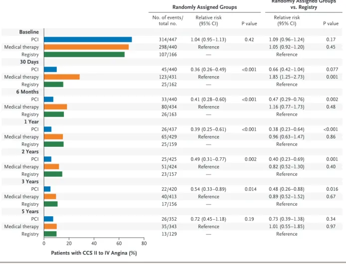
The percentage of patients with angina of CCS grade II, III, or IV was lower among pa tients in the PCI group than among those in the medicaltherapy group at all time points during the first 3 years of followup. However, this difference was no longer significant at 5 years (Fig. 3).
Discussion
This 5year followup of the FAME 2 trial showed that, among patients with stable angina, FFRguided PCI led to a significantly lower rate of the prespecified primary composite end point of death, myocardial infarction, or urgent revas cularization than medical therapy alone. This difference was driven by a significantly lower rate of urgent revascularization in the PCI group than in the medicaltherapy group. Patients in whom all coronary stenoses were hemodynami cally nonsignificant had an event rate with medical therapy alone that did not differ sig nificantly from the rate among patients with hemodynamically significant stenoses who un derwent FFRguided PCI. There was no evidence of convergence of event rates between groups in the long term. Patients who had originally been assigned to undergo FFRguided PCI reported significantly less angina up to 3 years after ran domization than did patients who had been as signed to receive medical therapy alone. How ever, this difference was no longer significant at 5 years, by which time 51% of the patients who had been initially assigned to medical therapy alone had undergone revascularization.
Guidelines recommend that revascularization be considered in patients with stable coronary disease when signs of reversible myocardial ischemia are present.1012 In routine clinical prac
noninvasive functional testing before elective PCI.14 FFR quantifies the impediment of myocar
dial flow with a higher spatial resolution than noninvasive testing and is currently the refer ence standard to guide revascularization. In the FAME 2 trial, patients underwent randomization only if they had at least one hemodynamically significant stenosis (FFR, ≤0.80) in a large artery. In addition, multivessel disease was observed on angiography in almost 45% of the patients who had undergone randomization, and more than 60% of the patients had a hemodynamically significant stenosis in the proximal or middle left anterior descending artery (Table S3 in the Supplementary Appendix). Patients in whom all angiographically significant stenoses were found to be hemodynamically nonsignificant were not included in the randomized trial, given that no benefit regarding the end points was expected in such patients.15
In previous trials comparing PCI with medical therapy in patients with stable coronary artery disease, patients were included mainly on the basis of symptoms and angiography without measurement of FFR.6,7 A sizable proportion of
these patients had no objective signs of reversible ischemia. Such patients would not be expected to benefit from revascularization.
Our results contradict the general belief that abrupt coronary occlusions occur predominantly at sites of mild stenosis and hence that the treat ment of severe lesions may not prevent myocar dial infarction. This belief was also questioned in the PROSPECT (Providing Regional Observa tions to Study Predictors of Events in the Coro nary Tree) study, which showed that the main determinants of future events in stable lesions were a small luminal area and a large plaque burden.16
In the FAME 2 trial, the physicians, who were
Figure 2. Kaplan–Meier Curves for Death from Any Cause, Myocardial Infarction, and Urgent Revascularization.
Hazard ratios below 1.00 denote a lower incidence of events in the PCI group than in the medical-therapy group. The 95% confidence intervals for secondary end points were not adjusted for multiple testing, and any inferences drawn from the intervals as reported may not be reproducible. Insets show the same data on an enlarged y axis. Cumulative Incidence (%) 100 80 90 70 60 40 30 10 50 20 0 0 1 2 3 4 20 15 5 10 0 0 1 2 3 4 5 20 15 5 10 0 0 1 2 3 4 5 30 20 10 0 0 1 2 3 4 5 5 Years since Randomization
B Myocardial Infarction A Death from Any Cause
Hazard ratio, 0.98 (95% CI, 0.55–1.75)
No. at Risk Medical therapy PCI 441447 432439 426431 416422 347360 343352 Medical therapy PCI Cumulative Incidence (%) 100 80 90 70 60 40 30 10 50 20 0 0 1 2 3 4 5
Years since Randomization Hazard ratio, 0.66 (95% CI, 0.43–1.00)
No. at Risk Medical therapy PCI 441447 408421 399410 387399 315340 301328 Medical therapy PCI C Urgent Revascularization Cumulative Incidence (%) 100 80 90 70 60 40 30 10 50 20 0 0 1 2 3 4 5
Years since Randomization Hazard ratio, 0.27 (95% CI, 0.18–0.41)
No. at Risk Medical therapy PCI 441 447 367 424 357 412 345 402 276 344 264 332 Medical therapy PCI
aware of the treatment assignments, might have been more likely to recommend a subsequent PCI procedure for patients in the medicaltherapy group than for those in the PCI group, thus in troducing a risk of bias for the end point of any revascularization. To limit the risk of such bias, the FAME 2 trial included only urgent revascu larizations in the primary end point. Revascular ization was considered to be urgent if a patient was readmitted to the hospital unexpectedly and revascularization was performed during that same admission. The majority of urgent revascu larizations were triggered by worsening angina,
ischemic changes observed on electrocardiog raphy, or myocardial infarction.17 After 5 years,
225 patients (51.0%) who had originally been assigned to receive medical therapy alone had undergone revascularization. Given the high rate of crossover to PCI among patients who had been originally assigned to medical therapy, an intentiontotreat analysis may underestimate the potential benefit of PCI as compared with medi cal therapy with regard to death, myocardial infarction, and severity of angina.
Some limitations must be taken into account. First, enrollment was stopped prematurely by
Figure 3. Angina Class in Patients in the Trial Groups and Registry Cohort over Time.
Shown are the numbers of patients in the two trial groups and the registry cohort who had angina of class II to IV on the Canadian Cardio-vascular Society (CCS) scale (which ranges from I to IV, with higher classes indicating greater limitations on physical activity owing to angina) at various time points. The 95% confidence intervals for secondary end points were not adjusted for multiple testing, and any inferences drawn from the intervals as reported may not be reproducible.
20 40 60 80
Patients with CCS II to IV Angina (%) Baseline PCI Medical therapy Registry 30 Days PCI Medical therapy Registry 6 Months PCI Medical therapy Registry 1 Year PCI Medical therapy Registry 2 Years PCI Medical therapy Registry 3 Years PCI Medical therapy Registry 5 Years PCI Medical therapy Registry No. of events/ total no.
Randomly Assigned Groups Randomly Assigned Groupsvs. Registry
0 P value Relative risk (95% CI) 1.09 (0.96–1.24) 1.05 (0.92–1.20) Reference 0.66 (0.42–1.04) 1.85 (1.25–2.73) Reference 0.47 (0.29–0.76) 1.16 (0.77–1.73) Reference 0.38 (0.23–0.64) 0.96 (0.63–1.47) Reference 0.40 (0.23–0.69) 0.82 (0.52–1.30) Reference 0.48 (0.26–0.88) 0.89 (0.52–1.52) Reference 0.73 (0.39–1.38) 1.01 (0.55–1.85) Reference 0.17 0.45 0.077 0.001 0.002 0.48 <0.001 0.86 0.001 0.40 0.016 0.67 0.34 0.97 314/447 298/440 107/166 45/440 123/431 25/162 33/440 80/434 26/163 26/437 65/429 25/159 25/425 51/424 23/157 22/420 40/413 17/156 26/352 35/343 13/129 P value Relative risk (95% CI) 1.04 (0.95–1.13) Reference — 0.36 (0.26–0.49) Reference — 0.41 (0.28–0.60) Reference — 0.39 (0.25–0.61) Reference — 0.49 (0.31–0.77) Reference — 0.54 (0.33–0.89) Reference — 0.72 (0.45–1.18) Reference — 0.42 <0.001 <0.001 <0.001 0.002 0.014 0.19
the data and safety monitoring board because of a large excess of primary endpoint events in the medicaltherapy group. The early termination of clinical trials has been shown to exaggerate treatment effects.18 Second, the sponsor of the
trial decided to close the trial after completion of the 3year followup. The academic steering committee subsequently invited all 28 sites to participate in an additional 5year followup, but only 19 sites participated. Taken together, these two points resulted in a relatively low number of events with limited statistical precision. Third, patients, physicians, and nurses were aware of the assigned treatment. Even though the blinded adjudication of clinical events may have reduced the risk of detection bias, we cannot rule out that betweengroup differences in clinical man agement biased our results regarding urgent re vascularization. Fourth, in stenoses that were estimated to be less than 50% in diameter, no FFR measurements were performed. A sizable number of stenoses with a 30 to 50% diameter are associated with FFR values below 0.80, espe
cially in proximal segments of large coronary arteries.19,20 Therefore, it is possible that some
stenoses that were deemed to be nonsignificant at angiography (and therefore left untreated) might have been hemodynamically significant.
In conclusion, in patients with stable coro nary artery disease, an initial FFRguided PCI strategy resulted in a sustained clinical benefit, as compared with medical therapy alone, with regard to the composite primary end point of death, myocardial infarction, or urgent revas cularization at 5 years. Patients without hemo dynamically significant stenoses had a favor able longterm outcome with medical therapy alone.
The FAME 2 trial was originally supported by St. Jude Medi cal. The present analysis was supported by research grants from the European Association of Percutaneous Cardiovascular In terventions and the Hellenic Cardiological Society (to Dr. Xa planteris), by a research grant from the Swiss National Science Foundation (to Dr. Fournier), and by a Tier 1 Canada Research Chair in Clinical Epidemiology of Chronic Diseases and the Canada Research Chairs Programme (to Dr. Jüni).
Disclosure forms provided by the authors are available with the full text of this article at NEJM.org.
Appendix
The authors’ full names and academic degrees are as follows: Panagiotis Xaplanteris, M.D., Ph.D., Stephane Fournier, M.D., Nico H.J. Pijls, M.D., Ph.D., William F. Fearon, M.D., Emanuele Barbato, M.D., Ph.D., Pim A.L. Tonino, M.D., Ph.D., Thomas Engstrøm, M.D., Ph.D., Stefan Kääb, M.D., JanHenk Dambrink, M.D., Ph.D., Gilles Rioufol, M.D., Ph.D., Gabor G. Toth, M.D., Zsolt Piroth, M.D., Nils Witt, M.D., Ole Fröbert, M.D., Petr Kala, M.D., Axel Linke, M.D., Nicola Jagic, M.D., Martin Mates, M.D., Kreton Mavromatis, M.D., Habib Samady, M.D., Ph.D., Anand Irimpen, M.D., Keith Oldroyd, M.D., Gianluca Campo, M.D., Martina Rothenbühler, Ph.D., Peter Jüni, M.D., and Bernard De Bruyne, M.D., Ph.D.
The authors’ affiliations are as follows: the Cardiovascular Center Aalst, OnzeLieveVrouw Clinic, Aalst, Belgium (P.X., S.F., E.B., G.G.T., B.D.B.); the Department of Cardiology, Catharina Hospital, and the Department of Biomedical Engineering, Eindhoven Univer sity of Technology, Eindhoven (N.H.J.P., P.A.L.T.), and Isala Klinieken, Zwolle (J.H.D.) — all in the Netherlands; Stanford University Medical Center and Palo Alto Veterans Affairs (VA) Health Care Systems, Stanford, CA (W.F.F.); Rigshospitalet University Hospital, Copenhagen (T.E.); Klinikum der Universität München–Campus–Innenstadt, Munich (S.K.), Heart Center Leipzig, Leipzig (A.L.), and Heart Center Dresden, Dresden (A.L.) — all in Germany; the Cardiovascular Hospital, Lyon, France (G.R.); Gottsegen Hungarian Insti tute of Cardiology, Budapest, Hungary (G.G.T., Z.P.); Karolinska Institutet at Södersjukhuset, Stockholm (N.W.), and Örebro Univer sity Hospital, Örebro (O.F.) — both in Sweden; Masaryk University and University Hospital, Brno (P.K.), and Na Homolce Hospital, Prague (M.M.) — both in the Czech Republic; Clinical Center Kragujevac, Kragujevac, Serbia (N.J.); Atlanta VA Medical Center, Decatur (K.M.), and Emory University School of Medicine, Atlanta (H.S.) — both in Georgia; Tulane University Heart and Vascular Institute, New Orleans (A.I.); Golden Jubilee National Hospital, Glasgow, United Kingdom (K.O.); Cardiology Unit, Azienda Ospedalieria Uni versitaria di Ferrara, Ferrara, and Maria Cecilia Hospital, Gruppo Villa Maria Care and Research, Cotignola — both in Italy (G.C.); Clinical Trials Unit Bern, University of Bern, Bern, Switzerland (M.R.); and the Applied Health Research Centre, Li Ka Shing Knowledge Institute of St. Michael’s Hospital, Department of Medicine and Institute of Health Policy, Management, and Evaluation, University of Toronto, Toronto (P.J.).
References
1. Damman P, Hirsch A, Windhausen F, Tijssen JG, de Winter RJ. 5Year clinical outcomes in the ICTUS (Invasive versus Conservative Treatment in Unstable coro nary Syndromes) trial: a randomized com parison of an early invasive versus selec tive invasive management in patients with nonSTsegment elevation acute coronary syndrome. J Am Coll Cardiol 2010; 55: 858 64.
2. Fox KA, PooleWilson PA, Henderson RA, et al. Interventional versus conserva tive treatment for patients with unstable angina or nonSTelevation myocardial infarction: the British Heart Foundation RITA 3 randomised trial: Randomized In tervention Trial of unstable Angina. Lan cet 2002; 360: 74351.
3. Ibanez B, James S, Agewall S, et al. 2017 ESC Guidelines for the management
of acute myocardial infarction in patients presenting with STsegment elevation: the Task Force for the Management of Acute Myocardial Infarction in Patients Present ing with STSegment Elevation of the Eu ropean Society of Cardiology (ESC). Eur Heart J 2018; 39: 11977.
4. Roffi M, Patrono C, Collet JP, et al. 2015 ESC Guidelines for the management of acute coronary syndromes in patients
presenting without persistent STsegment elevation: Task Force for the Management of Acute Coronary Syndromes in Patients Presenting without Persistent STSegment Elevation of the European Society of Car diology (ESC). Eur Heart J 2016; 37: 267 315.
5. Wallentin L, Lagerqvist B, Husted S, Kontny F, Ståhle E, Swahn E. Outcome at 1 year after an invasive compared with a noninvasive strategy in unstable coronary artery disease: the FRISC II invasive ran domised trial: Fast Revascularization dur ing Instability in Coronary artery disease. Lancet 2000; 356: 916.
6. Boden WE, O’Rourke RA, Teo KK, et al. Optimal medical therapy with or without PCI for stable coronary disease. N Engl J Med 2007; 356: 150316.
7. AlLamee R, Thompson D, Dehbi HM, et al. Percutaneous coronary intervention in stable angina (ORBITA): a double blind, randomised controlled trial. Lancet 2018; 391: 3140.
8. Hachamovitch R, Rozanski A, Shaw LJ, et al. Impact of ischaemia and scar on the therapeutic benefit derived from myo cardial revascularization vs. medical ther apy among patients undergoing stressrest myocardial perfusion scintigraphy. Eur Heart J 2011; 32: 101224.
9. Barbato E, Toth GG, Johnson NP, et al. A prospective natural history study of coronary atherosclerosis using frac tional flow reserve. J Am Coll Cardiol 2016; 68: 224755.
10. Kolh P, Windecker S, Alfonso F, et al.
2014 ESC/EACTS guidelines on myocar dial revascularization: the Task Force on Myocardial Revascularization of the Euro pean Society of Cardiology (ESC) and the European Association for CardioThoracic Surgery (EACTS): developed with the spe cial contribution of the European Associ ation of Percutaneous Cardiovascular In terventions (EAPCI). Eur J Cardiothorac Surg 2014; 46: 51792.
11. Montalescot G, Sechtem U, Achenbach S, et al. 2013 ESC guidelines on the man agement of stable coronary artery disease: the Task Force on the Management of Stable Coronary Artery Disease of the Eu ropean Society of Cardiology. Eur Heart J 2013; 34: 29493003.
12. Patel MR, Calhoon JH, Dehmer GJ, et al. ACC/AATS/AHA/ASE/ASNC/SCAI/SCCT/ STS 2017 appropriate use criteria for cor onary revascularization in patients with stable ischemic heart disease: a report of the American College of Cardiology Ap propriate Use Criteria Task Force, Amer ican Association for Thoracic Surgery, American Heart Association, American Society of Echocardiography, American Society of Nuclear Cardiology, Society for Cardiovascular Angiography and Inter ventions, Society of Cardiovascular Com puted Tomography, and Society of Tho racic Surgeons. J Am Coll Cardiol 2017; 69: 221241.
13. De Bruyne B, Pijls NH, Kalesan B, et al. Fractional flow reserveguided PCI versus medical therapy in stable coronary dis ease. N Engl J Med 2012; 367: 9911001.
14. Lin GA, Dudley RA, Lucas FL, Malenka DJ, Vittinghoff E, Redberg RF. Frequency of stress testing to document ischemia prior to elective percutaneous coronary intervention. JAMA 2008; 300: 176573.
15. Zimmermann FM, Ferrara A, Johnson NP, et al. Deferral vs. performance of per cutaneous coronary intervention of func tionally nonsignificant coronary steno sis: 15year followup of the DEFER trial. Eur Heart J 2015; 36: 31828.
16. Stone GW, Maehara A, Lansky AJ, et al. A prospective naturalhistory study of cor onary atherosclerosis. N Engl J Med 2011; 364: 22635.
17. De Bruyne B, Fearon WF, Pijls NH, et al. Fractional flow reserveguided PCI for stable coronary artery disease. N Engl J Med 2014; 371: 120817.
18. Bassler D, Briel M, Montori VM, et al. Stopping randomized trials early for ben efit and estimation of treatment effects: systematic review and metaregression analysis. JAMA 2010; 303: 11807.
19. Toth G, Hamilos M, Pyxaras S, et al. Evolving concepts of angiogram: frac tional flow reserve discordances in 4000 coronary stenoses. Eur Heart J 2014; 35: 28318.
20. Ciccarelli G, Barbato E, Toth GG, et al. Angiography versus hemodynamics to pre dict the natural history of coronary steno ses: Fractional Flow Reserve Versus Angiog raphy in Multivessel Evaluation 2 substudy. Circulation 2018; 137: 147585.
Copyright © 2018 Massachusetts Medical Society.
receiveimmediatenotificationwhenanarticle ispublishedonlinefirst
To be notified by email when Journal articles are published online first, sign up at NEJM.org.