DOTTORATO DI RICERCA IN BIOTECOLOGIE VEGETALI
XX Ciclo
Identification and characterisation of new members of
pectin methylesterase/invertase inhibitor family in tomato
(Solanum lycopersicum)
(AGR/07-BIO/04)
Coordinatore: Prof. Stefania Masci
Tutor: Prof. Daniela Bellincampi
Dottorando: Ida Barbara Reca Dipartimento di Biologia Vegetale
Tesi in cotutela con/Thèse an co-tutelle avec
L’UNIVERSITE PAUL CEZANNE (AIX-MARSEILLE III)
Identification and characterisation of new members of pectin
methylesterase/invertase inhibitor family in tomato
(Solanum lycopersicum)
En vue d’obtenir le grade deDOCTEUR de L’UNIVERSITE Paul CEZANNE Faculté des Sciences et Techniques
Discipline : Nutrition ; Aspects Moléculaires et Cellulaires Présentée et soutenue publiquement par Mme Ida Barbara RECA
Vendredi 14 Mars
Directeur de thèse : Prof. Daniela Bellincampi Co-Directeur de thèse : Dr. Thierry Giardina
JURY
Pr. Daniela Bellincampi Directeur de thèse
Dr Thierry Giardina Co-Directeur de thèse
Dr. Josette Perrier Examinateur
Pr. Stefania Masci Examinateur
Dr. Nathalie Juge Rapporteur
Pr. Rodolfo Federico Rapporteur
E' difficile in poche righe ricordare tutte le persone che, a vario titolo, hanno contribuito a rendere "migliori" questi anni.
Desidero ringraziare la Pr. Daniela Bellincampi e il Dr Thiery Giardina per la pazienza, la disponibilità, per avermi aiutata nei momenti difficili e per avermi indirizzata sempre verso la strada giusta da intraprendere. Ringrazio la Dr Laura Camardella per la collaborazione e la simpatia tipica napoletana. Vorrei inoltre ringraziare il Prof. Felice Cervone e la Prof Giulia De Lorenzo, il Prof. Renato D’ovidio e la Prof. Stefania Masci per la loro gentilezza e disponibilità. Grazie a Gianni per il prezioso supporto tecnico e a Vera per le lezioni di stile.
Un grazie particolare va a Lucia, Fedra e Flavio, insostituibili compagni con cui ho condiviso gioie e dolori di questo periodo così impegnativo della mia vita. Vorrei ringraziare le compagne di bancone Roberta e Danielina per i consigli e il supporto morale. Ringrazio Lorenzo Manuel e Daniel per le discussioni, costruttive e distruttive su tecniche e attitudini del laboratorio. Un ringraziamento va ad Enzo collega e conterraneo, per la simpatia e per i valori che ci accomunano. Grazie a Francesca per la gentilezza e la simpatia. Vorrei ringraziare anche Alexandra e Andrea, per la collaborazione e tutti i ragazzi di Viterbo per la loro amicizia e disponibilità. Ringrazio in modo particolare Sameh e Sophie indimenticabili amiche francesi, per avermi accompagnata con affetto ed entusiasmo in quella che è stata un’esperienza per me indimenticabile.
Un grandissimo ringraziamento va a mio marito, Alexandre, per essermi stato sempre vicino con amore in questi anni, per avermi sopportata anche nei momenti di gran difficoltà e per avermi convinto a non gettare mai la spugna.
Ringrazio mio padre che, fin da piccola, ha permesso che m’innamorassi delle scienze, mia madre che mi ha insegnato come portare i pantaloni senza dimenticare di essere una donna. Sono grata a nonna Grazia e Zia Nettina, per aver sempre creduto in me e per tutte le volte che mi hanno cucinato le orecchiette. Ringrazio mio fratello Ciro mia cognata Federica e Giovannino, per aver ricreato qui a Roma quell’insostituibile ambiente familiare che mi ha dato la forza di andare avanti. Ringrazio inolre Regine, Faustine e tutta la famiglia francese per avermi fatta sentire sempre parte della “famille”.
Grazie a tutti Barbara
Riassunto
La pectina metilesterasi (PME) e l’invertasi (INV) sono due enzimi chiave del metabolismo dei caboidraiti nelle piante. Oltre al controllo a livello trascrizionale, un importante meccanismo di regolazione dell’attività di questi enzimi è svolto da specifici inibitori proteici. Gli inibitori di invertasi (INH) e di pectina metilesterasi (PMEI) sono degli inibitori proteici raggruppati nella famiglia Pf 04043 in quanto condividono numerose proprietà strutturali. Nonostante queste similitudini strutturali, i diversi inibitori interagiscono in modo specifico con enzimi funzionalmente e strutturalmente diversi. Entrambi gli inhibitori non agiscono su enzimi prodotti da patogeni, indicando un ruolo prevalente nei processi di sviluppo, anche se sembrano partecipare nella difesa delle piante da patogeni.
In questa tesi, attraverso un approccio di genomica funzionale, un inibitore di PME (SolyPMEI) e un inibitore di INV (SolyCIF) sono stati identificati e caratterizzati in pomodoro (Solanum lycopersicum).
SolyPMEI ha mostrato essere molto espresso in frutto rosso e, a bassi livelli, anche in fiori e
polline. Il cDNA del SolyPMEI è stato clonato ed espresso in Pichia pastoris. Tutti i tentativi di produrre una proteina ricombinante attiva sono stati vani sebbene, l’analisi CD e dei ponti dusilfuro, rivelassero un corretto ripiegamento della proteina in vitro. Al fine di purificare l’inibitore naturale da bacca di pomodoro, è stata realizzata una colonna d’immuno-affinità. L’eluizione sia dell’inibitore sia della PME-1 di pomodoro, indica che in vivo, SolyPMEI è coinvolto nella formazione di un complesso stabile con le PME endogene.
SolyCIF è una proteina apoplastica espressa nelle foglie, nei fiori e nei frutti verdi. Il cDNA del
SolyCIF è stato clonato ed espresso in Pichia pastoris e la proteina ricombinante, è stata purificata e
caratterizzata biochimicamente L’invertasi di pomodoro era fortemente inibita in maniera dose dipendente dal SolyCIF. Il partner naturale dell’inibitore (l’invertasi vacuolare TIV-1) è stato purificato con una colonna di affinità e caratterizzato biochimicamente. TIV-1 mostrava diversi siti di proteolisi naturali. I frammenti che compongono TIV-1 interagiscono tra loro permettendo all’enzima di funzionare. Il sequenziamento N-terminale dei frammenti e analisi di modeling di TIV-1 mostrano che il sito attivo del’enzima è diviso in due, confermando che l’idrolisi del saccarosio avviene solo quando i frammenti sono associati tra loro.
Abstract
Pectin methylesterase (PME) and invertase (INV) are key enzymes in plant carbohydrate metabolism. An important post-transcriptional mechanism of regulation of these enzymes is represented by proteinaceous inhibitors. PME inhibitors (PMEI) and INV inhibitors (INH), belong to the same structural family Pf 04043. Despite the structural similarity, these two inhibitors act on two structurally and functionally different enzymes, but never both. Both inhibitors do not inhibit fungal enzymes indicating that they are mainly involved in growth and development even if they seems to be also involved in defence against pathogens.
In this thesis through a functional genomics approach a pectin methylesterase inhibitor (SolyPMEI) and an invertase inhibitor (SolyCIF) of Solanum lycopersicum have been identified and characterised.
SolyPMEI was mainly expressed in red fruits and, albeit at lower levels, in flowers and pollen. The SolyPMEI cDNA was cloned and expressed in Pichia pastoris. All attempts to produce active
recombinant SolyPMEI protein appeared to be unsuccessful, neverthless CD spectra and disulfide bridges indicated a correct folding of the protein in vitro. The immuno-affinity fishing approach was used to purify the natural SolyPMEI from tomato fruits. The isolation of both natural SolyPMEI and PME-1 indicated that SolyPMEIs is in vivo engaged in the formation of a stable complex with endogenous PMEs.
SolyCIF was mainly expressed in leaves, flowers and green fruits of the plant and localized in the cell wall compartment. The SolyCIF cDNA was cloned and expressed in Pichia pastoris. The purified recombinant protein was biochemically characterized. The invertase activity was strongly inhibited in a dose-dependent manner by recombinant SolyCIF.With an affinity chromatography approach, the natural ligand of the inhibitor (the vacuolar invertase namely TIV-1) was purified and characterised. TIV-1 has been shown to be naturally proteolyzed and it was established that the fragments produced have to be tightly associated for its enzymatic activity to occur. N- terminal sequencing of fragments and the molecular model of TIV-1 shows that the fragmentation splits the catalytic site of the enzyme into two halves, which confirms that the enzymatic activity is possible only when the fragments are tightly associated.
SUMMARY
ABREVIATIONS 13
FIGURES 15
TABLES 16
1. INTRODUCTION
17
1.1. Sugars: energy molecules and cell wall components
19
1.2. Plant proteinaceous inhibitors directed against exogenous
carbohydrate-active
enzymes
(CAE)
20
1.2.1. Xyloglucan endoglucanase inhibitor protein (XEGIPs) 20
1.2.2. Xylanase Inhibitors 20
1.2.3. Inhibitors of pectic enzymes 22
1.2.4. Pectin lyase inhibitor protein 22
1.2.5. Polygalacturonase-inhibitor proteins (PGIPs) 22
1.3. Plant proteinaceous inhibitors directed against endogenous
carbohydrate-active
enzymes
(CAE)
24
1.3.1. Pectin methylesterase/invertase inhibitor family (PMEI/INH family)
Pfam 04043 24
1.3.2.
The
Pectin
methylesterase
(PME)
25
1.3.2.1. Biochemical features and types of plant PMEs 25
1.3.2.2. The role of the PRE-PRO domain in PME targeting and function 26
1.3.2.3. Three-dimensional structures of PMEs 28
1.3.2.4. Mode of action of pectin methylesterase 30
1.3.2.5. Multiple roles of PMEs in plants 32
1.3.2.6. Role of PMEs in plant growth 32
1.3.2.7. PMEs and tomato fruit ripening 33
1.3.2.8. PMEs and plant defence 35
1.3.2.9. PME activity regulation 36
1.3.3.
Pectin
methylesterase
inhibitors
(PMEIs)
37
1.3.3.1. Proteinaceous inhibitor of PME (PMEI) 37
1.3.3.4. PMEIs expression and physiological roles 42
1.3.4. PME and PMEI biotechnological application
44
1.4.Invertase
45
1.4.1 Cytosolic invertases 46
1.4.2. Acid invertases 47
1.4.3. Cell wall invertase tree-dimensional structure 48
1.4.4. Acid invertase and plant development 50
1.4.5. Vacuolar invertase in tomato fruit ripening 51
1.4.6. Expression pattern and regulation of acid invertases 51
1.5. Regulation of invertases by proteinaceous inhibitors
52
1.5.1. Molecular structure of NtCIF 54
1.5.2. Expression pattern and physiological functions of invertase inhibitors 55
1.6. Biotechnological approaches using invertase and invertase inhibitors
56
1.7. Aim of the work
57
2.
MATHERIALS
AND
METHODS
59
2.1.
Microbiological
techniques
61
2.1.1. Escherichia coli and Pichia pastoris 61
2.1.2. Media and antibiotics 61
2.1.3. Preparation of electrocompentent E. coli, P pastoris and
Agrobacterium tumefaciens cells and transformation by électroporation 62
2.1.4. Agrobacterium- mediated transformation of Nicotiana tabacum leaves 62
2.2.
Molecular
biology
techniques
62
2.2.1. Agarose gels 62
2.2.3. Oligonucleotides 63
2.2.4. Isolation and determination concentration of total RNA 63
2.2.5. Real-time RT-PCR 64
2.2.6. 3’-RACE technique 64
2.2.7. Isolation of plant genomic DNA 64
2.3.
Cloning
procedures 65
2.3.1. Gel extraction and PCR purification 65
2.3.2. Cloning using restriction digestion 65
2.3.4. Cloning and expression of SolyPMEI and SolyCIF in Pichia pastoris 66
2.3.5. Site-directed mutagenesis of SolyPMEI 66
2.4.
Biochemical
techniques
66
2.4.1. SDS-PAGE 66
2.4.2. Coomassie staining 67
2.4.3. Silver staining 67
2.4.4. Periodic acid-Schiff staining 67
2.4.5. Western Blot 67
2.4.6. Molecular mass determination and N-terminal sequencing 68
2.4.7. Circular dichroism measurements 68
2.4.8. Protein alkylation 68
2.4.9. Mass spectrometry 68
2.5.
Protein
purification
69
2.5.1. Purification of recombinant from Pichia pastoris 69
2.5.2. Affinity purification of pSolyN101YPMEI antisera 69
2.5.3. CnBr-pSolyN101YPMEI affnity- chromatography 70
2.5.4. Partial purification and immuno affinity column of SolyPMEI 70
2.5.5. Purification of invertase from tomato (TIV-1) 71
2.6. Protein assay
71
2.6.1. Protein concentrations 71
2.6.2. Invertase activity determination and inhibition assay 71
2.6.3. PME activity determination and inhibition assay 72
2.7.
Bioinformatic
analysis
73
2.7.1. Sequence alignments and phylogenetic tree 73
2.7.2. Modeling analisys 73
2.7.3. GFP imaging 73
3.
RESULTS
AND
DISCUSSION
75
3.1. Identification of putative SolyINH/PMEI members in
Solanum lycopersicum
77
3.2. Pectin Methylestersase Inhibitor from tomato Solanum lycopercicum
3.2.1. Identification, complete gene sequencing and expression of the SolyPMEI 79
3.2.2. Heterologous expression of recombinant pSoly-PMEI, purification and
biochemical characterization 81
3.2.3. Isolation from tomato red fruit of the natural pSoly-PMEI in complex with the
partner 87
3.3. Invertase inhibitor from Solanum Lycopercum
(SolyCIF)
91
3.3.1. Identification, expression and subcellular localization of the SolyCIF 91
3.3.2. Heterologous expression of recombinant SolyCIF, purification and functional characterization 94
3.3.3. Isolation of a vacuolar tomato invertase interacting with SolyCIF 97
3.3.3. Molecular modeling of tomato vacuolar invertase 100
3.3.4. Enzymatic properties of TIV-1 and enzyme inhibition by recombinant SolyCIF 100
4. CONCLUSIONS 103
ABREVIATIONS
Amp: Ampicilline
AOX1: Alcool oxydase 1
ATP: Adenosin triphosphate
bp : Base pair
CIF: Cell wall Inhibitor of beta fructosidase
DNA: Desoxy ribonucleic Acid
DNS: 3-5 dinitrosalicylic acid
dNTP: Desoxynucleotid triphosphate
DTT: Dithiothreitol
EDTA: Ethylen diamine tetraacetic acid
GFP: Green Fluorescent Protein
GH: Glycosyl-hydrolase
GUS. β-Glucuronidase
HG: Homogalacturonan
INH: Invertase inhibitor
INV: Invertase
kDa : kiloDalton
LB : Luria Bertani
LRR: Leucin rich repeat
PAGE: Poly-acrylamide gel electrophoresis
PCR: Polymerisation chain reaction
PG: Polygalacturonase
PGIP: Polygalacturonase inhibitor protein
PME: Pectin methylesterase
PMEI: Pectin methylesterase inhibitor protein
PNLIP: Pectate lyase inhibitor protein
PNLP: Pectate lyase
PR: Pathogenesis related (protein)
SDS: Sodium dodecylsulfate
Soly: Solanum lycopersicon
SuSy: Sucrose synthase
Taq: Polymerase from Thermus aquaticus
TAXI : Triticum aestivum xylanase inhibitor
TLXI: Thaumatin like xylanase inhibitor
Tris: Tris-(hydroxyméthyl) aminomethane
VIF: Vacular Inhibitor of beta fructosidase
XEGIP: Xylo-Endoglucanase inhibitor protein
XIP: Xylanase inhibitor protein
FIGURES
Figure 1. Schematic representation of plant cell wall
Figure 2. Pectin methylesterase/invertase inhibitor family (PMEI/INH) Pfam 04043 3D-structures
Figure 3. Demethylesterification of pectins by pectin methylesterases (PME)
Figure 4. Pectin methylesterase (PME) structural motifs
Figure 5. Hypothesis for pectin methylesterase (PME) excretion into the apoplasm and maturation of the protein.
Figure 6. Comparison of the known structures of PMEs.
Figure 7. PMEs 3D structure
Figure 8. Stereo-view of the active site residues in the Michaelis complex.
Figure 9. Close-Up View of the Tomato PME Active Site
Figure 10. Modes of action of pectin methylesterases (PMEs). Figure 11. Phenotypes of detached transgenic PME
Figure 12. Amino acid sequence alignment of AtPMEI-1 and AtPMEI-2 with AcPMEI Figure 13. PMEIrp alignment
Figure 14. Sequence alignment of PMEI with PME pro-peptides sequence numbering refers to
PMEI
Figure 15. AtPMEI1 3D structure
Figure 16. Structure of the PME-PMEI complex
Figure 17. Analysis of flower-specific expression of AtPMEI1 and 2,using promoter: :GUS
fusions
Figure 18. Sucrose cleavage in plants by invertase and sucrose synthases Figure 19. Schematic comparison of cell wall invertase and vacuolar invertase. Figure 20. Arabidopsis thaliana invertase.
Figure 21. Invertase active site
Figure 22. NtCIF and AcPMEI 3D structure
Figure 23. Sequence comparison of PMEIs form Kiwi, PMEIs from Arabidopsis, and invertase
inhibitor from tobacco
Figure 24. Dendrogramof 28 protein sequences belonging to the PMEI-INH family Figure 25. Multiple alignment of SolyPMEI and known PMEIs.
Figure 26. SolyPMEI structural modeling Figure 27. Real-time PCR of SolyPMEI mRNA.
Figure 28. PME specific activity in different tomato tissues Figure 29. E. coli growth rate
Figure 30. SolyPMEI purification: Figure 31. Agar diffusion assay :
Figure 32. SDS-PAGE performed in reducing and not reducing conditions. Figure 33. SolyPMEI glycosylation analysis.
Figure 34. SolyPMEI model in complex with tomato PME-1 Figure 35. Asn101 site-directed mutagenesis
Figure 36. Circular Dichroism analysis
Figure 37. Gel diffusion assay of fraction eluted after affinity chromatography
Figure 38. Western blot analysis on total protein extract from different tomato tissues with
purified polyclonal antibodies generated against N101Y.
Figure 39. SDS-PAGE analysis of fractions elueted from after immuno-affinity chromatography Figure 40. SDS-PAGE analysis after immuno affinity column.
Figure 42. Gel filtration
Figure 43. Amino acid sequence alignment of SolyCIF and other invertase inhibitors Figure 44. Nucleotide and deduced amino acid sequences of the SolyCIF gene.
Figure 45. Real-time PCR of SolyCIF mRNA and invertase activity.
Figure 46. Localization of transiently expressed SolyCIF-GFP and PvPGIP2-GFP in tobacco
leaves under the control of the CaMV 35S promoter.
Figure 47. SDS-PAGE of P. pastoris culture filtrate
Figure 48. SDS-PAGE performed in reducing and not reducing conditions
Figure 49. Inhibitory effect of recombinant SolyCIF on tomato vacuolar invertase. Figure 50. Electrophoresis analysis of TIV-1 purified with affinity chromatography Figure 50. Analysis of the tomato vacuolar invertase fragments
Figure52. Tomato vacuolar invertase molecular model.
TABLES
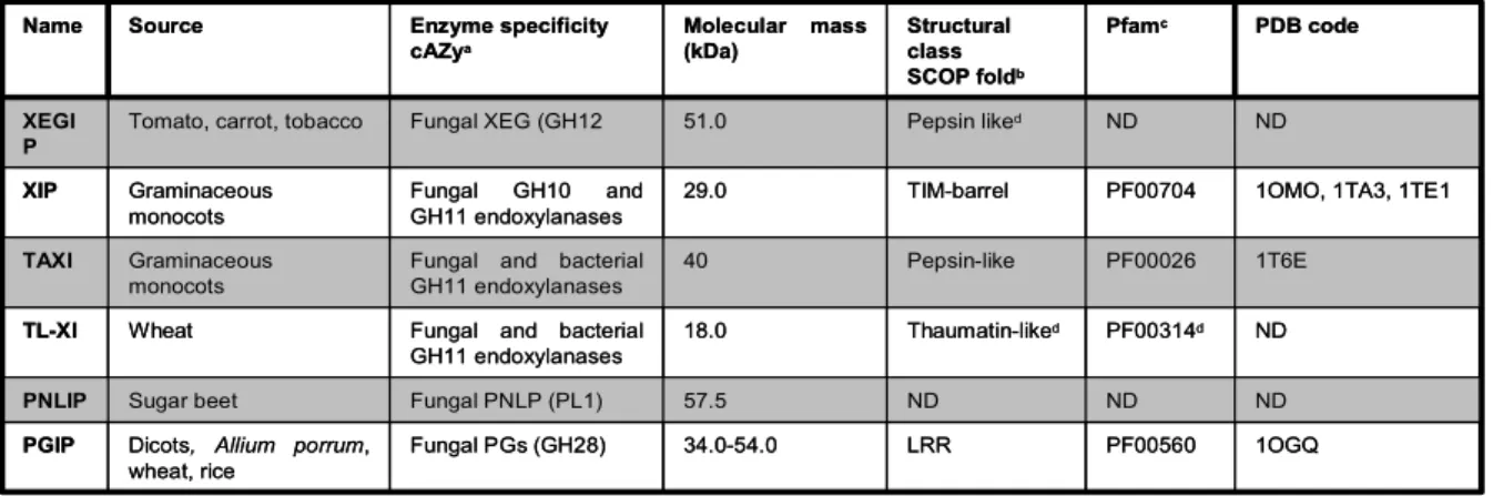
Table 1. Different classes of inhibitors directed against carbohydrate-active enzymes
Table 2. Oligonucleotides sequence in 5' to 3' direction
1.1. Sugars: energy molecules and cell wall components
Carbohydrates play fundamental roles in plants. They are the principal components of the cell wall as well as energy molecules which, depending on the physiological needs, can be channeled into various pathways in different subcellular compartments for their utilization as source of carbon and energy or converted in polymers for long-term storage. Sugars can also act as signalling molecules whose transduction pathways control gene expression and influence plant growth and development as well as defence responses against pathogens (Côté and Hahn, 1994; Smeekens, 2000; Loreti et al., 2002).
Cell wall is a dynamic interface that changes during the growth and differentiation of the cell and that also participates in cellular responses to exogenous stimuli. Primary cell wall is essentially constituted by cellulose, hemicellulose and pectin. Primary cell wall (Fig.1) is formed during cell division and is continuously modified during cell extension. The middle lamella constitutes the interface between the primary walls of neighbouring cells (Fig. 1).
Figure 1. Schematic representation of plant cell wall
Plants produce a number of carbohydrate-active enzymes including glycosidases, transglycosidases, glycosyltransferases, polysaccharide lyases and carbohydrate esterases, which activity and regulation have been a main focus in plant biology. Carbohydrate-active enzymes are also produced by microbial pathogens at the early stages of infection to invade plant tissue and sustain their growth and are important factors of pathogenicity (D'Ovidio et al., 2004a). Plants
synthesize different classes of proteinaceous inhibitors against endogenous carbohydrate-active enzymes as well as against the enzymes produced by pathogens.
1.2. Plant proteinaceous inhibitors directed against exogenous
carbohydrate-active enzymes (CAE)
1.2.1. Xyloglucan endoglucanase inhibitor protein (XEGIPs)
Xyloglucan is the quantitatively predominant hemicellulosic polysaccharide in the primary walls of dicots and non-graminaceous monocots. Xyloglucans consist of a β-1,4-linked D-glucose backbone substituted by D-xylose. Xyloglucans interact with cellulose microfibrils by forming hydrogen bonds, thus contributing to the structural integrity of the cellulose network. Endoglucanases (EGs) hydrolyse both xyloglucan and cellulose or carboxymethyl cellulose (CMC) (Jahr et al., 2000; Lev and Horwitz, 2003). A novel class of EGs, the xyloglucan-specific endoglucanases (XEGs) has been discovered whose specific activity toward xyloglucan exceeds the activity toward CMC and/or linear β-glucan by at least ten times (Grishutin et al., 2004). All known XEGs belong to the GH12 and GH74 families (Coutinho and Henrissat, 1999) but information on their properties is scarce (Grishutin et al., 2004).
It has been recently purified from suspension-cultured tomato cells a protein that binds an inhibits fungal GH12 XEGsv (Qin et al., 2003). This protein, termed XEG inhibitor protein (XEGIP), represents the newest class of plant-derived proteins that inhibit carbohydrate-active enzymes. XEGIP is widely expressed in tomato vegetative tissues and is down regulated during ripening. Genes that are homologous to tomato XEGIPs are present in soybean, lotus, carrot, cotton, maize, rice, sorghum, Medicago trunculata and Arabidopsis. XEGIPs and related proteins do not show any capacity to inhibit other carbohydrate-active enzymes of either plant or fungal origin (Qin et al., 2003). Today, XEGIP is the only protein inhibitor of EGs, including the cellulases, to have been reported and it remains to be established whether the ability to interact with XEG is a strict specificity of XEGIP-related proteins.
1.2.2. Xylanase Inhibitors
The major hemicellulose polymer in cereals and hardwood is xylan. Xylan consists of a β-1,4-linked D-xylose backbone and can be substituted by different side groups (Zuppini et al., 2005). Endo-β-1,4-xylanase (EC 3.2.1.8, xylanase) is a crucial component that carries out the initial breakdown of the xylan backbone into smaller oligosaccharides. Endoxylanases are classified into six GH different families (GH5, GH7, GH8, GH10, GH11 and GH43). All the family include plant
enzyme with the exception of GH11 family which is exclusively composed by microbial xylanases. The first direct evidence for a role of xylanase in pathogenesis was recently obtained by deleting the gene encoding an endoxylanase from the fungal plant pathogen B. cinerea, which severely affected virulence by delaying the appearance of secondary lesions and significantly reducing the average lesion size on tomato leaves and grape berries (Brito et al., 2006). Reintroducing the wild-type gene into the mutant strains the altered phenotype reversed back to wild type (Brito et al., 2006).
Three structurally different classes of endoxylanase inhibitors have been documented in the literature, the TAXI-type (Gebruers et al., 2004), the XIP-type (Juge et al., 2004) and the thaumatin-like (TL-XI) xylanase inhibitor. The two first inhibitors are widely represented in cereals (rye, barley, maize, rice, durum and bread wheat) as multi isoform families (Goesaert et al., 2004). The third type of inhibitor, TL-XI, was recently identified in wheat (Fierens et al., 2007). These are specific inhibitors of microbial (bacterial and fungal) endoxylanases and do not inhibit plant endoxylanases. XIP-type inhibits both GH10 and GH11 microbial endoxylanases (Flatman et al., 2002; Brutus et al., 2005; Furniss et al., 2005) whereas TAXI-type and TL-XI exclusively inhibit GH11 endoxylanases (Gebruers et al., 2001; Fries et al., 2007). Sequence and structural similarities indicate that XIP-I is related to chitinases in the GH18 family, despite its lack of enzymatic activity (Elliott et al., 2002; Payan et al., 2003).
XIP-I in complex with GH10 Aspergillus nidulans (PDB code 1TA3) and GH11 P.
funiculosum (PDB code 1TE1) endoxylanases showed the presence of two independent
enzyme-binding sites. The strategy for inhibiting endoxylanases involves substrate-mimicking contacts and interactions occluding the active site (Tahir et al., 2002; Payan et al., 2004).
The structures of TAXI-I, free or in complex with GH11 endoxylanase, were also recently determined (Sansen et al., 2004). TAXI-I is highly similar to the core structure of Rhizomucor
miehei aspartic proteinase but is proteolytically non-functional; the inhibitor probably divergently
evolved at an early stage from a pepsin-like aspartic protease ancestor (Sansen et al., 2004).
The structure of TL-XI has not been determined. Like other PR proteins, thaumatin is predicted to have a mainly β structure, with a high content of β-turns and little helix (Fierens et al., 2007).
Although a direct role remains to be demonstrated, increasing lines of evidence suggest that endoxylanase inhibitors are part of the network of plant defence against pathogens. They inhibit the activity of GH11 endoxylanases secreted by pathogens such as B. cinerea (Brutus et al., 2005) and
1.2.3. Inhibitors of pectic enzymes
Pectins are highly heterogeneous polysaccharides whose backbones are highly enriched in α-1,4-linked-D-galacturonic acid residues, with various degrees of methyl esterification. Their structure is continuously modified by pectic enzymes such as pectin lyases (PL), polygalacturonases (PGs), pectin methylesterases (PME). In healthy plants, pectic enzymes participate in the remodelling of the cell wall during plant growth and development. At the early stages of plant infection, pectic enzymes are the first cell wall degrading enzymes produced by fungal and bacterial pathogens and have been demonstrated to be important virulence factors (D'Ovidio et al., 2004a). A number of proteinaceous inhibitors against pectic enzymes have been discovered in plants.
1.2.4. Pectin lyase inhibitor protein
The first evidence of a proteinaceous inhibitor of fungal pectin lyase was obtained in extract of been and cucumber (Bock et al., 1975). Pectin lyases (PNLs; EC 4.2.2.10), catalyse β-elimination cleavage of methylesterified polygalacturonic acid in pectin, the methyl ester, rather than pectate, the anion which is the preferred substrate of pectate lyases, (PeLs; EC 4.2.2.2). Pectin lyases belong to the family 1 of polysaccharide lyases, of which 18 families are known (Henrissat et al., 1995). Although pectate lyases produced by fungal and bacterial pathogens have been shown to be important factors of virulence (Akimitsu et al., 2004), little information is available on the role of PNLs during pathogenicity, apart from a study reporting PNL activity in mallow leaves during the biotrophic and necrotrophic phases of infection (Wei et al., 2002). Sugar beet produces a cell wall PNLIP that inhibits a PNL from Rhizoctonia solani, which is a fungal pathogen of sugar beet (Bugbee, 1993). The presence of the PNLIP has been shown to correlate with resistance to root rot caused by R. solani (Bugbee, 1993). The inhibitor, which the mode of inhibition was uncompetitive, is also active against other fungal PNLs from Phoma betae and Aspergillus japonicus (Bugbee, 1993). No investigations have yet be performed on the inhibitor structure, and its physiological role.
1.2.5. Polygalacturonase-inhibitor proteins (PGIPs)
Polygalacturonases (PG) are the first enzymes to be secreted by pathogens when they encounter plant cell walls and their contribution to the pathogenicity of same fungi and bacteria has been assessed. PGs (EC 3.2.1.15), cleave the linkages between D-galacturonic acid residues in non-methylated homogalacturonan, a major component of pectins. Based on their sequences and their structurally related catalytic folding, PGs have been classified in family 28 of glycoside hydrolases, GH28 (Henrissat and Davies, 1997). To accommodate pathogenesis a variety of different conditions, PGs are highly variable in term of primary structure, specific activity, pH optimum and
mode of action (D'Ovidio et al., 2004b). To counteract the action of PGs, plants have evolved cell wall associated polygalacturonase inhibiting proteins (PGIPs) displaying a high degree of polymorphism. PGIPs are organized into multigene families, clustered in specific chromosomal regions, and encode PGIP isoforms with homologous structure but different specificities (De Lorenzo et al., 2001; D'Ovidio et al., 2004b). PGIPs are typically effective against fungal PGs and ineffective against other pectic enzymes of either bacterial or plant origin. Thibition can be either competitive, non-competitive or mixed, a further indication of the versatility of PGIPs to evolve different recognition specificities (Sicilia et al., 2005).
PGIPs belong to the leucine-rich repeat (LRR) superfamily of proteins whose structure is conserved in many disease resistance genes (R-genes) (Bergelson et al., 2001) and is characteristic of protein–protein interactions (Shanmugam, 2005).
The role of PGIPs as important players in the defence response is now clear: they are induced early during infection, retard PG function, prevent cell wall degradation, and limit fungal growth and colonization. Long-chain oligogalacturonides (OGs), which elicit many defence responses in plants, are produced when fungal PGs activity is controlled by PGIP. For this reason PGIPs are included among the microbe-detecting molecules that are part of the plant immune system (Federici et al., 2006).
Table 1. Different classes of inhibitors directed against carbohydrate-active enzymes (Juge, 2006).
Abbreviation: ND, not determined
a Carbohydrate Active Enzyme database (http://www.cazy.org/CAZY) b Structural Classification of Proteins (http://www.scop.berkeleiy.edu) c Pfam protein families database (http://www.sanger.ac.uk/Sotftware.Pfam/ ) d Based on sequence and homology modelling
1OGQ PF00560
LRR 34.0-54.0
Fungal PGs (GH28) Dicots, Allium porrum,
wheat, rice PGIP ND ND ND 57.5 Fungal PNLP (PL1) Sugar beet PNLIP ND PF00314d Thaumatin-liked 18.0 Fungal and bacterial GH11 endoxylanases Wheat TL-XI 1T6E PF00026 Pepsin-like 40 Fungal and bacterial GH11 endoxylanases Graminaceous
monocots
TAXI
1OMO, 1TA3, 1TE1 PF00704 TIM-barrel 29.0 Fungal GH10 and GH11 endoxylanases Graminaceous monocots XIP ND ND Pepsin liked 51.0 Fungal XEG (GH12
Tomato, carrot, tobacco
XEGI P PDB code Pfamc Structural class SCOP foldb Molecular mass (kDa) Enzyme specificity cAZya Source Name 1OGQ PF00560 LRR 34.0-54.0 Fungal PGs (GH28) Dicots, Allium porrum,
wheat, rice PGIP ND ND ND 57.5 Fungal PNLP (PL1) Sugar beet PNLIP ND PF00314d Thaumatin-liked 18.0 Fungal and bacterial GH11 endoxylanases Wheat TL-XI 1T6E PF00026 Pepsin-like 40 Fungal and bacterial GH11 endoxylanases Graminaceous
monocots
TAXI
1OMO, 1TA3, 1TE1 PF00704 TIM-barrel 29.0 Fungal GH10 and GH11 endoxylanases Graminaceous monocots XIP ND ND Pepsin liked 51.0 Fungal XEG (GH12
Tomato, carrot, tobacco
XEGI P PDB code Pfamc Structural class SCOP foldb Molecular mass (kDa) Enzyme specificity cAZya Source Name
B
kiwi A. thaliana tobacco
1.3. Plant proteinaceous inhibitors directed against endogenous
carbohydrate-active enzymes (CAE)
1.3.1. Pectin methylesterase/invertase inhibitor family (PMEI/INH family) Pfam 04043
Recent plant genome sequencing projects have identified a novel structural protein family Pfam04043 (http://pfam.wustl.edu/), formed by invertase inhibitors (INH) and the pectin methylesterase inhibitors (PMEI), the first involved in the carbohydrate metabolism and sugar signalling (Rausch and Greiner, 2004) and the latter involved in the control of pectin metabolism. Common features of this plant-specific family include an N-terminal signal peptide, four conserved cysteine residues involved in the formation of two disulfide bridges and a molecular mass of about 18 kDa. The determination of their three dimensional structures also shown that they are both folded in an asymmetric up-and-down four-helix bundle (Hothorn et al., 2004a; Di Matteo et al., 2005).
Figure 2. Pectin methylesterase/invertase inhibitor family (PMEI/INH) Pfam 04043 3D-structures. (A)
PMEI from kiwi. (B) PMEI from Arabidopsis; (C ) CIF from Nicothiana tabacum; disulfide bridges are in pink (Hothorn et al., 2004a; Hothorn et al., 2004b; Di Matteo et al., 2005).
Despite this structural similarity these two inhibitors act on two structurally and functionally different carbohydrate modifying enzymes, but never both.
A B C
1.3.2. Pectin methylesterase (PME)
1.3.2.1. Biochemical features and types of plant PMEs
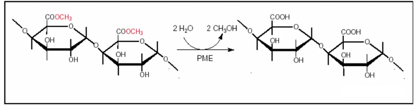
Pectic polysaccharides are synthesized in the Golgi apparatus and secreted into the wall as highly methylesterified forms. Subsequently, they can then be modified by pectinases such as pectin methylesterases (PMEs, EC 3.1.1.11), which catalyse the demethylesterification of homogalacturonans (HGA), the main pectic cell wall component, releasing acidic pectins, methanol and proton (Fig. 3) (Moustacas et al., 1991).
Figure 3. Demethylesterification of pectins by pectin methylesterases (PME) (Micheli et al., 2000).
Several PME isoforms differing in molecular weight, pI and biochemical activity were detected in all higher plants examined so far as well as in a number of plant pathogenic fungi and bacteria (Collmer and Keen, 1986; Brady, 1987). Pectin methylesterase, belong to class 8 (CE-8) of the carbohydrate esterase (CAZy, http://www.cazy.org/fam/CE8.html)(Henrissat, 1991). Plants PMEs belong to large multigene families. For instance, in Arabidopsis thaliana, 66 ORFs have been annotated as putative full-length PMEs. The PME ORFs of Populus trichocarpa are 89 (http://genome.jgi-psf.org/Poptr1_1/Poptr1_1.home.html; (Geisler-Lee et al., 2006) whereas those numbers is substantially lower (35 ORFs) in Oryza sativa (http://www.tigr.org/tdb/e2k1/osa1/), probably due to the low level of pectins in their cell wall.
Higher plant PMEs are frequently organised in pre-pro-proteins. The PRE domain leading to the export of PMEs to the cell wall, is formed by a common type signal peptide (SP) and by a transmembrane domain (TM or signal anchor). Different PMEs possess one, both, or neither of these motifs. Those with neither motif are classed as putatively soluble isoforms. The mature, active part of the protein (PME domain, Pfam01095, IPR000070) is preceded by an N-terminal extension (PRO region Pfam04043, IPR006501) that share similarities with the pectin methylesterase inhibitors. The PRO-regions can vary in length and shows a relatively low level of amino acids
PME domain tends to be basic whereas that of the PRO region is neutral or acidic (Bosch et al., 2005; Bosch and Hepler, 2005). Similar differences in length and isoelectric points between PRO and PME domains have also been found in several other species (Al Qsous et al., 2004; Bosch et al., 2005), and might influence the optimal pH activities of specific isoforms.
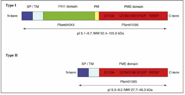
PMEs can be classified on the presence or absence of the PRO domain in PMEs type I (500– 900 amino acids; 52–105 kDa) which contain 1–3 PRO domains and two or tree introns (Fig. 4), or PMEs type II (250 to 400 amino acids; 27–45 kDa) without PRO domain and with five or six introns (Fig. 4) (Micheli, 2001; Tian et al., 2006). The type II sequences have a structure close to that of the PMEs identified in phytopatogenic organisms (bacteria, fungi).
Figure 4. Pectin methylesterase (PME) structural motifs. Type I and Type II PMEs possess a conserved PME
domain (Pfam01095) with characteristic, highly conserved amino acid fragments. Type I PMEs possess an N-terminal extension designated the PRO domain and a processing motif (PM) that might be a putative target for subtilisin –like proteases.The targeting to the endomembrane system leading to the export of PMEs to the cell wall is mediated either by a signal peptide (SP) or a transmembrane domain (TM or signal anchor). Different PMEs possess one, both, or neither of these motifs (Pelloux et al., 2007).
1.3.2.2. The role of the PRE-PRO domain in PME targeting and function
PMEs, type I and II, have been found to be localized in Arabidopsis pollen tubes (Jiang et al., 2005; Tian et al., 2006) and tobacco cell wall (Li et al., 2002) respectively. The PRE-PRO domain of the protein, has been shown to be necessary for the targeting of Nicotiana tabacum tPPME1, a type I protein, to the pollen tube cell wall via the exocytotic pathway (Bosch et al., 2005). However, the absence of a PRO domain in type II proteins does not preclude cell wall targeting (Tian et al., 2006). As initially described for Phaseolus vulgaris PME, tobacco PME Q9LEB0 has been shown to possess TM domain (Dorokhov et al., 2006b). Whereas the TM domain
PRO PRO
is required for transient binding of the tobacco Q9LEBO PME to endoplasmic reticulum membranes, its transport to the cell surface and export to the cell wall, the PRO-domain between the TM and the PME domain (Fig. 4), does not affect the targeting (Dorokhov et al., 2006b). Because NtPPME1 and Q9LEBO tobacco protein sequences both have TM domains but differ in the presence or absence of the common type signal peptide, the TM domain might be sufficient for the targeting of the protein to the cell wall.
SP/TM and PRO-domain are processed to yield the mature active PMEs. Protein sequence analysis has revealed that the processing motif (MP) might correspond to the sequence R(RK)L(L/M) conserved in plant PMEs. PMEs are likely to be processed inside cells or immediately after export in the apoplast (Fig. 5) (Dorokhov et al., 2006b). In the first case, the pro-domain might be degraded or play a role either inside the cell or in the apoplast (Fig. 5).
Figure 5. Hypotheses for pectin methylesterase (PME) excretion into the apoplasm and maturation of the protein. The cleavage of the pro region (red) from the mature PME (green) occurred either early on, before excretion of
the mature PME into the apoplasm (a,c), or afterwards (b). In the first case, the pro region could be degraded (d) or play a role either inside the cell (e) or in the apoplasm (f)(Micheli et al., 2000).
Although the role of the pro region is not known, several hypotheses are possible. It might play a role: in targeting PMEs towards the cell wall, as an inhibitor of the enzyme activity to prevent pectin demethylesterification before its secretion into the wall. (Bosch et al., 2005). This last
hypothesis agree with the fact that, bacterial PMEs, do not have a pro-peptide, because they did not secrete pectins (Lord, 2000).
1.3.2.3. Three-dimensional structures of PMEs
Major advances facilitating attempts to elucidate the mechanism(s) of action and substrate specificity of PMEs were achieved when 3D crystallographic structures were obtained. Since now three 3D structures have been solved: a bacterial PME, from Erwinia chrysantemi (PDB code 1QJV), the same PME in complex with pectin (Jenkins et al., 2001; Fries et al., 2007) and two plant PMEs, are from Daucus carota (PDB code 1GQ8) and the other from Solanun Lycopersicon (Fig. 7A) (Di Matteo et al., 2005). The enzyme folds into a right handed parallel β-helix, first observed in pectate lyase C (Yoder et al., 1993) and typical of pectic enzymes (Jenkins and Pickersgill, 2001).
The 3D structures of the carrot and tomato PMEs show striking similarities, and are almost entirely structurally superimposable (Fig. 6/A) (Di Matteo et al., 2005) In addition, both of these plant PMEs show similar folding topology to PME from Erwinia chrysanthemi, although one major difference lies in the length of turns protruding from the β-helix in proximity of the substrate cleft (Fig. 6/B), in particular, turns that protrude out of the β-helix are much longer in the bacterial enzyme, making its putative active site cleft deeper and narrower than that of plant PMEs (Fig. 6).
Figure 6. Comparison of the known structures of PMEs. (A) Overlay of the Cα trace of PME from tomato (green) and PME from carrot (orange). Structures are almost completely superimposable. (B) Superimposition of PME from tomato (green) and PME from E. chrysanthemi (violet). Although the β-helices are completely superimposable, main differences (showed by arrow) are located in the length of the turns protruding out from the β-helix in proximity of the putative active site cleft (Di Matteo et al., 2005).
In tomato PME-1 the β-helix consists of seven complete coils, which have different lengths because the number of amino acids located in the loops connecting the β-strands is variable. Each
A B
A B
A B
coil consists of three β-strands that line up to form three extended parallel β-sheets called PB1, PB2, and PB3 (Fig.7). The N-terminal region of PME is composed by a short α-helix followed by a β-strand that lines up with PB1. The C-terminal region has an extended conformation in which a long tail and four short and distorted α-helices protrude out of the parallel β-helix flanking PB1 (Fig. 7/A).
Figure 7: PMEs 3D structure (A) Three-dimensional structure of PME-1 from tomato; (B) Surface representation of Erwinia chrisantemi PME together with stick representation of hexasacharide II showing the shape and position of the
substrate-binding cleft (Di Matteo et al., 2005; Fries et al., 2007).
The putative active site of PME is located on the PB3. Many aromatic residues (Phe80, Tyr135, Phe156, Tyr218, Trp223, and Trp248) putatively involved in substrate binding are located in this pocket (Johansson et al., 2002). These residues are well conserved in plant PMEs (Markovic and Janecek, 2004). Tyr135, Phe156, and Trp223 are also conserved in PME of E. chrysanthemi (Jenkins et al., 2001). The structures of the Michaelis complexes of PME from E. chrysanthemi with methylated hexagalacturonates, suggest the following residues are involved in the reaction mechanism: Asp 199, Asp 178 and Gln 177 (Fig. 8). Arg 267, conserved amongst all PMEs, is not a direct participant in catalysis (Fries et al., 2007).
Figure 8. Stereo-view of the active site residues in the Michaelis complex (Fries et al., 2007).
A B
In analogy with the proposed mechanism of action of PME from carrot and from E. chrysantemi (Johansson et al., 2002; Fries et al., 2007), a mechanism of catalysis of PME-1 from tomato, have been described: in particular Asp153, polarized by the proximity with Arg221, performs a nucleophilic attack on the carboxymethyl group of the substrate. The tetrahedral anionic intermediate formed is stabilized by the interaction with two conserved Gln residues (Gln109 and Gln131). Afterwards, Asp132 likely acts as a proton donor in the cleavage step where methanol is released. The resulting carboxylate group of Asp132 then behaves as a base and receives a proton from an incoming water molecule (W227), thus restoring the active site of the enzyme (Fig. 9).
Figure 9. Close-Up View of the Tomato PME Active Site. (A) Structure of tomato PME in which residues involved
in catalysis (violet), in stabilization of the catalytic intermediate (orange), and in substrate binding (blue) are shown in ball and stick representation. (B) Further close-up view representation of amino acid residues and a water molecule (blue ball) putatively involved in catalysis; H-bond pattern is highlighted (Di Matteo et al., 2005).
1.3.2.4 Mode of action of pectin methylesterase
After their secretion into the cell wall, mature PMEs could exhibit three different modes of action: (I) a single-chain mechanism where the enzyme converts all substrate sites on the polymeric chain, (II) a multiple-chain mechanism where the enzyme catalyses only one reaction and then dissociates from the substrate and, (III) a multiple-attack mechanism where the enzyme catalyses a number of reaction cycles before the enzyme-polysaccharide complex dissociates. Plant and bacterial PMEs produce products with contiguous regions of galacturonic acid and both a single-chain and multiple-attack mechanism have been proposed (Dongowski G and Bock W, 1984; Kohn R et al., 1985; Christensen et al., 1998). In contrast, fungal PMEs attack more randomly and a multiple-chain mechanism has been proposed for those enzymes (Limberg et al., 2000; van Alebeek et al., 2003; Duvetter T et al., 2006).
When PMEs act randomly on pectic polymer, the demethylesterification releases protons that promote the action of endopolygalacturonases (Moustacas et al., 1991) and contribute to cell
wall loosening (Fig 10). When PMEs act linearly on methylated pectin, PMEs give rise to blocks of free carboxyl groups that could interact with Ca2+, creating the so called junction zones. Because the action of endopolygalacturonases in such a gel is limited, this action pattern of PMEs contributes to cell wall stiffening (Fig. 10).
Figure 10. Modes of action of pectin methylesterases (PMEs). Mature PMEs (green) can act randomly (a), promoting
the action of pH-dependent cell wall hydrolases such as endopolygalacturonases (PG) and contributing to cell wall loosening, or can act linearly (b), giving rise to blocks of free carboxyl groups that interact with bivalent ions (Ca2+), so
rigidifying the cell wall. Methylesterified galacturonic acids are represented in blue and demethylesterified galacturonic acids in yellow (Micheli et al., 2000)
Because acidic PMEs were thought to be essentially confined to fungi, the simplest hypothesis is that random demethylesterification is produced by acidic fungal PMEs, whereas linear demethylesterification is determined by alkaline plant and bacterial PMEs. However, more recent studies have shown that PME activity also depends on pH and on the initial degree of methylesterification of the pectins. Some isoforms can act randomly at acidic pH but linearly at alkaline pH. At a given pH, some isoforms are more effective than others on highly methylesterified pectins (Catoire et al., 1998; Denes et al., 2000).
1.3.2.5. Multiple roles of PMEs in plants
The diversity of the putative roles of PMEs in plants can be illustrated by the diversity of their expression patterns. In both Arabidopsis and Populus, as revealed by microarray analyses, PME isoforms cluster into several distinct groups, highlighting their putative redundancy of function, and organ or stress-specific expression patterns.
1.3.2.6. Role of PMEs in plant growth.
Various experiments have demonstrated that PMEs are involved, directly and indirectly, in diverse physiological processes associated with both vegetative and reproductive plant development. It has been shown that PMEs play roles in cell wall extension and stiffening (Moustacas et al., 1991; Jenkins et al., 2001), microsporogenesis and pollen tube growth (Wakeley et al., 1998; Futamura et al., 2000; Bosch et al., 2005), cellular separation (Wen et al., 1999), seed germination (Ren and Kermode, 2000), root development (Pilling et al., 2004), leaf growth (Hasunuma et al., 2004), hypocotyls elongation (Bordenave and Goldberg, 1993), fruit ripening and loss of tissue integrity (Brummell and Harpster, 2001).
Cellular adhesion and tissue elongation: during the separation of the border cells of the
root cap of pea PME activity increase and is correlated with an increase of acid pectic and decrease of cell wall pH (Stephenson and Hawes, 1994). Transgenic plants expression of the PME gene
rcpme1 in pea root caps influences cell-shape, root growth, and border cell separation (Wen et al.,
1999). Transgenic potato plants over-expressing a Petunia inflata PME, showed increased elongation rates (Pilling et al., 2000). The same authors have obtained an inverse phenotype with the silencing of PME (Pest2) gene in potato plants.
Microsporogenesis and pollen tube growth: recent analysis of pollen-specific
transcriptome of Arabidopsis indicated that several PMEs are specifically expressed in floral buds, and pollen (Pina et al., 2005). In Arabidopsis, it has been shown that QUARTET1 (QRT1, encoded by AT5G55590), which is expressed in pollen and surrounding anther tissues, has a role in pollen tetrad separation during floral development and has PME activity when expressed in E. coli (Francis et al., 2006). QRT3, one of 66 putative PGases identified in Arabidopsis, might act in tandem with QRT1, leading to the degradation of demethylesterified polygalacturonic acid in pollen mother cell primary walls. Thus, pectin degradation might involve a precise interplay between highly specific PME-PMEI-PGase isoforms that might be temporally or spatially regulated. Disruption of the Arabidopsis gene encoding VANGUARD1 (VGD1), demonstrated to be a functional PME, causes a slight reduction in pollen PME activity and retarded pollen tube growth within the style transmitting tract (Jiang et al., 2005). The mutant phenotype has lower levels of
pollen fertility than wild-type and, hence, smaller siliques with fewer seeds. Similar effects, albeit weaker, have been observed in AtPPME1 (AT1G69940) mutants (Tian et al., 2006). Among PME genes expressed in pollen in Arabidopsis (Francis et al., 2006), some are able to complement the phenotype of VGD1, whereas others are not (Jiang et al., 2005). In tobacco different authors have suggested that the degree of pectin esterification of the apex might control oscillatory pollen tube growth (Bosch et al., 2005; Bosch and Hepler, 2005).
Wood development: emerging roles for PMEs in vegetative development include their
putative involvement in wood development. In Arabidopsis, five different PME encoding genes are highly expressed in the xylem. Several PME isoforms have been observed at specific stages of xylogenesis, implying that specific isoforms may have different functions at different stages during the course of xylem cell differentiation (Micheli et al., 2000). Another emerging role of PME is played by the extensive pectin modifications that occur during secondary wall deposition in xylem cells also mediated by pectate lyases and polygalacturonases (PGases) (Hertzberg et al., 2001). Furthermore the presence of homogalacturonan (HGA) and its de-esterification are likely to be essential for xylem lignification.
1.3.2.7. PMEs and tomato fruit ripening
The role of PME in fruit ripening has been intensively examined in tomato in an attempt to relate changes in PME activity to modifications in the cell wall structure of the pericarp (Brummell and Harpster, 2001).
Tomato PMEs belong to small gene family consisting of at least four genes, some of which show high level of identity (Ray et al., 1988; Harriman et al., 1991; Hall et al., 1994). Tomato PMEs characterized so far are type I and are typically represented by multiple isoforms (Pressey R and Avants JK, 1972; Delincee, 1976; Tucker et al., 1982; Warrilow et al., 1994). Three immunologically related isoforms are specific to fruit, plus several other isoforms found in all tissues including fruit (Gaffe et al., 1994). PME protein and activity, present throughout fruit development, increase from the early stages of green fruit to red ripe fruit (Harriman et al., 1991; Tieman et al., 1992). PME mRNA shows a different pattern of accumulation, increasing to a maximum in mature green fruit and declining as ripening progresses (Ray et al., 1988; Harriman et al., 1991).
Transgenic tomato plants, silenced with the insertion of antisense cDNA of a fruit-specific
PME2 were generated (Tieman et al., 1992; Hall et al., 1993). Although PME2 mRNA and
immunodetectable protein were reduced to undetectable or trace levels in fruit, PME activity was present at almost 10% of wild type. In leaf and root PME activity was not reduced by introduction
of this PME2 transgene, although no mRNA or immunodetectable protein homologous to the PME2 gene were detected (Hall et al., 1993). The coding region of PME2 hybridizes with the more highly expressed PME1 (Hall et al., 1994), thus mRNA of neither gene was detected in fruit of antisense plants. This suggests that the antisense PME2 transgene suppressed mRNA accumulation of PME2 and homologous genes including PME1, and that the residual activity in fruit and activity in other tissues was due to the presence of more divergent PME gene products. The degree of pectin methylesterification in transgenic antisense PME fruits was higher than controls by 15–40% throughout ripening (Tieman et al., 1992; Hall et al., 1993), however the fruit otherwise ripened normally (Tieman et al., 1992). On the contrary, in over-ripe fruit PME-silencing, caused an almost complete loss of tissue integrity (Tieman and Handa, 1994).
Figure 11. Phenotypes of detached transgenic PME (A) wild-type Rutgers and (B) transgenic PME-silencing fruits
after 7 weeks storage (Tieman and Handa, 1994).
Increased pectin methylesterification resulted in reduced polyuronide depolymerization in red ripe fruit, and decreased the amount of chelator-soluble pectin during ripening by 20–30% (Tieman and Handa, 1994). The changed ionic and physical conditions in the wall may also have affected the activity of other cell wall modifying enzymes, including PG (Chun and Huber, 1998). Raw juice prepared from antisense PME fruit showed an almost 20% increase in soluble solids content (Tieman et al., 1992). Processed juice showed significantly high total and soluble solids, serum viscosity, paste viscosity and reduced serum separation (Thakur et al., 1996). This was associated with a large increase in polyuronide molecular weight relative to controls, larger than the difference in ripening fruit, presumably due to the high degree of pectin methyl-esterification protecting pectin from PG-mediated hydrolysis during fruit homogenization (Thakur et al., 1996). PME activity thus plays little role in fruit softening during ripening, but substantially affects tissue integrity during senescence and fruit processing characteristics.
1.3.2.8. PMEs and plant defence
Because cell wall constitutes a physical barrier between the environment and the internal contents of plant cells, its modifications are often associated with plant defence responses (Vorwerk et al., 2004). Arabidopsis microarray database has revealed that the expression levels of about 75% of predicted PME sequences vary in response to biotic and abiotic stresses. For example, some PME transcripts are regulated by cold , wounding, ethylene, OGAs and phloem-feeding insects (Lee and Lee, 2003; De Paepe et al., 2004; Moscatiello et al., 2006; Thompson and Goggin, 2006).
PME and microbial pathogens: PME activity is necessary for complete degradation of
pectin by PGs and PLs, since these enzymes are not able to cleave highly methyl-esterified pectin. The disruption of pmea gene in the bacterium Erwinia chrysantemi, reduces strongly its virulence on Saintpaulia ionanta (Boccara and Chatain, 1989; Beaulieu et al., 1993). Decreases and increases in PME activities observed in antisense or overexpressing plant lines, and associated changes in the degree of methylesterification (DM) of cell wall pectins, have been correlated with changes in the susceptibility of plants to pathogens and abiotic stresses (Marty et al., 1997; Dorokhov et al., 2006a; Lionetti et al., 2007). For example, the potato resistance to soft-root Erwinias, was related to cell wall pectin esterification (McMillan et al., 1993a).
Many pectin-derived compounds have been reported to be involved in plant defence. Following PME processing, polygalacturonic acid glycosidic bonds can be cleaved by glycoside hydrolases, such as polygalacturonases, allowing the subsequent formation of eliciting active oligogalacturonides (OGs). The induction of plant defences by OGs has been widely studied, and its efficacy has been shown to depend upon the degree of OGs polymerization and methylesterification (Vorwerk et al., 2004; Moscatiello et al., 2006). The mode of PME action itself might modulate the susceptibility of a plant to stress. Indeed, significant differences in HGA methylesterification patterns have been found between stem rust fungus-resistant (block-wise distribution of methylesters) and susceptible (nonblock-wise distribution) lines of wheat (Wietholter et al., 2003).
PME and virus: The binding of functional plant PMEs to the movement proteins (MPs) of
Taboma virus is required for cell-to-cell diffusion of the viruses via plasmodesmata (Chen et al., 2000). However, although it is known that (i) PMEs are associated with cell wall-embedded plasmodesmata, (ii) MPs colocalize with plasmodesmata and (iii) PME-like proteins bind to MPs in cell wall extracts, the mechanism allowing PMEs to facilitate viral movement remains unclear. From inoculated non-vascular tissues, virions are loaded into the phloem in a PME-independent manner and subsequently spread systematically from source to sink organs (Chen and Citovsky, 2003). Phloem unloading is partly PME-dependent and systemic infection by viruses in tobacco can be delayed by antisense mRNA reductions in PME expression and activity (Chen and Citovsky,
2003). Unfortunately, a plant protection strategy based on this approach might only delay infection but not fully suppress it. These results indicate that cell wall PMEs are only one of the targets for virus-induced infection. It has been shown that co-agroinjection of functional proPME (type I proteins) cDNA and a viral vector stimulates plant virus defence via virus-induced gene silencing mechanisms that repress virus reproduction in tobacco (Dorokhov et al., 2006a). The roles of PME in viral infections are still unclear, but tight regulation of different tissue-specific isoforms, and the balance between PME and MP levels, is likely to be involved. The abolition of the pro-PME anti-viral effect following overexpression of the Tobacco Mosaic Virus MP gene indicates that the PME:MP ratio is important (Dorokhov et al., 2006a).
PME and plant-herbivore interaction: Methanol production has been observed in
plant-herbivore interactions (Penuelas et al., 2005; von Dahl et al., 2006b). Rapid and sustained increases in methanol emissions have been observed from tobacco after wounding, and are enhanced in the presence of caterpillar oral secretions, owing to up-regulation of the activity and expression of PMEs (von Dahl et al., 2006a). PME genes have also been found to be specifically regulated by aphids in phloem tissues of celery (Divol et al., 2005), indicating that PME-induced methanol production might be more generally involved in plant-stress signalling.
1.3.2.9. PME activity regulation
In addition to the translational control, PME activity is regulated by different post-translational mechanisms. It was postulated that PME activity in the cell wall is regulated by H+ concentration in a cyclic manner during cell growth (Ricard and Noat, 1986). The enzyme optimal activity occurs at pH close to neutrality and is reduced by the local decrease of pH generated by protons released as a consequence of the reaction. In turn, the pH decrease activates glycosidases and glycosyl-transferases, involved in cell wall extension and building up. The consequent dilution of negative charges and increase of local pH result in reactivation of PME, starting a new cycle (Giovane et al., 2004). In addition, a mechanism of regulation on PME activity is played by specific PME inhibitors (PMEIs).
1.
3.3. Pectin methylesterase inhibitors
The first PMEI was isolated from the sap extract of potato tuber (Ricard and Noat, 1986). It was extremely thermostable, and its molecular mass was in the range of 158–232 kDa. The purified inhibitor contained no detectable protein, whereas the presence of uronic acid and neutral sugars was detected. The inhibitory molecule was suggested to have a pectic configuration since it was inactivated by the PG and PNL. The inhibitor was active against PME from potato and from different plant species and exhibited an uncompetitive mode of action, whereas it did not affect bacterial and fungal PMEs. An inhibitory PME activity was also found in banana fruit (Musa
sapientum L.) (Wu et al., 2002) in particular in rubbery fruits. The molecule was active against its
own banana PME and the pea pod (Pisum sativum) PME, and was extremely thermostable. The chemical nature of this inhibitor was not elucidated. A highly thermostable inhibitory activity was found in intact jelly fig (Ficus awkeotsang) achenes. This inhibitor, fractionated by gel filtration and concanavalin A Sepharose chromatography, was found to consist of polypeptides with molecular mass ranging from 3.5 to 4.5 kDa (Jiang et al., 2002).
1.3.3.1. Proteinaceous inhibitors of PME (PMEIs)
Since now three proteinaceous inhibitors of PME have been identified and characterized. The first, AcPMEI (accession number P83326), was discovered in fully ripe kiwi fruit (Actinidia
chinensis) during studies focused on PME characterisation (Balestrieri et al., 1990). Protein micro
heterogeneities were detected at five positions of the amino acid sequence (Ala/Ser56, Tyr/Phe78, Ser/Asn117, Asn/ Asp123, and Val/Ile142), indicating that several isoforms of the protein were present in the fruit (Camardella et al., 2000). The presence of three cDNA sequences in Actinidia
deliciosa, other kiwi specie, (AB091088, AB091089 and AB091090 ) confirmed this hypothesis
(Irifune et al., 2004).
Two others genes (AtPMEI-1/At1g48020 and AtPMEI-2/At3g17220), encoding for PMEI sequences have been more recently identified in Arabidopsis (Wolf et al., 2003; Raiola et al., 2004). The intronless AtPMEI-1 and AtPMEI-2 genes share 64% identity at the nucleotide level. The encoded AtPMEI-1 and AtPMEI-2 share about 38% identity at the amino acid level with AcPMEI, and 45% amino acid identity, respectively, with each other. Removal of the predicted N-terminal signal peptide generates mature AtPMEI-1 and AtPME2 proteins of 151 amino acids (16.266 Da, predicted pI =7.7) and 148 amino acids (15.615 Da, pI = 9.0) respectively (Camardella et al., 2000; Raiola et al., 2004). PMEIs show a conserved C-terminal hydrophobic region (CxIxLVISN) and five conserved Cys residues, the first four of which have been shown to be engaged in disulfide bridges. The fifth Cys residue has a free thiol group. AcPMEI is not glycosilated while AtPMEI-1
shows one N-glycosylation and AtPMEI-2 shows two N-glycosylations (Fig. 12) (Raiola et al., 2004).
Figure 12. Amino acid sequence alignment of AtPMEI-1 and AtPMEI-2 with AcPMEI. Asterisks and dots indicate
100 and 66% amino acid identity, respectively. The five conserved Cys residues and the putative N-glycosylation sites are grey shadowed. Signal sequence of Arabidopsis proteins is underlined. The conserved C-terminal hydrophobic region is in bold (Raiola et al., 2004).
Search for homology in databank revealed similarity of PMEI with a sharp number of sequences. The identity level is not high, however, the position of the four Cys residues engaged in disulfide bridges is strictly conserved. We can distinguish two groups of related sequences, the first comprising cDNAs encoding proteins belonging to Pfam04041 (Fig. 13); members of this group include pectin methylesterase and invertase inhibitors, making difficult the functional characterization of the different inhibitors on the basis of their amino acidic sequence.
Figure 13. PMEIrp alignment Alignment of kiwi fruit PMEI (P83326), AtPMEI-1 (Q9LNF2), AtPMEI2 (Q9LUV1)
with the apoplasmic (O49908) and vacuolar (AAN60076) INH isoforms from N. tabacum, with INH from Lycopersicon esculentum (O82001), with INH-like protein from Ipomoea batatas (Q8LJU6), and with INH-like proteins (Q9LSN2, Q9LSN3), INH homolog (O49603). Conserved Cys residues are black-shadowed. Other conserved residues are grey-shadowed (Giovane et al., 2004).
The second group contains the N-terminal pro-domains, PME precursors which are removed in mature PMEs (a representative alignment is shown in Fig.14). These sequences are quite variable; however, they maintain the conservation of the four Cys residues (Fig. 14). Secondary structure predictions on the basis of the deduced amino acid sequence indicate in the pro-domain region a prevalence of a-helix structure, in contrast with the mature protein, which is rich in β-sheets (Gaffe et al., 1997). The amino acid sequence and the predicted structure similarity of the PME pro-domain with the PMEIs are in favour of the hypothesis that a genome rearrangement occurred during evolution producing the physical separation of the pro-domain, initially linked to the catalytic domain, leading to a distinct protein with PME inhibitory activity (Giovane et al., 2004).
Figure 14. Sequence alignment of PMEI with PME pro-peptides sequence numbering refers to PMEI. Four Cys
residues conserved in all sequences are black-shadowed. Strongly similar residues (:) and weakly similar residues (.) are indicated. The conserved R(R/K)L(L/M) sequence, which is close to the cleavage site of PME precursor, is underlined (Giovane et al., 2004).
PMEIs are typically active against plant PMEs, however they typically do not inhibit fungal and bacterial PMEs (Balestrieri et al., 1990; Raiola et al., 2004). The inhibition occurs through the formation of a reversible 1:1 stoichiometric complex. The stability of the complex between AcPMEI and PME-1 from tomato is strongly influenced by pH (Di Matteo et al., 2005), indicating that PME activity can be modulated by pH either directly or by modulation of the affinity between the enzyme and its inhibitor. Kinetic parameters of PMEIs/PME interaction have been investigated