UNIVERSITA’ DEGLI STUDI DI VERONA
DEPARTMENT OF
NEUROLOGICAL, BIOMEDICAL AND MOVEMENT SCIENCES
GRADUATE SCHOOL OF LIFE AND HEALTH SCIENCES
DOCTORAL PROGRAM IN
APPLIED SCIENCES OF LIFE AND HEALTH XXXI CYCLE
CELLULAR RESPONSE TO PHYSICAL EXERCISE:
ANALYSIS OF SERUM PROTEINS MODULATION AND EXPRESSION PROFILES IN CIRCULATING PROGENITOR CELLS
S.S.D. BIO/13
Coordinator: Prof. Giovanni Malerba
Tutor: Prof. Monica Mottes
Co-tutor: Dr. Maria Teresa Valenti
This work is licensed under the Creative Commons Attribution-NonCommercial-NoDerivatives 4.0 International License.
To view a copy of this license, visit http://creativecommons.org/licenses/by-nc-nd/4.0/ or send a letter to Creative Commons, PO Box 1866, Mountain View, CA 94042, USA.
Attribution — You must give appropriate credit, provide a link to the license, and indicate if changes were made. You may do so in any reasonable manner, but not in any way that suggests the licensor endorses you or your use.
NonCommercial — You may not use the material for commercial purposes.
NoDerivatives — If you remix, transform, or build upon the material, you may not distribute the modified material.
To my best friend and my partner in crime. To Michela.
Index
Index
Abstract pag. 6
1. Introduction pag. 7 2. Aim of the study pag. 14
3. Results pag. 15
4. Discussion pag. 26 5. Materials and methods pag. 29 6. Bibliografy pag. 34
Abstract
Abstract
Physical activity plays an important role against pathological degenerative conditions and metabolic diseases. In particular, it works as a modulator of the mutually exclusive osteogenic or adipogenic fates of mesenchymal stem cells through a direct action on differentiation-related gene expression. On the other hand, it has also been reported that oxidative stress generated by strenous physical efforts (e.g. marathon running) can affect cell functions.
The purpose of this study was to investigate the effects induced by a half marathon in male amateur runners. In particular the investigation focused on: i) serum proteins modulation in response to the oxidative environment, ii) the modulation of circulating progenitor cells commitment, monitored in terms of gene expression; iii) progenitor cells proliferation and homeostasis, monitored through the expression levels of genes related to telomerase activity and autophagic induction, respectively; iv) the effects of soluble factors present in runners’ sera on differentiation process in an in vitro cellular model.
The shotgun proteomic approach applied to runners’ sera confirmed the production of reactive oxygen species, counteracted by an increased production of detoxifying and scavenger proteins. Overall, the proteome modulation profile suggests a consequent positive effect of the trained condition.
Gene expression analyses showed an upregulation of osteogenesis related genes in Circulating Progenitor cells (CPs) after training, in particular RUNX2 and BMPs. In addition, chondrogenesis related genes such as SOX9, COMP and COL2A1 were upregulated after the run. At the same time, the higher expression of BMP3 suggests a stimulation of CPs proliferation which justifies as well the increased expression of telomerase-related genes, TERT and TERF1. The enhanced expression of autophagy-related genes (ATG3 and ULK1) correlates positively with the induction of MSCs differentition.
Data based on an in vitro model (i.e. Bone Marrow-derived MSCs supplemented with pre- and post-run sera), suggest that intense physical exercise enhances BM-MSC potential for osteo-chondrogenic commitment at the expense of the mutually exclusive adipogenesis. The in vitro deposition of calcium salts demonstrates mineralization, i.e. complete maturation of osteoblasts promoted by soluble factors in runners’ sera. In conclusion, changes induced by physical activity may be considered positive in terms of: i) oxidative stress management during oxigen reactive species production; ii) progenitor cells proliferation, under autophagy-mediated positive selection; iii) osteo-chondrogenic induction of CPs; iv) production of circulating soluble factors which support complete maturation of committed osteoblasts. All data seem to suggest that physical activity has positive effects on overall health.
Introduction
1. Introduction
1.1. Physical activity
The World Health Organization (WHO) defines physical activity as “any bodily movement produced by skeletal muscles that requires energy expenditure, including activities undertaken while working, playing, carrying out household chores, travelling, and engaging in recreational pursuits”.
The Lancet Global Health has shown that one in four adults are inactive in many countries, in particular in high income ones [3]. WHO promotes and recommends
exploiting moderate and vigorous intensity physical activity to improve overall health. An adequate level of activity is fundamental to: i) maintain a good cardiorespiratory fitness; ii) reduce the onset of many cardiovascular diseases, like hypertension, heart diseases, stroke; iii) reduce the risk of falls and fractures; iv) improve bone functionality and prevent bone quality deterioration. In contrast, an insufficient physical activity is one of the risk factors involved in the global mortality levels worldwide. Inactive people have an about 25% risk of death compared to active people. In addition, the onset of chronic diseases, also known as Noncommunicable diseases (NCDs), is linked to the activity rate and to unhealthy diet. Data from adolescents showed that globally 81% of them are insufficiently active [4]. The situation is underestimated worldwide and
WHO has developed a global action plan scheduled for twelve years to perform effective and feasible actions to increase physical activity [5].
The University of Verona and its department of Neurosciences, Biomedicine and Movement Sciences (DNBM), thanks to an inter-academic organization (Universities of Turin, Rome, Milan, Brescia, Kent and Jyvaskyla-Finland), promotes yearly the “Run For Science” (R4S) event, which is one of the most important scientifically monitored marathons in Europe. During R4S, male amateur runners were investigated to evaluate the modulations induced by a half-marathon (21 km), in relation to ageing; the purpose was to better understand the contribution of sport positive effects on the ageing process. Athletes were examined for muscular, cardiovascular and metabolic parameters by a staff of medical doctors during the preliminary training, before and after the half-marathon run. Moreover, blood samples were collected to assess biochemical consequences in sera and to isolate circulating mesenchymal stem cells (c-MSCs). That, to investigate the effects of sport activity on progenitor cells, combined with ageing. It is known that training promotes the proliferation and mobilization of stem cells, as it is also known (from animal models) that training influences their osteogenic potential [6]. Physical exercise plays a crucial role in maintaining bone
quality, in terms of bone mass [7], and it is recommended for the prevention of bone
deterioration [8]. Probably, the enhanced bone quality in trained subjects is due to the
induction of osteo-chondrogenesis and the positive modulation of bone homeostasis
[9].
1.2. Mesenchymal Stem Cells
Mesenchymal stem cells (MSCs) were first discovered in 1970 [10]. They can be defined
as undifferentiated cells capable of self-renewal, adherent to laboratory plasticware, presenting a spindle shape. MSCs can be isolated from many adult tissues, such as bone marrow, placenta, umbilical cord, and others. Circulating MSCs, also known as Circulating Progenitors (CPs), can also be recovered from peripheral whole blood thanks to a multi-step enrichment method [11]. MSCs do not have specific surface
Introduction
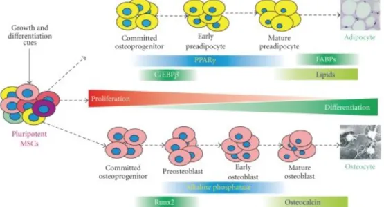
CD90, CD105) and the absence of others, such the haematopoietic and endothelial ones (CD45, CD34, CD19, CD31). In most laboratories, the gold standard procedure for the assessment MSCs identity, is their capability to differentiate in several cell lineages, such as bone, cartilage and adipose tissue cells. The differentiation of MSCs is regulated by multiple microenvironmental factors, which promote their transformation in a two-step stimulation: a first commitment of MSCs to progenitors, followed by a maturation stage where progenitors develop specific cell type characteristics. Once the differention induction is completed, gene expression profiles of MSCs shift towards specific patterns which are to be found in mature cell types.
1.2.1. Osteo-chondrogenesis
Osteogenesis, the formation, development and maintenance of bone, is a life-long process. Bone formation is finely regulated by systemic and local factors, which promote the commitment of MSCs towards osteoblastogenesis. Expression of the mastergene Runt-related transcription factor 2 (RUNX2) is the conditio sine qua non for triggering the osteogenic differentiation [12, 13], while inhibiting the adipogenic
commitment [14] of MSCs. The Runt-related transcription factor (RUNX) family
includes three proteins with a DNA-binding domain, called Runt, which works in cooperation with the transcriptional coactivator core binding factor b (CBFb) to promote stem cells differentiation. The Runx family members are structurally similar, but only Runx2 regulates osteogenesis.
RUNX2 gene is the target of several molecular pathways, such as Wingless(Wnt), Hedgehogs (HH), Bone Morphogenetic Proteins (BMPs) and Bone Morphogenetic
Protein Receptors (BMPRs). Wnt signalling is triggered by the binding of Wnt ligand to Frizzled receptor complexed with co-receptors LRP5/6. It initiates a signal cascade resulting in the stabilization and nuclear translocation of the activator, promoting cell proliferation. Wnt signalling has both pro-osteogenic and antiadipogenic activities through the bcatenin-dependent canonical pathway and also a non-canonical one [13].
bcatenin alone acts on peroxisome proliferator-activated receptor gamma (PPARγ, the
Figure 1: The lineage-specific differentiation is a multiple-stage and well-coordinated process regulated by master regulators, such as PPARγ for adipogenesis and Runx2 and Osterix for osteogenesis. Osteogenic differentiation can be staged by measuring RUNX2 gene expression (early marker) and osteocalcin and osteopontin (late markers). Production of lipids are indicators of terminal adipogenic differentiation. Adapted from [2].
Introduction
adipogenic commitment master regulator) preventing the adipogenic differentiation
[14]; this effect is reciprocal: an upregulation of PPARγ inhibits the bcatenin cascade [15-17]. These data are confirmed by many studies which evaluate the effects produced by
the interruption of that pathway: impaired osteogenesis and increased adipogenesis in
vitro and in vivo [15, 18].
Hedgehog signalling occurs when a hedgehog family ligand binds to the receptor Patched, releasing Smoothened from suppression and allowing it to transduce signals through G proteins, ultimately leading to nuclear translocation of a Gli zinc finger transcription factor. Several studies have investigated the HH signalling effects on MSC differentiation, and have evidenced its crucial role in early stages of osteoblastogenesis [19, 20]. The osteogenic differentiation effect of HH requires the
cooperation of BMPs signalling and involves primarily Gli transcriptional factor activity [21, 22]. The committed cells, in order to complete their differentiation and
maturation, must express other genes such as Osterix (SP7) [23]. Only mature
osteoblasts, further on, do express mineralization-related genes: Osteocalcin (BGLAP), Osteopontin (SPP1) and Osteonectin (SPARC); while at this late
maturation stage, the expression of commitment genes - RUNX2 and SP7 - is switched off (Fig. 1).
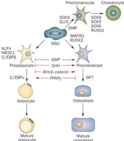
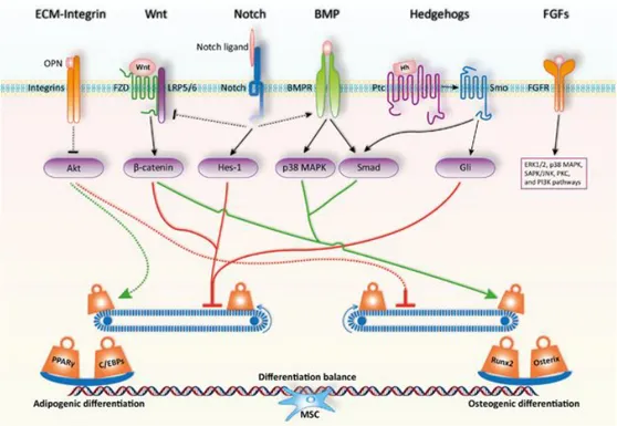
Figure 2: A number of regulators control MSC lineage fate. Humoral factors like Wnt ligands and BMP transmit their signals through cognate cell membrane receptors expressed by the differentiating cells. Transcription factors often govern the final cell lineage decision during MSC differentiation, and their transcriptional activities are modulated through crosstalk with cell-membrane receptor-mediated signals. Adapted from [1].
Introduction
1.2.2. Adipogenesis
The expression of osteogenesis-related genes and adipogenesis-related ones are mutually exclusive. The fat tissue master transcription factor PPARγ triggers adipogenic differentiation of MSCs. PPARγ is expressed in two isoforms, PPARγ1 and
PPARγ2, but the second one is predominant in adipose tissue. Evidence from animal
models demonstrates its central role in adipogenesis: PPARg deficient mice show a reduction in adipocytic commitment and fat storage; at the same time, they present increased bone mass as a consequence of upregulated osteoblastogenesis [21]. Coherent
evidence can be obtained using PPARγ agonists or ligands, which confirm how it inhibits ostegenesis, while inducing adipogenesis [24, 25]. While Runx2 must be
downregulated for the accomplishment of osteoblast maturation, complete differentiation and subsistence of adipocytes, instead, is supported by the continuous high-level expression of PPARγ [21].
1.2.3. Osteogenic-adipogenic balance
The balance between adipogenic and osteogenic commitment of MSCs is regulated by many molecular cascades and signalling pathways with mutually exclusive effects. Together, the two processes are controlled by multiple factors. These factors attract significant attention since their dysregulation is involved in several pathological conditions. In osteoporosis patients for instance: increased bone marrow adipose tissue occurs at the expense of reduced osteoblastogenesis from mesenchymal stem cells [26]. Some of the involved factors, e.g. insulin, dexamethasone and ascorbic acid,
are well known and they are used in in vitro models to induce differentiation. Also physical factors, characteristic of MSCs microenvironment play an important role in controlling progenitors’ commitment: extra cellular matrix (ECM) and growth supports influence the differentiation, in vitro and in vivo [27]. In fact, many inducing
proprieties of different ECMs are used to improve and control commitment and differentiation in laboratory studies. Integrins and other mechano-transducer molecules act in the control of physical property-dependent stimulation, such as the activation of Runx2 through the mechanical signal transduced by the cytoskeleton [28, 29] and consequent osteogenic upregulation. The pro-osteogenic effect of mechanical
forces exerts an antiadipogenic effect, confirming the mutual exclusion of the two processes. In fact, through Fyn/FAK/mTORC2 induced pathways, which enhance bcatenin signalling, the stimulated cytoskeleton promotes the osteogenic way [30, 31]. The
involvement of mTORC in the remodelling skeleton-dependant osteogenesis is confirmed by the observation that its knockdown in MSCs facilitates the adipogenic commitment along with an impairment of osteogenesis [32]. Osteopontin (OPN), an
organic component of bone which is synthesized by many cell types including osteocytes, acts on the integrin-mediated signal promoting osteogenesis; in fact, OPN gene knockdown results in robust adipogenesis at expense of osteogenesisis [33, 34]. In
contrast, other molecular cascades, e.g. the BMP pathways promote both adipogenesis and osteogenesis of MSCs. BMP4, which induces cartilage and bone formation, stimulates also the commitment of MSCs to adipogenesis [35]. BMP2, which acts like a
pro-osteogenic factor at high concentrations, promotes adipogenesis at lower ones [36, 37]. In addition, mature adipose and bone tissues, behave like endocrine organs, which
secrete active molecules, adipokines and osteokines, respectively, resulting in a reciprocal modulation [38].
Introduction
Figure 3: Signaling pathways and key transcription factors in regulating the adipo-osteogenic differentiation of MSCs. The fine balance of adipogenic and osteogenic differentiation of MSCs is achieved by the actions of critical signaling pathways and key transcription factors. MSCs exist in specific microenvironments or niches, which is composed of various extracellular matrix components, growth factors, cytokines, and chemokines. Upon interaction with MSCs, these components activate or inhibit the lineage commitment of MSCs. In addition, the initiated cellular signaling pathways can also interfere each other to form a fine regulatory network. Ultimately, this signaling network maintains a delicate differentiation balance through regulating key transcription factors such as PPARγ and C/EBPs or Runx2 and Osterix for adipogenesis or osteogenesis respectively. OPN, osteopontin; FZD, Frizzled receptor; Hh, Hedgehog; Ptc, Patched; Smo, Smoothened. Adapted from [39].
It is worth mentioning that all the processes, pathways and molecular flows involved in osteogenesis and/or adipogenesis do not work separately; and that also the microenvironment plays an essential role in commitment fate.
1.3. Ageing
Ageing is a progressive physiological process, where cellular damage accumulation, with consequent organ and tissue deterioration, causes the loss of homeostasis [40].
Effects of ageing are found in the MSC population, whose beneficial functions, such as self-renewal and multilineage differentiation capacity, result compromised. During ageing, bone marrow MSCs show a reduction of repair and regenerative capacity: they have a reduced osteogenic differentiation competence versus an increased adipogenic one; they show reduced proliferation too [41, 42]. Many factors concur to MSCs
senescence, including telomeres shortening and accumulation of reactive oxygen species [43, 44].
Bone tissue undergoes a continuous self-regeneration process, which eliminates old bone and produces new tissue, called bone remodelling. The final balance is finely tuned by numerous actors and may be affected by many variables, including age. In growing adolescents and young adults, bone formation exceeds bone resorption. Conversely, in elderly people the balance is inverted, causing a reduction in bone
Introduction
mineral density. Ageing is the process which causes bone quality disruption accompanied by adipose infiltration in the bone marrow niche; in fact, the lineage commitment balance shifts from osteogenesis to adipogenesis [45]. In an aged scenario,
expression of the adipogenic transcription factor PPARγ is upregulated compared to a young MSC environment [45]. Furthermore in old animal models the Runx2 co-factor
Cbfb is underexpressed; old mice show a decrease in bone density and an adipose infiltration in marrow [46]. On the other hand, studies have confirmed the effects of
Cbfb in osteoblast maintaining and in adipogenesis inhibition, by enhancing Wnt/bcatenin signalling [46, 47]. Interestingly, conditioned media from young bone
marrow aspirates act like “rejuvenating” factors which rescue the age-related decrease of osteogenesis in in vitro experiments [48]. Not only soluble factors act in this way: other
results suggest that also old or young ECM plays an essential role in adipogenic vs. osteogenic fate [49]. The resulting age-related commitment alteration clearly appears in
some pathological conditions, e.g. osteoporosis [50].
An important and well studied cause for the impairment of MSCs proliferation and differentiation potential is telomere shortening. This process is continuosly active in proliferating somatic cells, and erodes linear DNA molecules ends [51, 52]. In stem cells
the action of telomerase reverse transcriptase (TERT) counteracts chromosomes shortening by a continous addiction of repetitive elements at their ends. Shelterins, such as TRF1 and TRF2, cooperate with telomerase in order to prevent age-related senescence in the progenitors population [53, 54].
1.4. Autophagy
Autophagy is a process in which cellular components such as proteins and damaged mitochondria are engulfed by autophagosomes and delivered to lysosomes to be degraded and recycled in order to maintain cellular homeostasis [55].
Variations in cell-intrinsic mechanisms, such as autophagy, epigenetic modifications, et al., contribute to produce the “aged” bone phenotype [56]. A hallmark of ageing is
the reduction of autophagy activity. In fact, a reduction in the elimination of dysfunctional progenitors causes a decline of MSCs stemness, proliferation and their osteogenic differentiation capacity [45, 57]. Basal autophagy is a crucial mechanism in the
maintenance of the healthy state of progenitors; failure of autophagy, on the contrary, causes cell senescence characterized by a decline in number and functionality of MSCs
[58]. Recently, the important role of autophagy was confirmed by experiments, where
aged mice restored their bone mineral density after pharmacological stimulation of the autophagic process [59]. Molecularly, specific genes are expressed during autophagy:
after stimulation, in particular through the mTOR1 axis and Akt/PI3k signalling cascade, autophagy is triggered by the interaction of Unc-51 Like Autophagy Activating Kinase 1 (Ulk1) with AMP kinase (AMPK) [60]. The autophagic cascade
continues with interactions in many pathways, including PI3P-mediated activation of others autophagy-related proteins (Atg family), such as Atg5 and Atg7, and LC3 precursor [61]. In conclusion, the activated LC3-B promotes a cytosolic activity: the
formation of autophagosome [59, 62].
1.5. ROS
The production of oxygen reactive species (ROS) and consequent induction of oxidative stress plays a significant role in the adipogenic vs osteogenic fate of MSCs. The concept that ROS lead to cellular damage in ageing emerged a long time ago [63].
Introduction
production is trusted to promote some essential signalling pathways, which control cell survival, proliferation and differentiation [64, 65]. MSCs produce survival factors with
antioxidant properties in ischemic tissue; they are known to have high levels of intracellular antioxidants and low levels of ROS [66, 67]. Numerous recent studies show
the influence of oxidants on MSCs differentiation trough osteo-chondrogenesis or adipogenesis [68-70]. Many studies employ hydrogen peroxide to investigate the effects
of oxidation on cells; in vitro experiments show that H2O2: decreases alcaline
phosphatase activity in osteogenically-induced hMSCs [71]; abolishes osteogenesis in
osteoblast progenitors [72]; it impairs osteogenesis downregulating Gli protein levels by
Hh signalling inhibition [73]; it promotes a general adipogenic induction on MSCs [68] in
a dose-dependent manner [74]. Interestingly, intracellular ROS levels increased after
exposure of MSCs to an adipogenic cocktail [75]. On the other hand, by antioxidant
administration, osteogenic differentiation could be restored [76-78]. In conclusion, an
excessive amount of ROS prevents osteogenesis; loss or reduction of defence mechanisms against oxidative stress promotes this phenomenon. In this scenario, the balance between oxidative species production and ROS scavenger response must be evaluated carefully. The signalling pathways involved in the ROS-mediated shift include Wnt and PPARγ [79, 80]. ROS production in osteoblasts, together with enhanced
mitochondrial metabolism and glucose intake affect the differentiation balance and stimulate the adipogenic way; mitochondrion-driven adipogenesis may be switched off by mitochondrion-targeted antioxidants, which deactivate the adipogenic transcription factors [75]. In addition, the increased mitochondrial metabolism results necessary for
adipogenesis [81]. Is clear that a positive energy balance promotes the
Aim of the study
2. Aim of the study
Physical activity is widely recognized as being associated with healthy ageing and reduced risk for a number of chronic conditions, such as obesity, diabetes, bone disease, hypertension. Moreover, it is known that training regulates the balance between bone and fat tissue formation. However, it has also been demonstrated that strenuous physical exercise may trigger adverse effects since it increases the production of free radicals, such as reactive oxygen species. This may enhance inflammation and induce DNA damage.
The aim of this study was to investigate the various aspects of cellular response to physical exercise (half-marathon performance by amateur male runners).
In particular we intended to:
1. evaluate serum proteins modulation in response to the oxidative products by a proteomic approach;
2. investigate the effects of physical activity on gene expression profiles in circulating progenitor cells (CPs) by means of gene array analyses;
3. confirm and investigate further the preliminary data obtained in step 2. concerning: • effects of physical activity on osteogenic/chondrogenic/adipogenic
commitment of progenitor cells;
• effects of physical activity on the autophagic activity in CPs; • effects of physical activity on telomerase-related genes;
4. verify the effects of soluble factors present in runners’ sera before and after the marathon on an in vitro model (using a bone marrow-derived hMSC cell line).
Results
3. Results
3.1. Investigations on sera
3.1.1. Biochemical sera analysis
All runners and controls were evaluated for some biochemical parameters, such as vitamin D and electrolytes. In particular, sodium, potassium, iron and ferritin levels appeared significantly increased in post-run condition (Table 1).
Table 1: biochemical analysis on runners’ sera [82].
3.1.2. Modulation of sera proteins
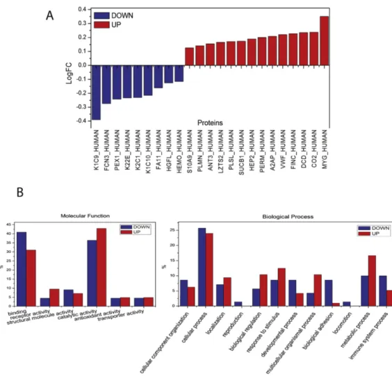
The proteome of all the athletes before and after the half marathon was investigated for quantitative changes. In order to eliminate possible inter-individual variability which can affect protein analysis, each athlete was analysed singularly in pre- and post-run condition. The analysis identified modulations of many proteins and, to better understand their molecular function, a gene ontology classification was conducted (Fig. 4). The two most abundantly represented functions were: catalytic activity (39%) and binding function (37%), respectively. Proteins associated with antioxidant activity were also detected (2%). Proteins related to cellular (23%) and metabolic processes (17%), together with the response to stimulus (9%) were also well represented in the samples. The gene ontology classification based on protein class showed that enzyme modulator (13%), hydrolase (10%) and signaling molecule (9%), respectively, were the three most represented classes (Fig. 4). By analyzing the log fold change of the regulated proteins we observed that 23 proteins showed variations after the marathon: 9 proteins were underexpressed (K1C9, FCN3, PEX1, K22E, K2C1, K1C10, FA11,
Results
LZTS2, PLSL, SUCB1, HEP2, PERM, A2AP, VWF, FINC, DCD, CO2, MYG) (Fig.
4). In order to reduce individual variability and to highlight thedifferences between pre- and post- athletic performance, we compared the proteomic profile of each athlete before and after the competition.
Common regulated proteins among athletes where then analyzed using gene ontology classification with the aim to highlight the main molecular functions and biological processes involved in sport activity. Gene ontology analysis (Fig. 5) showed an up-regulation of proteins related to catalytic activities, metabolic processes and response to stimulus, consequent to physical exercise. Conversely, there was a decrease of proteins related to the immune system processes and to binding functions such as some coagulation factors.
Figure 4: Log fold changes of the regulated proteins between the pre- and post- marathon classes (FC ≥ 1.30 or FC≤ 0.77; p-value< 0.05) (A). Gene Ontology analysis of regulated proteins from individual athlete, according to PANTHER classification. Bars represent the percentage of hits (proteins) in the functional category (some proteins are placed in multiple functional GO categories). The molecular functions (left) and the biological processes (right) analyses are represented (B).
Results
Figure 5: gene ontology classification based on protein class.
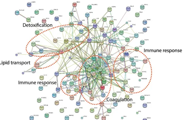
STRING analysis of regulated proteins between pre- and post-marathon single athletes showed that detoxification pathway as well as immune response, lipid transport, and coagulation were affected by physical activity (Fig. 6).
Figure 6: STRING analysis for the regulated proteins between pre- and post- marathon from individual athletes.
Results
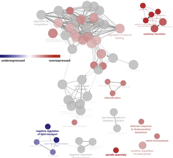
In addition, employing Cytoscape software and the ClueGO plug-in, we performed the Pathway enrichment analysis (Fig. 7). Increased secretion of cytokines, and a negative regulation of lipid transport could be observed after the half marathon.
Figure 7: Pathway enrichment analysis through Cytoscape software and the ClueGO plug-in. In the functionally grouped networks, terms are linked based on к-score (≥0.4). Edge thickness indicates the association strength. Node size corresponds to the statistical significance for each term. Red term indicates an abundance increase after the half marathon, blue term indicates an abundance decrease after the half marathon.
Results
3.2. Circulating progenitors analysis 3.2.1. Osteogenesis
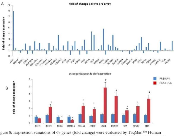
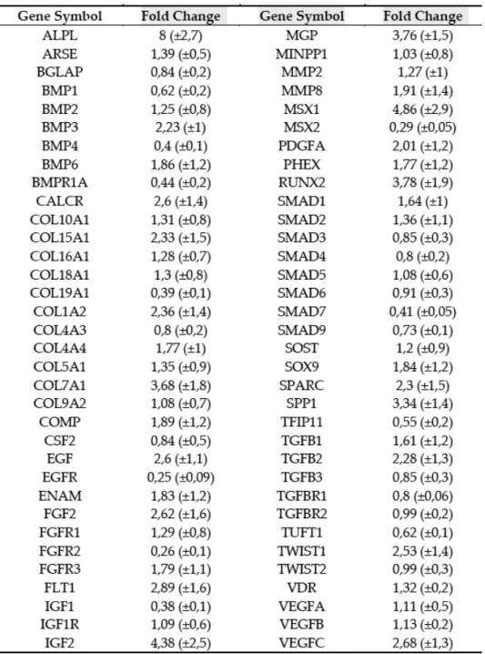
The fold changes of expression (Fig. 8A) disclosed by the osteogenesis array are listed in Table 2. In order to validate these findings, we performed Real Time RT PCR assays for several genes. We investigated the expression of SP7, the RUNX2 downstream gene, as well. Most genes investigated were expressed at higher levels in post-run CPs compared to pre-run CPs (Fig. 8B). The higher expression levels indicated a strong commitment of CPs to osteogenic differentiation. In particular, RUNX2 expression in post-run was >3 fold higher compared to pre-run CPs (p<0.01); RUNX2 downstream genes: COL1A2 (p<0.05), SPARC (p<0.05), SP7 (p<0.05) and SPP1(p<0.01) in turn confirmed the osteogenic trend, as their expression was higher too. However, we observed lower post-run expression levels of BMP4 (p<0.05) and BMPR1A (p<0.05), compared to pre-run levels. To exclude any bias due to technical procedures, we enclosed also 10 controls in our study. In control samples obtained at time 0 and 120’ no differences were observed in Array profiles as well as in data obtained by Real time RT PCR (data not shown).
Figure 8: Expression variations of 68 genes (fold change) were evaluated by TaqMan™ Human Osteogenesis Array (A). Real Time PCR Single assay validation of BMP1, BMP3, BMP4, BMPR1A, COL1A2, COMP, MSX1, RUNX2, SOX9, SPARC and SSP1 (B). Fold change of expressions are reported as normalized 2−dCT values. *p < 0.05; #p<0.01.
Results
Table 2: TaqMan™ Human Osteogenesis Array results.
3.2.2. Chondrogenesis
To investigate further the chondrogenic commitment in CPs, we analyzed, by Real Time RT PCR, the expression of COL2A1 and COMP genes, in addition to the chondrogenic transcription factor SOX9. In particular, the higher expression of COL2A1 (p<0.01) as well as of COMP (p<0.05) in post-run CPs corroborated clues from the chondrogenic determinant SOX9 (p<0.05) (Fig. 9).
Results
Figure 9: Physical activity improved chondrogenic differentiation. Besides SOX 9 upregulation, we observed also higher expression of COL2A1 and COMP in circulating progenitors after the run.
3.2.3. Adipogenesis
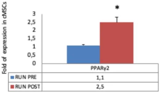
To evaluate CPs adipogenic commitment we analyzed gene expression of the adipogenic transcription factor PPARγ2. Data obtained by Real Time RT PCR showed that PPARγ2 gene expression was higher in post-run CPs (Fig. 10) (p<0.05).
Figure 10: Adipogenic differentiation. PPARY2 gene expression was higher in post-run cMSCs. Conversely, the BM-MSC line treated with post-run sera showed a reduced adipogenic differentiation.
3.2.4. Telomerase-related gene expression
Telomerase-associated genes, evaluated by gene array analysis in CPs, were differentially expressed in CPs after physical activity (p<0.01) (Fig. 11A). In particular, Table 3 reports post-run fold changes of expression. Thereafter we performed Real Time PCR assays to validate array data, which were confirmed as shown in Figure 7B. Notably both TERT and TERF1 post-run expression levels were higher than pre-run levels (p<0.01), while post-run expression of several DNA repair genes (e.g. RAD50 and HNRNPA1), was lower (p<0.05) (Fig.11B).
Results
Figure 11: Gene expression variations are plotted for 19 selected candidates, evaluated by TaqMan™ Human Telomerase Array (A). Real Time PCR Single assay validation of HNRNPA1, MRE11A, RAD50, TERF1 and TERT are shown in B). Fold changes of expression are reported as normalized 2−dCT values. *p < 0.05; #p<0.01.
Results
3.2.5. Autophagy
We analyzed by Real time RT PCR the expression of autophagy related genes in pre- and post-run CPs in order to investigate autophagy modulation during physical activity. Autophagy related genes appeared to be upregulated in post-run CPs (Fig. 12A). To confirm these findings, we treated a BM-MSC cell line with pre- and post-run sera. By immunofluorescence assay we found that cells treated with post-post-run sera expressed higher LC3 levels, compared to cells treated with pre-run sera (Fig. 12B). Interestingly, we found a positive correlation between ATG3 and SOX9 expression (R=0.95) as well as ATG3 and RUNX2 expression (R=0.98). Similarly, positive correlations between ULK1 and SOX9 expression (R=0.96); ULK1 and RUNX2 expression (R=0.97) were observed.
Figure 12: Physical activity induced the upregulation of autophagy related genes in cMSCs (A). To
confirm this finding, we treated a BM-MSC line with pre- and post-run sera and we analyzed LC3 expression by fluorescence confocal microscopy (magnification 63X). (B) BM-MSCs treated with post-run sera expressed higher LC3 levels compared to cells treated with pre-run sera. Scale Bar 20µm.
Results
3.3. Bone marrow-MSC cell line treated with sera 3.3.1. Osteogenesis on treated cells
We analyzed the effects of sera obtained from runners before and after the marathon on a Bone Marrow-derived Mesenchymal Stem Cell line. Findings from the BM-MSC line culture supplemented with runners’ sera confirmed the data obtained in CPs. In particular, the gene expression profiles (Fig. 13A) as well as osteocalcin expression (Fig. 13B) and calcium deposition, evaluated by alizarin red staining (Fig. 13C), indicated the increased osteogenic differentiation and maturation in cells treated with post-run sera.
Figure 13: Effects of sera on a Bone Marrow-derived Mesenchymal Stem Cell line. The gene expression profiles confirmed the osteogenic commitment observed in circulating progenitors (A); Osteocalcin expression (Scale Bar 40µm) (B); calcium deposition, evaluated by alizarin red staining, (Scale Bar 100 µm), (C), indicated the increased osteogenic maturation in cells treated with post-run sera. Representative cell fields are shown. *p<0.05
Results
3.3.2. Adipogenesis on treated cells
To evaluate the BM-MSC cell line adipogenic commitment after runners’sera addition, we analyzed gene expression of the adipogenic transcription factor PPARγ2. Data obtained by Real Time RT PCR showed that PPARγ2 gene expression was lower in post-run sera treated cells, compared to that observed with pre-run sera treatment (Fig. 14A) (p<0.05). These latter data were confirmed by oil red O staining of cells after 14 days culture: a reduction of lipid droplets (Fig. 14B) and a lower expression of the adipogenic marker Peripilin1, evaluated by immunofluorescence, (Fig. 14C) were observed.
Figure 14: the BM-MSC line treated with post-run sera showed a reduced adipogenic differentiation. In fact, PPARY2 gene expression levels were lower (A) and the number of lipid droplets (B) as well as PLIN1 expression (C) were reduced in BM-MSCs treated with post-run sera. Representative cell fields are shown. Scale Bar 40µm\; *p<0.05; #p<0.01.
Discussion
4. Discussion
Physical activity is widely considered a promoter of overall health: it reduces the onset of metabolic pathologies, e.g. diabetes and obesity, and of diseases such as hypertension and cardiovascular failure [83]. The metabolic regulation has been finely
studied, effects on glucose homeostasis, mitochondrial activity and adipocyte-related modulation topics have been reviewed extensively [84-86]. Therefore exercise is
recommended not only for its prophylactic value, but also as an effective therapy for many conditions and diseases [87]. Its role in preventing degenerative conditions is
associated to its intrinsic stem cell mobilization capability [6]. In fact, mesenchymal stem
cells have promising therapeutic potential against diseases caused by unbalanced cell metabolism/renewal in bone, cartilage and adipose tissue. Clinicians recognize the phenotypical effects on health induced by sport activity, such as bone mass increment, which reflects osteocytes viability (WHO). Some bone-related degenerative conditions (e.g. osteoporisis) represent a substancial burden for the public health service. An active lifestyle opposed to a sedentary one makes a big difference, since it has been demonstrated that customary physical activity delays age-related bone deterioration and bone loss-related fractures [87-89].
Comments on biochemical data
We saw an overall electrolyte iperconcentration values due to dehydration in athletes after the run; that result is characteristic of the high-intensity run condition. In addition, the analysis highlights an increment in iron and ferritin. The iron release is probably linked to tissue injury, which is a typical condition of training [88-90]. At the
same time, serum ferritin increase is probably consequent to phlogosis induced by the run [91-93]; it also suggests its implication in macrophage-mediated autophagy, which is
recorded during exercise [94].
Comments on proteomic analysis
It is known that physical activity produces not only positive effects on the organism. In particular, intense training, characterized by an increment of aerobic metabolism, triggers massive release of free radicals, i.e. reactive oxygen species [95]. To prevent
damages from ROS, the organism reacts with the production of antioxidants, ROS scavengers and with damage repair mechanisms [95-97]. In this scenario, a thorough
investigation of possible negative effects induced by sport activity is fundamental. The whole proteomic approach described in this dissertation allowed an extended evaluation of plasma proteins variations. The upregulation - in terms of protein expression - of myoglobin, myeloperoxidase and S100A9 confirmed that, even in the half-marathon endurance, the body responds to the oxidative challenge with the production of antioxidants [98-101]. It is appropriate to recall here that exercise causes
damages in muscle tissue cells [102]. In fact, an increased production of myoglobin, along
with complement system proteins, was registered. The inflammatory system responds to exercise in the same way, e.g. stimulating the production of plastin-2 and dermcidin, as seen. Marathon running stimulates also the coagulation system, modulating the balance between procoagulant von Willebrand factor, a2-antitripsin, fibronectin and anticoagulant molecules, such as heparin cofactor II, antithrombin III, plasminogen
[103, 104]. Fibronectin also plays an important role on MSCs: it enhances osteoblastic
differentiation [105-108]. The depletion of peroxisome biogenesis factor 1 (PEX1) and
Discussion
production. Furthermore, PEX1 is a proadipogenic factor through PPARγ signalling and it results upregulated both in in vivo and in vitro experiments [109].
Comments on Bone Marrow MSCs data
Physical activity plays a preeminent role in osteogenesis; its mechanical stimulation-mediated effects on MSCs osteogenic commitment are well known [29]. In order to
assess the stimulating effects of sera from runners and controls, we analyzed gene expression profiles variations in a Bone Marrow-derived MSC cell line (BM-MSC) cultured with differentiating media enriched with sera. After seven days culture, BM-MSCs supplemented with post-run sera showed a significant increase in RUNX2 gene expression and they showed a significant reduction in PPARγ gene expression. These data confirm that, during sport activity, serum levels of soluble factors involved in osteogenesis are incremented, at the expense of the adipogenic ones. Further on, in long term cultures (3 wks) we were able to assess correct osteoblastic maturation and the initial mineralization process. In fact mineralization-related SPP1 and SPARC genes expression was increased. Alizarin Red S staining was performed, in order to measure calcium salts deposition by developing osteoblasts. Stained slides showed an augmented red-marked calcium deposition in cells treated with post-run sera from trained subjects; this result confirms the correct in vitro mineralization of committed cells. Furthermore, to better assess osteogenesis in those long-term cultures immunofluorescent staining for Osteocalcin (BGLAP) was performed. Accordingly, cells cultured in adipogenic medium were stained with Oil Red O, to highlight the deposition of oil droplets in the cytoplasmic compartment. From stained slides, the significant reduction of lipid droplets inside differentiated stem cells treated with post-run sera confirmed a reduced adipogenic differentiation.
Comments on Circulating Progenitors results
The selection of circulating progenitors from whole blood (CPs) can be used to evaluate in real time the effects of inducing factors on the Mesenchymal Stem Cells (MSCs) population. We applied this technique to investigate promptly modulations occurring during the marathon, comparing post-run data with individual pre-run data and with sedentary controls data. Using the TaqMan™ Human Osteogenesis Array, we assessed the gene expression modulation of 68 osteogenesis-related genes. Several genes from the array, evaluated also in single assays, confirmed their expression modulation towards osteogenic commitment. A similar approach confirmed upregulation of cartilage-related genes, indicating pro-chondrogenic induction as well. The gene expression analysis also evidenced some peculiar modulations. We observed, for instance, lower post-run expression levels of BMP4 and BMPR1A which codes for its receptor [92], compared to pre-run levels in CPs. BMPs, in general, do promote
osteogenesis, nevertheless some of them may have a noteworthy double action. BMP4 actually plays an important role during adipogenesis, since it acts switching the expression of the two isoforms of PPARγ. In particular, it shifts the expression of the second isoform to PPARγ1, thus directing progenitors towards white adipocytes differentiation at the expense of brown adipocytes [110-113]. BMP4 downregulation
results associated to PPARγ2 increase in CPs. Supposedly it enhances insulin caption by cells and consequent glucose intake [114]. BMP4 decrease and PPARγ2 upregulation
suggest that, during training, the brown/white adipogenic maturation balance leans towards the first one [112]. The upregulation of BMP3 gene expression can be associated
Discussion
to its ability in promoting MSCs proliferation through Wisp1 signalling pathway [115-117].
Progenitors proliferation is also supported by data concerning TERT and TERF1 genes enhanced expression [118-120]. Telomere lenght maintainance is required for the
preservation of proliferation and self-renewal of MSCs [121, 122]. The TaqMan™ Human
Telomerase Array showed also a downmodulation of DNA repair-related genes (RAD50, HNRNPA1), confirmed by single amplification assays. Those repair systems prevent DNA damage accumulation-mediated induction of apoptosis. On another hand, they also prevent the elimination of genetically damaged MSCs subpopulations
[123, 124]. In particular, the DNA repair capacity of MSCs is related to their differentiation
stage: committed and pre-differentiated cells silence the elevated repair activity of their progenitors [125, 126]. Our overall results suggest that physical activity promotes the
proliferation of MSCs and it also fosters a selection of undamaged progenitors. To prevent the accumulation of damaged organelles, misfolded proteins and other intracellular anomalies, the quality control system acts regulating the autophagic process inside the cell. We highlighted the upregulation of autophagy-related genes (ATG3, ATG7, ULK1) in post-run samples. Supporting data come from the increased immunofluorescence staining of LC3-B in post-run sera treated cells. Autophagy plays a crucial role in counteracting oxidative stress effects; its defensive role is necessary for progenitors’ differentiation properties. Combining these data with telomerase-related results, we assume that the commitment and differentiation to osteo-chondrogenesis in a stressful environment is better safeguarded by a system that can rule out defective progenitors.
In conclusion, we confirm that a production of antioxidants occurs during the half-marathon endurance. Importantly, our data also suggest that the half-half-marathon physical commitment promotes osteogenic and chondrogenic differentiation as well as autophagy, through a complex biological interplay.
We are aware that our observations, based mainly on gene expression analyses, could be corroborated by further data on protein products analysis. Paucity of biological samples due to the low number of circulating progenitors was a factual limitation. Further technical approaches may hopefully contribute to data implementation
Materials and Methods
5. Materials and methods
5.1. Enrollment and samples collection
The study was conducted during a sport event called “Run for Science”, held in Verona (Italy), which was specifically planned to investigate the effects of distance running on recreational athletes. Twenty-two amateur male runners (median age 41,4 ± 10,1) carried out a 21.1 Km half marathon. Blood samples, obtained by venipuncture, were collected before the run and immediately after. For controls, we harvested samples from ten sedentary male subjects at time 0 and after a half-marathon running-time. The control samples were used only in differentiating cell culture experiments; peripheral blood-MSCs gene expression analysis. Proteomic and biochemical evaluations were performed for runners’ samples only.
Written informed consent was obtained from all participants and the study was approved by the Ethical Committee of Azienda Ospedaliera Universitaria Integrata of Verona, Italy (number 1538; 3th December 2012; Local Ethical Committee of AOUI di Verona).
5.2. Sera collection
Sera were obtained from 6 ml of fresh blood after a centrifugation at 1800g for 15’ at 4°C, using a vacutainer-mediated collection. Then, sera were aliquoted and frozen at −80 °C for further investigations.
5.3. Peripheral blood-circulating progenitors isolation
Circulating Progenitor cells (CPs) were isolated from 25 ml of heparinized blood by a depletion method including two Ficoll procedures to deplete hematopoietic cells by antibodies cocktail. Firstly, Peripheral Blood Mononuclear Cells (PBMCs) were obtained by a gradient centrifugation at 800g for 30’ at 20°C (first Ficoll procedure). Then, to deplete unwanted hematopoietic cells, Rosette-Sep antibodies cocktail was used against 5 ml of whole blood mixed with the PBMCs obtained by the first Ficoll; in particular, the antibody cocktail was incubated with samples for 20’, at room temperature. Then, a second Ficoll procedure was performed to remove the unwanted CD3, CD14, CD19, CD38 and CD66b positive cells crosslinked to red blood cells (glycophorin A) as reported in Valenti et al [11]. Collected cells were then washed in
phosphate-buffered saline (PBS), pelleted and stored dried at -80°C. To evaluate cell phenotype we analysed gene expression for CD3, CD14, CD19, CD45, and CD34 markers, as in Valenti et al [127].
5.4. Bone marrow-MSC cell line culture
Sera, obtained from each runner before and after the competition, were pooled in two groups: pre-run and post-run. To investigate sera effects, we used BM-hMSC (Bone Marrow-human Mesenchymal Stem Cells, PromoCell) in order to study osteogenic and adipogenic differentiation, after and before physical activity. Pooled sera were added to the Mesenchymal Stem Cell Grow Medium (PromoCell) at 10% of final concentration. Cells were plated at density of 5 × 10⁴ cells per well into 24-well plates
and cultured until 21 days. In particular, the osteogenic differentiation was performed with osteogenic medium containing osteogenic stimulatory supplements (10%, StemCell), 10nM dexamethasone, 2mM β-glycerophosphate, 100µM ascorbic acid (StemCell Technologies Inc). The adipogenic differentiation was performed by using 500nM isobutilmetilxantine, 200 µM indomethacin, 1µM dexamethasone and 10
Materials and Methods
µg/ml insulin in basal medium. For both osteogenic and adipogenic differentiation, the medium was changed every 3 days after initial plating. Pellets from all types of culture were harvested and stored dried a-80°C after three, seven, fourteen and twenty-one days of differentiation.
5.5. RNA extraction and reverse-transcription
Pellets from differentiated BM-hMSC cells and from CPCs were collected and stored at -80°C. Then, nucleic acid extraction was performed using “RNeasy Protect Mini Kit” by Qiagen, following the manufacturer’s protocol. RNA samples were quantified by Qubit 3 Fluorometer using “Qubit RNA HS Assay Kit” by Invitrogen. Two micrograms of the extracted RNAs were reverse transcribed with “High-Capacity cDNA Reverse Transcription Kit” by ThermoFisher Scientific, according to producer’s instructions. RNA and cDNA samples obtained were stored at -80°C.
5.6. Gene expression analysis
5.6.1. PCR arrays were performed by using TaqMan™ Array Human Osteogenesis (ThermoFisher, cat# 4414096) and TaqMan™ Array Human Telomere Extension by Telomerase (ThermoFisher, cat# 4414187) according to manufacturer’s instructions.
5.6.2. Real time PCR analyses were performed in a total volume of 20 µl containing TaqMan Universal PCR Master mix and 2 µl of cDNA from each sample. Pre-designed, gene-specific primers and probe sets for each gene were obtained from Assay-on-Demand Gene Expression Products (Applied Biosystems, cat# at 5.6.2.1 section). In order to analyze the autophagy genes expression we used SyBR green primers (Invitrogen, oligo sequences at 5.6.2.2). Real Time TaqMan PCR reactions were carried out in multiplex. The amplification reaction program included a 10’ at 95°C for the enzyme activation step, followed by 40 cycles at 95°C for 15’’ and at 60°C for 1’.
Thermocycling and signal detection were performed with ABI Prism 7300 Sequence Detector (Applied Biosystems). Ct values for each reaction were obtained using TaqMan SDS analysis software. For each amount of RNA tested triplicate Ct values were averaged. Since Ct values vary linearly with the logarithm of the amount of RNA, this average represents a geometric mean. In addition, we considered a fold change < 0,5 and> 1,2 to be relevant, even if smaller changes were statistically significant.
5.6.2.1. TaqMan Probes: CD3, Hs00174158_m1; CD14, Hs02621496-s1; CD19, Hs00174333_m1; CD45, Hs00174541_ m1; CD34, HS00156373_m1; CD73, Hs00159686_m1, CD105, Hs00923996_m1; RUNX2, Hs00231692_m1; SP7, Hs00541729_m1; COL1A2, Hs01028956_m1; SPARC, Hs00234160_m1; SPP1, Hs00167093_m1; BMP1, Hs00241807-m1; BMP3, Hs00609638-m1; BMP4, Hs03676628-s1; BMPR1A, Hs1034913-g1; SOX9, Hs01107818_m1; COMP, Hs004359-m1; MSX1, Hs00427183- m1; HNRNPA1, Hs 01656228-s1; MRE11A, Hs00967437-m1; RAD50, Hs00990023-m1; TERF1, Hs 00819517-m1; TERT, Hs 00972650-m1; COL2A1, Hs00264051_m1; PPARG2, Hs01115513_m1; B2M, Hs999999_m1; GAPDH, 0802021; by Applied Biosystems (Thermo Fisher corporation, Waltham, MA, USA).
Materials and Methods
5.6.2.2. SyBR oligos:
ATG3 (Fw GGAAGAATATGAAGAGAGTGG; Rv CTCATCATAGCCAAACAACC); ATG7 (Fw AGATTGTCCTAAAGCAGTTG; Rv CCATACATTCACTGAGGTTC); ULK1 (Fw TCAAAATCCTGAAGGAACTG; Rv ACCAGGTAGACAGAATTAGC); ACTIN (Fw GATGTATGAAGGCTTTGGTC; Rv TGTGCACTTTTATTGGTCTC); by Invitrogen (ThermoFisher corporation, Waltham, MA, USA).
5.7. Alizarin Red S staining
To evaluate calcium deposition, we performed the Alizarin Red S staining. Briefly, after 21 days culture in the presence of osteogenic medium, cells were fixed with formalin for 30’, washed three times with tap water, and stained for 45’ with 40 mM Alizarin red S at pH 4.1. The cells were then gently rinsed three times with distilled water and visualized under optical microscope.
5.8. Oil Red O staining
The BM-hMSCs, cultured in adipogenic medium in presence of pre-run or post-run sera, were fixed as in 5.7 then stained with Oil Red O after two washings with distilled water. Fixed cells were stained with Oil Red O solution for 20’, and then washed three times with distilled water. Nuclei were stained with Haematoxylin for 1’ then cells were rinsed for additional three times with distilled water and kept in PBS. The total area of red pixels in the Oil Red O-stained droplets/cell was detected by using the IMAGE J image analysis. In particular, the mean ±SD of red stained areas, evaluated at 40× magnification was calculated in three different field/slide and expressed as percentage with respect to total area.
5.9. Immunofluorescence
After 21 days culture, BM-hMSCs were fixed and processed according to the manufacturer’s protocols. Primary antibodies were diluted (as reported in the datasheet) in Antibody Dilution Buffer and incubated overnight at 4°C. To evaluate osteogenic differentiation and autophagy we used Osteocalcin C-8 antibody (SC74495, Santa Cruz) and LC-3B (Cat. #2775, Cell Signaling) respectively. For the assessment of adipogenic stimuli promoted by sera, we tested Perilipin D1D8 (Cat. #12589 Cell Signaling). Slides were then incubated with secondary antibodies Alexa Fluor® 594 anti rabbit (Cat. # R37117), Alexa Fluor® 488 anti rabbit (Cat. #A-11034), goat mouse fluorescein coniugated (Cat. AP124F, Millipore). Nuclear staining was performed by ProLong™ Gold Antifade Mountant with DAPI. Images were recorded using a Leica TCS SP5 AOBS inverted confocal microscope at 63X.
5.10. Statistical analysis
Results were expressed as mean ± S.E. Statistical analysis was assessed by Student's paired t test. We considered significant differences with p < 0,05. We performed also correlation analyses by Pearson test to measure a linear dependence between the expression of a single autophagy gene and the transcription factor RUNX2 or SOX9, respectively. For in vitro data, analyses were applied to experiments carried out at least three times. Statistical analyses were performed using SPSS for Windows, version 22.0 (SPSS Inc, Chicago, IL, USA).
Materials and Methods
5.11. Proteomic analysis 5.11.1. Sera sample preparation
Twelve microliters of sera were depleted of high abundant proteins using the Seppro IgY14 spin column kit (Sigma-Aldrich Inc., St. Louis, MO, USA) following the manufacturer’s protocol. The sample was transferred into an Amicon Ultra-0,5 ml 3 kDa centrifugal filter (Millipore, Billerica, MA, USA) following the manufacturer’s protocol, to collect the high molecular weight proteins. The sample were then subjected to I) denaturation with TFE, II) reduction with DTT 200 mM, III) alkylation with IAM 200 mM and IV) the complete protein trypsin digestion with 2 µg of Trypsin/Lys-C (Promega, Madison, WI, USA). The peptide digests were desalted on the Discovery® DSC-18 solid phase extraction (SPE) 96-well Plate (25 mg/well) (Sigma-Aldrich Inc., St. Louis, MO, USA). The adsorbed proteins were eluted with 800 µl of acetonitrile:water (80:20) [128]. After the desalting, the sample was vacuum
evaporated and reconstituted with 20 µl of 0,05% formic acid in water. Lastly, 2 µl of stable-isotope-labeled peptide standard (DPEVRPTSAVAA, Val- 13C5 15N1 at V10, Cellmano Biotech Limited, Anhui, China) was spiked into the samples before the LC-MS/MS analysis and used for instrument quality control.
5.11.2. LC-MS/MS analyses
Serum proteins were analyzed with a micro-LC Eksigent Technologies (Eksigent Technologies, Dublin, CA, USA) system that included a micro LC200 Eksigent pump with flow module 5–50 µl, interfaced with a 5600+ TripleTOF system (Sciex, Concord, ON, Canada) equipped with DuoSpray Ion Source and CDS (Calibrant Delivery System). The stationary phase was a Halo C18 column (0.5 Å~ 100 mm, 2,7 µm; Eksigent Technologies, Dublin, CA, USA). The mobile phase was constituted with a mixture of 0,1% formic acid in water (A) and 0,1% formic acid in acetonitrile (B), eluting at a flow-rate of 15 µl min−1 at an increasing concentration of solvent B from
2% to 40% in 30’. The injection volume was 4 µl and the oven temperature was set at 40°C. For identification purposes the samples were subjected to a data dependent acquisition (DDA): the mass spectrometer analysis was performed using a mass range of 100–1500 Da (TOF scan with an accumulation time of 0,25’’), followed by a MS/MS product ion scan from 200 to 1250 Da (accumulation time of 5 ms) with the abundance threshold set at 30 cps (35 candidate ions can be monitored during every cycle). The ion source parameters in electrospray positive mode were set as follows: curtain gas (N2) at 25 psig, nebulizer gas GAS1 at 25 psig, and GAS2 at 20 psig, ionspray floating voltage (ISFV) at 5000V, source temperature at 450°C and declustering potential at 25V. For the label-free quantification the samples were subjected to cyclic data independent analysis (DIA) of the mass spectra, using a 25- Da window: the mass spectrometer was operated such that a 50-ms survey scan (TOF-MS) was performed and subsequent MS/MS experiments were performed on all precursors. These MS/MS experiments were performed in a cyclic manner using an accumulation time of 40 ms per 25-Da swath (36 swaths in total) for a total cycle time of 1.5408’’. The ions were fragmented for each MS/MS experiment in the collision cell using the rolling collision energy. The MS data were acquired with Analyst TF 1.7 (Sciex, Concord, ON, Canada). Two DDA and three DIA acquisitions were performed
Materials and Methods
5.11.3. Protein database search
The DDA files were analized using Protein Pilot software v. 4.2 (Sciex, Concord, ON, Canada) and Mascot v. 2.4 (Matrix Science Inc., Boston, MA, USA). Trypsin as digestion enzyme was specified for both the software. For Mascot we used 2 missed cleavages, the instrument was set to ESI-QUAD-TOF and the following modifications were specified for the search: carbamidomethyl cysteins as fixed modification and oxidized methionine as variable modification. A search tolerance of 50 ppm was specified for the peptide mass tolerance, and 0,1 Da for the MS/MS tolerance. The charges of the peptides to search for were set to 2+, 3+ and 4+, and the search was set on monoisotopic mass [131]. The UniProt Swiss-Prot reviewed database containing
human proteins (version 2015.07.07, containing 42,131 sequence entries) was used and a target-decoy database search was performed. False Discovery Rate was fixed at 1%
[132].
5.11.4. Protein quantification
The quantification was performed by integrating the extracted ion chromatogram of all the unique ions for a given peptide. SwathXtend was employed to build an integrated assay library, built with the DDA acquisitions, using a protein FDR threshold of 1% [133]. The quantification was carried out with PeakView 2.0 and
MarkerView 1.2. (Sciex, Concord, ON, Canada). Six peptides per protein and six transitions per peptide were extracted from the SWATH files. Shared peptides were excluded as well as peptides with modifications. Peptides with FDR lower than 1% were exported in MarkerView for the t-test [134].
5.11.5. Bioinformatics and statistics software
The identified proteins were classified with PANTHER Classification System [132]. The
regulated proteins were analyzed by using STRING software [135], which is a database
of known and predicted proteinprotein interactions. For functional annotation clustering and network analysis of proteins, the Cytoscape 3.1.0 plug-ins ClueGO v2.0.8 was also used [134]. Graphical representations were performed by Origin 8.0
Bibliography
6. Bibliography
1. Takada, I., A.P. Kouzmenko, and S. Kato, Wnt and PPARγ signaling in osteoblastogenesis and
adipogenesis. Nature Reviews Rheumatology, 2009. 5: p. 442.
2. Wagner, E.R., et al., Therapeutic Implications of PPARgamma in Human Osteosarcoma. PPAR Res, 2010. 2010: p. 956427.
3. Guthold, R., et al., Worldwide trends in insufficient physical activity from 2001 to 2016: a pooled
analysis of 358 population-based surveys with 1·9 million participants. The Lancet Global Health, 2018.
6(10): p. e1077-e1086.
4. Organization, W.H., Noncommunicable diseases country profiles 2018. September 2018. 5. WHO, Global action plan for the prevention and control of NCDs 2013-2020. 2013: p. 55. 6. Maredziak, M., et al., Physical Activity Increases the Total Number of Bone-Marrow-Derived
Mesenchymal Stem Cells, Enhances Their Osteogenic Potential, and Inhibits Their Adipogenic Properties. Stem Cells
Int, 2015. 2015: p. 379093.
7. Bagur-Calafat, C., et al., The impact of high level basketball competition, calcium intake, menses, and
hormone levels in adolescent bone density: a three-year follow-up. J Sports Med Phys Fitness, 2015. 55(1-2): p.
58-67.
8. Banfi, G., et al., Bone Metabolism Markers in Sports Medicine. Sports Medicine, 2010. 40(8): p. 697-714.
9. Sansoni, V., et al., Bone turnover response is linked to both acute and established metabolic changes in
ultra-marathon runners. Endocrine, 2017. 56(1): p. 196-204.
10. Friedenstein, A.J., R.K. Chailakhjan, and K.S. Lalykina, The development of fibroblast colonies in
monolayer cultures of guinea-pig bone marrow and spleen cells. Cell Tissue Kinet, 1970. 3(4): p. 393-403.
11. Valenti, M.T., et al., Gene expression analysis in osteoblastic differentiation from peripheral blood
mesenchymal stem cells. Bone, 2008. 43(6): p. 1084-92.
12. Otto, F., et al., <em>Cbfa1</em>, a Candidate Gene for Cleidocranial Dysplasia Syndrome, Is
Essential for Osteoblast Differentiation and Bone Development. Cell, 1997. 89(5): p. 765-771.
13. Ducy, P., et al., Osf2/Cbfa1: A Transcriptional Activator of Osteoblast Differentiation. Cell, 1997. 89(5): p. 747-754.
14. Komori, T., Regulation of osteoblast differentiation by Runx2. Adv Exp Med Biol, 2010. 658: p. 43-9.
15. Ross, S.E., et al., Inhibition of adipogenesis by Wnt signaling. Science, 2000. 289(5481): p. 950-3. 16. Liu, J. and S.R. Farmer, Regulating the balance between peroxisome proliferator-activated receptor gamma
and beta-catenin signaling during adipogenesis. A glycogen synthase kinase 3beta phosphorylation-defective mutant of beta-catenin inhibits expression of a subset of adipogenic genes. J Biol Chem, 2004. 279(43): p. 45020-7.
17. Muruganandan, S., A.A. Roman, and C.J. Sinal, Adipocyte differentiation of bone marrow-derived
mesenchymal stem cells: cross talk with the osteoblastogenic program. Cell Mol Life Sci, 2009. 66(2): p. 236-53.
18. Holmen, S.L., et al., Essential role of beta-catenin in postnatal bone acquisition. J Biol Chem, 2005. 280(22): p. 21162-8.
19. van der Horst, G., et al., Hedgehog stimulates only osteoblastic differentiation of undifferentiated KS483
cells. Bone, 2003. 33(6): p. 899-910.
20. Spinella-Jaegle, S., et al., Sonic hedgehog increases the commitment of pluripotent mesenchymal cells into
the osteoblastic lineage and abolishes adipocytic differentiation. J Cell Sci, 2001. 114(Pt 11): p. 2085-94.
21. Tzameli, I., et al., Regulated production of a peroxisome proliferator-activated receptor-gamma ligand
during an early phase of adipocyte differentiation in 3T3-L1 adipocytes. J Biol Chem, 2004. 279(34): p.
36093-102.
22. Yuasa, T., et al., Sonic hedgehog is involved in osteoblast differentiation by cooperating with BMP-2. Journal of Cellular Physiology, 2002. 193(2): p. 225-232.
23. Peng, Y., et al., Characterization of Osterix Protein Stability and Physiological Role in Osteoblast
Differentiation. PLoS ONE, 2013. 8(2): p. e56451.
24. Ali, A.A., et al., Rosiglitazone causes bone loss in mice by suppressing osteoblast differentiation and bone
formation. Endocrinology, 2005. 146(3): p. 1226-35.
25. Lazarenko, O.P., et al., Netoglitazone is a PPAR-gamma ligand with selective effects on bone and fat. Bone, 2006. 38(1): p. 74-84.
26. Valenti, M., L. Dalle Carbonare, and M. Mottes, Osteogenic Differentiation in Healthy and
Pathological Conditions. International Journal of Molecular Sciences, 2017. 18(1): p. 41.
27. Engler, A.J., et al., Matrix elasticity directs stem cell lineage specification. Cell, 2006. 126(4): p. 677-89.
![Table 1: biochemical analysis on runners’ sera [82].](https://thumb-eu.123doks.com/thumbv2/123dokorg/8249758.129503/14.892.190.753.320.752/table-biochemical-analysis-runners.webp)