Alma Mater Studiorum – Università di Bologna
DOTTORATO DI RICERCA IN
BIOLOGIA CELLULARE, MOLECULARE E INDUSTRIALE:
BIOLOGIA FUNZIONALE DEI SISTEMI
CELLULARI E MOLECOLARI
Ciclo XXIV
Settore Scientifico Disciplinare: 05/E2
Settore Concorsuale di afferenza: BIO/11
TITOLO TESI
Trascriptome analysis and functional genomics
of Neisseria meningitidis in human blood
Presentata da:
Elena Del Tordello
Coordinatore Dottorato
Relatore
Chiar.mo Prof. Chiar.mo Prof.
Vincenzo Scarlato Vincenzo Scarlato
Dott.
Davide Serruto
Il mio progetto di dottorato, presentato in questo lavoro di tesi, si è focalizzato principalmente sullo studio dei cambiamenti dell'espressione genica di Neisseria meningitidis durante l'incubazione in sangue intero umano, per capire come il batterio si adatta a crescere nel sangue, dato che questo rappresenta una fase fondamentale della patogenesi. La prima parte del progetto presentato è stata già pubblicata, mentre per la seconda parte, un articolo è in preparazione:
Del Tordello E, Bottini S, Muzzi A, Serruto D. Identification of novel Neisseria meningitidis transcripts
differentially expressed in human whole blood using high density tiling microarrays. In preparazione
Echenique-Rivera H, Muzzi A, Del Tordello E, Seib KL, Francois P, Rappuoli R, Pizza M, Serruto D. Transcriptome analysis of Neisseria meningitidis in human whole blood and mutagenesis studies identify virulence factors involved in blood survival. PLoS Pathog. May;7(5):e1002027. (2011)
Parallelamente all’analisi di trascrittomica e genomica funzionale, ho anche collaborato alla caratterizzazione immunologica e funzionale di antigeni del vaccino che Novartis Vaccine & Diagnostic in Siena sta sviluppando contro il sierogruppo B di Neisseria meningitidis. In particolare mi sono occupata della generazione di ceppi ricombinanti di meningococco, esprimenti diverse varianti dell'antigene fHbp, e della caratterizzazione genetica dei ceppi batterici ottenuti. I ceppi ricombinanti sono stati poi utilizzati in saggi di battericidia per valutare l’influenza della variabilità di sequenza sull’attività battericida indotta dall’antigene del vaccino. Il lavoro svolto è stato parte di una pubblicazione scientifica:
Brunelli B, Del Tordello E, Palumbo E, Biolchi A, Bambini S, Comanducci M, Muzzi A, Pizza M, Rappuoli R, Donnelly JJ, Giuliani MM, Serruto D. Influence of sequence variability on bactericidal activity sera induced by Factor H binding protein variant 1.1. Vaccine, Jan 29;29(5):1072-81. (2011)
Table of Contents
Abstract………..7
1. Introduction……….….9
1.1 Neisseria meningitidis, a strictly human pathogen………...9
1.2 Meningococcal colonization and carriage………..11
1.3 Invasive meningococcal disease………...12
1.4 Meningococcal disease: epidemiology………...15
1.5 The survival in blood as an important step of the pathogenesis……….17
1.6 Global changes in the gene expression profile of meningococcus in human whole blood………18
1.7 A broader view of transcriptional adaptation of bacteria to the environment……….…………...21
1.8 Regulatory RNAs………...22
1.9 Trans-acting small RNAs………...24
1.10 Cis-encoded antisense RNAs………...26
1.11 5’ and 3’ untranslated regions………..30
2. Results ……….33
2.1 Identification of genes that contribute to survival of Nm in an ex vivo blood model of infection...33
2.1.2 Survival experiment of wild type and knock out strains in an ex vivo model of bacteremia………...36
2.1.3 Complementation experiments were able to restore the survival in blood………. 40
2.2 Identification of new Nm transcript, differentially expressed during incubation in human whole blood………..42
2.2.1 Analysis of the whole transcriptome of Nm using an oligonucleotide high density tiling microarray ..42
2.2.2. Identification of new transcribed regions specifically expressed during incubation in human whole blood………..44
2.2.3 Intergenic small RNAs……….………46
2.2.4 Antisense RNAs……….………52
2.2.5 5’ and 3’ untranslated regions (UTRs) ……….…..58
2.2.6 Differentially expressed operons………..……….63
3. Discussion……….67
4. Materials and Methods………...71
4.1 Construction of isogenic deletion mutants and complementing strains……….71
4.2 Southern blot analysis……….72
4.3 Ex vivo whole blood model of meningococcal bacteremia………73
4.4 Bacterial strains and growth conditions ………73
4.5 Human whole blood………...74
4.6 Isolation and enrichment of bacterial RNA………74
4.7 RNA amplification and labeling……….75
4.8 Microarray design and analysis ……….75
4.9.1 Sequence conservation, promoter prediction and target prediction of the identified small intergenic
RNA………....76
4.9.2 Motif analysis of the identified UTRs with Rfam database………..………77
4.9.3 Comparison of the identified operons with the in silico prediction by Door algorithm…………...…….77
4.10 Simultaneous mapping of 5’- and 3’-ends of RNA molecules (5’-3’ RACE)……….78
4.11 Northern Blot analysis………..78
4.12 RT-PCR to detect antisense RNAs, 5’ and 3’UTRs and operons………79
5. References……….………...………81
Abstract
Neisseria meningitidis (Nm) is the major cause of septicemia and meningococcal meningitis. During the course of infection, it must adapt to different host environments as a crucial factor for survival. Despite the severity of meningococcal sepsis, little is known about how Nm adapts to permit survival and growth in human blood.
A previous time-course transcriptome analysis, using an ex vivo model of human whole blood infection, showed that Nm alters the expression of nearly 30% of ORFs of the genome: major dynamic changes were observed in the expression of transcriptional regulators, transport and binding proteins, energy metabolism, and surface-exposed virulence factors. Starting from these data, mutagenesis studies of a subset of up-regulated genes were performed and the mutants were tested for the ability to survive in human whole blood; Nm mutant strains lacking the genes encoding NMB1483, NalP, Mip, NspA, Fur, TbpB, and LctP were sensitive to killing by human blood.
Then, the analysis was extended to the whole Nm transcriptome in human blood, using a customized 60-mer oligonucleotide tiling microarray. The application of specifically developed software combined with this new tiling array allowed the identification of different types of regulated transcripts: small intergenic RNAs, antisense RNAs, 5’ and 3’ untranslated regions and operons. The expression of these RNA molecules was confirmed by 5’-3’RACE protocol and specific RT-PCR.
Here we describe the complete transcriptome of Nm during incubation in human blood; we were able to identify new proteins important for survival in human blood and also to identify additional roles of previously known virulence factors in aiding survival in blood. In addition the tiling array analysis demonstrated that Nm expresses a set of new transcripts, not previously identified, and suggests the presence of a circuit of regulatory RNA elements used by Nm to adapt to proliferate in human blood.
1. Introduction
1.1 Neisseria meningitidis, a strictly human pathogen
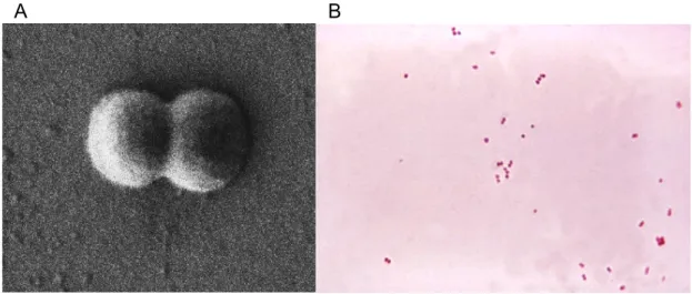
Neisseria meningitidis (Nm) is a Gram-negative B-proteobacterium. and member of the bacterial family of Neisseriaceae. It is a fastidious bacterium, dying within hours on inanimate surfaces, and is either an encapsulated or unencapsulated, aerobic diplococcus with a “kidney” or “coffee-bean” shape (Figure 1).
Figure 1. Neisseria meningitidis diplococcus. A. Scanning electron microscopy image of the gram negative bacterium Neisseria meningitidis, showing a diplococcus morphology. The size of the bacterium is about 1 micrometer (from http://www.stockholmmicro.se; owner Niklas Söderholm). B. N. meningitidis grown on an agar plate.
(from http://bioweb.uwlax.edu/bio203/s2008/bingen_sama)
Nm is a commensal and pathogen only of humans that are the unique reservoir for this bacterium. Virulence determinants include the polysaccharide capsule, outer membrane proteins including pili, the porins (PorA and B), the adhesion molecule Opc, iron sequestration mechanisms and endotoxin (lipooligosaccharide) [1]. N. meningitidis is now classified into 13 serogroups based on the immunogenicity and structure of the
polysaccharide capsule. At least 13 distinct meningococcal groups have been defined on the basis of their immunological reactivity and structure of the capsule’s polysaccharide [2]. These serogroups are the following: A, B, C, E-29, H, I, K, L, W-135, X, Y, Z, and Z’ (29E). Only six serogroups (A, B, C, W-135, X, Y) cause life-threatening disease. Further classification into serosubtype, serotype and immunotype is based on class 1 outer membrane proteins (PorA), class 2 or 3 (PorB) outer membrane proteins and lipopoly[oligo]saccharide structure, respectively [3], [1]. A genetic typing system based upon polymorphisms in multiple housekeeping genes (Multilocus Sequence Typing, MLST) is now the gold standard for molecular typing and has defined hyper-virulent meningococcal lineages [4].
Based on sequencing of eight genomes, the chromosome is between 2.0 and 2.1 megabases in size and contains about 2000 genes [73], [5], [6]. Each new strain sequenced has identified 40–50 new genes and the meningococcus shares about 90% homology at the nucleotide level with either N. gonorrhoeae or N. lactamica. Mobile genetic elements including IS elements and prophage sequences make up ~10% of the genome [5]. Other than the capsule locus, no core pathogenome has been identified suggesting that virulence may be clonal group dependent. Given that transformation is an efficient mechanism of genetic exchange and that meningococci have acquired DNA from commensal Neisseria spp. and other bacteria (e.g. Haemophilus) as well as phages, the gene pool for adaptation and evolution is quite large. Genome plasticity and phenotype diversity through gain and loss of DNA or, for example, through DNA repeats, is a characteristic of meningococcal evolution. This is in contrast to the relatively conserved genomes of for example Bacillus anthracis. The acquisition of the capsule locus by horizontal transfer possibly from Pasteurella multocida or P. hemolytica [6] appears to be a major event in the evolution of the pathogenicity of the meningococcus. Several repetitive sequence and polymorphic regions are present, usually in large heterogeneous arrays, suggesting active areas of genetic recombination. Another characteristic of the meningococcal genome is the presence of multiple genetic switches (e.g., slipped-strand misparing, IS element movement), contributing to the expression of pathogen-associated genes [7].
1.2 Meningococcal colonization and carriage
Colonization of the upper respiratory mucosal surfaces by N. meningitidis is the first step in establishment of a human carrier state and invasive meningococcal disease. Meningococcal transmission among humans occurs largely through respiratory droplets and secretions, but the inoculum size needed for transmission is unknown. Acquisition of N. meningitidis in the upper respiratory tract may be asymptomatic or may infrequently result in local inflammation, invasion of mucosal surfaces, access to the bloodstream and fulminant sepsis or focal infections such as meningitis [1]. Meningococcal disease usually occurs 1–14 days after acquisition of the pathogen [3]. Acquisition may also result in upper respiratory and pharyngeal meningococcal carriage. The duration of carriage can vary from days to months. From an evolutionary perspective, the interactions of meningococci and the human nasopharynx are key. Meningococcal carriage and transmission, not disease, determine the global variation and composition of the natural population of meningococci. The biological role of capsular polysaccharide in carriage/transmission is not well understood. Capsule-deficient strains may be transmitted efficiently, so the theory of resistance to dessication during transit or capsule antiadhesive properties promoting loss does not seem strong. Capsule is not required for efficient carriage. However, there is evidence of differences in propensity for carriage associated with the different capsular polysaccharides (serogroups), e.g. low carriage of serogroup C meningococci. The adaptive advantage of switching between the capsulate and non-capsulate state seems likely to provide a fitness advantage possibly for close adherence and initial steps in cell invasion. As noted, the redundancy of adhesins possessed by meningococci, e.g., pilus, Opa, NadA and their striking allelic variation, is impressive. The source of this variation is strongly influenced by lateral transfer of genetic information (recombination) and meningococci are naturally transformable. Nevertheless, most individuals are colonized with only one meningococcal strain (at least based on carriage studies) and this must place some constraints on the opportunity for genetic exchange between heterologous strains. Meningococcal adhesins are not known to be characteristic of other commensal Neisseria although this is not well studied. The relationship between meningococcal carriage rates and meningococcal disease and even the use of carriage prevalence as a proxy for predicting outbreaks of meningococcal disease has received considerable study. The important measure in terms of disease is the rate of acquisition of meningococci of
hypervirulent lineages, not overall meningococcal carriage [8]. The probability of meningococcal disease after the acquisition of N. meningitidis declines very sharply, such that invasive disease becomes unlikely 10–14 days after acquisition. Too little research has been done on the interactions of N. meningitidis with other upper respiratory tract commensals/pathogens. For example, carriage of pneumococci appears to be a risk factor for meningococcal carriage [9].
1.3 Invasive meningococcal disease
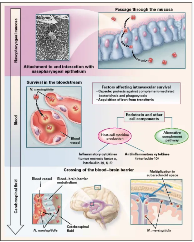
Meningococci that are transmitted by aerosol or contact with secretions colonize upper respiratory mucosal surfaces (e.g., the nasopharynx), may spread to adjacent mucosal surfaces (e.g., lower respiratory tract), and may invade epithelial surfaces and gain access to the bloodstream to produce systemic and focal infections (Figure 2).
Figure 2. Colonization of Neisseria meningitidis in the nasopharynx and entry into the bloodstream and cerebrospinal fluid. N. meningitidis enters the nasopharynx and attaches to nonciliated epithelial cells, probably through the binding of the pili to the CD46 receptor (a membrane cofactor protein) and the subsequent binding of opacity-associated proteins, Opa and Opc, to the CD66e (carcinoembryonic antigen) and heparan sulfate proteoglycan receptors, respectively. The attached organisms are engulfed by the cells, enter phagocytic vacuoles, and may then pass through the cells. IgA1 protease (an outer-membrane protein) cleaves lysosome-associated membrane protein and may promote the survival of N. meningitidis in epithelial cells.
PorB (another outermembrane protein) crosses the cell membrane and arrests the maturation of the phagosome. In the bloodstream, the organisms release endotoxin in the form of blebs (vesicular membrane structures) that contain 50 percent lipooligosaccharide and 50 percent outer-membrane proteins, phospholipids, and capsular polysaccharide. The endotoxin and probably other components stimulate cytokine production and the alternative complement pathway. N.
meningitidis crosses the blood–brain barrier endothelium by entering the subarachnoid space,
possibly through the choroid plexus of the lateral ventricles (from [3]).
Overcoming specific and nonspecific mucosal host defenses, crossing of the upper respiratory mucociliary blanket and attachment to human epithelial cells appears to be required for the colonization of the nasopharynx by meningococci. Twitching motility may allow meningococci to penetrate mucus and attach to epithelial cells. Meningococci can enter human nasopharyngeal epithelial cells by a process of parasite-induced endocytosis; bacteria may penetrate between epithelial or endothelial cells, transcytoses through them or are carried across the epithelial and endothelial barriers within cells (Trojan horse theory). Bacteria enter and accumulate in phagocytic vacuoles of nasopharyngeal epithelial cells, which may be released into interepithelial spaces below epithelial cell tight junctions. Alternatively, meningococci may invade upper respiratory epithelium damaged by smoking coinfections or other environmental factors. Entry of meningococci into the bloodstream is likely much more frequent than clinically recognized, but is usually transient. Once access to the bloodstream is obtained, meningococci may multiply rapidly to high levels. Meningococcal bacteremia can result in the seeding of the meninges, pericardium and large joints. Up to one third of patients with meningococcal disease present with meningitis or other closed space infections without signs of sepsis. Meningococci may also translocate across the blood-meningeal barrier, proliferate in the CNS and cause meningitis. How meningococci traverse the blood brain barrier and enter the cerebrospinal fluid (CSF) or reach other closed sites is unclear. Meningococci have been shown to invade endothelial cells both experimentally and in vivo. The chorioid plexus is also a potential site of entry of meningococci into the CSF. The inflammatory cytokines, TNF-α and IL-1, released in meningococcal bacteremia [10], may also enhance the permeability of the blood brain barrier and may allow meningococcal entry into the CSF. Meningitis and other closed space infections (e.g., arthritis, pericarditis) are the result of bacterial survival and multiplication at these sites.
The ability to cause invasive disease depends on environmental factors, meningococcal virulence factors and lack of a “protective immune response.” Environmental factors that impair the integrity of the human nasopharyngeal mucosa such as tobacco [1], exposure to low humidity, dust and co-infections, expecially influenza and mycoplasma, [11]; increase the incidence of invasive meningococcal disease. There is epidemiologic, biological and pathological evidence for each of these events. Major meningococcal contributors to the invasive meningococcal disease include: capsular polysaccharide, other surface structures [pili, OMPs (e.g. PorA, PorB, Opa, Opc), lipooligosaccharide (LOS)] and genotype. Resistance to complement-mediated lysis and phagocytosis is determined by the expression of the capsule and lipooligosaccharide [12]. Meningococcal endotoxin released in blebs also plays a major role in the inflammatory events of meningococcemia and meningococcal meningitis [13]. LOS plays a role in the adherence of the meningococcus [13] and activation of the innate immune system. Severity of meningococcal sepsis has been correlated with circulating levels of meningococcal LOS [5]. Pili and other OMPs, facilitate the adherence of the meningococcus to endothelial surfaces. In coclusion, multiple meningococcal invasion virulence factors influence invasive disease and some are the focus of new vaccines [14].
1.4 Meningococcal disease: epidemiology
Invasive meningococcal disease results from the interplay of: (1) microbial factors influencing the virulence of the organism, (2) environmental conditions facilitating exposure and acquisition, and (3) host susceptibility factors favoring bacterial acquisition, colonization, invasion, and survival. In the pre-serum therapy and pre-antibiotic eras, 70– 85% of meningococcal disease cases were fatal; today, the overall mortality rate in invasive meningococcal disease still remains high, at between 10 and 15% [15]. Meningococcal disease is also associated with marked morbidity including limb loss, hearing loss, cognitive dysfunction, visual impairment, educational difficulties, developmental delays, motor nerve deficits, seizure disorders, and behavioral problems [3]. Although rates of sporadic disease can reach ~5–10/100,000 population, a key characteristic of meningococcal disease are epidemic outbreaks. Seasonal epidemics
(usually due to serogroup A) occur yearly in sub-Saharan Africa and cyclical pandemics have occurred there every 8–10 years for the last 100 years. During seasonal epidemics and cyclical pandemics the incidence can climb to >1/1000 population for weeks before the frequency of disease declines in the immediate outbreak area. Serogroups B, C and Y are associated with sporadic disease, case clusters and outbreaks seen in the United States, Canada, New Zealand, South America, Europe and other parts of the world [3]. Serogroup W-135 is responsible for recent worldwide outbreaks associated with pilgrims returning from the Hajj [16]. The different characteristics of outbreaks are caused by hypervirulent lineages as defined by MLST. The worldwide W-135 outbreaks were caused by W-135 strains of the ST-37 clonal complex most often associated with serogroup C disease and outbreaks. The introduction of new virulent clones into a population can change the epidemiology and the clinical spectrum of meningococcal disease and the recent emergence of serogroup X meningococci in Niger [17] highlights the need for continued surveillance for new clonal complexes.
Meningococcal disease has the highest incidence in infants and children aged <4 years and adolescents [1]. Two-thirds of meningococcal disease in the first year of life in the US occurs in infants less than 6 months of age [18]. Worldwide, the rates of meningococcal disease are also highest for young children due to waning protective maternal antibody, but in epidemic outbreaks, older children and adolescents can have high rates of disease. Even though peak incidence occurs among infants and adolescents; one-third to one-half of sporadic cases are seen in adults older than 18 years. The early stages of disease can mimic viral infections such as influenza, but the disease course may be fulminant. Thus, it can be difficult to identify and treat the disease quickly. Rapid progression of the disease from bacteremia and/or meningitis to life-threatening septic shock syndrome or meningitis can occur within the first few hours after initial symptoms appear. Because of these parameters, prevention through vaccination is the best option for the control of this disease in a community. While significant progress is being made in understanding meningococcal pathogenesis and in new meningococcal vaccines and vaccine strategies, challenges remain. Dissecting the molecular basis of meningococcal pathogenesis also has important scientific lessons for understanding bacterial emergence, pathogenic genome structure, horizontal genetic exchange, and innate and adaptive immune responses [8].
1.5 The survival in blood as an important step of the pathogenesis
After breaching the mucosal barrier in the upper respiratory tract, invasive meningococci enter the circulation and start to proliferate. The growth velocity in the vasculature is a major determinant of the clinical presentation. A majority of patients reveal a comparatively low-grade meningococcemia, which subsequently seeds the subarachnoid space, where meningococci proliferate rapidly, leading to meningitis without shock symptoms. A minority of patients develop fulminant meningococcal septicemia, characterized by very rapid bacterial growth in the circulation [10], [1], [19] . The real bacterial loads in patients with fulminant meningococcemia have been quantified to be 105 to 108 bacteria/ml by determining the numbers of N. meningitidis DNA molecules, using robotized magnetic bead extraction of the bacterial DNA and real-time PCR [20], [21]. Survival and multiplication of meningococci in the bloodstream are directly dependent on the ability of the meningococcus to circumvent humoral and, to a lesser extent, phagocytic immune defenses. Multiplication of meningococci correlates with the systemic release of inflammatory cytokines (IL-1, IL-6, TNF-α), which are key elements in the pathogenesis of meningococcemia. In fact, the massive bacterial growth is reflected in high levels of endotoxin (lipopolysaccharide [LPS]), complement activation products, and cytokines in plasma [22], [23], [24]. Meningitis and its sequelae are due to the induction of local inflammatory cytokines and other mediators (e.g., nitric oxide), leukocyte infiltration across the blood brain barrier, breakdown of the blood brain barrier with edema, release of metaloproteases, induction of cellular apoptosis, coagulation of vessels and ischemia [22]. The ability of meningococcus to survive and grow in human whole blood is a crucial step of the bacterial pathogenesis and the establishment of an invasive disease. In fact, even in case of the development of meningitis, with a low grade of meningococcemia and no shock symptoms, the bacterium must survive and multiply in blood to reach the blood-brain barrier and enter in the subarachnoid space. To survive, Nm must adapt to the blood environment and to different interactions with host cells and factors; to do that, it has evolved molecular and cellular mechanism to protect itself from host immune system and to exploit the host to obtain nutrients. The knowledge of how Nm responds to adaptation in blood could be helpful to develop diagnostic and therapeutic strategies to control the devastating disease caused by this bacterium.mutagenesis (STM) approach to identify genes essential for bacteremia [25]. However, since Nm is an exclusively human pathogen, existing animal models may not accurately simulate meningococcal disease. For this reason, human whole blood has been used as an ex vivo model of sepsis for studying the pathogenesis of Nm in terms of complement activation, cytokine production and immunity [86], [26], [27], [28].
1.6 Global changes in the gene expression profile of meningococcus in
human whole blood
In order to evaluate the transcriptional response of Nm during growth in blood, in our research group we have analyzed the global changes in the transcriptional profile of a virulent Nm serogroup B strain in an ex vivo model of bacteremia, using incubation in human whole blood and a time-course oligo-microarray experiment [29]. The ex vivo human whole blood model enabled meningococcal responses to both host cellular and humoral bactericidal mechanisms to be analyzed and has shown potential to examine a number of parameters that are likely to be important in the cascade of events associated with acute systemic meningococcal infection [95] and to characterize Nm factors involved in the survival of the bacterium during infection [30], [31].
Freshly isolated whole venous blood collected from four healthy human volunteers (2 male
and 2 female) was used. Nm MC58 bacteria (approximately 1x10⁸, grown in GC medium
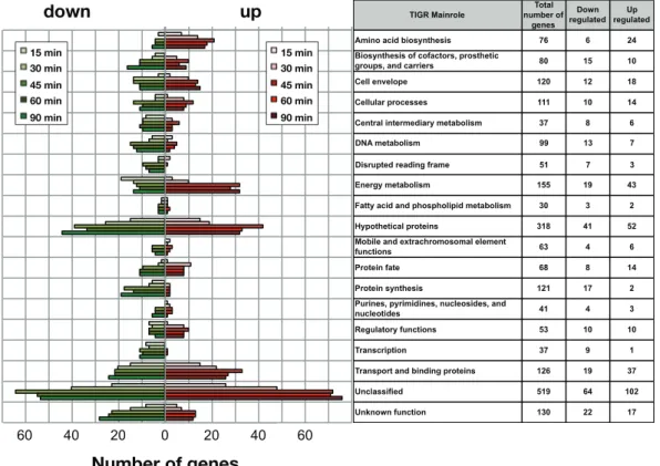
to early exponential phase) were mixed with blood from each donor in order to mimic disease. In order to evaluate the adaptation of Nm to human blood, samples were collected at six different time points: we then applied an in vitro transcription amplification/labeling step [32] to produce amplified-labeled cRNA that was then used in competitive hybridization experiments using a 60-mer Nm oligo-microarray. Transcriptional changes throughout the course of Nm incubation in human blood were defined by comparison of expression levels at various time points against time 0. The results showed that Nm alters the expression of nearly 30% of ORFs of the genome with 360 genes up-regulated and 277 genes down-regulated compared to the reference time 0. Interestingly, gene expression profiles clustered by K-means partitioning were modularly organized with respect to TIGRFAM functional classes [33] or KEGG metabolic pathways (Figure 3).
Figure 3. Time course distribution of up- and down- regulated genes within TIGRFAM main roles. The plot reflects the dynamics of Nm metabolic adaptation to blood, and the number of regulated genes within each TIGR family is shown for each time point. The total number of genes in each class and the number of up- and down-regulated genes are listed in the table (from [29]).
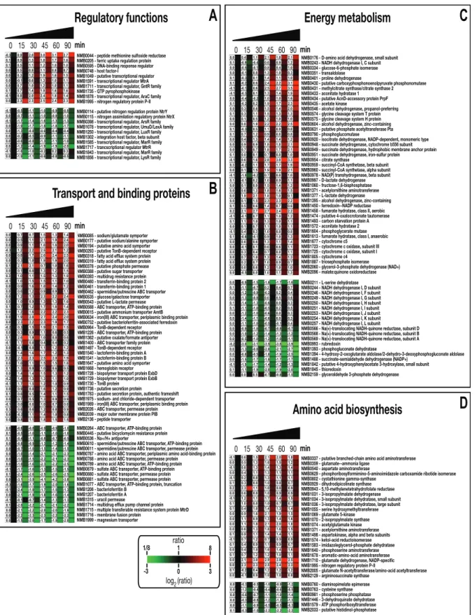
A wide range of hypothetical, unclassified ORFs and ORFs with unknown function was differentially regulated and the analysis of the unclassified ORFs regulated in blood may aid in their functional characterization. The major groups of differentially regulated genes are involved in energy metabolism, transport and binding, amino acid biosynthesis and regulatory functions (Figure 4).
TIGR Mainrole number of Total genes
Down regulated regulatedUp Amino acid biosynthesis 76 6 24 Biosynthesis of cofactors, prosthetic
groups, and carriers 80 15 10 Cell envelope 120 12 18 Cellular processes 111 10 14 Central intermediary metabolism 37 8 6
Disrupted reading frame 51 7 3 DNA metabolism 99 13 7
Energy metabolism 155 19 43 Fatty acid and phospholipid metabolism 30 3 2 Hypothetical proteins 318 41 52 Mobile and extrachromosomal element
functions 63 4 6
Protein fate 68 8 14 Protein synthesis 121 17 2 Purines, pyrimidines, nucleosides, and
nucleotides 41 4 3
Regulatory functions 53 10 10 Transcription 37 9 1 Transport and binding proteins 126 19 37 Unclassified 519 64 102 Unknown function 130 22 17 0 20 40 60 60 40 20 Number of genes 15 min 30 min 45 min 60 min 90 min 15 min 30 min 45 min 60 min 90 min down up
Figure 4. (see next page).
A
0 15 30 45 60 90min Transport!and!binding!proteins!C
Amino!acid!biosynthesis!B
Energy!metabolism!D
Regulatory!functions! NMB0085!-!sodium/glutamate!symporter NMB0177!-!putative!sodium/alanine!symporter NMB0194!-!putative!amino!acid!symporter NMB0293!-!putative!TonB-dependent!receptor NMB0318!-!fatty!acid!efflux!system!protein NMB0319!-!fatty!acid!efflux!system!protein NMB0378!-!putative!phosphate!permease NMB0388!-!putative!sugar!transporter NMB0393!-!multidrug!resistance!protein NMB0460!-!transferrin-binding!protein!2 NMB0461!-!transferrin-binding!protein!1 NMB0462!-!spermidine/putrescine!ABC!transporter NMB0535!-!glucose/galactose!transporter NMB0543!-!putative!L-lactate!permease NMB0588!-!ABC!transporter,!ATP-binding!protein NMB0615!-!putative!ammonium!transporter!AmtB NMB0634!-!iron(III)!ABC!transporter,!periplasmic!binding!protein NMB0752!-!putative!bacterioferritin-associated!ferredoxin NMB0964!-!TonB-dependent!receptor NMB1226!-!ABC!transporter,!ATP-binding!protein NMB1362!-!putative!oxalate/formate!antiporter NMB1400!-!ABC!transporter!family!protein NMB1497!-!TonB-dependent!receptor NMB1540!-!lactoferrin-binding!protein!A NMB1541!-!lactoferrin-binding!protein!B NMB1647!-!putative!amino!acid!symporter NMB1668!-!hemoglobin!receptor NMB1728!-!biopolymer!transport!protein!ExbD NMB1729!-!biopolymer!transport!protein!ExbB NMB1730!-!TonB!protein NMB1738!-!putative!secretion!protein NMB1783!-!putative!secretion!protein,!authentic!frameshift NMB1975!-!sodium-!and!chloride-dependent!transporter NMB1989!-!iron(III)!ABC!transporter,!periplasmic!binding!protein NMB2026!-!ABC!transporter,!permease!protein NMB2039!-!major!outer!membrane!protein!PIB NMB2136!-!peptide!transporter NMB0264!-!ABC!transporter,!ATP-binding!protein NMB0445!-!putative!bicyclomycin!resistance!protein NMB0536!-!Na+/H+!antiporter NMB0610!-!spermidine/putrescine!ABC!transporter,!ATP-binding!protein NMB0611!-!spermidine/putrescine!ABC!transporter,!permease!protein NMB0787!-!amino!acid!ABC!transporter,!periplasmic!amino!acid-binding!protein NMB0788!-!amino!acid!ABC!transporter,!permease!protein NMB0789!-!amino!acid!ABC!transporter,!ATP-binding!protein NMB0879!-!sulfate!ABC!transporter,!ATP-binding!protein NMB0880!-!sulfate!ABC!transporter,!permease!protein NMB0881!-!sulfate!ABC!transporter,!permease!protein NMB1077!-!ABC!transporter,!ATP-binding!protein,!truncation NMB1206!-!bacterioferritin!B NMB1207!-!bacterioferritin!A NMB1315!-!uracil!permease NMB1714!-!multidrug!efflux!pump!channel!protein NMB1715!-!multiple!transferable!resistance!system!protein!MtrD NMB1716!-!membrane!fusion!protein NMB1999!-!magnesium!transporter NMB0337!-!putative!branched-chain!amino!acid!aminotransferase NMB0359!-!glutamate--ammonia!ligase NMB0540!-!aspartate!aminotransferase NMB0629!-!phosphoribosylformimino-5-aminoimidazole!carboxamide!ribotide!isomerase NMB0802!-!cystathionine!gamma-synthase NMB0929!-!dihydrodipicolinate!synthase NMB0943!-!5,10-methylenetetrahydrofolate!reductase NMB1031!-!3-isopropylmalate!dehydrogenase NMB1034!-!3-isopropylmalate!dehydratase,!small!subunit NMB1036!-!3-isopropylmalate!dehydratase,!large!subunit NMB1055!-!serine!hydroxymethyltransferase NMB1069!-!glutamate!5-kinase NMB1070!-!2-isopropylmalate!synthase NMB1074!-!acetylglutamate!kinase NMB1371!-!acetylornithine!aminotransferase NMB1498!-!aspartokinase,!alpha!and!beta!subunits NMB1574!-!ketol-acid!reductoisomerase NMB1583!-!imidazoleglycerol-phosphate!dehydratase NMB1640!-!phosphoserine!aminotransferase NMB1678!-!aromatic-amino-acid!aminotransferase NMB1710!-!glutamate!dehydrogenase,!NADP-specific NMB1995!-!nitrogen!regulatory!protein!P-II NMB2005!-!glutamate!N-acetyltransferase/amino-acid!acetyltransferase NMB2129!-!argininosuccinate!synthase NMB0760!-!diaminopimelate!epimerase NMB0763!-!cysteine!synthase NMB0981!-!phosphoserine!phosphatase NMB1446!-!3-dehydroquinate!dehydratase NMB1579!-!ATP!phosphoribosyltransferase NMB2033!-!putative!histidinol-phosphatase NMB0176!-!D-amino!acid!dehydrogenase,!small!subunit NMB0243!-!NADH!dehydrogenase!I,!C!subunit NMB0334!-!glucose-6-phosphate!isomerase NMB0351!-!transaldolase NMB0401!-!proline!dehydrogenase NMB0430!-!putative!carboxyphosphonoenolpyruvate!phosphonomutase NMB0431!-!methylcitrate!synthase/citrate!synthase!2 NMB0433!-!aconitate!hydratase!1 NMB0434!-!putative!AcnD-accessory!protein!PrpF NMB0435!-!acetate!kinase NMB0546!-!alcohol!dehydrogenase,!propanol-preferring NMB0574!-!glycine!cleavage!system!T!protein NMB0575!-!glycine!cleavage!system!H!protein NMB0604!-!alcohol!dehydrogenase,!zinc-containing NMB0631!-!putative!phosphate!acetyltransferase!Pta NMB0790!-!phosphoglucomutase NMB0920!-!isocitrate!dehydrogenase,!NADP-dependent,!monomeric!type NMB0948!-!succinate!dehydrogenase,!cytochrome!b556!subunit NMB0949!-!succinate!dehydrogenase,!hydrophobic!membrane!anchor!protein NMB0951!-!succinate!dehydrogenase,!iron-sulfur!protein NMB0954!-!citrate!synthase NMB0959!-!succinyl-CoA!synthetase,!beta!subunit NMB0960!-!succinyl-CoA!synthetase,!alpha!subunit NMB0978!-!NAD(P)!transhydrogenase,!beta!subunit NMB0997!-!D-lactate!dehydrogenase NMB1060!-!fructose-1,6-bisphosphatase NMB1371!-!acetylornithine!aminotransferase NMB1377!-!L-lactate!dehydrogenase NMB1395!-!alcohol!dehydrogenase,!zinc-containing NMB1450!-!ferredoxin--NADP!reductase NMB1458!-!fumarate!hydratase,!class!II,!aerobic NMB1474!-!putative!4-oxalocrotonate!tautomerase NMB1493!-!carbon!starvation!protein!A NMB1572!-!aconitate!hydratase!2 NMB1604!-!phosphoglycerate!mutase NMB1613!-!fumarate!hydratase,!class!I,!anaerobic NMB1677!-!cytochrome!c5 NMB1723!-!cytochrome!c!oxidase,!subunit!III NMB1725!-!cytochrome!c!oxidase,!subunit!I NMB1805!-!cytochrome!c4 NMB1887!-!triosephosphate!isomerase NMB2060!-!glycerol-3-phosphate!dehydrogenase!(NAD+) NMB2096!-!malate:quinone!oxidoreductase NMB0211!-!L-serine!dehydratase NMB0244!-!NADH!dehydrogenase!I,!D!subunit NMB0246!-!NADH!dehydrogenase!I,!F!subunit NMB0249!-!NADH!dehydrogenase!I,!G!subunit NMB0250!-!NADH!dehydrogenase!I,!H!subunit NMB0251!-!NADH!dehydrogenase!I,!I!subunit NMB0253!-!NADH!dehydrogenase!I,!J!subunit NMB0254!-!NADH!dehydrogenase!I,!K!subunit NMB0257!-!NADH!dehydrogenase!I,!L!subunit NMB0566!-!Na(+)-translocating!NADH-quinone!reductase,!subunit!D NMB0568!-!Na(+)-translocating!NADH-quinone!reductase,!subunit!B NMB0569!-!Na(+)-translocating!NADH-quinone!reductase,!subunit!A NMB0993!-!rubredoxin NMB1393!-!phosphogluconate!dehydratase NMB1394!-!4-hydroxy-2-oxoglutarate!aldolase/2-dehydro-3-deoxyphosphogluconate!aldolase NMB1488!-!succinate-semialdehyde!dehydrogenase!(NADP+) NMB1842!-!putative!4-hydroxyphenylacetate!3-hydroxylase,!small!subunit NMB1845!-!thioredoxin NMB2159!-!glyceraldehyde!3-phosphate!dehydrogenase NMB0114!-!putative!nitrogen!regulation!protein!NtrY NMB0115!-!nitrogen!assimilation!regulatory!protein!NtrX NMB0398!-!transcriptional!regulator,!ArsR!family NMB1078!-!transcriptional!regulator,!UmuD/LexA!family NMB1250!-!transcriptional!regulator,!LuxR!family NMB1302!-!integration!host!factor,!beta!subunit NMB1585!-!transcriptional!regulator,!MarR!family NMB1717!-!trancscriptional!regulator!MtrR NMB1843!-!transcriptional!regulator,!MarR!family NMB1856!-!transcriptional!regulator,!LysR!family -3 0 3 1/8 1 8 NMB0044!-!peptide!methionine!sulfoxide!reductase NMB0205!-!ferric!uptake!regulation!protein NMB0595!-!DNA-binding!response!regulator NMB0748!-!host!factor-I NMB1049!-!putative!transcriptional!regulator NMB1591!-!transcriptional!regulator!MtrA NMB1711!-!transcriptional!regulator,!GntR!family NMB1735!-!GTP!pyrophosphokinase NMB1878!-!transcriptional!regulator,!AraC!family NMB1995!-!nitrogen!regulatory!protein!P-II ratio log2(ratio) -0.3 0.9 2.0 3.0 3.1 2.7 0.1 0.9 1.3 1.8 1.7 1.6 0.1 0.5 1.1 1.2 1.0 1.0 0.0 0.5 0.8 1.1 0.8 0.7 0.0 0.3 1.2 1.9 1.6 1.5 0.0 -0.1 0.6 1.3 1.0 1.1 0.0 0.7 1.0 1.2 1.0 1.1 0.1 0.7 1.4 1.9 1.6 1.6 -0.1 0.8 2.0 2.4 1.8 1.4 0.0 2.3 2.3 2.5 2.4 2.3 -0.1 -0.5 -1.1 -1.0 -0.9 -0.8 0.0 -0.7 -1.1 -1.0 -0.9 -1.0 -0.1 -1.7 -1.4 -1.6 -1.3 -1.3 0.0 -1.9 -1.7 -1.8 -1.8 -1.7 0.0 -1.3 -0.8 -0.6 -0.4 -0.4 0.0 -0.2 -0.5 -0.9 -1.1 -1.1 0.0 -0.6 -0.8 -1.3 -1.1 -1.0 0.0 -1.2 -1.4 -1.9 -1.9 -2.0 -0.1 -1.6 -1.0 -0.2 0.2 0.3 0.0 -1.9 -1.1 -0.6 -0.8 -0.5 0 15 30 45 60 90min 0 15 30 45 60 90 min 0 15 30 45 60 90 min 0.0 0.7 0.7 1.5 1.3 1.4 0.0 0.5 1.1 2.1 1.9 2.4 0.0 0.9 1.1 1.8 1.8 2.0 -0.3 1.2 2.2 2.5 2.1 1.2 0.0 0.8 1.7 2.6 2.4 2.5 0.0 0.3 1.0 1.9 1.9 2.0 0.1 2.1 2.1 0.7 1.0 1.6 0.0 0.1 0.8 1.3 1.2 1.5 0.3 1.0 1.1 1.5 1.3 1.9 -0.1 1.0 1.5 1.5 1.1 1.0 0.0 0.9 1.6 1.8 1.4 1.2 0.0 0.6 1.2 0.9 0.9 1.1 0.0 1.2 1.3 2.3 2.2 2.4 0.0 1.0 0.9 1.3 1.3 1.6 0.1 0.7 1.2 1.7 1.8 1.4 0.0 1.2 1.6 2.2 2.2 2.3 0.0 1.9 2.8 2.7 2.4 2.0 0.0 2.3 2.2 2.3 1.9 1.7 -0.2 0.3 1.5 1.9 2.1 1.5 0.0 0.3 0.9 1.1 0.6 0.5 0.1 2.6 1.9 1.0 0.8 0.6 -0.1 1.7 0.8 0.4 0.4 0.2 0.0 -0.1 0.8 1.1 1.2 1.2 0.0 1.4 1.6 1.7 1.2 1.3 0.2 2.3 2.3 2.5 2.0 1.9 0.0 1.0 1.1 1.5 1.5 1.8 0.1 1.3 0.9 1.0 0.6 0.5 0.0 1.4 1.4 1.5 1.3 1.2 -0.2 1.4 1.3 1.3 1.1 1.0 -0.1 1.7 1.5 1.2 1.0 0.9 0.0 0.9 0.8 1.6 1.2 0.9 0.1 0.6 0.5 1.4 1.1 1.5 0.0 0.7 0.5 1.3 0.9 0.9 -0.1 0.0 0.6 1.0 0.7 0.6 0.3 0.6 0.6 1.2 0.8 0.9 0.0 -0.2 1.1 1.4 1.0 0.8 0.0 0.8 1.1 1.8 1.5 1.5 0.0 -0.6 -0.8 -0.9 -0.9 -1.0 0.1 -0.5 -1.0 -1.1 -1.0 -1.0 0.1 -0.4 -1.1 -0.8 -0.7 -0.5 0.0 -0.8 -0.9 -1.3 -1.2 -1.4 0.0 -0.7 -1.0 -1.5 -1.4 -1.5 0.0 -1.0 -1.4 -2.1 -2.1 -2.3 0.0 -1.0 -1.5 -2.2 -2.1 -2.3 0.0 -1.0 -1.6 -2.3 -2.2 -2.5 -0.1 -1.7 -1.6 -1.3 -1.5 -1.5 0.1 -2.4 -2.6 -2.5 -2.6 -2.5 0.0 -3.1 -3.9 -4.1 -4.2 -3.8 -0.3 -1.6 -1.7 -1.9 -2.0 -1.6 -0.1 -2.4 -1.9 -1.7 -1.5 -1.3 0.0 -2.1 -1.9 -1.8 -1.6 -1.4 -0.1 -0.1 -0.7 -1.4 -1.3 -1.3 0.1 -2.0 -2.2 -2.1 -2.1 -2.2 0.0 -2.1 -2.2 -2.0 -2.0 -1.9 0.0 -2.2 -2.3 -2.3 -2.2 -2.2 0.0 -1.5 -2.0 -1.7 -1.9 -2.0 0.0 0.4 1.1 2.1 1.8 2.1 0.1 -0.9 -0.4 0.6 0.9 1.2 0.0 -0.5 -0.1 0.8 0.4 1.0 0.0 -0.1 0.8 1.3 1.0 0.3 0.1 -0.3 0.1 1.4 1.1 1.0 0.1 -0.2 0.6 2.4 2.2 3.1 -0.1 -0.1 0.6 2.5 2.7 3.5 -0.1 -0.3 0.1 1.3 1.4 1.9 0.0 -0.4 -0.2 1.0 1.0 1.6 0.1 -0.2 0.2 1.9 1.7 2.3 0.0 0.3 0.6 1.5 1.4 2.0 0.1 0.3 1.1 1.5 1.3 1.3 0.0 0.1 0.5 1.2 1.0 1.0 0.0 0.8 1.9 2.2 2.2 2.5 0.1 -0.3 0.5 1.1 0.9 0.8 0.1 0.7 1.2 0.9 0.8 0.8 0.0 0.4 1.3 1.4 1.4 1.2 0.0 0.9 0.9 1.8 1.8 2.1 0.0 0.7 0.7 1.8 1.7 2.1 0.1 0.0 0.1 0.7 0.8 1.0 -0.1 0.6 1.6 2.8 3.0 3.3 0.3 -0.2 1.1 1.7 1.7 1.9 0.0 0.1 1.5 1.9 1.9 2.1 -0.1 -1.0 -0.2 0.7 0.8 1.0 0.1 -0.1 0.2 1.0 0.9 1.1 0.0 0.1 0.9 1.5 1.4 1.3 0.0 0.6 1.3 1.8 1.6 1.5 0.0 1.9 2.1 1.8 1.0 0.1 0.0 0.1 1.5 2.3 2.3 2.2 0.1 0.5 0.6 1.4 0.7 0.6 0.2 2.1 2.7 2.7 2.4 2.2 -0.1 -0.1 0.7 1.1 1.0 1.2 0.1 1.1 1.0 1.3 1.0 1.1 0.0 0.1 0.7 1.1 1.0 1.1 -0.1 0.4 0.3 1.0 0.8 0.8 0.1 -0.6 -0.1 0.9 1.0 1.3 0.0 0.6 0.6 1.3 1.1 1.4 0.0 -0.1 0.1 0.6 0.6 1.1 0.0 0.3 0.4 0.7 0.8 1.1 0.0 0.7 1.0 1.3 1.4 1.3 0.0 0.2 0.7 1.0 0.9 0.7 0.0 0.0 0.6 1.2 1.0 1.0 0.0 -0.5 0.0 1.0 1.1 1.3 0.0 -0.9 -1.5 -1.8 -1.7 -1.6 0.1 -1.1 -0.7 0.2 0.4 0.7 0.0 -1.2 -0.9 -0.2 0.2 0.4 0.0 -1.3 -0.8 0.0 0.5 0.6 0.1 -1.4 -1.3 -0.6 0.0 0.3 0.1 -1.1 -1.0 -0.5 0.2 0.5 0.0 -1.2 -1.0 -0.6 0.0 0.3 0.0 -1.4 -1.1 -0.6 0.0 0.3 0.0 -1.1 -1.0 -0.5 -0.1 0.4 0.0 -0.9 -1.0 -0.7 -0.8 -0.7 0.0 -1.0 -1.2 -1.4 -1.3 -1.2 0.0 -0.5 -1.0 -1.2 -1.0 -1.1 -0.2 -3.0 -3.1 -3.5 -3.8 -3.9 0.0 -1.3 -0.8 -2.0 -2.1 -2.7 0.0 -0.8 -0.5 -1.8 -1.9 -2.5 0.0 -1.5 -1.7 -1.8 -1.7 -1.7 -0.1 -1.9 -1.7 -1.4 -1.2 -1.3 0.1 -1.1 -1.9 -2.0 -1.9 -2.0 0.0 0.0 0.5 -0.7 -0.8 -1.5 -0.1 0.6 1.5 2.1 2.0 1.8 0.0 0.5 1.3 1.6 1.5 1.2 -0.1 0.8 1.3 1.4 1.3 1.4 0.0 0.4 1.1 1.3 1.1 1.1 -0.1 0.2 0.8 1.3 1.1 1.1 0.1 0.3 0.8 1.0 0.8 0.7 0.2 2.3 1.1 1.0 0.8 0.9 0.0 0.6 0.8 1.0 0.9 0.9 -0.1 1.2 1.1 1.7 1.4 1.5 0.1 1.0 0.8 1.0 0.7 0.7 0.1 0.4 1.0 1.4 1.3 1.4 0.0 0.9 1.6 2.1 1.9 1.5 0.0 1.1 0.9 1.3 1.4 1.6 0.0 1.1 1.8 2.1 2.0 1.8 0.0 0.6 1.3 1.8 1.6 1.5 0.1 0.9 1.1 1.4 1.4 1.1 -0.1 0.7 1.2 0.7 0.9 0.9 0.1 0.4 0.9 1.1 0.9 0.7 0.0 0.4 1.0 1.6 1.4 1.6 0.1 0.7 1.2 1.2 1.0 1.1 0.0 2.1 3.0 2.9 2.8 2.5 0.0 2.3 2.3 2.5 2.4 2.3 0.1 -0.2 0.4 1.2 1.2 1.2 0.0 -0.1 0.9 1.0 1.0 0.8 0.0 -0.3 -0.5 -1.0 -0.9 -1.2 0.0 -1.2 -0.4 -0.4 -0.6 -0.5 0.0 -0.6 -0.7 -1.1 -1.0 -1.4 0.0 -0.3 -0.8 -1.2 -1.2 -1.3 0.0 -1.1 -0.9 -0.5 -0.5 -0.3 0.1 -0.3 -1.0 -2.2 -2.2 -2.2Figure 4. Transcriptional profile of differentially regulated genes grouped by functional TIGRFAM family main roles. Detailed expression profiles of functionally related genes during the time course of Nm in human whole blood. Clusters were created using TMEV. (A) Regulatory functions (B) Transport and binding proteins (C) Energy metabolism (D) Amino acid biosynthesis. Each gene is represented by a single row and each time point by a single column; gene identification numbers (based on the MC58 annotation) and gene definitions are reported on the right. Gene expression is displayed in fold change represented by the color bar under the figure. The numerical gene expression values are shown for all the genes at the different time points (from [29]).
These groups are predominantly upregulated, suggesting a high degree of metabolic adaptation occurs in blood, enabling uptake of different substrates and induction of alternative metabolic pathways. In particular, we found that the gene encoding the regulator Fur, as well as all genes encoding iron uptake systems, were significantly up-regulated. Analysis of regulated genes encoding for surface-exposed proteins involved in Nm pathogenesis allowed us to better understand mechanisms used to circumvent host defenses. During blood infection, Nm activates genes encoding for the factor H binding proteins, fHbp and NspA, genes encoding for detoxifying enzymes such as SodC, Kat and AniA, as well as several less characterized surface-exposed proteins that might have a role in blood survival.
1.7 A broader view of transcriptional adaptation of bacteria to the
environment
To survive and thrive in an often hostile environment, a bacterium has to monitor its surroundings and adjust its gene expression and physiology accordingly. This is especially important for pathogenic bacteria, which continuously interact with the host during an infection and need to tighly regulate virulence factors at gene expression level, in order to control the various steps of pathogenesis. Traditionally, this regulation have been
accredited to the activity of transcriptional factors, but studies in the past years have largely demonstrated the expression in bacteria of RNA regulatory molecules. Moreover, regulation mediated by RNAs and their associated proteins is more common than previously thought. Genome-wide analyses based on tiling arrays and high-throughput RNA-sequencing technologies have revealed the transcriptomes of several bacteria, including pathogenic bacteria [34]. Both techniques allow the unbiased visualization of all RNA molecules transcribed during specific conditions (for example, during stress, high osmolarity and low oxygen) without taking into account the positions of annotated ORFs. Therefore, the positions of all RNAs transcribed in a cell — namely, mRNAs (including operons), tRNAs, ribosomal RNAs, cis-acting antisense RNAs and trans-acting sRNAs — can be mapped with 1-nucleotide resolution. These studies have discovered unexpected numbers of small RNAs and other species of RNA regulators, that act to control the expression of genes encoding virulence factors at multiple levels and in a temporal manner, according to the stress [35], [36], [37], [38] and have shown that the transcriptional landscape of all organisms is much more complex than expected.
With this regard, it is conceivable that Neisseria meningitis can possess a circuit of regulatory RNAs involved in the regulation of different steps of pathogenesis. Indeed, three research groups independently demonstrated that the RNA chaperone Hfq, which is up regulated during incubation in human whole blood [29], is involved in stress response and virulence in Nm and is a pleiotropic regulator of protein expression, suggesting the presence in Nm of a large small RNA network, not yet defined [30], [39], [40].
1.8 Regulatory RNAs
RNAs are excellent regulatory molecules that can carry out a plethora of regulatory tasks [41]. For instance, RNAs can directly sense environmental cues, such as differences in temperature, pH and nutrient availability, through regulatory regions that lie upstream of the coding sequence on the same transcript, leading to an altered transcriptional read-through or translation initiation of that downstream coding sequence. Furthermore, bacteria can regulate transcript expression through cis-acting RNAs, which function in an antisense manner and control the expression of mRNAs encoded on the opposite DNA strand, or
trans-acting small non-coding RNAs (sRNAs), which function at a distance to alter the expression of target RNAs through an antisense mechanism. The fate of RNA transcripts can also be controlled by proteins, including RNases and RNA chaperones, which can degrade both cis- and trans-acting antisense regulatory RNAs and their target mRNAs or can facilitate the interaction of trans-acting sRNAs with the target mRNAs, respectively (Figure 5).
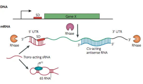
Figure 5. Control of mrNA activity and stability. The fate of an mRNA is controlled by several factors. 5′ untranslated regions (UTRs) lie upstream of the coding sequences and include thermosensors, pH sensors and riboswitches. 3′ UTRs lie at the other end of coding RNAs and, in many cases, determine transcription termination and stability. Cis-acting antisense RNAs are expressed from the opposite strand of the DNA. Trans-acting small non-coding RNAs (sRNAs) are expressed from a different location on the chromosome and most commonly bind to the Shine– Dalgarno (SD) sequence of a target mRNA in an antisense manner. Trans-acting sRNAs can also sequester target proteins; for example, 6S RNA sequesters the housekeeping RNA polymerase σ70 (also known as RpoD). The fate of transcripts can also be controlled by RNases, which function either exonucleolytically (orange), by targeting transcripts from the 5′ or 3′ end (such as RNase J1 and RNase J2, which are found in some Gram-positive bacteria) or endonucleolytically (purple), by targeting sequences in the middle of the transcript (from [42]).
Because pathogenic bacteria encounter diverse environmental conditions, they require rapid regulatory circuits to survive, making regulatory RNAs particularly suitable for controlling bacterial virulence. Together with regulatory proteins and two-component systems, regulatory RNAs integrate environmental stimuli into outputs that are important for pathogenicity. In fact, there is evidence that regulatory RNAs are more suitable than proteins for controlling gene expression. First, the energy cost of transcription (that is, generating regulatory and target RNAs) is much lower than that of translating regulatory proteins. Second, regulatory RNAs can control gene expression much faster than regulatory proteins; this is especially true for 5′ untranslated regions (UTRs), which directly dictate the expression of downstream mRNAs on sensing an environmental cue. Third, regulatory RNAs are generally much less stable than regulatory proteins, which allow their rapid clearance when they are no longer needed. Fourth, many regulatory RNAs act at post-transcriptional level and can therefore modify mRNAs that have already been expressed; they can thereby dictate and possibly overcome effects at the transcriptional level.
1.8.1 Trans-acting small RNAs
RNA molecules acting in trans on distant targets are commonly denoted as trans-acting sRNAs and are perhaps the best-studied form of regulatory RNA [43]. Trans-acting sRNAs, traditionally identified in intergenic regions, are encoded distally from their targets and share only limited complementarity with their target mRNAs.
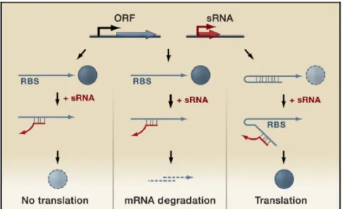
These sRNAs regulate the translation and/or stability of target mRNAs and are, in many respects, functionally analogous to eukaryotic miRNAs. The majority of the regulation by the known trans-encoded sRNAs is negative [43]. Base pairing between the sRNA and its target mRNA usually leads to repression of protein levels through translational inhibition, mRNA degradation, or both (Figure 6). The bacterial sRNAs primarily bind to the 5’ UTR of mRNAs and most often occlude the ribosome binding site, inhibiting translation through base pairing far upstream of the AUG of the repressed gene ([44]; [45]). The sRNA-mRNA duplex is then frequently subject to degradation by RNase E. However, sRNAs can also activate expression of their target mRNAs through an anti-antisense mechanism
whereby base pairing of the sRNA disrupts an inhibitory secondary structure, which sequesters the ribosome binding site [46], [47], [48] (Figure 6).
Given that sRNAs are often degraded along with the mRNAs they regulate, in other words they are consumed in the process of regulation, the use of RNA regulators might be advantageous over regulatory proteins in achieving a fast and irreversible transition, which is often required during pathogenesis [49].
Figure 6. Regulatory functions of trans-acting small RNAs. Genome loci encoding trans-acting small RNAs (red) are located separate from the genes encoding their target RNAs (blue) and only have limited complementarity. These small RNAs can act negatively by base pairing with the 5’ UTR and blocking ribosome binding (left panel) and/or targeting the sRNA-mRNA duplex for degradation by RNases (middle panel). Trans-acting sRNA can act positively by preventing the formation of an inhibitory structure, which sequesters the ribosome binding site (RBS) (right panel) (from [43]).
For trans-acting sRNAs, there is little correlation between the chromosomal location of the sRNA gene and the target mRNA gene. In fact, each trans-encoded sRNA typically base pairs with multiple mRNAs. The capacity for multiple base pairing interactions results from imperfect base-pairing with target mRNAs, rather than extended stretches of perfect complementarity, as for cis-encoded antisense sRNAs. The region of potential base pairing between trans-acting sRNAs and target mRNAs typically encompasses ~10–25 nucleotides, but in all cases where it has been examined only a core of the nucleotides
seem to be critical for regulation. In many cases, the RNA chaperone Hfq is required for trans-acting sRNA-mediated regulation, presumably to facilitate RNA-RNA interactions due to limited complementarity between the sRNA and target mRNA. Hfq-associated small RNAs are diverse in length (50-250 nucleotides); in several cases, Hfq binds in a AU-rich single strand region located upstream of the terminator and this binding may then expose the target binding domain of the sRNA to the potential mRNA partners [50]. Beyond facilitating base pairing, Hfq contributes to sRNA regulation through modulating sRNA levels. Once base paired with target mRNAs, many of the known sRNA-mRNA pairs are subject to degradation by RNaseE. Nevertheless, there are also cases of Hfq-independent sRNAs, for example, the recently characterized sRNA VrrA in V. cholerae, which modulates colonization of the host small intestine [51]. In general, longer stretches of base pairing, as is the case for the cis-encoded antisense sRNAs, usually do not require Hfq for function, and/or high concentrations of the sRNA may obviate a chaperone requirement.
Most of the trans-encoded sRNAs are synthesized under very specific growth conditions. In E. coli for example, these regulatory RNAs are induced by low iron (Fur-repressed RyhB), oxidative stress (OxyR-activated OxyS), outer membrane stress (σE-induced MicA and RybB) and elevated glycine (GcvA-induced GcvB) [52], [53], [54]. Most trans-acting sRNAs are involved in responding to rapidly changing environmental conditions, and there are cases of sRNAs with a role in virulence and pathogenesis, for example: Rli38 of L. monocytogenes [36], induced in human blood, RNAIII of S. aureus [55,56] and Qrr of V. cholera [57] involved in quorum-sensing and SprD of S. aureus, which negatively regulates the expression of the Sbi immune-evasion molecule [58].
The fact that a given base pairing sRNA often regulates multiple targets means that a single sRNA can globally modulate a particular physiological response, in much the same manner as a transcription factor, but at the post-transcriptional level [43].
1.8.2 Cis-encoded antisense RNAs
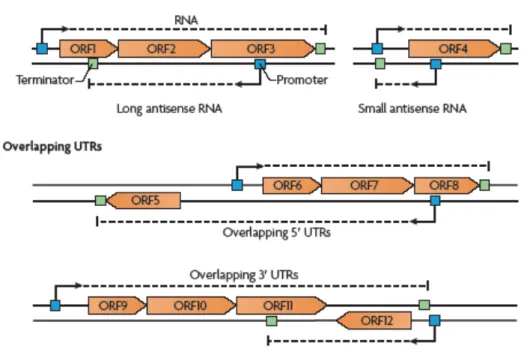
Cis acting antisense RNAs consist of two subtypes: bona fide antisense RNAs, which are present on the complementary strand to one or several ORFs, or overlapping UTRs, which
consist of a long 5’ or 3’ UTR of an mRNA that overlaps with the mRNA encoded by the other DNA strand (Figure 7).
Figure 7. Cis-acting antisense RNAs. Schematic representation of the different types of antisense RNA molecules. These include bona fide antisense RNAs and overlapping 5′ and 3′ untranslated regions (UTRs). The antisense RNA may be either a long antisense RNA covering more than one ORF (in the example, the long antisense RNA overlaps ORF1, ORF2 and ORF3) or a small antisense RNA that overlaps the Shine–Dalgarno sequence (which lies between the promoter and the start codon) and affects mRNA stability and/or protein translation (for example, the antisense RNA overlapping ORF4). Overlapping 5′ UTRs are generated when the transcription of a certain gene (for example, ORF5) starts from a promoter located on the DNA strand opposite divergent genes (in this case, ORF6, ORF7 and ORF8). As a result, the ORF5 mRNA has a long 5′ UTR that overlaps the ORF6, ORF7 and ORF8 mRNA. Overlapping 3′ UTRs are generated when transcription of a certain gene ends in or after a gene located on the opposite DNA strand; for example, the transcription of the operon encoding ORF9, ORF10 and ORF11 ends after ORF12, so the long 3′ UTR completely overlaps the ORF12 mRNA (from [42]).
Until a few years ago, the understanding of cis-acting antisense RNAs was limited to studies in bacteriophages, plasmids and transposons. Recent technological advancements have shown that antisense transcription occurs in all species, including bacteria. Many
antisense transcripts have been found in Helicobacter pylori (46% of all ORFs were associated with at least one antisense transcription start site [38], Listeria monocytogenes [59] but also Mycoplasma pneumoniae, Pseudomonas syringae and Bacillus subtilis [60,61] [62]. It therefore seems that cis acting antisense RNAs are extremely abundant and the size of antisense transcripts can range from few bases to several kilobases, so one particular antisense RNA may overlap several genes (as reported for L. monocytogenes and B. subtilis).
Plasmid encoded antisense RNAs involved in the regulation of replication, are expressed constitutively. By contrast, chromosomally encoded antisense RNAs have been found to be expressed only under certain conditions, for example in stationary phase (e.g. GadY) [63] or under iron stress (e.g. IsrR) [64]. In H. pylori, seveval cis-acting antisense RNAs and their corresponding mRNA targets are induced by the same signal (in this case, acid stress). This is in contrast to most trans-acting sRNAs, which are typically controlled by a different regulator than their mRNA target.
Antisense RNAs could provide an advantage when the levels of a particular protein need to be repressed very tightly and expressed under very select circumstances, as in the case of transposase or toxin. Indeed, many of the characterized antisense RNA targets are subject to extensive regulation, where the antisense RNAs provide yet one more level of control [65]. In general, it is conceivable that only mRNA that is present at higher levels than its cis-acting antisense antagonist is translated, so translation of certain genes starts only when the mRNA concentration reaches a certain level [34].
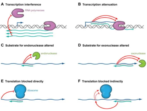
In the majority of cases, antisense RNA action entails post-transcriptional inhibition of target RNA function but, in few cases, activating mechanisms have been found too. The currently known mechanisms employed by cis-encoded antisense RNAs are (Figure 8) [65]:
• Transcription interference: when transcription from one promoter is suppressed by a second promoter present in cis [66];
• Transcription attenuation: generally found on plasmids, when base pairing of the antisense RNA to the mRNA induce the formation of a terminator structure in the target mRNA;
• RNA cleavage: antisense RNA can also impact the stability of a target RNA by either promoting or blocking cleavage by endoribonucleases or exoribonucleases. In many bacteria, two major endoribonucleases have been linked to antisense RNA-induced target mRNA cleavage: RNase III that cleaves double-stranded RNA
and Rnase E, which cleaves single-stranded RNA. It is also possible that base pairing might block an RNaseE recongnition site, thus leading to increased stability of the target RNA. Moreover, the base pairing might impact the ability of an exonuclease to degrade a particular target RNA, in a way similar to the presence of stem-loops within a transcript.
• Translation block: many trans-encoded sRNAs base pair with the Shine-Dalgarno sequences of their target mRNAs thereby preventing ribosome binding and protein translation. Antisense RNAs for which the complementarity extends into the 5’UTR of the mRNA target may act by a similar mechanism.
In addition, some antisense RNAs are reported to encode for protein (e.g. alr1690 anti furA) [67], or antisense RNA could act as both cis and trans-encoded base pairing RNAs, as for E.coli GadY RNA [63].
Figure 8. Mechanisms of action of antisense RNAs. Antisense RNAs can induce transcription interference (A), where transcription from one promoter blocks transcription from a second promoter by preventing RNA polymerase from either binding or extending a transcript encoded on the opposite strand. Transcription interference does not involve basepairing and does not occur when the antisense RNA is provided in trans. In transcription attenuation (B), base pairing of the antisense RNA to the target RNA causes changes in the target RNA structure ultimately affecting
![Table 2. Nm genes up-regulated in human blood and encoding known and putative virulence factors, that were selected for a further characterization in an ex vivo model of bacteremia (from [29])](https://thumb-eu.123doks.com/thumbv2/123dokorg/8194644.127726/34.892.89.704.118.405/regulated-encoding-putative-virulence-factors-selected-characterization-bacteremia.webp)
