UNIVERSITY OF TOR VERGATA
Faculty of Sciences
PhD school in Cellular and Molecular Biology
XIX cycle
POLYMORPHISMS AND
DNA METHYLATION: TWO WAYS FOR
FUNCTIONAL DIFFERENCES
IN THE 3’ REGULATORY REGION
OF THE IGH LOCUS
by Vincenzo Giambra
Mentors:
Prof. Domenico Frezza
ABSTRACT
The IgH locus in mouse and human has a 3' regulatory region (3'RR) with multiple DNaseI hypersensitive sites. In the human, but not in the mouse, the sites (HS3, HS1.2 and HS4) are duplicated. One unit is downstream of the Cα-1 gene and a second unit is downstream of the Cα-2 gene. Human HS1,2 enhancers show polymorphic features.
In the mouse, HS3A, HS1.2, HS3B and HS4 are enhancers involved in the expression and class switching of immunoglobulin heavy chain genes. A recently identified downstream region, which contains hypersensitive sites HS5, HS6 and HS7, has been hypothesized to serve as an insulator of the Igh locus. This downstream region is associated with marks of active chromatin throughout B cell development and contains binding sites for CTCF, a protein associated with mammalian insulators. CTCF binding to many of its cognate DNA sites is prevented by DNA methylation. Previous studies using genomic Southern analysis have shown changes in DNA methylation in the upstream region of the murine 3' RR during B cell development.
In the first part of this work I identified the polymorphic structure of human HS1,2, and its distribution in some populations and in some immunological diseases. The data suggest that the HS1,2 enhancer that lies downstream of the Cα-1 gene has four alleles, one of which, allele *2, is more frequent in some immunological disorders and less frequent in the sub-Saharan region. I have also observed using EMSA that protein binding is different in the four alleles.
Furthermore I have studied changes in DNA methylation in the murine 3'RR during B cell development by digesting genomic DNA with methylation-sensitive restriction enzymes, such as HpaII and MaeII, followed by PCR. The data revealed that the 3’RR is methylated in embryonic stem cells. ES cells derived from histone H1 depleted mice showed a reduction in methylation as compared to their respective wild-type counterparts. I have detected a progressive loss of DNA methylation during B cell development. DNase I HS sites HS4, HS5 and HS7 are the earliest regulated and unmethylated sites in cell lines reflecting early stages of B development, while the HS1.2 and HS3B enhancers are unmethylated
only in plasma cell lines. DNA methylation is also reduced in splenic B cells stimulated with LPS, and LPS plus IL4 to undergo class switch recombination.
These experiments suggest that the DNA methylation pattern of the 3'RR is regulated by the H1 linker histone and related to B cell development and activation.
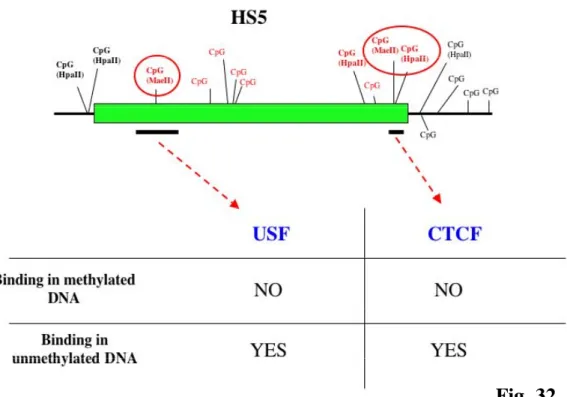
I have also used EMSA to detect the influence of DNA methylation on protein binding to HS5. Results indicate that USF and CTCF proteins bind HS5 in vitro when their specific binding sites are unmethylated, but not when these sites are methylated.
These observations suggest that DNA methylation and polymorphic regions could change the binding of insulator and/or transcription factors to the 3’RR, thereby impacting on the functions of the 3’RR during B cell development.
ABBREVIATIONS
3’RR 3’ Regulatory Region
AID Activation-induced cytidine deaminase BCR B cell receptor
C Constant region
CSR Class Switch Recombination GB Genbank
HS DNAse I hypersensitive site I I-region promoter
Ig Immunoglobulin
IgH Immunoglobulin heavy chain locus LCR Locus Control Region
RFLP Restriction fragment length polymorphism S Switch region
SHM Somatic Hypermutation V Variable region
TABLE OF CONTENTS
ABSTRACT... 2
ABBREVIATIONS... 4
INTRODUCTION... 8
1. Immune system and evolution... 8
2. Structure of the immunoglobulin... 11
3. The murine IgH locus and 3’ Regulatory Region (3’RR)... 12
4. The human IgH locus and 3’ Regulatory Region (3’RR)... 15
5. B cell development and immunoglobulin gene rearrangements... 19
5.1. V(D)J recombination and early B cell development... 19
5.2. Somatic hypermutation (SHM)... 21
5.3 Class switch recombination (CSR)... 22
6. Regulatory elements of immunoglobulin heavy chain locus.... 25
6.1 Variable- and I-region promoters... 25
6.2 The Eµ enhancer ... 26
6.3 The enhancers in the murine 3’ Regulatory Region ... 27
6.4 The enhancers in human 3’ Regulatory Region ... 29
7. The enhanceosome model... 32
8. Gene regulation at distance and DNA insulators... 33
9. Disorders related to immunoglobulin production: the autoimmune diseases... 36
10. Immunoglobulin production in response to neuroleptics in schizophrenic disorder... 38
AIM OF PROJECT... 40
MATERIAL AND METHODS... 41
1. In silico analysis... 41
2. DNA extraction... 41
3. The selective PCRs for the human HS1.2-A and HS1.2-B... 42
4. DNA amplification of HS1.2 alleles from monkeys and apes... 43
6. Human and mouse cell lines... 43
7. Mouse primary B cells... 44
8. LPS or LPS plus IL4 induced class switch recombination in mouse B cells... 44
9. DNA extraction from cell lines and splenic B cells... 45
10. Amplification by PCR of HpaII and MaeII sites... 45
11. Electrophoretic mobility shift assays (EMSA)... 48
12. ds DNA competitors and Antibodies... 48
13. Synthesis and methylation of probes... 49
14. 32P labeling of DNA fragments... .... 49
RESULTS... 50
1. In silico analysis of contigs from GenBank... 50
2. The polymorphisms of the HS1.2-A and HS1.2-B enhancers... 54
3. Changes of protein binding in the different HS1.2-A alleles by EMSA... 58
4. Analyses of HS1.2 in monkeys and apes... 60
5. The frequency of the HS1.2 alleles... 62
6. The frequency of the HS1.2-A alleles in some autoimmune diseases... 66
7. The frequency of the HS1.2-A alleles in antibody response to neuroleptics... 69
8. DNA unmethylation patterns of 3’RR in murine cell lines reflecting different stages of B development... 71
9. DNA methylation patterns of 3’RR in unstimulated and stimulated spleenic B cells... 75
10. DNA methylation patterns of 3’RR in non B cells... 77
11. DNA methylation at specific sites in 3’RR requires histone H1... 79
12. EMSA shows CTCF and USF binding to unmethylated HS5... 81
DISCUSSION... 85
1. The polymorphism of HS1.2 enhancer in the human 3’RRs... 85
2.
The patterns of DNA methylation in the murine 3’RR... 87
3.
Conclusions... 92
INTRODUCTION
1. Immune system and evolution
The immune system of mammals is composed of a complex constellation of cells, organs and tissues, arranged in an elaborate and dynamic communications network and equipped to optimize the response against invasion by pathogenic organisms. The immune system is, in its simplest form, a cascade of detection and adaptation, culminating in a system that is remarkably effective.
The immune system protects the body from infection by employing three basic strategies:
• Creating and maintaining a barrier that prevents bacteria and viruses from entering the body.
• If a pathogen breaches the barriers, and gets into the body, the innate immune system is equipped with specialized cells that detect, and often eliminate, the invader before it is able to reproduce and cause potentially serious injury to the host.
• If a pathogen is able to successfully evade the innate immune cells, the immune system activates a second, adaptive immune response against the pathogen. It is through the adaptive immune response that the immune system gains the ability to recognize a pathogen, and to mount an even stronger attack each time the pathogen is encountered. The principal elements of human adaptive immune system are well known. These are called T (for thymus-derived) and B (for bursa- or bone-marrow-derived) lymphocytes (Cooper et al., 1965). T and B lymphocytes can specifically recognize and respond to antigenic determinants of potentially hazardous pathogens and toxins.
Throughout evolution, the immune system has used a remarkably extensive variety of solutions to meet fundamentally similar requirements for host protection (Fig.1). Adaptive and innate immunity were thought to be temporally separable processes involving different cell types. While innate immunity is generally considered to be more phylogenetically ancient, the adaptive system has diversified and refined its functions over evolutionary time such that it persists in multiple forms and shows lineage-specific diversity (Litman et al., 2005).
Fig.1-Immune phylogeny of immunoglobulin heavy and light chain in selected jawed-vertebrate models. Throughout evolution, the immune system has used a remarkably extensive variety of solutions to meet fundamentally similar requirements for host protection, combinatorial rearrangement, junctional diversity, somatic hypermutation and gene conversion (Litman et al., 2006).
Phylogenetic studies of immune molecules clearly indicate that various mechanisms are used to diversify relatively limited amounts of genetic material to create a diverse set of receptor structures, the complexity of which is immense. The events that are likely to have occurred during the evolution of immunoglobulin and other complexes, as well as some of the other diverse immune receptors, show the successful integration of pathways that share important properties with those that are used to achieve genetic variation in viruses, bacteria, fungi and protozoan parasites. There is a co-evolutionary struggle in which selection acts on DNA in the host and in pathogens to mediate sequence variation and diversity (Fig.2). In this co-evolutionary struggle, the host and the
pathogen use similar mechanisms to modify selection and outcome. In various pathogens, several fundamental mechanisms of DNA change (as DNA recombination and gene hypermutation) can lead to the evasion of host detection and to the subsequent clonal selection of the pathogenic variant. In the host, analogous processes can increase the capacity of the immune system to recognize and to defeat the pathogens (Litman et al., 2006).
This view is supported by the diversity of immune mechanisms that are now being discovered in a lineage- and taxon-dependent manner. It is likely that understanding the complex mechanisms that affect immune function in multicellular organisms will also provide valuable information about different mechanisms of pathogenic activity.
Fig.2-Mechanistic similarities between pathogenicity and protection. In a co-evolutionary struggle, the host and the pathogen use similar mechanisms to modify selection and outcome. In various pathogens, several fundamental mechanisms of DNA change (such as DNA recombination and gene hypermutation) can lead to the evasion of host detection and to the subsequent clonal selection of the pathogenic variant. In the host, analogous processes can increase the capacity of the immune system to recognize and defeat the pathogens (Litman et al., 2006)
2. Structure of the immunoglobulin
Antibodies (immunoglobulin) are the key effector molecules of the adaptive immune system. Only differentiated B cells, the plasma cells, produce soluble antibodies (they are ∼ 20% of total serum protein) by a tightly regulated process.
All antibodies are composed of two identical heavy chains and two identical light chains. Both the light chains and the heavy chains have a variable (V) and a constant (C) region and are held together by disulfide bonds.
The variable regions contain the antigen binding site of the antibody molecule (Fig.3). Although the specificity of the antibody depends on both the heavy and light chains, properties such as half life, complement fixation and placental transfer, depend only on the heavy chain. The gene encoding heavy chain can be modified by DNA rearrangements using a process called class switch recombination (CSR).
We can distinguish five different isotypes for the constant region (α, δ, ε, γ and µ); each is encoded from different DNA segments and defines the specific class of the antibodies, i.e. IgA, IgD, IgE, IgG and IgM.
Fig.3 Immunoglobulin Structure. Schematic representation of an antibody molecule with two identical heavy and light chains. Both chains have an N-terminal variable region and a C-terminal constant region and are held together by disulfide bonds. The variable regions contain the antigen binding site of the antibody molecule.
3. The murine IgH locus and 3’ Regulatory Region
(3’RR)
In the mouse the Immunoglobulin Heavy Chain gene (IgH) cluster is localized at Chromosome 12F1 and spans ∼ 3Mb (Fig.4A). The variable region is close to the telomere and has ∼101 VH segments, followed by ∼13 DH and 4 JH segments. Downstream of the variable region gene segments, we can distinguish eight heavy chain constant region genes (Cµ, Cδ, Cγ3, Cγ1, Cγ2b, Cγ2a, Cε, and Cα). Regulatory elements are present upstream and downstream of constant region genes. The intronic enhancer, Eµ, is located upstream and a set of four enhancers (hs3A, hs1.2, hs3B and hs4) contained in a 3’ Regulatory Region (3’RR) is downstream of the constant region genes (Khamlichi et al., 2000).
The 3’RR can be subdivided into two units (Fig.5). The first unit includes an extensive palindrome (∼25 kb) containing three of the murine 3’ enhancers, hs3A, hs1.2 and hs3B and families of locally repetitive elements (Chauveau and Cogne, 1996; Saleque et al., 1997). Although no biological function has yet been associated to this palindromic structure, it has been reported that under certain circumstances a cruciform structure can form in a DNA sequence with an inverted repeat or palindromic sequence (Howell et al., 1996; Shlyakhtenko et al., 2000). A palindromic structure is also present in the human 3’ Regulatory Region and spans ∼2.5 kb (Sepulveda et al., 2005). However the implications of this finding are still controversial. The second unit of murine 3’ RR contains hs4, the most distal of the 3’ enhancers.
A recently identified downstream region, which contains hypersensitive sites HS5, HS6 and HS7, has been hypothesized to serve as an insulator of the IgH locus. This downstream region is associated with marks of active chromatin throughout B cell development and contains binding sites for CTCF, a protein associated with mammalian insulators (Garrett at al., 2005).
Other experiments identified hole as the nearest known non-IgH gene, followed by crip1, crip2 and mta1 (Sepulveda et al., 2005). Another landmark of the region downstream of Cα is an origin of replication (Ori), which is located ∼76 kb downstream of Cα in mouse erythroleukemia (MEL) cells. This origin marks the end of ∼500 kb replicon that extends 3’ to 5’ through C, J, and D genes to the most proximal VH genes (Zhou et al.,
Recently, a cluster of hypersensitive sites was revealed 30 kb upstream of the most 5' VH gene. One of these sites, HS1, is restricted to pro-B cell lines and is accessible to restriction enzyme digestion exclusively in normal pro-B cells, the stage defined by actively rearranging IgH-V loci (Pawlitzky et al., 2006). Additional description of mouse and human 3’enhancers is presented later (See section 6)
Fig.4 Schematic map of mouse (A) and human (B) IgH locus. The depicted orientation reflects the order followed by genome projects in which the sequence is presented starting near the centromere and proceeding towards the telomere. Regulatory elements, such as Eµ and the 3’RR, are present near the constant genes. In humans the 3’ Regulatory Region (RR) is duplicated, one unit downstream of each of the two alpha genes, α1 and α2.
Fig.5 Schematic map of 3’ Regulatory Region (3’RR) in murine IgH locus. In the mouse IgH locus the 3' regulatory region (3'RR) has multiple DNaseI hypersensitive sites. HS3A, HS1.2, HS3B and HS4 are enhancers involved in the expression and class switching of immunoglobulin heavy chain genes. HS5, HS6 and HS7 have been hypothesized to serve as an insulator of the Igh locus. This downstream region contains binding sites for CTCF, a protein associated with mammalian insulator regions.
4. The human IgH locus and 3’ Regulatory Region
(3’RR)
The human IgH locus is located at chromosome 14q32.33, spans ∼3 Mb and has the same general organization of the murine IgH cluster with the variable region close to the telomere (Fig4B). There are ∼39 functional VH gene segments, followed by 26 DH and 6 JH segments.
In contrast to mouse, there is a partial duplication of constant region genes. Each unit includes four heavy chain genes, an 3’ Regulatory Region (3’RR), and a downstream elk-pseudogene (Pinaud et al., 1997; Sadhu et al., 1997, Max et al. 2000).
Thus, the human constant region can be subdivided in 3 clusters (Fig.6):
1) Eµ – Cµ – Cδ;
2) Cγ3 – Cγ1 − ψε − Cα1 – 3’RRα1 – ψelk;
3) Cγ2 – Cγ4 − Cε − Cα2 – 3’RRα2 - ψelk.
The first cluster is not involved in the ancestral duplication. The second and the third clusters are separated by the ψγ gene (Flanagan et al., 1984; Lefranc et al., 1982).
In contrast to mouse, each human 3’ Regulatory Region contains a set of three enhancers: hs3, hs1.2 and hs4 (Fig.6). Humans lack an orthologue for the mouse hs3B enhancer, and the hs1.2 enhancers are inverted with respect to each other (Chen and Birshtein, 1997; Mills et al., 1997). However, the genomic structure of the two 3’RRs is extensively conserved (Sepulveda et al., 2005). Because the differences between them are very limited, it is technically difficult to identify sequences unique to 3’RR (α1) or 3’RR (α2) (Pinaud et al., 1997).
The 3’RR also shows RFLPs (Frezza et al., 1998) and reflects deletions or duplications, involving large DNA fragments (Bottaro et al., 1991; Rabbani et al., 1996). These rearrangements could change the 3’RR action and so influence the Ig expression (Rabbani et al., 1995).
There is limited sequence similarity between rodent and primates in 3' Igh regulatory regions. Both human and murine regulatory regions contain a palindrome and locally repetitive elements. In primates, repetitive elements are blocks of "switch-like" sequences that differ from the families of inverted and tandem repeats that are present in rodents. Together with enhancers, these "conserved" structural features are predicted to be essential for the activity of the 3' Igh regulatory region in vivo (Fig.7) (Sepulveda et al., 2005).
Fig.6 Schematic map of 3’ Regulatory Regions (3’RR) in human IgH locus. In the human IgH locus, the 3' regulatory region (3'RR) is duplicated and each has three DNaseI hypersensitive sites: HS3, HS1.2, and HS4. The genomic structure and sequence of the two 3’ RRs are extensively conserved. However the hs1.2 enhancers are inverted with respect to each other
Fig.7A Schematic representation of B cell development. Progression from stem cell to mature naïve B cell is associated with V(D)J recombination in the heavy chain locus and VJ rearrangement in the light chain genes.
Fig.7B Schematic representation of B cell development. After antigen dependent activation, B cells proliferate and continue to differentiate into memory or antibody-producing plasma cells. Activated B cells show class Switch recombination (CSR) and somatic hypermutation.
5. B cell development and immunoglobulin gene
rearrangements
B cell development is tightly associated with immunoglobulin gene rearrangements (Manis et al., 1998; Pinaud et al., 2001; Manis et al., 2003.). Three molecular mechanisms contribute to B cell development and to the diversity of the immune repertoire of B cells: V(D)J recombination, class-switch recombination (CSR) and somatic hypermutation (SHM). These three mechanisms involve marked DNA modification and require a fully competent cellular DNA-repair machinery.
5.1 V(D)J recombination and early B cell development
The various lineages of the immune system arise from a common lymphoid progenitor (CLP), which differentiates from a haematopoietic stem cell (HSC) in the bone marrow. T cells further mature in the thymus, whereas B cells develop in the bone marrow. V(D)J recombination is the first rearrangement that characterizes the early B cells and creates productive heavy and light chain alleles. Early in B cell development, at the pro-B cell stage, the DH and JH regions of the heavy chain locus are
joined together to generate a DJ segment. Then the VH region is joined to
the rearranged DJ region. After a productive VDJ recombination, the rearranged heavy chain allele is transcribed (VDJ-Cm), translated and paired with a surrogate light chain to create a pre B cell (Fig. 8).
During the initial phase of V(D)J recombination, the lymphoid-specific recombinase-activating gene (RAG1)/RAG2 factors, together with ubiquitous DNA architectural proteins (high mobility group, HMG, proteins), recognize and bind to recombination signal sequences (RSSs) that flank all V, D, and J gene segments and introduce a DNA double-strand break at the border of the RSS. On the chromosome, coding ends are left as hairpin-sealed structures, whereas signal ends, which are excised from the chromosome, are blunt and 5' phosphorylated. The subsequent steps are executed by the DNA-repair machinery of the non-homologous end-joining (NHEJ) apparatus (Bassing et al., 2002).
After V(D)J recombination in the heavy chain locus and VJ rearrangement of light chain genes, the B cell receptor (BCR) with two identical heavy and two identical light chains is assembled in the membrane. B cells then leave the bone marrow and move to the secondary
lymphoid organs as mature B cells. Mature B cells express IgM and IgD molecules from the same transcript by alternative splicing.
If B cells are not activated by antigen, they will die in few days, but after positive selection by antigen, the B cells will proliferate and continue to differentiate into memory or antibody-producing plasma cells. Some activated B cells undergo class switch recombination (CSR) and somatic hypermutation in germinal centers.
Fig.8 Rearrangements at the immunoglobulin heavy chain locus. The variable region of the immunoglobulin heavy chain is assembled from component variable (VH), diversity (DH), and joining (JH) gene segments by V(D)J recombination. The process of rearrangement involves cleavage of the recombination signal sequences in the DNA, which flank the rearranging gene segments. Transcription across the locus is driven by a promoter upstream of the rearranged VDJ segment (blue arrow), which facilitates the synthesis of a µ heavy chain. This then associates with a light chain, thereby forming an IgM molecule, which is displayed on the cell-surface of a B cell. Subsequently, secondary isotypes are produced by class-switch recombination (CSR), a process that exchanges the constant region of the heavy chain (CH) with a set of downstream constant-region genes (CSR to IgE is shown). (Chaudhuri J. C. et al. 2004).
5.2 Somatic hypermutation (SHM)
Class switch recombination (CSR) and somatic hypermutation (SHM) are two distinct immunoglobulin gene diversification processes. CSR involves recombination between switch (S) regions to alter the C region of the IgH and therefore the effector function of the immunoglobulin molecule. By contrast, SHM introduces non-templated point mutations in the variable region of rearranged immunoglobulin heavy and light chain genes. SHM underlies the process of affinity maturation, which results in the preferential outgrowth of B cells expressing an immunoglobulin that has high affinity for its cognate antigen.
The mutations introduced by SHM are predominantly point mutations. Transition mutations occur about twice as frequently as transversion mutations and a high proportion of mutations arise in the hotspot motif DGYW (where D denotes adenosine (A), guanosine (G) or thymidine (T); Y denotes cytidine (C) or T; and W denotes A or T) or its reverse complement WRCH (where R denotes A or G; and H denotes T, C or A), showing that SHM is influenced by the primary sequence of the DNA (Odegard et Schatz, 2006).
Several cis and trans acting elements have been implicated in the regulation of SHM (Li et al., 2004). SHM is regulated by activation-induced cytidine deaminase (AID), Rad51 family (XRCC2 and XRCC3; involved in the regulation of chromosome stability), and several error prone DNA polymerases.
Both SHM and CSR require transcription and AID-mediated deamination of cytidine residues on the non-template DNA strand, which is exposed as single-stranded DNA during the elongation reaction.
Some evidence, based on transgenic mouse experiments, suggest that the 3’ enhancers, hs3 and hs4, are involved in regulation of SHM (Terauchi et al., 2001). However more recent data showed that in the hs3-and hs4 knock out mice, SHM was not influenced (Morvan et al., 2003). Therefore, it is possible that hs3 and hs4 can cooperate with other regulatory factors to be involved in this process (Odegard et al., 2006).
5.3 Class switch recombination (CSR)
Much is known about the mechanism and regulation of CSR in mice and humans, although this process is still not understood. CSR occurs by intrachromosomal deletional recombination between switch (S) region sequences located upstream of each of the CH genes. Before CSR, usually
B cells express IgM and the VDJ-Cµ gene is transcriptionally active. If cells are treated to induce CSR, a specific promoter (I) for germline transcription is induced (e.g. Iγ3). Thus the active Sµ and specific S regions (e.g. Sγ3) become accessible to AID, which deaminates dC resulting in dU. The dU residues are substrates for uracil DNA glycosylase (UNG) which excises dU, leaving “abasic” residues. Endonucleases attack the ”abasic” residues, creating nicks. Double-strand breaks are created and ligated by unknown mechanisms (Fig.9) (Stavnezer et al., 2004)
CSR regulation involves cis and trans acting elements. Although B cell development continues to progress after selected individual enhancer elements have been knocked out by targeted deletion (e.g. Eµ (Chen et al., 1993), hs3A (Manis et al., 1998), hs1.2 (Manis et al., 1998)), CSR is severely impaired in hs3B-hs4 KO animals (Pinaud et al., 2001) (Fig.10). There is a downregulation of the levels of Cm expression in resting, but not in LPS stimulated mature B cells in the hs3B-hs4 KO (Pinaud et al., 2001). In vitro stimulation of splenic B cells from hs3B-hs4 KO animals showed reduced production of Cγ3, Cγ2b, Cγ2a, Cα and Cε isotypes, while Cγ1 and Cµ secretion remained normal. This correlated with significant reduction in serum levels of Cγ3, Cγ2b, Cγ2a, Cα and Cε (Pinaud et al., 2001) (Dunnik WA. et al., 2005).
A recent study suggests that in mouse all elements necessary for recruitment of the recombination machinery are present in the transgene containing HS3B and HS4. These enhancers probably provide something more specific than mere increased accessibility of switch regions (Laurencikiene et al., 2006).
Hs3-hs4 KO mice show a decrease of germline transcription class switching. This implies that the 3’ enhancers create contact and regulate the activity of I region promoters. However, CSR to γ1 is not impaired in these knockout mice, suggesting that the γ1 heavy chain genes are regulated in a different way.
Fig.9 Model for class switch recombination (CSR). The top line shows a portion of the Ig heavy chain locus in mouse B cells expressing IgM. The VDJ-Cµ gene is transcriptionally active. If cells are treated to induce CSR to IgG3, the promoter (I) for germline transcripts is induced. Thus the active Sµ and Sγ3 regions become accessible to AID, which deaminates dC to dU. The dU residues are substrates for the uracil DNA glycosylase (UNG), which could excise dU, leaving abasic residues. The endonucleases attack the abasic residues, creating nicks. Then double-strand breaks are created and ligated by unknown mechanisms (Stavnezer et al., 2004).
Fig.10 In vivo phenotypes for murine knockout and NeoR replacement of 3’
enhancers. A) Clean knockout of hs3A and hs1.2 enhancers in the mouse has no effect on B cell development and class switching, suggesting that they are dispensable (Manis et al., 1998) However, the deletion of both hs3B and hs4 impaired class switch recombination to all isotypes, except γ1(Pinaud et al., 2001). B) The replacement of the 3’ enhancers by NeoR affects class switching (Cogne et al., 1994; Manis et al., 1998) but the insertion of a
Neo-cassette downstream of hs4 does not affect B cell development or class switching (Manis et al., 2003)
6. Regulatory elements of immunoglobulin heavy chain
locus
Regulation of transcription is mediated partially by the interaction of trans-acting DNA-binding proteins with specific motifs in promoter and enhancer elements. Although enhancers are defined by their ability to augment the level of transcription initiated from promoter elements, dissection of motifs within enhancers has revealed a more complex regulation including both positive and negative components (Ernst et al., 1995). Many of these motifs are present in the regulatory elements of disparate genes and bind to ubiquitous trans-acting DNA-binding proteins; yet, transcriptional regulation can be restricted in a tissue-, lineage-, and stage-specific manner. This has led to the notion of “combinatorial regulation” of transcription in which the net regulatory effect is due to a unique combination of relatively ubiquitous trans-acting DNA-binding proteins that interact with the regulatory elements of a specific gene (Ernst et al., 1995).
The loss (~90%) of immunoglobulin expression in a plasma cell line by the deletion of the 3’ regulatory region (Michaelson et al., 1995) and, as discussed above, the decrease of germline transcription during class switching in hs3-hs4 KO mice (Pinaud et al., 2001) suggest that the 3’ enhancers might regulate both VH and I-region promoters in vivo.
6.1 Variable- and I-region promoters
Each V gene segment and each heavy chain (except δ) is associated with a promoter region. B cell specificity depends only on a conserved octamer motif (ATGCAAAT) and a TATA-box (Sleckman et al., 1996). Other motifs, such as E boxes, C/EBP and Ets sites, contribute to Ig promoter activity (Hatada et al., 2000).
I region promoters respond to a variety of stimuli. As discussed above, I region-driven germline transcription is regulated by the 3’ enhancers (Pinaud et al., 2001). However the mechanism of these enhancers under different stimulatory conditions is not clear.
In a hypothetical model, the activation of the I-region promoters involves the 3’ enhancers. Some observations indicate that the I-region promoters are required for AID to target the non-coding DNA strand (Li et al., 2004). Transcription permits the formation of R-loops between RNA
and DNA. In fact the transcribed DNA hybridizes temporarily with the RNA. It was hypothesized that AID might modify RNA or DNA structures in some way to make them more stable or better substrates for repair endonucleases (Manis et al., 2002).
Finally more recent data suggest that in the absence of UNG and MSH2, AID may occasionally act at the µ switch region in an apparently processive manner, but there is no marked preference for targeting of the transcribed versus nontranscribed strand (even in areas capable of R loop formation) (Xue et al., 2006).
Deamination of cytidine by AID results in a C to U transition, recognized by the enzyme uracil N-glycosylase, which removes the uracil from DNA. Thus the expression of germline transcripts precedes the activity of AID and uracil N-glycosylase. It is possible that the 3’ enhancers might cooperate with I-region promoters in recruiting several transcription factors.
6.2 The Eµ enhancer
The murine intronic enhancer, Eµ, was the first mammalian enhancer to be described in the heavy chain locus (Fig.4) (Gillies et al., 1983; Banerji et al., 1983). It lies between the JH gene segments and the Cµ gene and is
closely associated with two matrix attachment regions (MARs) (Cockerill et al., 1987). Motifs within Eµ that potentially bind to trans-acting DNA binding proteins have been mapped in detail (Staudt et al., 1991). A 220-bp region of Eµ defines the minimal region required for transcriptional activity. Mutational analysis in transfection assays indicated that most of the E motifs are functionally redundant. However, EMSA experiments determined that the E motifs were bound by different protein complexes, in spite of their sequence similarity (Weinberger et al., 1986). The role of individual binding motifs has been analyzed (Fernex et al., 1995). For instance, the E-box motif, µE3, in conjunction with motifs upstream of µE3, appears to be essential for Eµ -mediated accessibility to the V(D)J recombinase.
Targeted mutation studies have definitively shown that Eµ plays a role in regulating rearrangement of the heavy chain locus. In one study, hit-and-run replacement of Eµ with a short oligonucleotide resulted in slightly diminished DH-to-JH rearrangement (70% of normal) but more substantially
inhibited VH to-DJH rearrangement of the targeted allele (Serwe et al.,
1993). In a second study, replacement of Eµ with a neomycin resistance (neor) gene resulted in a dramatic decrease in the ability of the J
H locus to
undergo recombination. Notably, insertion of the neor gene upstream of
Eµ, without deletion of endogenous sequences, resulted in a similar cis-acting inhibition of JH rearrangement (Chen J. et al., 1993). However,
plasma cell lines that show a spontaneous deletion of Eµ are still capable to express high levels of immunoglobulin (Aguilera et al., 1985; Eckhardt et al., 1985). These observations suggest that there might be regulatory elements other than Eµ that are responsible for the upregulation of IgH expression at later stages of B cell development, likely, the 3’ enhancers HS4 (Eckhardt et al., 1985; Pettersson et al., 1990). It has been suggested that there is a temporal shift in enhancer function between the Eµ enhancer and the 3’ enhancers. In this model, early in B cell development, Eµ is active and as B cell development progresses, the 3’ enhancers start to synergize with each other and with Eµ. In mature B cells, in which the 3’ enhancers are active, Eµ activity begins to diminish. Finally, at the plasma cell stage, the combined activity of the 3’ enhancers would be roughly the same as for Eµ (Ong et al., 1998).
6.3 The enhancers in the murine 3’ Regulatory Region
Soon after the identification of Eµ, it became clear that other enhancers might exist outside the JH-Cµ intron. In fact cell lines were reported that
efficiently transcribed their IgH genes despite deletion of Eµ (Aguilera et al., 1985; Eckhardt et al., 1985). Search for additional regulatory elements based on a DNase I hypersensitivity assay led to the discovery of other murine enhancers called: HS3A, HS1.2, HS3B and HS4 (Pettersson et al., 1990; Giannini et al., 1993; Madisen et al., 1994; Michaelson et al., 1995) (Fig.5).
Activity of the 3’ IgH enhancers was mainly assayed by transient transfection in cell lines thought to represent different stages of B cell differentiation and by transgenic models. These observations showed that the activity of the murine 3’ enhancers is B cell specific and developmentally regulated (Khamlichi et al., 2000). In fact while HS1.2, HS3A and HS3B are active at late B cell differentiation stage, HS4 seems to be active throughout B cell development (Madisen et al., 1994; Michaelson et al., 1995). This expression pattern correlates quite well with
the DNA methylation pattern of the 3’ region. In fact, the 3’ regulatory region is hypermethylated at the pre-B cell stage and becomes demethylated at the plasma cell stage, but it is not clear whether this applies to HS4 (Giannini et al., 1993). One aim of my work has been to define better the DNA methylation pattern of the 3’ region in mouse.
An important question concerning the multiple enhancers is their potential synergy in CSR and transcription activation. To understand better the complexity of problem, one has to bear in mind that the 3’ regulatory region is ~200kb from Eµ (before class switching) and that the four HSs are kilobases distant from each other within a palindromic structure (except for HS4). It has been proposed that the activity of the 3’ enhancers is down-regulated during early stages of B cell development. In vitro, HS3A and HS3B enhancers are regulated negatively by heterodimers composed by Bach2 and a small Maf protein (Muto et al., 1998). Pax5 has been proposed to repress the HS1.2 activity (Singh et al., 1996). Pax5 is expressed from early stages of B cell development but is not expressed at the plasma cell stage (Urbanek et al., 1992). Thus, the HS1.2 enhancer is activated at late B cell stage. Although the deletion of HS3A and HS1.2 has no effect on B cell development and class switching, it is possible that the activities of the 3’ enhancers in vivo are redundant (Saleque et al., 1999). In fact both GL ε and γ2b promoters synergized strongly with the HS1.2 enhancer in activated primary B cells, a mature B cell line, and a plasma cell line. The principal activity of HS1.2 in activated primary B cells occurs within a 310-bp fragment that includes NF-κB, OCT, and nuclear factors of activated B cells (Ets/AP-1) sites. By mutating the consensus sequences for various transcription factors, it was determined that sites in HS1.2 are important for synergy with the GL ε and γ2b promoters. It suggests that different sites in HS1.2 might selectively interact with the GL e and γ2b promoters (Laurencikiene et al., 2001).
Reporter assays showed that HS4 is active at all stages of B cell development. However, the function of HS4 in vivo remains unknown, especially at early stages of B cell development. It is possible that it is involved in early regulation of chromatin changes at the 3’ end of IgH locus because it is associated with markers indicative of open chromatin (Garrett et al., 2005). Murine HS4 is regulated by NF-κB, octamer binding proteins, and Pax5. It appears that Oct-1 and NF-κB binding activities positively regulate HS4 activity, whereas Pax5 is a repressor (Michaelson et al.1996). Recently it has been reported that human hs4 is regulated differently. EMSAs and Western analysis of human B cells before and after
stimulation with anti-IgM plus anti-CD40 showed a complex binding pattern formed by NF-κB, Oct-1, and Oct-2, but not by Pax5 (Sepulveda et al., 2004). This opens the possibility that other human 3’ enhancers (HS3 and HS1.2) are also regulated differently from the mouse.
It has been suggested that the mouse 3’ enhancers have a common regulatory mechanism because they share binding of a group of transcription factors, i.e. Pax5, NF-κB and octamer binding proteins (Michaelson et al., 1996).
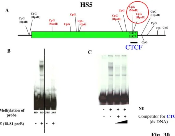
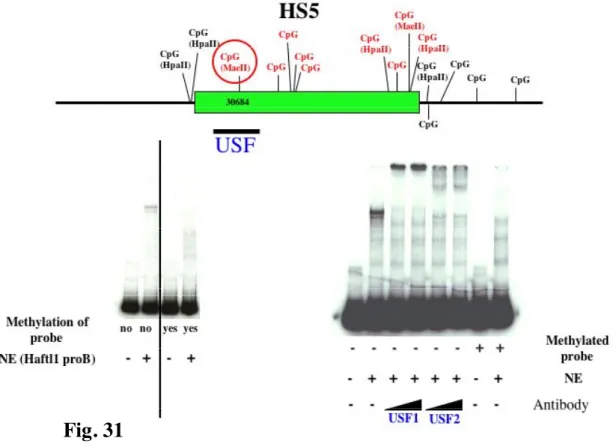
Recently it was suggested that a boundary for the CSR was located within close proximity to HS4 involving DNase I hypersensitivity sites HS5, HS6 and HS7 (Garrett et al., 2005). These three segments are positioned between the IgH locus and the next non-IgH genes in a region with potential for insulator function. Moreover, preliminary experiments i.e. testing HS5 and HS6 in reporter constructs, did not detected enhancer activity in a plasma cell line. Insulators are DNA elements that protect genes from inappropriate neighboring regulatory signals by enhancer blocking activity and/or by delineating chromatin domains. All vertebrate insulators tested so far have been associated with CTCF binding sites (West et al., 2002). In this work, it was shown that CTCF binding sites are occupied in vivo in HS5, HS6 and HS7 in cell lines representing various stages of B cell development and normal splenic B cells (Garrett et al., 2005). The presence of a constitutively undermethylated region (Giannini et al., 1993), located in the vicinity of the mapped CTCF binding sites, suggests that these sites are accessible to CTCF binding. However the role of individual CTCF sites in the 3’ RR remains to be elucidated. One aim of this thesis is to define better the correlation between DNA methylation and binding of specific proteins like CTCF.
6.4 The enhancers in human 3’ Regulatory Region
In contrast to murine 3’ enhancers, the human 3’ enhancers, HS3, HS1.2 and HS4, are duplicated as described above (Chen et al., 1997; Mills et al., 1997; Pinaud et al., 1997) (Fig.6).
The human α1 and α2 HS1.2 enhancers both reside near the centers of ~10kb palindromes, with each palindrome closely flanked by a single copy of HS3 immediately adjacent to the 5’ end and an HS4 unit located ~4 kb downstream (Pinaud et al., 1997). By comparison, mouse HS1.2 is centrally positioned in a considerable larger (~24 kb) palindrome that
contains a copy of HS3 on each end, with HS4 once again located ~4 kb downstream of the palindrome (Chauveau et al., 1996). Certain functional elements in murine enhancers, including Pax5 sites, do not appear to be conserved in the human HS1.2 or HS4 (Mills et al., 1997).
The 3’ enhancers are also responsible for the upregulation of C-myc expression in human Burkitt’s lymphoma and in mouse plasmacytomas. In these cells, a reciprocal translocation juxtaposes 3’ enhancers with c-myc. It has been observed that the mouse 3’ enhancers could increase the expression and the histone acetylation of the translocated c-myc (Madisen et al., 1994).
Similar to the enhancers comprising the murine 3’ regulatory region, human HS3, HS1.2 and HS4 interact synergistically with each other in transient transfections and all three enhancer elements may be needed in the activation of Cα genes before switching (Hu et al., 2000). In addition, there is some evidence for specific enhancer – promoter interactions, as demonstrated by differential effects in the upregulation of the Iγ3 and Iα region promoters (Hu et al., 2000). There is a complex polymorphism of human α1 HS1.2 enhancers, in which multiple alleles have been generated through inversions and internal deletions and/or duplication (Denizot et al., 2001). This polymorphism is characterized by the presence of variable numbers of tandem repeats (VNTR) within the core enhancer. In addition the screening of patients with IgA nephropathy toward renal failure shows that one allele is significantly correlated with the disease and the α1 hs1.2 enhancer controls the level of IgA production in patients and the evolution of IgA nephropathy toward renal failure (Aupetit et al., 2000). This suggests that DNA repeated sequences could carry potential transcription factor binding sites that may boost the transcription of the α1 gene. Interestingly, minisatellites located within 3’ regulatory elements and binding transcription factors have been reported in a few other cases (Kominato et al., 1997; Maeng et al., 1998), and constitute one aspect through which the occurrence of each repeat helps improve gene expression in the course of evolution. Recently, it was shown that within hs1.2, three regions (1, 2, and 3) are all necessary, but individually not sufficient, for enhancement of transcription. In region 2, a HoxC4 site and a HoxC4/embedded octamer (HoxC4/Oct) site are conserved across human, mouse, rat, and rabbit. These two sites recruit HoxC4 and Oct-1/Oct-2, which act synergistically with the Oca-B coactivator to yield the full hs1.2-enhancing activity (Kim et al., 2004).
The distribution and the influence of this polymorphism in various diseases involving immunoglobulin production deserves to be investigated (Denizot et al., 2001). It is not clear if α2 HS1.2 is polymorphic. Thus one aim of my project has been to study the two HS1.2 enhancers and to see the allelic distribution of a1 HS1.2 in some healthy populations and in patients with various diseases involving immunoglobulin production.
While a complex restriction length polymorphism is documented for the human HS1.2 enhancer, a non polymorphic restriction pattern was found for human HS3 and HS4 showing the stability of regions encompassing these two elements within the human species (Guglielmi et al., 2004).
7. The enhanceosome model
One of the central problems in understanding gene regulation is to explain how specific sets of genes are selected for expression during cell growth, differentiation or in response to environmental cues. In molecular terms, gene activity is specified by cis-DNA elements, enhancers and promoters, which provide regulatory infrastructure. In recent years, a model has emerged of how the different transcription factors, general DNA binding proteins (as histones and HMG), co-activators, basal transcription factors and chromatin modifying activities work together to activate transcription. In several cases specificity in gene transcription is achieved by the assembly of higher-order three-dimensional transcription
factor/enhancer DNA complexes, termed enhanceosomes. The
enhanceosomes activate transcription by recruiting chromatin-modifying activities and basal transcription factors to a specific promoter. In this way, a specific gene is selected for activation only if all the enhanceosome components are present in the same nucleus. Therefore, a specific gene is expressed only if specific signals are sensed and appropriately interpreted by a cell. For this reason, genes responding to a single signal assemble the corresponding enhanceosome only in response to this signal, whereas genes responding to multiple signals could assemble multiple, but specific enhanceosomes. Each set of enhancers might show architectural differences in their organization and also cooperative interactions among the different components of the enhanceosome that are essential for its assembly and activity (Merika et al., 2001).
The IFN-β enhancer is an example. It is activated upon virus infection and contains three positive regulatory binding sites recognized by NF-κB, members of the IRF family, and the ATF-2/c-Jun heterodimer (Merika et al., 2001). After virus infection, enhanceosome formation is activated. The study of the IFN-β enhancer has suggested that there is a step-wise recruitment of transcription factors and chromatin modifiers, which in turn, triggers the opening of the promoter region.
8. Gene regulation at distance and DNA insulators
Eukaryotic genomes necessarily are organized in domains with distinct functions. In fact an active gene might be surrounded by constitutively silenced chromatin structures. To an extent, the identity of these domains is maintained by classical transcriptional regulatory elements, such as enhancers, silencers and upstream activating sequences (UAS). In other cases, however, specific DNA sequences and their associated binding proteins have a role in establishing or maintaining discrete inter-domain boundaries. Such sequence elements have been called insulators.
Insulators are DNA sequence elements that prevent inappropriate interactions between adjacent chromatin domains. We can distinguish two types of insulator activity: enhancer-blocking and barrier activity. The first protects from activation by enhancers. The second protects against heterochromatin-mediated silencing.
Some compound insulators possess both enhancer-blocking and barrier activities, and enhancer-blocking insulators also protect against certain types of transcriptional repressors (Gaszner et al., 2006).
The DNA insulators are involved in the transcriptional control and in the enhancer activity. In fact it is recognized their role in the long-range interactions with the enhancer elements and in the three-dimensional organizations of chromatin within the nucleus. Insulation has emerged as a major mechanism for epigenetic control of gene expression, in particular at imprinted loci (Bell et al., 2000).
Recently some studies suggested that both kinds of insulator elements exploit the function of the other regulatory elements within the nucleus by establishing divisions between various regulative elements (enhancers, silencers, promoters and insulators). In the past it has been difficult to explain the position dependence of enhancer-blocking insulators. Recently two models were created (Gaszner et al., 2006). The first is a direct contact model. In this case, enhancers might function by directly interacting with their designated promoters. The enhancer-blocking insulators might have a steric effect that prevents enhancers from contacting other promoters, either by favouring intra-loop enhancer-promoter interactions or preventing inter-loop contacts (Fig.11).
Alternatively, there could be an activating signal that travels processively from enhancer to promoter (the tracking model of enhancer action). This signal could be, for example, a helicase complex that modifies histones or alters nucleosome structure, or it could be RNA polymerase
itself, launched from the enhancer. Then this signal could be blocked by an enhancer-blocking insulator as it tries to traverse the nucleoprotein structure at the base of the loop that the insulator generates.
Therefore, the loop architecture that is connected with insulator action, might be one specialized application of a more general set of regulatory mechanisms that assist in bringing together distant regulatory elements and genes and stabilizing inter-chromosomal interactions. Proteins, such as CTCF and USF, are well suited for these mechanisms, which would be quite different from their modes of action at enhancer-blocking insulators (West et al., 2004).
Fig.11 Models for the enhancer-blocking activity A) The direct-contact model focuses on the formation of topologically closed looped chromatin domains. An assumption of this model is that the frequency of intra-loop enhancer-promoter interactions is higher than that of inter-loop interactions. This can be achieved by the existence of a mechanism that either facilitates intra-loop interactions or inhibits inter-loop interactions. B) Inherent to the tracking model is the idea that transcriptional activation involves the processive transfer of a signal from the enhancer to the promoter. Specific interactions between the activation signal and the enhancer-blocking complex disrupt the transfer (Gaszner et al., 2006).
9. Disorders related to immunoglobulin production:
the autoimmune diseases
Autoimmune diseases are complex multisystem disorders. In many cases, their etiology is unknown, but genetic, hormonal and environmental factors are important. The population burden of autoimmune disorders is large and underestimated, because the incidence and prevalence of individual autoimmune diseases are not high. Autoimmune diseases are preceded by a long preclinical phase in which patients can be identified by the presence of characteristic autoantibodies.
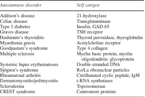
Autoimmune disease can be divided into either organ-specific illnesses, such as herpetiform dermatitis and Hashimoto disease, or systemic illnesses, such as rheumatoid arthritis, systemic lupus erythematosus, Crohn’s disease, psoriasis and celiac disease. Pathogenesis may be mainly mediated by autoimmune T lymphocytes. However virtually all autoimmune diseases are associated with circulating autoantibodies, which bind self-proteins (Table 1). Furthermore, for many diseases these autoantibodies are found in serum samples many years before disease onset (Scofield, 2004).
Numerous studies have found that autoimmune diseases have a genetic predisposition (Shepshelovich et al., 2006). The abnormal immune response probably depends upon interactions between susceptibility genes and various environmental factors. Evidence for genetic predisposition includes increased concordance for disease in monozygotic compared with dizygotic twins and an increased frequency of patients with more than one affected family member (Wanstrat et al., 2001). Susceptibility to autoimmune diseases is a multigenic phenotype affected by a variety of genetic and environmental or stochastic factors. The most potent genetic influence on susceptibility to autoimmunity is the major histocompatibility complex (MHC). Different HLA alleles are linked to different autoimmune diseases. However, there are also non-MHC susceptibility alleles related to autoimmune diseases. These are difficult to identify, predominantly due to extensive genetic heterogeneity and possible epistatic interactions among the multiple genes required for diseases development. An intriguing finding indicates that several alleles affect multiple autoimmune diseases, or simply that many immune system genes are clustered together (Wanstrat et al., 2001). It is consistent with the hypothesis that certain immunological pathways are common to multiple autoimmune diseases, whereas other pathophysiological mechanisms are specific to a particular disease. The
effect of genetic factors on the development of autoimmune disorders could be underestimated. Therefore, the presence of a specific autoantibody in an individual’s serum combined with disease prone MHC haplotypes or other susceptibility loci increases exponentially the risk for that person to develop an autoimmune disease in the future (Shepshelovich et al., 2006).
It has not been clear if there is a correlation between the 3’RR of immunoglobulin heavy chain locus and autoimmune diseases. The only linkage that was shown was between patients with IgA nephropathy toward renal failure and α1 hs1.2 enhancer (Aupetit et al., 2000). One aim of my project has been to study the distribution of the α1 hs1.2 polymorphic enhancer in different autoimmune disorders in order to understand the functional significance of different alleles of HS1.2.
Table 1 Selected autoimmune disease and characteristic autoantigens (Shepshelovich et al., 2006)
10. Immunoglobulin production in response to
neuroleptics in schizophrenic disorder
Schizophrenia affects 1% of the world’s population, but its cause remains obscure. Numerous theories have been proposed regarding the cause of schizophrenia, ranging from developmental or neurodegenerative processes or neurotransmitter abnormalities to infectious or autoimmune processes (Jones et al., 2005). However, nowadays, there is not a clear hypothesis about the etiology of this disorder. The immune alterations in schizophrenia have been described for decades. The unspecific, “innate” immune system shows signs of over-activation in non medicated schizophrenic patients. The question of whether some or all cases of schizophrenia have an immune or autoimmune etiology has been asked now for nearly 100 years. Many general immune abnormalities have been reported over this time. These include morphological changes in lymphocytes, altered levels of CD4+ CD45RA+ T cells, CD8+T cells,
CD5+B cells and γδ T cells, increased or decreased levels of γ-globulin in
serum, increased levels of circulating cytokines, particularly IL-2, IFN-γ and IL-6, and increased levels of antiviral antibodies (Rothermundt et al., 2001). Despite numerous reports of immune abnormalities in schizophrenia, the hypothesis that these abnormalities are related to the pathogenesis of the disease continues to be viewed with much skepticism. However associations of other autoimmune diseases with particular MHC haplotypes, increased serum levels of autoantibodies, and in vivo and in vitro replication of some of the functional and ultrastructural abnormalities of schizophrenia by transfer of autoantibodies from the sera of patients with schizophrenia suggest that, in some patients at least, autoimmune mechanisms could play a role in the development of disease (Jones et al., 2005).
Pharmacological treatments of schizophrenia alter also the immune system. In fact, during antipsychotic therapy with neuroleptics, the specific TH-1 cells become activated and, in addition, the B cell system and antibody production increase in some patients (Muller et al., 2000).Other evidences suggest that antipsychotic drugs, e.g. chlorpromazine and the atypical compound clozapine, influence the production of cytokines. Cytokines, organized in networks of related peptides with pleiotropic functions, are pivotal humoral mediators of infection and inflammation, and they play an important role in hematopoiesis, autoimmunity and immunoglobulin production (Pollmacher et al., 2000). Therefore, these
treatments can represent a model to study drug dependent immunological alteration.
AIM OF PROJECT
After these considerations, we want to know how HS1.2A and HS1.2B, the two enhancers of the human 3’RRs, could relate to immunoglobulin production, in terms of structure and allelic distribution in different populations. Finally we want also to see how the accessibility of the murine 3’RR changes during CSR and B cell development by studying DNA methylation state of this locus.
Therefore the principal aims of the project are:
1. To study the polymorphic structure of the two 3’ enhancers, HS1.2A and HS1.2B, in the human heavy chain locus of immunoglobulin; 2. To study the distribution of alleles of polymorphic HS1.2 in healthy
populations and in patients with different autoimmune disorders; 3. To study the pattern of DNA methylation in the murine 3’RR of
immunoglobulin heavy chain locus during B development and under specific stimulation for class switching recombination (e.g. LPS or LPS plus IL4).
MATERIAL AND METHODS
1. In silico analysis
Contings of the constant heavy chain region were rescued from GenBank with Blast programs of the NCBI and from maps of the
chromosome 14q32 telomeric region
(www.ncbi.nlm.nih.gov/mapview/maps.cgi).
Comparison of the sequences of the 3’RRα1 and 3’RRα2 was performed with Clustal W and Gene Jockey programs. A new contig at the 3’RR α1 sequence (called CHR77) was assembled from the junction of five genomic contigs (X76785, Y14407, AL928767, AL928765 and U64453), using the overlapping regions (Fig. 12).
The sequencing data of the different clones were also compared by Clustal W and Blast programs.
The WebGene program (in Cabibbo et al., 2002) at the web site http://125.itba.mi.cnr.it/~webgene/genebuilder.html was used to find the consensus sites for transcription factors on the polymorphic HS1.2-A and HS1.2-B enhancers.
2. DNA extraction
The DNA of some human donors was extracted from total blood or peripheral blood lymphocytes using standard methods.
The DNA of other human samples was extracted from buccal epithelial cells embedded in a sterile cotton plug (Becton Dickinson, Sparks, MD, USA) The cotton plugs were removed from the support with a sterile lancet and introduced in a sterile vial with 1.0 ml of TE with SDS 1%, RNAse (100 mg/ml), and 20 mg/ml of proteinase K, then incubated o.n. at 37 °C with gentle shaking. The DNA was harvested according to the standard protocol of Microcon (Millipore, Bedford MA, USA).
3. The selective PCRs for the human HS1.2-A and HS1.2-B
The 5.4-kb fragment of 3’RR-1 was selectively amplified by the primers SA2.5 (5’-GGA TCC CTG TTC CTG ATC ACT G-3’) and A2R (5’-GCC CTT CCT GCC AAC CTG-3’), respectively located in U2 and U8 sequences (Fig.13 and Fig.14) which are differently orientated in the two 3’RRs. Conversely, to amplify the 3’RR-2 specific fragment of 4.4 kb, the primer A2R was selected respectively within the U8r sequence and it was paired to primer A2F (5’-GCA CTG TCG GCT TAC AGA GG-‘) within UB2, which is unique in the 3’RR-2.
Reaction conditions were: 1.5 units of Taq Polymerase Platinum High Fidelity (Invitrogen, Carlsbad, CA, USA) buffer 1x Platinum High Fidelity
(200 mM Tris–HCl, pH 8.4, 500 mM KCl); dNTPs (0.2 mM), MgCl2 (1.5
mM), primers 15 pmol, water for final volume of 50 ml. The reaction was done at 94 °C for 2 min followed by 10 cycles at 94 °C for 30 s, 59 °C per 30 s, 68 °C per 5 min, followed by 20 more cycles at 94 °C for 30 s, 57 °C for 30 s, 68 °C for 5 min and one final extension at 72 °C for 10 min.
A nested PCR was performed for both loci with the same two primers, P3Frw (5’-GAC TCA TTC TGG GCA GAC TTG-3’) and D3Rev (5’-GTC CTG GTC CCA AAG ATG G-3’), in order to obtain the polymorphic region of the enhancer HS1,2. The amplification conditions were performed with 1/10 of the volume of the selective PCR to minimize DNA genomic carryover, using 1 unit of Platinum Taq Polymerase (Invitrogen) and 1x buffer platinum, dNTPs (0.2 mM), MgCl2 (1.5 mM), primers (15
pmol) and adding water to a final volume of 50 ml. The temperature steps of the reaction were as follows: 94 °C for 2 min followed by 30 cycles at 94 °C 30 s, 56 °C for 30 s, 72 °C for 1 min followed by a final extension at 72 °C for 5 min.
4. DNA amplification of HS1.2 alleles from monkeys and apes
Amplification of the HS1.2 enhancer from monkeys was performed with the same human primers (P3frw and D3rev), used for the direct nonselective PCR of the short fragment. The ape enhancers were amplified using the same nested PCRs used for humans except that the annealing temperature was 54°C. Southern blot analysis was performed on genomic DNA from five samples of Macacus fascicularis digested by XbaI and hybridized with the human enhancer probe with the fluorescence system (Roche Diagnostic, Germany). DNAs were extracted from blood using the standard protocol.
5. Cloning and analysis of the polymorphisms
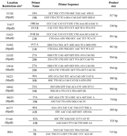
PCR products were cloned with TOPO XL PCR Cloning kit (Invitrogen) in the plasmid pCR-XL-TOPO and electroporated in E. coli TOP10 (Invitrogen) competent cells. The cells were plated on ampicillin selective agar medium, X-gal and incubated o.n. until white/blue colonies were visible. Minipreps were performed with SIGMA kit (Illinois, USA) and DNA from positive clones extracted, sequenced with M13 forward and reverse universal primers and analyzed by automatic AppliedBiosystem capillary 3700 apparatus at the BMR Sequencing Service CRIBI (Padova, Italy). DNA fragments particularly rich in GC repeats were resolved by use of DMSO in the sequencing reactions. Polymorphism frequency in the different populations was determined by electrophoresis of PCR products on agarose gels (3%) stained with ethidium bromide. The alleles of HS1.2-A and HS1.2-B were analyzed as shown in Fig.15.
6. Human and mouse cell lines
Mouse and human cell lines were maintained in complete-RPMI-1640. All cell culture media were supplemented with 10% FBS (Gemini Bio-Products), 1% Penicillin/Streptomycin (P/S) and β-mercapthoethanol. These media will be referred as “complete media” from here on. All cells were grown at log phase at 37°C in a humidified atmosphere with 5% CO2.
7. Mouse primary B cells
Normal mouse B cells were obtained from spleens of C57B/6 female animals 6-8 weeks of age. Briefly, spleens were surgically removed and placed in PBS buffer (1x) and then homogenized between two glass slides. The cell suspension was filtered through cheesecloth and centrifuged at 1,800 rpm for 5 min at 4-10°C (Beckman). The cells were resuspended in 3 ml of red blood cells (RBC) lysis solution (Puregene), incubated for 3 min at room temperature (RT), and recovered by cetrifugation. T cells were removed by negative selection using anti-CD43 (Ly-48) MACS Microbeads (Miltenyi Biotec, Auburn, CA) as indicated by the manufacturer. B cell purity was 95.3% as evaluated by FACS analysis of CD43(-), B220(+), CD3e(-) cells.
8. LPS or LPS plus IL4 induced class switch recombination in mouse B cells
Mouse primary B cells (106 cells/ml in 20ml RPMI) were induced to
undergo CSR in vitro by addition of LPS (50 µg/ml Calbiochem) or LPS plus IL4 (final concentration of 50 ng/ml- R&D Systems). Non-stimulated B cells and cells stimulated for 48h, 72h and 96h were analyzed for the expression of CTCF, USF1 and USF2. Total RNA prepared from 5 x 10⁶
cells per sample was used to monitor for germiline transcripts by RT-PCR using specific primer pairs for mouse γ1, γ2b and γ3 at 0, 48, 72 and 96h. Cell surface expression of the switched isotypes as well as cell cycle progression were monitored by FACS analysis.