Head and neck
Cost analysis in oral cavity and oropharyngeal
reconstructions with microvascular and pedicled flaps
Analisi dei costi nelle ricostruzioni orali e orofaringee con lembi microvascolari
e peduncolati
A. DegAnello, g. gitti, g. PArrinello, e. MurAtori, g. lArotonDA, o. gAllo Academic Clinic of otolaryngology and Head-neck Surgery, university of Florence, italy SummAry
reconstructive surgery of the head and neck region has undergone tremendous advancement over the past three decades, and the success rate of free tissue transfers has risen to greater than 95%. it must always be considered that not all patients are ideal candidates for free flap reconstruction, and also that not every defect strictly requires a free flap transfer to achieve good functional results. At our institution, free flap reconstruction is first choice, although we use pedicled alternative flaps for most weak patients suffering from severe comorbidities, and for pretreated patients presenting a second primary or a recurrent cancer. From July 2006 to may 2010, 54 consecutive patients underwent soft tissue reconstruction of oral cavity and oropharyngeal defects. We divided the cohort in three groups: group 1 (g1): 16 patients in good general conditions that received free radial forearm flap reconstruction; group 2 (g2): 18 high-risk patients that received a reconstruction with infrahyoid flap; group 3 (g3): 20 patients that received temporal flap (10 cases) or pectoral flap (10 cases) reconstruction. We must highlight that pedicled alternative flaps were used in elderly, unfavourable and weak patients, where usually the medical costs tend to rise rather than decrease. We compared the healthcare costs of the three groups, calculating real costs in each group from review of medical records and operating room registers, and calculating the corresponding Drg system reimbursement. For real costs, we found a statisti-cally significant difference among groups: in g1 the average total cost per patient was € 22,924, in g2 it was € 18,037 and in g3 was € 19,872 (p = 0.043). The amount of the refund, based on the Drg system, was € 7,650 per patient, independently of the type of surgery. our analysis shows that the use of alternative non-microvascular techniques, in high-risk patients, is functionally and oncologically sound, and can even produce a cost savings. in particular, the infrahyoid flap (g2) ensures excellent functional results, accompanied by the best economic savings in the worst group of patients. our data reflect a large disconnection between the Drg system and actual treatment costs. Key WorDS: Healthcare costs • Cost analysis • Pedicled flap • Microvascular free flap • Infrahyoid flap • Head and neck reconstruction
riASSunTo
La chirurgia ricostruttiva del distretto testa-collo è avanzata enormemente nel corso degli ultimi tre decenni. Il tasso di successo dei lembi liberi rivascolarizzati supera il 95%. Si deve però considerare che non tutti i pazienti sono dei candidati ideali per la ricostruzione con lembi liberi; inoltre, non tutti i difetti necessitano strettamente di una ricostruzione microvascolare per ottenere buoni risultati fun-zionali. Presso il nostro Istituto, la ricostruzione con lembi liberi è solitamente la prima scelta, tuttavia usiamo lembi peduncolati come alternativa in pazienti con gravi comorbidità generali, e in pazienti pre-trattati nei quali ci attendiamo una compromessa affidabilità dei vasi del collo. Da luglio 2006 a maggio 2010, 54 pazienti consecutivi sono stati sottoposti a ricostruzione dei tessuti molli del cavo orale e/o orofaringe. Abbiamo diviso i pazienti in tre gruppi: Gruppo 1 (G1): 16 pazienti in buone condizioni generali che hanno ricevuto una ricostruzione con lembo libero di avambraccio; Gruppo 2 (G2): 18 pazienti ad alto rischio sottoposti a ricostruzione con lembo infraioideo; Gruppo 3 (G3): 20 pazienti che hanno ricevuto un lembo temporale (10 casi) o un lembo pettorale (10 casi). È importante sottolineare che i lembi peduncolati sono stati utilizzati in pazienti anziani, compromessi da un punto di vista generale, in cui di solito le spese mediche tendono ad aumentare piuttosto che diminuire. Abbiamo confrontato i costi sanitari dei tre gruppi, sia esaminando le cartelle cliniche e i registri di sala operatoria, sia calcolando i rimborsi previsti dal Servizio Sanitario Nazionale tramite il sistema DRG. Per quanto riguarda i costi reali, abbiamo trovato una differenza statisticamente significativa tra i gruppi: in G1 il costo medio totale per paziente è stato di € 22.924, in G2 di € 18.037, ed € 19.872 in G3 (p = 0,043). L’importo del rimborso, basato sul sistema DRG, è stato di € 7.650 per ogni paziente, indipendentemente dal tipo di intervento chirurgico. La nostra analisi mostra come l’utilizzo di lembi peduncolati alternativi, in pazienti ad alto rischio, non sia soltanto adeguato dal punto di vista funzionale ed oncologico, ma come sia in grado di produrre un risparmio economico. In particolare, il lembo infraioideo (G2) garantisce ottimi risultati funzionali accompagnati dai migliori risultati economici, questo nel gruppo di pazienti più fragili. I nostri dati riflettono un divario significativo tra il sistema DRG e i costi effettivi del trattamento.
Parole CHIave: Costi sanitari • Analisi dei costi • Lembo peduncolato • Lembo libero microvascolare • Costo • Lembo infraioideo • Ricostruzione testa-collo
Introduction
The application of microvascular free flaps is the most widespread method currently employed for the recon-struction of extensive defects after resection of head and neck cancer because of their versatility and reliability. The success rate of free tissue transfers has risen to greater than 95%, and fascio-cutaneous free flaps (i.e. free radial forearm flap, free antero-lateral thigh flap) are currently considered the gold standard for soft tissue re-construction of oral cavity and oropharyngeal defects 1-3.
A recent report showed that in the United States free flap reconstruction of the head and neck is even profitable, and generates substantial revenue for the hospital 3. Is
such a scenario also valid in Italy? In fact, the complex-ity of modern head and neck reconstruction is paralleled by consumption of large amounts of resources, provided by both treating physicians as well as the institution. In times of increasing economic constraints, analysis of the financial value of providing these services seems worthwhile. Free flap reconstruction requires special knowledge and surgical skills, dedicated personnel and tools, careful postoperative monitoring 5 6. Accordingly,
it has been hypothesized that adopting microvascular reconstructive techniques could lead to an increase in healthcare costs 7 8. Our interest on this subject arises
from our institutional policy of treating, with alternative pedicled flaps, most weak patients suffering from severe comorbidities 9 10, pretreated patients presenting a second
primary or a recurrent cancer and patients with major vessel exposure 11 12. In fact, not all patients are ideal
can-didates for free flap reconstruction 13, and not every
de-fect strictly requires a free flap transfer to achieve good functional results 14 15, thereby minimizing medical
com-plications and mortality 16.
DRG is the acronym of “Diagnosis-Related Group”, and indicates the remuneration system to the hospital based on healthcare activities. The system was created in the early 1980s by Professor Fetter of Yale University 17, and
has been utilized in Italy since 1995. In Fetter’s proto-type, the hospital is defined as a company that provides numerous products. The first step is to classify each clin-ical case in one of 467 groups. Next, starting from inputs represented by the available resources, the hospital de-velops a defined number of outputs for each patient that are fitted on the starting health status. All these outputs are directed to obtain a final product: diagnosis and/or treatment (defined as the evaluation and/or any change in the state of health of the patient). Fetter developed a classification system for discharged patients, identifying subgroups of patients receiving a similar pattern of out-puts, and assuming that similar diseases, treated in simi-lar institutions, need a simisimi-lar consumption of human and material resources. With this system, the hospital is remunerated using predetermined rates. Each resigned
patient is attributed to a specific DRG, calculated using a Software Grouper that, through a process of hierarchi-cal combination of information contained in the hospi-tal discharge card (in Ihospi-taly called Scheda di Dimissione
Ospedaliera, SDO), automatically assigns each group. The SDO contains: the main discharge diagnosis (en-coded with ICD9-CM, a classification system in which diseases and traumas are ordered with an epidemiologi-cal aim), any received treatment or procedure and the patient’s general information.
The DRG code assignment is based on three steps: • assignment to one of 25 “Major Diagnostic
Catego-ries” (MDCS), based on the ICD9-CM encoded main discharge diagnosis; • assignment to a subgroup after surgical “Medical” or “Surgical”. Then consider: • type of intervention (for surgical DRG); • age;
• further disorders and/or complications related to the main discharge diagnosis;
• discharge status (alive, deceased, resigned against the advice of physicians, transferred to another Depart-ment).
Once codified, each DRG will have its weight, and the software will provide the fraction of DRG’s value com-pared to a full DRG. Each DRG corresponds to a tariff. To calculate the total reimbursement of a DRG, it is there-fore necessary to apply the following formula:
Cost = (fraction of DRG’s value) × DRG’s point
It must be specified that the DRG’s point value, in Ita-ly, varies from region to region, and that for each DRG there is a threshold value, expressed in days, which is the length of hospitalization considered outside the threshold. Outside this limit, the applied additional re-muneration per day is much less consistent than within the threshold. In this study, we compared the real costs of microvascular vs. alternative pedicled flap reconstruc-tions, and we calculated the reimbursement based upon the DRG system.
Materials and methods
From July 2006 to May 2010, 86 consecutive patients with oral cavity or oropharyngeal squamous cell car-cinomas underwent head and neck reconstruction by a single operator (AD), using microvascular free flaps or alternative pedicled flaps. We selected cases where the surgical defect (resulting from pull-through or trans-mandibular approaches) put the oral cavity and/or the oropharynx in communication with neck spaces, and we excluded reconstructions after segmental bony resec-tions (mandibular resecresec-tions/maxillectomy), thus
result-ing in a study population of 54 patients. After analysis of medical records and surgical registers, we recorded the following for each patient: all examinations and visits carried out during pre-operative evaluation; tumour site, clinical and pathological staging (in accordance with the 7th edition of TNM classification system) 18; type of
re-constructive procedure, surgical and rere-constructive time, materials and drugs used during surgery; days of hospi-talization in intensive care; global hospihospi-talization time, consultations, medications, blood transfusions, and ex-aminations performed in post-surgery or in protected resignation; time of tracheotomy closure, time of oral feeding restoration. The pre-operative risk of each pa-tient was evaluated using the Classification of the Amer-ican Society of Anesthesiology (ASA) 19. Postoperative
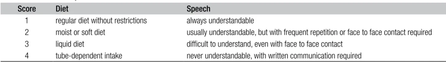
functional results were assessed by the physician at out-patient follow-up consultation and at 6 months after sur-gery using a score system; the type of diet was assessed in all cases. Options were numerically weighted from 1 to 4 as shown in Table I.
Patients
We divided patients into three groups. In Group 1 (G1), 16 patients in good general conditions receiving free radial forearm flap reconstruction; in Group 2 (G2), 18 high risk patients who received a reconstruction with infrahyoid flap; in Group 3 (G3), 20 patients who received temporal flap (10 cases) or pectoral flap (10 cases) reconstruction. G1 comprised 12 male and 4 female patients; 9 patients received a free radial forearm flap to reconstruct a defect of the oral cavity, while 7 patients had reconstruction of the oropharynx. The mean age in G1 was 58.2 years (me-dian 58, range 45-70 years), and all patients were classi-fied ASA I-II.
G2 included 12 male and 6 female patients, 12 receiv-ing infrahyoid flap for oral cavity and 6 for oropharyngeal reconstruction. All flaps were harvested from the same neck side of the primary tumour during homolateral neck dissection; 10 patients had bilateral neck dissection. The mean age in G2 was 69.6 years (median 72, range 55-83 years), 3 patients were classified ASA II, and the remain-ing ASA III. Contraindications for free flap reconstruction in G2 were: severe comorbidities (diffuse atherosclerosis, diabetes mellitus, heart failure) in 15 cases, and age ex-ceeding 80 years with moderate comorbidities in 3 cases. G3 had 16 male and 4 female patients, 11 reconstructions of the oral cavity (7 pectoralis major flaps and 4
tempo-ral flaps) and 9 reconstructions of the oropharynx (3 pec-toralis major flaps and 6 temporal flaps). The mean age in G3 was 69.6 years (median 70, range 64-81 years); 3 patients were classified ASA I, 14 patients ASA II, 2 pa-tients ASA III and 1 ASA IV. The contraindications for free flap and infrahyoid flap in G3 were: age exceeding 80 years with severe comorbidities and contraindications for infrahyoid flap reconstruction in 3 cases; post surgical vessel-depleted neck and previous radiation in 10 cases, and previous chemoradiation in 7 cases. Ten patients with vessel-depleted neck had no neck dissection. However, even in these cases, tumour resection created a commu-nication between the oral cavity or oropharynx and neck spaces.
Costs
We compared the healthcare costs of the three groups in two different ways:
• calculating the reimbursement following the DRG sys-tem;
• calculating real costs in each group from review of medical records and operating room registers.
To assess actual costs for each patient, we looked at: • the cost of main materials and drugs actually
con-sumed during diagnostic and therapeutic procedures, provided by the regional administrative institution for human and financial medical resources of Tuscany, Italy (ESTAV-Centro);
• the standard cost per hour of the physician and nurse (obtained by dividing the average salary per contrac-tual hours, € 55 and € 23 respectively);
• the cost of each diagnostic procedure, retrieved from the regional tariff list (including personnel expendi-ture);
• the average hospital stay, according to the Institutional Business Accounting (€ 420 per day, all inclusive); •
the average cost of hospital intensive care unit stay, ac-cording to Institutional Business Accounting (€ 1,300 per day, all inclusive);
• the cost of operating theatre, estimated according to the Institutional Business Accounting (€ 200 per hour including all fees except those of the medical/para-medical staff).
Costs were divided into three categories: preoperative, op-erative and postopop-erative. Preopop-erative costs include only those required by the anesthesiologist for undertaking the surgical procedure. All diagnostic procedures requested
Table I. Functional analysis.
Score Diet Speech
1 regular diet without restrictions always understandable
2 moist or soft diet usually understandable, but with frequent repetition or face to face contact required
3 liquid diet difficult to understand, even with face to face contact
by the surgeon to determine the specific characteristics of the disease (CT, MRI) were excluded, since these belong and are charged within the outpatient path. Postoperative costs were calculated until discharge.
Statistical analysis
Differences among groups were tested with the ANOVA; for categorical variables we used a chi-square test of Pear-son: P values less than 0.05 were considered statistically significant.
Results
Clinical results
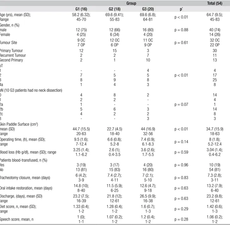
Patient characteristics and results are shown in Table II. All reconstructions were successful. In all cases, a separa-tion between oral cavity or oropharynx and neck spaces was obtained and none of the patients was re-admitted within 6 months from surgery. The mean operative time in G1 was 9 hr (range 7 h-12 h 40 min), in G2 it was 6 hr 40 min (range 5 hr 20 min-8 h), and in G3 it was 7 hr (range 5 hr 10 min-8 hr 30 min).
Table II. Patient characteristics and statistical analysis.
Group Total (54)
G1 (16) G2 (18) G3 (20) p*
Age (yrs), mean (SD);
Range 58.2 (6.32);45-70 69.6 (9.41);55-83 69.6 (6.8);64-81 p < 0.01 64.7 (9.5);45-83 Gender, n (%) male Female 12 (75)4 (25) 12 (66)6 (34) 16 (80)4 (20) p = 0.88 40 (74)14 (26) Tumour Site 9 OC7 OP 12 OC6 OP 11 OC9 OP p = 0.61 32 OC22 OP Primary Tumour Recurrent Tumour Second Primary 12 2 2 15 2 1 3 7 10 30 11 13 pT 1 2 3 4a -7 8 1 -5 9 4 4 5 8 3 p < 0.01 174 25 8 pN (10 G3 patients had no neck dissection)
0 1 2a 2b 2c 3 4 2 1 5 4 -8 2 -6 2 -2 -3 2 3 p = 0.07 14 4 1 14 8 3 Skin Paddle Surface (cm2)
mean (SD)
range 44.7 (15.5)20-63 22.7 (4.5)18-40 44 (16.9)32-56 p < 0.01 34.7 (15.9)18-63
Operating time, (h), mean (SD);
range 9.5 (1.6);7-12.4 6.6 (0.8);5.2-8 7.4 (0.9);6.1-8.3 p = 0.14 5.2-12.48 (1.8);
Blood loss (Hb g/dl), mean (SD); range 3.25 (1.4);1.1-6.2 2.6 (1);0.4-3.5 3.6 (2.6);1.7-5.5 p = 0.59 3.04 (1.4);0.4-6.2 Patients blood-transfused, n (%)
Yes
No 13 (81)3 (19) 15 (83)3 (17) 16 (80)4 (20) p = 0.96 10 (19)54 (81)
Tracheotomy closure, mean (days) 6 (4.2);3-9 7.4 (2.7);4-11 7 (2.1);5-10 p = 0.83 7.3 (2.8);3-11
Oral intake restoration, mean (days) 14.8 (10);8-40 11.5 (5.9);6-25 12.6 (4.7);9-18 p = 0.63 13.2 (7.9);6-40 Discharge, (days), mean (SD)
range 23.2 (7.5);16-39 21.8 (12);12-61 26.5 (9.9);16-38 p = 0.63 23.2 (9.8);12-61
Diet score, n, mean (SD);
range 1.33 (0.4);1-2 1.28 (0.4);1-2 1.6 (0.7);1-3 p = 0.29 1.42 (0.6);1-3
Speech score, mean, n 1 (0);1-1 1.07 (0.2);1-2 1.2 (0.4);1-2 p = 0.28 1.06 (0.2);1-2
SD: Standard deviation; ChT: Chemotherapy; RT: Radiotherapy; Hb: Haemoglobin; OC: Oral Cavity; OP: Oropharynx; * Differences in mean values among groups were tested with ANOVA, for categorical variables chi-square Pearson test was used.
Postoperative intensive care recovery was used in 4 pa-tients in G1 with a mean stay of 3.7 days, in 4 G2 papa-tients with a mean stay of 3 days and in 3 G3 patients with a mean stay of one day.
All patients were discharged with complete restoration of oral intake (mean time 15 days, range 7-18) and trache-otomy closure (mean time 7 days, range 3-11). Mean dis-charge time after surgery was 23 days (range 12-39) with no differences among groups (23.2 days G1; 21.8 days G2; 26.5 days G3). No significant differences were found with regards to verbal intelligibility and diet score among groups. Nevertheless, patients in G3 receiving TMF had minimal diet restrictions, while all patients with PM flap reconstruction required soft or liquid diets.
Economic results
The DRG system has assigned all 54 patients to the main diagnostic category (MDC) #3 “Diseases and disorders of the ear, nose, mouth and throat”, and class number 482: “Surgical tracheotomy for diagnosis concerning the face, the mouth and the neck”. Since our Hospital is a tertiary referral centre, it receives a 3% increase on 1st tariff level
for DRG high specialty (weight > 2.5). The amount of the refund, based on the DRG system, was € 7,650 for each patient. In fact, none of the patients had a hospital stay beyond the threshold of 72 days.
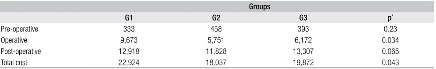
Looking at the real costs, we found a statistically signifi-cant difference among groups: in G1 the average total cost per patient was € 22,924, in G2 it was € 18,037, and € 19,872 in G3, (p = 0.043; Table III). Surgical expenses for G1 patients were significantly higher than those for G2 and G3 patients: € 9,673, € 5,751 and € 6,172 respec-tively (p = 0.034; Table III). No statistically significant differences were found for preoperative and postoperative costs among the 3 groups: € 333 and € 12,919, € 458 and € 11,828, € 393 and € 13,307, in G1, G2 and G3 respectively (p values were 0.23 and 0.065 respectively; Table III).
Discussion
The main goals in modern head and neck reconstructive surgery are restoration of form and function 20. In oral
cavity and oropharyngeal reconstructions, the surgeon is faced with several challenges: ensuring optimal healing;
increasing residual function; preventing scar formation and anchylosis of mobile structures; ensuring effective deglutition, intelligible speech, and airway patency. Fail-ure in some of these aspects, in addition to jeopardizing the patient’s quality of life, produces an increase in health care costs. In the present study, we analyzed reconstruc-tions performed by a single surgeon (AD) to avoid inter-operator differences, and focused on soft tissue recon-structions to obtain a homogeneous cohort. We selected oral cavity and oropharyngeal defects in communication with neck spaces to represent a similar level of complex-ity. In fact, transoral resections are mostly performed for small tumours, where the reconstruction in these cases is less difficult, employing primary closure, local flaps or skin grafts. Furthermore, since we focused our study on head and neck surgery, we excluded the costs of adjuvant therapies, since these are independent of the type of re-constructive procedure and could have created a bias (i.e. pre-irradiated patients). In recent years, at our Institution, the free radial forearm flap has represented the main re-constructive option for soft tissue reconstruction of oral cavity and oropharyngeal defects following cancer abla-tion. In fact, microvascular reconstructions represent a major advancement in the management of head and neck tumours; nevertheless, our philosophy of carefully con-sidering all anatomical and general conditions for each patient drove us to reconsider pedicled alternative flaps in selected cases. With this study, we wanted to verify our preliminary impression that this philosophy was not only oncologically sound, but also cost effective. Indeed, the infrahyoid flap has proven to be a valuable alternative in elderly patients suffering from severe comorbidities (G2 patients), ensuring excellent functional results 9 10 21 22. The
temporal flap and pectoralis major flap can still be use-ful in patients with a vessel depleted neck or when the expected quality of the recipient vessels is questionable (G3 patients) 11 23. Looking at our data, and calculating
the total real costs in the three groups, we immediately realized the inadequacy of the DRG system, which always assigned the highest hierarchical remuneration to the tra-cheotomy, rather than any other accompanying demoli-tion/reconstruction.
The advantages of the DRG system should consist in fix-ing an anticipated “price” for hospitalizations, but the DRG miserably fails when dealing with major head and
Table III. Real costs in euro.
Groups G1 G2 G3 p* Pre-operative 333 458 393 0.23 Operative 9,673 5,751 6,172 0.034 Post-operative 12,919 11,828 13,307 0.065 Total cost 22,924 18,037 19,872 0.043
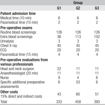
neck oncologic resections and reconstructions. In our se-ries, the obtained refund per patient, based on the DRG, was € 7,650; the gap between the real costs and the refund has been as high as € 15,274 for G1 patients, € 10,387 for G2 and € 12,222 for G3 patients. These data reflect a large disconnection between the DRG system and true treatment costs; the DRG seems undeniably unsuitable to calculate and compare healthcare costs, and therefore to be used as a parameter for policy choices. The results of our analysis showed a significantly increased cost for microvascular procedures vs. pedicled alternatives. We must highlight that pedicled alternative flaps were used in elderly, unfavourable and weak patients, where medical costs usually tend to rise rather than decrease. In fact, the average preoperative costs for the more “fragile” patients of Group 2 and Group 3, requiring specific additional pre-operative assessments were higher than prepre-operative costs in Group 1 (Table IV). These data show that our philoso-phy is not only valid from a medical point of view, but it is also economically sound. Nevertheless, our findings warrent further confirmation in a larger cohort of patients. It seems difficult to conduct a comparison with other stud-ies because there are significant differences due to: the different criteria for choosing the type of reconstruction, the diverse systems of remuneration and the various costs of human and material supplies among different institu-tions and countries.
Kroll 24 in 1997 compared 145 oral cavity and
oropharynge-al free flap reconstructions (using free radioropharynge-al forearm flaps or rectus abdominis free flaps) with 33 pectoralis major flap reconstructions. The operative costs were slightly higher for free flaps, but the total costs were lower: $ 37,314 for free flaps and $ 48,917 for pectoralis major flaps.
Ten years later, de Bree 25 matched 40 oral
cavity/oro-pharyngeal reconstructions with free radial forearm flap with 40 patients receiving the pectoralis major flap for similar defects; total costs were lower for the free radial forearm flap group: € 38,709 vs. € 42,733. However, in both these studies, free flaps were tested against the pectoralis major flap, which unfortunately is known to cause some healing delay for frequent necrosis of the most distal edge of the skin paddle; this usually doesn’t require further interventions, but it does increase hospi-tal stay and costs. In fact, where conservative transman-dibular approaches are employed, the bulkiness of the pectoralis major flap produces less than ideal functional outcomes, because the mandible presses upon the flap favouring hypovascularization and necrosis of the distal portion, and because the thickness and bulkiness of the flap hinders the motility of the preserved structures. Ac-cording to previous studies, the incidence range of total necrosis and partial necrosis for the pectoralis major flap has been reported to be from 0-2.7% and 4-29%, respec-tively 26-34.
It is our policy, however, to use the pectoralis major flap for defects mainly lying below an imaginary line between the labial commissure and tragus; instead, the temporal flap is chosen for defects mainly lying above this line. Furthermore, for reconstructions following mandibular sparing procedures, we prefer to use the pectoralis ma-jor flap as myofascial transposition, reducing its bulk, and consequently reducing the pressure of the mandible. These two specific indications decrease the occurrence of distal marginal necrosis and the related costs.
In our series, the mean length of hospitalization was 23.2 days in G1, 21.8 days in G2 and 26.5 in G3, which was not significantly different (p = 0.63). The intraoperative costs for G1 patients were significantly higher (p = 0.034) than costs for G2 and G3 patients: € 9,673, € 5,751, and € 6,172 respectively (Table V). The highest intra-operative costs for G1 patients are due to longer opera-tive time, and, above all, to the simultaneous work of a double medical and paramedical team (flap harvest during tumour resection; Table V). Longer operative times in G1 were mainly dependent on the microvascular reconstruc-tion times, not only technically related to preparareconstruc-tion of the recipient vessels under microscopic magnification and revascularization times, but also to “meticulous” and “pa-tient/delayed” surveillance of microanastomosis patency prior to definitive skin closure (of course this step could be omitted or quickened, but we feel that “it is better to be safe than sorry”). On the other hand, higher operative costs in G1 were less dependent on operative times and mainly related to personnel-related costs (medical and paramedical).
The analysis of postoperative expenses (Table VI) showed a substantial parity between G1 and G3, with slight best performance again for G2. The inappropriate use of
post-Table IV. Pre-operative costs in euro.
Group
G1 G2 G3
Patient admission time Medical time (10 min)
Paramedical time (10 min) 62 62 62
Pre-operative exams Routine blood screenings Extra blood screenings Urinalysis
Chest X-ray ECG
Paramedical time (15 min)
126 36 3 45 20 4 126 113 3 45 20 4 126 102 3 45 20 4 Pre-operative evaluations from
various professionals Head and neck surgeon Anaesthesiologist (20 min) Nurse
Specific additional preoperative assessments 22 11 9 6 11 11 4 53 11 11 6 6 Other costs
15% direct and indirect costs 43 60 51
operative intensive care recovery (ICU) in 4 G1 patients did deny a saving in this group of healthier patients, and instead raised postoperative costs (Table VI). Posttive ICU monitoring was not related to protracted opera-tive times, but only for the lack of the appropriate sub-in-tensive facility and it was no longer used for the 12 more recent cases.
Our reconstructive philosophy has provided successful results in functional terms, also in terms of “cost-effec-tiveness”. The use of alternative pedicled flaps in high-risk patients probably reduced the high-risk of flap failure, with consequent expenditure restraints. The use of microvas-cular techniques for these patients might have led to an increase in production costs linked to the increase of indi-rect costs arising from possible complications. The limits of our study are mainly represented by the retrospective setting and the small cohort. It would be beneficial, for subsequent analyses, a perspective evaluation with a larg-er cohort, possibly multi-institutional. In our opinion, sat-isfaction and quality of life of the patient must, however, precede any economical concern 35-38.
Conclusions
Our analysis shows that the use of alternative non-micro-vascular techniques in high-risk patients, does not affect
the result in oncologic and functional terms, and can even produce a cost saving. In particular, the infrahyoid flap ensures excellent functional results accompanied by the best economic performance in the most fragile patients.
This paper was awarded with the 3rd SIO Price at the 99th
National Congress of the Italian Society for Otorhinolar-yngology and Head and Neck Surgery, Bari 2012.
References
1 Suh JD, Sercarz JA, Abemayor E, et al. Analysis of outcome
and complications in 400 cases of microvascular head and
neck reconstruction. Arch Otolaryngol Head Neck Surg
2004;130:962-6.
2 Novak CB, Lipa JE, Noria S, et al. Comparison of
anterolat-eral thigh and radial forearm free flap donor site morbidity.
Microsurgery 2007;27:651-4.
3 Tarsitano A, Pizzigallo A, Sgarzani R, et al. Head and
neck cancer in elderly patients: is microsurgical
free-tis-sue transfer a safe procedure? Acta Otorhinolaryngol Ital
2012;32:371-5.
4 Momeni A, Kattan A, Lee GK. Is microsurgical head and
neck reconstruction profitable?: analysis at an academic
medical center. Ann Plast Surg 2012;68:401-3.
5 Ferguson RE Jr, Yu P. Techniques of monitoring buried
fascio-cutaneous free flaps. Plast Reconstr Surg 2009;123:525-32.
6 Pellini R, Pichi B, Marchesi P, et al. External monitor for
buried free flaps in head and neck reconstruction. Acta
Otorhinolaryngol Ital 2006;26:1-6.
7 Thoma A, Veltri K, Archibald S, et al. Microsurgical
recon-struction of the through-and-through defect in head and neck
cancer: is it worth it? J Reconstr Microsurg 1999;15:401-8.
8 Pfister, DG, Ruchlin HS, Elkin EB. Economic considerations
in the care of patients with head and neck malignancies. Curr
Opin Oncol 1997;9:241-6.
9 Deganello A, Manciocco V, Dolivet G, et al. Infrahyoid
fascio-myocutaneous flap as an alternative to free radial
forearm flap in head and neck reconstruction. Head Neck
2007;29:285-91.
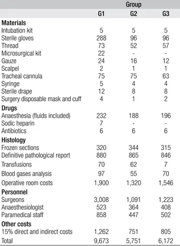
Table V. Operative costs in euro.
Group G1 G2 G3 Materials Intubation kit Sterile gloves Thread Microsurgical kit Gauze Scalpel Tracheal cannula Syringe Sterile drape
Surgery disposable mask and cuff 5 288 73 22 24 2 75 5 12 4 5 96 52 -16 1 75 4 8 1 5 96 57 -12 1 63 4 8 2 Drugs
Anaesthesia (fluids included) Sodic heparin Antibiotics 232 7 6 188 -6 196 -6 Histology Frozen sections
Definitive pathological report 320880 344865 315846
Transfusions 70 62 7
Blood gases analysis 97 55 70
Operative room costs 1,900 1,320 1,546
Personnel Surgeons Anaesthesiologist Paramedical staff 3,008 523 858 1,091 364 447 1,223 408 502 Other costs
15% direct and indirect costs 1,262 751 805
Total 9,673 5,751 6,172
Table VI. Postoperative costs in euro.
Group
G1 G2 G3
Ordinary hospital stay 9,744 9,156 11,130
Hospital stay in ICU 1,219 867 195
Medications (hospital ward) Materials Medical time Paramedical time 97 31 16 79 14 7 81 22 19
Other specialists in consultation 63 98 38
Exams Imaging, ECG 30 26 38 Rehabilitation Speech therapy Physiotherapy 278 2414 3116 Other costs 1,684 1,543 1,737 Total 12,919 11,828 13,307
10 Deganello A, Gitti G, Parrinello G, et al. Infrahyoid flap re-construction of oral cavity and oropharyngeal defects in
el-derly patients with severe general comorbidities. Head Neck
2012;34:1299-305.
11 Deganello A, Gallo O, De Cesare JM, et al. Surgical
man-agement of surgery and radiation induced peristomal neck
ulcerations. B-ENT 2008;4:169-74.
12 Deganello A, Gitti G, Struijs, et al. Palliative combined
treatment for unresectable cutaneous basosquamous cell
carcinoma of the head and neck. Acta Otorhinolaryngol Ital
2013;33:353-356.
13 Colletti G, Autelitano L, Tewfik K, et al. Autonomized flaps
in secondary head and neck reconstructions. Acta
Otorhi-nolaryngol Ital 2012;32:329-35.
14 van der Putten L, Spasiano R, de Bree R, et al. Flap
recon-struction of the hypopharynx: a defect orientated approach.
Acta Otorhinolaryngol Ital 2012;32:288-96.
15 Montemari G, Rocco A, Galla S, et al. Hypopharynx
recon-struction with pectoralis major myofascial flap: our
experi-ence in 45 cases. Acta Otorhinolaryngol Ital 2012;32:93-7.
16 Bhattacharyya N, Fried MP. Benchmarks for
mortal-ity, morbidmortal-ity, and length of stay for head and neck
sur-gical procedures. Arch Otolaryngol Head Neck Surg
2001;127:127-32.
17 Fetter RB, Freeman JL, Mullin RL. DRGs: how they evolved
and are changing the way hospitals are managed.
Patholo-gist 1985;39:17-21.
18 Sobin LH, Gospodarowicz M, Wittekind Ch, editors. TNM
classification of malignant tumors. 7th ed. International
Un-ion Against Cancer. New York: Wiley-Liss; 2009.
19 American Society of Anesthesiologists Relative Value Guide.
A Guide for Anesthesia Values 2012. Park Ridge, Ill.
Ameri-can Society of Anesthesiologists; 2012;.
20 Jones NF, Jarrahy R, Song JI. Postoperative medical
com-plications – not microsurgical comcom-plications –negatively influence the morbidity, mortality, and true costs after
mi-crosurgical reconstruction for head and neck cancer. Plast
Reconstr Surg 2007;119:2053-60.
21 Gangloff P, Deganello A, Lacave ML, et al. Use of the infra
hyoid musculo-cutaneous flap in soft palate reconstruction.
Eur J Surg Oncol 2006;32:1165-9.
22 Deganello A, De Bree R, Dolivet G, et al. Infrahyoid myocuta-neous flap reconstruction after wide local excision of a Merkel
cell carcinoma. Acta Otorhinolaryngol Ital 2005;25:50-3.
23 You YS, Chung CH, Chang YJ, et al. Analysis of 120
pec-toralis major flaps for head and neck reconstruction. Arch
Plast Surg 2012;39:522-7.
24 Kroll SS, Evans GR, Goldberg D, et al. A comparison of
re-source costs for head and neck reconstruction with free and
pectoralis major flaps. Plast Reconstr Surg 1997;99:1282-6.
25 de Bree R, Reith R, Quak JJ, et al. Free radial forearm flap versus pectoralis major myocutaneous flap reconstruction of
oral and oropharyngeal defects: a cost analysis. Clin
Otolar-yngol 2007;32:275-82.
26 Baek SM, Lawson W, Biller HF. An analysis of 133
pec-toralis major myocutaneous flaps. Plast Reconstr Surg
1982;69:460-9.
27 Shah JP, Haribhakti V, Loree TR, et al. Complications of the pectoralis major myocutaneous flap in head and neck
recon-struction. Am J Surg 1990;160:352-5.
28 Kroll SS, Goepfert H, Jones M, et al. Analysis of
com-plications in 168 pectoralis major myocutaneous flaps
used for head and neck reconstruction. Ann Plast Surg
1990;25:93-7.
29 Mehta S, Sarkar S, Kavarana N, et al. Complications of
the pectoralis major myocutaneous flap in the oral cavity:
a prospective evaluation of 220 cases. Plast Reconstr Surg
1996;98:31-7.
30 IJsselstein CB, Hovius SE, ten Have BL, et al. Is the pec-toralis myocutaneous flap in intraoral and oropharyngeal
reconstruction outdated? Am J Surg 1996;172:259-62.
31 Milenovic A, Virag M, Uglesic V, et al. The pectoralis major
flap in head and neck reconstruction: first 500 patients. J
Craniomaxillofac Surg 2006;34:340-3.
32 Corten EM, Schellekens PP, Hage JJ, et al. Clinical outcome after pedicled segmental pectoralis major island flaps for head
and neck reconstruction. Ann Plast Surg 2009;63:292-6.
33 Pinto FR, Malena CR, Vanni CM, et al. Pectoralis major
my-ocutaneous flaps for head and neck reconstruction: factors influencing occurrences of complications and the final
out-come. Sao Paulo Med J 2010;128:336-41.
34 Vartanian JG, Carvalho AL, Carvalho SM, et al. Pectoralis
major and other myofascial/myocutaneous flaps in head and neck cancer reconstruction: experience with 437 cases at a
single institution. Head Neck 2004;26:1018-23.
35 Gisquet H, Gangloff P, Graff P, et al. [Microsurgical recon-struction and full management of patients with head and neck cancer: importance of a quality approach and a patient care
team]. Rev Laryngol Otol Rhinol (Bord) 2009;130:249-54.
36 Pellini R, Mercante G, Spriano G. Step-by-step mandibular
reconstruction with free fibula flap modelling. Acta
Otorhi-nolaryngol Ital 2012;32:405-9.
37 Giordano L, Bondi S, Ferrario F, et al. Radial forearm free flap surgery: a modified skin-closure technique improving
donor-site aesthetic appearance. Acta Otorhinolaryngol Ital
2012;32:158-63.
38 Mura F, Bertino G, Occhini A, et al. Advanced carcinoma
of the hypopharynx: functional results after
circumferen-tial pharyngolaryngectomy with flap reconstruction. Acta
Otorhinolaryngol Ital 2012;32:154-7.
Address for correspondence: Alberto Deganello, Academic Clinic of Otolaryngology and Head-Neck Surgery, University of Florence, Largo Brambilla 3, 50134 Florence, Italy. Tel. +39 055 7947054. Fax +39 055 435649. E-mail: [email protected]