University of Sassari
Department of Biomedical Sciences
INTERNATIONAL PHD SCHOOL IN BIOMOLECULAR AND
BIOTECHNOLOGICAL SCIENCES
XXVIII Cycle
APPLICATION OF MOLECULAR TECHNIQUES TO
STUDY THE ETIOLOGY AND EPIDEMIOLOGY OF
CANDIDA ssp. INFECTIONS IN CENTRAL VIETNAM
Director: Prof. LEONARDO A. SECHI
Tutor: Prof. PIERO CAPPUCCINELLI Co-tutor: Dr. TON NU PHUONG ANH
Dr. ANTONELLA SANTONA PhD thesis of
Dott.ssa NGO THI MINH CHAU
ATTESTATION OF AUTHORSHIP
I hereby declare that this submission is my own work and that, to the best of my knowledge and belief, it contains no material previously published or written by another person except that which appears in the citations and acknowledgements. Nor does it contain material, which to a substantial extent I have submitted for the qualification for any other degree of another university or other institution of higher learning.
TABLES OF CONTENTS
ACKNOWLEDGEMENTS LIST OF ABBREVIATION LIST OF TABLES LIST OF FIGURES ABSTRACT………1 1. INTRODUCTION ... 2 1.1. Candida species ... 21.2. Candida spp. virulent factors ... 15
1.3. Molecular epidemiology of Candida spp... 18
1.4. Molecular mechanism to resistant drugs in Candida spp. ... 21
1.5. Candida spp. and bacteria interaction ... 24
2. RESEARCH OBJECTIVES ... 27
3. MATERIALS AND METHODS ... 29
3.1. Study site ... 29
3.2. Study population ... 29
3.3. Sample collection and Candida strains isolates ... 30
3.4. Candida spp. identification ... 32
3.5. Antifungal susceptibility testing ... 37
3.6. Detection Candida virulence genes by multiplex PCR and sequencing ... 38
3.7. Detection of erg11 gene mutations associated to fluconazole resistance in C. tropicalis by PCR and sequencing ... 40
3.8. Candida albicans Multi Locus Sequence Typing ... 41
3.9. Detection H. pylori from Candida spp. by nested PCR and sequencing ... 43
3.10. Data analysis ... 44
4. RESULTS ... 45
4.1. Study population and Candida spp. identification by MALDI-TOF MAS and ITS sequencing ... 45
4.3. Detection H. pylori from Candida spp. by nested PCR and sequencing ... 61 5. DISCUSSION ... 64
5.1. Candida species identification by MALDI - TOF MS and ITS sequencing . 64 5.2. Phenotypic and genotypic characterization of Candida spp. isolates ... 67 5.3. H. pylori detection from Candida spp. ... 71 6. CONCLUSION ... 73 REFERENCES
ACKNOWLEDGEMENTS
“No one who achieves success does so without the help of others” Alfred North Whitehead This thesis and my research were supported by Hue Universiy of Medicine and Pharmacy-Vietnam, Sassari University-Italy, and Carlo Urbani Project-Italian Government. Many people and organizations contributed to the completion of my PhD study and I acknowledge to everyone, who gave me contributions and assistances during last three years.
Firstly, I sincerely express my great gratitude to my supervisor, Professor. Piero Cappuccinelli, for the continuous support of my study and research process, for his excellent guidance, patience, immense knowledge.
I am thankful for my co-supervisor, Dr. Ton Nu Phuong Anh, my believable tutor, the first person leading me to real science. I am extremly thankful and indebted to her for sharing expertise, sincere and valuable guidance, and encouragement extended to me.
A special thanks goes to Dr. Antonella Santona, a enthusiasm tutor, who trained and let me experience the research of molecular techniques, encouraged and helped me in research process, and patiently corrected my writing. I would like to thank Dr. Maura Fiamma, Dr. Bianca Paglietti, and Dr. Silvana Sanna for their supports in my research. I also would like thank to Professor. Leonardo A. Sechi, Rector of International PhD School, Professor. Rubino Salvatore, and everyone in Microbiology Department, Sassari University, who gave me warmly environment to study and helped me to interact in Italian culture. My sincere thanks also goes to Dr. Vito Astone, who supported me to do fungal molecular diagnostic in Nuoro Hospital, Italy. I want to say thank to Professor. Bruno Masala, Professor. Claudia Crosio, who facilitated my attendance to the PhD program at Sassari University.
I would like to thank Dr. Giovanni Sini and Ms. Giustina Casu, who gave me strong supports to fullfil all documents relating my PhD course.
My sincere thanks also goes to Rector of Hue University of Medicine and Pharmacy, Professor. Cao Ngoc Thanh, who provided me an opportunity to apply document to enrolle PhD programme and helped me to complete this study program.
I am sincerely grateful to all my colleagues at Parasitology department, especially to enthusiastic supports of Mrs. Do Thi Bich Thao and Ms. Tran Thi Diem Na, who spent a lot of time to help me do my research.
I express my warm thanks to staffs in Hue University of Medicine and Pharmacy Hospital and Hue Central Hospital for their supports and their contributions in my collecting samples period. In particular, I am grateful to Dr. Tran Xuan Thinh and Dr. Phan Thi Hong Diep, who gave me good cobollarations.
I would like to thank others Vietnamese PhD students at cycle XXVIII, who supported me in study, writing, and helping me to achieve the final goal in this course.
A special thanks to my family. Words cannot express how grateful I am to my parent, parent-in law for all of their sacrifices. I would also like to thank to my brothers and sisters for their supports. I want to express my gratitude to my cousin, Ms. Anh Tran, who spent a lot of time to correct my writing. At the end I would like express appreciation to my beloved husband, who gave me continuous encouragement, shared with me the most difficult time and was always my support. My deepest love and special thanks are also to my son and daughter. They are my motivation, encouragement, and their loves help me face the difficulties and made me become stronger.
I would like to say thank and share my gratitude again for everything. Ngo Thi Minh Chau
LIST OF ABBREVIATION
AAT1a Aspartate aminotransferase ACC1 Acetyl-coenzyme A carboxylase
ADP1 ATP-dependent permease
AIDS Acquired immunodeficiency syndrome
ALS Agglutinin like sequence
BFIs Bacteria fungi interactions
BMD Broth microdilution
BSI Blood stream infection
C Clade
C. albicans Candida albicans C. non albicans Candida non albicans Candida spp. Candida species
CC Cluster Clade
CDC Centers for Disease Control and Prevention
CDR Candida drug resistance
CLSI Clinical and Laboratory Standards Institute
DSTs Diploide sequence typing
EDTA Ethylenediaminetetraacetic acid
EPA Epithelial adhesin
ERGs Ergosterol biosynthetic
EUCAST European Committee on Antimicrobial Susceptibility Testing
HC Hospital Hue Central Hospital
H. pylori Helicobacter pylori
HIV Human Immunodeficiency Virus
HUMP Hue University of Medicine and Pharmacy
HWP Hyphae wall protein
ITS Internal transcribed spacer
LOH Loss of heterozygosity
MALDI - TOF MS Matrix assisted laser desortion ionization time-of- flight mass spectrometry
MDR Multidrug resistance
MIC Minimum inhibitory concentration
MLST Multi Locus Sequence Typing
MP11b Mannose phosphate isomerase
PCR Polymera chain reaction
PFGE Pulsed field gel electrophoresis
RAPD Random amplified fragment length polymorphism RFLP Restriction fragment length polymorphism
rRNA Ribosom RNA
SDA Sabouraud dextrose agar
SYA1 Alanyl-RNA synthetase
SVP13 Vacuolar protein sorting protein
TAE Tris-acetate -EDTA
UK United Kingdom
USA United States of America
UV Ultraviolet
ZWF Glucose-6-phosphate dehydrogenase
LIST OF TABLES
Table 1.1. Summary of recommendations by Candida disease, specimen and test
evaluated [42] ... 12
Table 3.1 . Distribution of patients in hospital wards ... 30
Table 3.2. Zone diameter interpretive follow Liofilchem Laboratory's instructions 38 Table 3.3. Primers of 7 housekeeping genes (http://calbicans.mlst.net/) ... 42
Table 4.1. Population distribution by wards, age and gender ... 45
Table 4.2. Candida spp. identification ... 48
Table 4.3. Antifungal susceptibility test by disk diffusion ... 51
Table 4.4. Frequency Candida species resistance to azole group drugs (%) ... 52
Table 4.5. Cross - resistance to multiple azole drugs ... 53
Table 4.6. erg11 gene mutations in 15 C.tropicalis strains ... 55
Table 4.7. Source of C. albicans strains detecting virulent genes... 57
Table 4.8. The DST assignments at each of seven MLST loci for all 15 C. albicans isolates ... 57
Table 4.9. Clades corresponding to the clonal Cluster by E.burst and UPGMA ... 60
LIST OF FIGURES
Figure 1.1. Candida spp. morphology ... 4
Figure 1.2. Structure of the C. albicans cell wall [19]. ... 5
Figure 1.3. The human mycobiota [31] ... 7
Figure 1.4. The steps of C. albicans tissue invasion [19]. ... 8
Figure 1.5. Hospital - acquired Candida infections [37] ... 10
Figure 1.6. An overview of selected virulent factors distribute to C.albicans pathogenicity mechanism [32]. ... 16
Figure 1.7. Main mechanisms of azole and polyens resistance [78] ... 23
Figure 1.8. The bacteria fungal interaction: the combination of physical associations and molecular interactions [88] ... 25
Figure 3.1. Colonies of C. albicans and C. glabrata on Brillant Candida medium32 Figure 3.2. Possitive germ tube test and chlamydospore assay [10] ... 33
Figure 3.3. Principle of MALDI-TOF Mass Spectrometry system [120] ... 34
Figure 4.1. Distribution of samples sources by hospital ward ... 47
Figure 4.2. Candida identification result by MALDI –TOF MS and ITS sequence 48 Figure 4.3. Distribution Candida spp. in hospital wards ... 49
Figure 4.4. Distribution of Candida spp. in colonization and candidiasis ... 50
Figure 4.5. Resistance to at least one type of azole drug ... 52
Figure 4.6. Frequency Candida spp. resistance to 5-Fluorocystocine ... 53
Figure 4.7. Frequency Candida spp. resistance to caspofungin ... 54
Figure 4.8. ERG11 missense mutations in C. tropicalis strain no. 41 and no.189 .. 55
Figure 4.9. ERG11 silent mutations in C. tropicalis strain no. 193 ... 56
Figure 4.10. C. albicans virulent genes: sap4, hwp1 and als1 gene amplification products in positive samples ... 56
Figure 4.11. Loss of heterozygosity in loci VPS13 (W to A) in strain no. 19A ... 58
Figure 4.12. Population snapshot of the 2973 C. albicans distinct DSTs currently in the MLST database (www.calbicans.mlst.net) defined using eBURSTv3. ... 59
Figure 4.14. (1) Specific band (183bp) and (2) unspecific band (300 bp) ... 61 Figure 4.15. Candida spp. distribution in H. pylori presence ... 62
ABSTRACT
The overall objective of this study was to apply a variety of molecular techniques to determine the etiology and epidemiology of Candida spp. infections in Central Vietnam.
MALDI-TOF Mass Spectrometry correctly identified 94.9% of 196 Candida spp. isolated strains. The remaining unidentified species (5.1%) were detected by ITS gene amplification and sequencing. A variety of Candida species were isolated,
with the most common species being C. albicans (47.96%), followed by C. tropicalis (16.33%), C. parapsilosis (10.71%), C. glabrata (8.67%), C. orthopsilopsis (5.61%), C . krusei (3.57%) and others species.
Antibiotic susceptibility showed higher rates of resistance to fluconazole in C. tropicalis (56.67%). Two specific missense mutations in the ERG11 protein
(Y132F and S154F) were detected from 26.67% of fluconazole resistant C. tropicalis isolates.
96.81% of C. albicans isolates carried all three virulence genes (als1, hwp1, sap4) regardless of the sample source, that was concordant with a higher frequency of C. albicans in mucosal candidiasis compared to a higher frequency of C. non albicans in candida colonizations.
A total of 12 C. albicans diploid sequence types (DSTs) were identified from ten different wards by Multi Locus Sequence Typing (MLST). Of the 12 identified DSTs, six were new DSTs assigned by this study. Based on C. albicans cluster analysis, 66.67% of the isolates clustered with previously known clades in global or Asian data, and 33.33% isolates were singleton. MLST results suggested a potential nosocomial transmision of C. albicans since the same DTS clones were found in different wards.
Amplification of specific H. pylori gene (ureA) was applied to find a presence of this bacteria in Candida cells. Positivity was detected in 15.27% of Candida spp. (C. albicans, C. tropicalis, C. orthopsilosis) isolated from oral mucosa, sputum, vagina, and gastric fluid.
1. INTRODUCTION
1.1. Candida species 1.1.1. History
The original name of Candida comes from the Latin term “candidus”, meaning “glowing white,” which relates to the creamy and glistening white characteristic of yeasts colonies on culture media. This fungi is responsible for oral thrush, an infection that has been recognized for over 2000 years [1]. Candida infection, or „candidiasis‟, is also known as „candidosis‟ or „moniliasis‟.
Candida albicans (C. albicans) was identified in the nineteenth century from three independent sources. First, in 1841, Fredrick Berg, a Swedish medical pratitioner, discovered that thrush was caused by fungus with filaments that dispersed into epithelial cells. Then in 1842, David Gruby, a medical practitioner in Paris, fully described the cells of thrush fungus and compared to that causing tinea. Thrush fungus was later named in 1853 as Oidium albicans by Charles Phillipe Robin. In 1868, Charles Quinquaud renamed the fungus to Syringospora robinii. The name was changed again to Monilia albicans in 1890 by Wilhelm Zopf. Around this time, detailed drawings of the fungus revealed the following characteristics: budding cell, pseudohyphae, hyphae, and dimorphism. In 1923, Christine Berkhout, a Dutch mycologist, changed the name of the fungus to Candida albicans [2, 3].
In addition to infecting the oral cavatities, Candida infects other parts of the body, including oesophageal, vaginal, cerebral, and intestinal lesions, etc. [2].
1.1.2. Taxonomy and mycology
The heterogeneous genus Candida belongs to the Kingdom: Fungi, Phylum: Ascomycota, Subphylum: Ascomycotina, Class: Ascomycetes, Order: Saccharomycetales, Family: Saccharomycetaceae, Genus: Candida. The genus contains approximately 200 species [4]
The taxonomy of the genus Candida is increasing overtime because of the reclassification of certain species (e.g., Torulopsis glabrata has been correctly identified as C. glabrata), and the discovery of new species such as C. dubliniensis, C. orthopsilosis, and C. metapsilosis. C. orthopsilosis, and C. metapsilosis were previously classified as part of the C. parapsilosis complex [5]. Futhemore, some species have been classified under C. albicans, including C. claussenii and C. langeronii. Recently, some new Candida species were distinguished from C. albicans, such as C. albicans var. africana, a new sucrose-negative variant of C. albicans that is closely related to C. stellatoidea type II [6].
More than 200 species of Candida have been described, most of which exist as saprophytes orgnisms. Half of the described species can not grow at 370C, making them unsuccesful as human pathogens. Approximately 30 species can infect humans. C. albicans is the most prevalent species, followed by other pathogenic species which include C. glabrata, C. tropicalis, C. parapsilosis, C. krusei, C. guilliermondii, C. lusitaniae, C. kefyr, C. inconspicua, C. famata, C. rugosa, C. dubliniensis, C. norvegensis, C. lipolytica, C. sake, C. pelliculosa, C. apicola, C. zeylanoides, C. valida, C. intermedia, C. pulcherrima, C. haemulonii, C. stellatoidea, C. utilis, C. humicola, C. lambica, C. ciferrii, C. colliculosa, C. holmii, C. marina, and C. sphaerica ect [7].
C. albicans, C. glabrata, C. parapsilosis, C. tropicalis and C. krusei account for 90-92% of all cases of candidiasis [8, 9]. The Candida pathogenic species list is expanding rapidly [7].
1.1.3. Morphogenesis
Genus Candida constitute a heterogeneous group of eukaryotic, dimorphic, or polymorphic organisms [10, 11]. All Candida species grow as yeast cells or blastoconidia under general culture conditions between 250C and 350C, with growth augmented by increased sugar or fat content in the media. Yeast cells are approximately 2-10µm in the largest dimension, round to oval, and reproduce by budding. C. albicans is among the larger yeast at 4-6 x 6-10µm, whereas C.
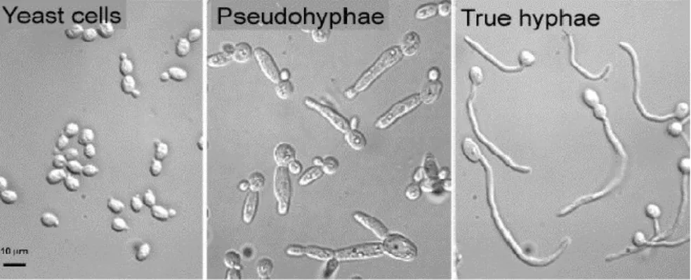
glabrata and C. parapsilosis are among the smallest at 1-4 x 9µm and 4 x 2-9µm, respectively [5]. They multiply principally by the production of blastoconidia (buds). As normally, blastoconidia of Candida spp. in microbiota gut of being human or animal exits in round, oval shape, but they could have different morphogenesis depending on the species [10, 12]. When blastoconidia are produced from one another in a linear fashion without separating, a structure termed a pseudohypha is formed. Under certain circumstances, such as growth under reduced oxygen tension, some yeasts may produce true hyphae. Most members of the genus produce filamentous forms (pseudohyphae or true hyphae). C. parapsilosis forms pseudohyphae but not true hyphae. C. dubliniensis, C.tropicalis and C. albicans form true hyphae [5, 12]. The presence of budding yeasts, pseudohyphae or hyphae in infected tissue are usually indicative of candidiasis [13, 14]. C. glabrata is the only pathogenic species that does not produce filamentous forms, existing exclusively as blastoconidia [12].
Figure 1.1. Candida spp. morphology
(www.tcd.ie/Biology_Teaching_Centre/assets/pdf/by2205/by2205- webgalleries2011/by2205-gallery1/candida.pdf)
1.1.4. Cell biology and enzymology
Candida spp. growth characteristics, metabolic features and enzymology characteristics are the same to those of eukaryotes and especially similar to Saccharomyces cerevisiae [10].
Polysaccharides are an essential compound in the cell walls of Candida species [10, 15]. Candida cell walls are composed of mannans, glucans and a small amount of chitin (Figure1.2) [10, 16, 17]. These components are closely bound to polypeptides and proteins found on the cell membrane [16]. Three types of adhesion molecules were observed [18]: (1) glycoproteins which are expressed specifically on the surface of hyphae form, (2) the protein moiety of glycoproteins which binds to host glycosides containing fucose or N-acetyl glucosamine, and (3) the polysaccharide portion of a mannoprotein. Furthermore, the structure of mannan polysaccharides found on the walls of Candida plays a potent role in its pathogenicity [10, 19]. C. albicans mannan masking of glucan and yeast hypha morphogenesis are required for disruption of host processes that function to inactivate pathogens, leading to survival and escape of this fungal pathogen from within host phagocytes [20].
Phospholipids and sterols are dominant in lipids structure of Candida spp. Ergosterol is the major membrane sterol. These lipids provide the site of action for the synthesis of enzymes involved in cell wall morphogenesis and antifungal action. Lipid alterations can occur during a yeast to mycelium transition [21]. Additionally, Candida spp. are constantly changing the structure of enzymically active proteins such as enolase and N-acetyl glucosaminidase, ubiquitin like epitopes and a protein related to the heat shock protein family (hsp70 and hsp 90) [10, 11].
Candida spp. can grow in wide a pH range, from below 2.0 to nearly 10 [22], and under microaerophilic and even anaerobic conditions as well as the more normal aerobic atmospheres of incubation. Glucose, galactose and sucrose are all substrates for growth of the fungus, and nitrogen requirements can be met by relatively low concentrations of ammonium ions.
Many enzymes of C. albicans and C. non albicans have been characterized. Secreted aspartyl proteinases (SAP), one of the most studied enzymes, produce by C. albicans, C. parapsilosis, C. tropicalis, C. dubliniensis, C. guilliermondii, C. kefyr, C. lusitaniae, and C. krusei [23-25]. These enzymes produce non specific proteolysis of host proteins involved to defend against infection. Their different profiles of pH dependent irreversible denaturation may partially explain differences in virulence of Candida species [25]. The sap gene family in C. albicans includes at least 10 isoenzymes which are referred to as SAP1 through SAP10 [25]. Different SAPs are associated with a different location within the yeasts and different pathogenocity [25]. SAP1-3 expression is dominant in mucosal and cutaneous candidosis, whereas SAP 4-6 may be important for systemic disease [10, 25] and could induce apoptosis of epithelial cells by a novel Trojan horse mechanism [26].
C. albicans, C. dubliniensis, C. glabrata, C. krusei, C. lusitaniae, C. parapsilosis, and C. tropicalis also produce phospholipases [10, 27]. These enzymes play an important role in controlling of yeast growth, remodeling of fungal cell membranes and spreading in host tissues through hydrolysis of phospholipids [28]. Phospholipase B is important in C. albicans virulence, and it is secreted by the yeast during the infection process [28, 29].
1.1.5. Candida spp. from gut commensal to pathogen
Candida species are human commensals [30], commonly found on the mucosal surfaces of gastrointestinal and genitourinary tracts, skin, and under fingernails, and belong to human mycobiota (Figure 1.3) [5, 10, 31]. Moreover, C. albicans is also isolated from various sources, such as the atmosphere, fresh water, sea water and soil [10].
The prevalence of Candida colonization varies depending on site and population sampled, and sampling method. It is estimated that between 25-40 % of people are colonized by C. albicans at any given point [11], this rate is approximately 6% (2-37%) among healthy person for oral Candida colonization and approximately 47% (13-76%) in hospitalized patients [10]. These figures could be higher in patient having high risks factors. For example, oral carriage rates may be higher in certain situations such as in HIV infected patients with low CD4 counts, denture users with denture stomatitis, diabetic patients, patients receiving antineoplastic chemotherapy and children [10]. It is believed that 100% of humans may carry one or more Candida species in the gut from the duodenum to the colon. The numbers of yeasts carried at any point in the gut can increase to levels that may become detectable in the mouth and feces in illness or other situations where the host‟s microbial suppression mechanisms become reduced.
Figure 1.3. The human mycobiota [31]
Candida spp. become pathogens in situations where the host‟s resistance to infection is lowered locally or systemically [11, 32]. As opportunistic pathogens,
they can damage local mucosal epithelium and sometimes, systemic infections in which they can spread to all major organs and colonize in these organs. There are several steps in tissue invasion by C. albicans: (1) adhesion to the epithelium; (2) epithelial penetration and invasion by hyphae; (3) vascular dissemination, which involves hyphal penetration of blood vessels and seeding of yeast cells into the bloodstream; and (4) endothelial colonization and penetration during disseminated diseases [19]. This process is shown in Figure 1.4.
.
The switch from commensalism to pathogenesis in Candida spp. depends on both fungal and host factors. Some essential fungal factors include adhesion, dimorphism, biofilm formation and hydrolytic enzyme [32].
1.1.6. Candidiasis epidemiology
Candidiasis refers to infection caused by any of more than 200 species of the genus Candida. This fungi is capable of causing several different types of diseases, ranging from commonly encountered superficial infections (oral candidiasis, intertriginous candidiasis, ungual and periungual candidiasis, angular chelitis, vaginitis, chronic mucocutaneous candidiasis) to rare, candidaemia, systemic diseases [32-34]. Systemic infections are normally caused by endogenous Candida spp. escaping the gastrointestinal tract and circulating via the bloodstream to infect deep tissues such as the lungs, kidney, and liver [5]. Candidiasis is usually caused by C. albicans, and some time by other species (C. parapsilosis, C. tropicalis, C. krusei, C. glabrata, C. stellatoides etc.).
Candida spp. are the fourth most common cause of nosocomial bloodstream infections (BSI) in the United States with a 35% to mortality rate. According to Centers for Disease Control and Prevention (CDC), approximately 46.000 healthcare associated Candida infections occur among hospitalized patients in the United States each year with 30% of patients with candidemia with drug resistant Candida die during hospitalization. Additionally, drug resistant Candida infections results in millions of dollars in excess costs to U.S. healthcare expenditures each year[35].
In the world, it estimated that there are 72.8 million Candida opportunistic infection cases per year [36]. The increasing rate of invasive fungal pathogens is related to the increasing number of critically ill patients and the use of a broad spectrum of antibiotics, surgical procedures, cytotoxic therapy with prolonged neutropenia, other immune suppressive therapies, indwelling invasive devices, and intensive care support. Candida spp. are the most prominent invasive fungal infection in critically ill adult patients as well as in neonatal patients treated in the
Intensive care units (ICU), and the second cause of invasive fungal infection in severely immunocompromised patients such as those with cancer and recipients of hematopoietic stem cell and bone marrow or solid organ transplantation. It is estimated that 0.5-1% patients having high risk factors will contract Candida bloodstream infection, which contributed to 8-10% of all nosocomial bloodstream infections. Approximately 20-50% of these patients will die as a result of the infection, and an additional 10-40% will die from underlying disease (Figure 1.5) [37].
Figure 1.5. Hospital - acquired Candida infections [37]
Although C. albicans is the most common cause of invasive fungal infections, the increasing number of infections from C. non albicans species is reported as a major source of infection [7, 38]. The ARTEMIS Global Antifungal Surveillance Program determined that C. albicans was the most common cause of
invasive fungal infections (63-70%), followed by C. glabrata (44%), C. tropicalis (6%), and C. parapsilosis (5%) [39]. However, the species distribution of C. non albicans varies by location and istitution of source reports. In most surveys conducted in US and Europe, C. glabrata is the second most common Candida species leading to invasive fungal infections [9] [40]. By comprison, in Asian - Pacific countries and Latin America, C. tropicalis and C. parapsilosis are the second and third most common Candida species, respectively [40]. Worldwide, there is a decrease in frequency of C. albicans and an increase in C. parapsilosis and C. tropicalis, while the frequency of C. glabrata and C. krusei has remained unchanged. In addition, patient characteristics and time period using antifungal therapy also have an influence on the species distribution of this genus. C. albicans is more frequent in patients aged 18 years and younger, the frequency of C. parapsilosis decreases with age, and C. glabrata is more common in the elderly [9].
1.1.7. Diagnosis of candidiasis Candidiasis diagnosis
For Candida spp. isolated from sterile sites, including blood and peritoneal fluids, intravenous line tips and tissue, should be identified to species level by sending to a specialized laboratory if necessary, exception of bronchoscopy fluid [41]. It is very important to classify between candida colonization and candidiasis for Candida spp. isolated from unsterile sites, such as urine, vaginal secretion, gastrointestinal fluid, stool, ect. In these cases, the observation of hyphae or pseudohyphae by direct microscopy can help to detect the infection. In addition, not all Candida spp. form filaments during infection (e.g. C. glabrata), and microscopy in such cases will show only yeast cells [42].
According to The European Society of Clinical Microbiology and Infectious Diseases (ESCMID) [42], candidiasis diagnostic was recommended as follow:
Table 1.1. Summary of recommendations by Candida disease, specimen and test
evaluated [42]
Disease Specimen Test Recommendation Candidaemia Blood Serum Blood culture Mannan/anti mannan β-D glucan Essential investigation Recommended Recommended Invasive candidiasis Blood Serum Blood culture B-D glucan Essential investigation Recommended Tissue and sterile
body fluid Direct microscopy and histopathology Culture Essential investigation Essential investigation Chronic disseminated Candidiasis Blood Serum Blood culture Mannan/anti mannan B-D glucan Essential investigation Recommended Recommended Tissue and sterile
body fluid Direct microscopy and histopathology Culture Essential investigation Essential investigation Oropharyngeal and oesophagic candidiasis Swab Biopsy Culture Direct microscopy and histopathology Culture Essential investigation Essential investigation Essential investigation Vaginal candidiasis
Swab Direct microscopy Culture
Commercial test
Essential investigation Essential investigation Use validated test only Candida spp. phenotypic identification
Different methods have been developed and used since 1950s for Candida spp. identification [34]. The gold standard technique of phenotype identification is based on culturing strains followed by identification of different phenotypic characteristics. The detection techniques includes germ tube test, chromogenic test, enzymatic test, and fermentation tests [43, 44].
The most convenient and common methods for Candida species identification is carbohydrate assimilation and/or enzyme detection. This method proceed in strips or plates, which are commercially available. These commercial availbe test include the API 20C AUX (bioMerieux-Vitek, France), the API Candida (bioMerieux, France), the Auxacolor (Sanofi Diagnostics Pasteur, France), and the Uni Yeast Tek kit (Remel Laboratories, Lenexa, Kansas, USA). These tests use an increase in turbidity (API 20C AUX) or the production of color (API Candida, Auxacolor, Uni YeastTek) in each of a series of wells containing different substrates to produce a particular biochemical profile. The profile produced is read and translated into a numerical code that is deciphered using the manufacturer‟s reference manual. However, a disadvantage of these tests is that they are unable to differentiate between C. albicans and several species such as C. dubliniensi, C. tropicalis, C.lusitaniae [45].
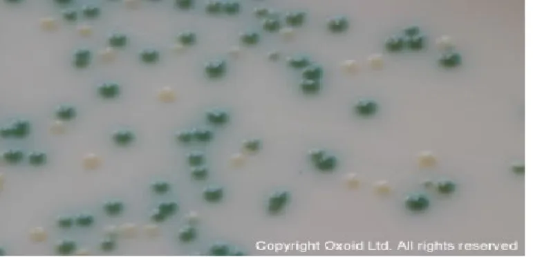
Additional methods for Candida spp identification was chromogenic media such as CHROMagar Candida (France), Oxoid Chromogenic Candida Agar (USA), HiCrome Candida agar (HiMedia, Mumbai, India). These media help to differentiate Candida spp. by combine substrates linked to chemical dyes in a solid medium to made different color depend on species: C. tropicalis (dark blue colonies), C. albicans/C. dubliniensis (green colonies) and C. krusei (dry, irregular, pink-brown colonies). This medium is very useful for the identification of common Candida pathogenic species [46-48].
Unfortunately, two major limitions of current test methods are test time and test sensitivity. It is known that current tests are time consuming and are not sensitive enough to give the accurate results. This results in the delay of necessary antifungal therapy. Additionally, current test methods may not be sensitive enough to identify strains from different tissue specimens due to low number of cells present in different internal organs especially in case of invasive candidiasis. In summary, the limitations of Candida spp. identification based on phenotype includes long testing time requirement and inability to differentiate between several species.
Another method to diagnose candidose is immunological test which is the detection of antigen or antibody. The detection of antibodies against differentCandidaantigens may help in thediagnosis, but may not be able to differentiate between species. Futhermore, the methods traditionally used for the detection of antibodies have been based on crude antigenic fungal extracts, which generally have low reproducibility and have problems with cross reactivity [49]. Candida spp. genotypic identification
The limitations described above for phenotypic identification techniques have led to the development of technologies to quickly and accurately identify Candida strains that will result in early diagnosis, treatment and management of candidemia and other infections caused by Candida species. Currently, three are three advanced methods that have been developed to identify Candida. These are polymerase chain reaction based Candida detection, MALDI - TOF MS and DNA Microarray for Candida detection [34, 43].
Polymerase chain reaction (PCR). A large number of different protocols have been developed over the last five decades to identify different fungal strains present in clinical specimens by polymerase chain based Candida detection techniques. Various Candida DNA markers include 5.8S rRNA genes, 18S rRNA gene, small unit rRNA gene, noncoding internal transcribed spacer (ITS) of rRNA genes, and lanosterol demethylase gene have been used for detection of Candida species. This technique also help to identify species base on specific primers. In addition, real-time PCRs is more sensitive and less time consuming techniques for rapid and accurate identification of different Candida species [34].
Matrix assisted laser desorption ionization time-of-flight mass spectrometry (MALDI-TOF MS). This technique is very useful in medical diagnostic for the rapid identification of clinically important bacteria and yeasts. In recent years, this technology has been applied to Candida biology in save timme manner and accurate identification. MALDI-TOF MS has been useful for identifying Candida species that are not easy to differentiate in the phenotypic
identification. Base on this technology, it could be discrinimate closed species such as C. parapsilosis, C. orthopsilosis, and C. metapsilosis as well as closely related species like C. dubliniensis /C. albicans , C. glabrata/C. bracarensis [50]
DNA Microarray. This techniques has revolutionized the understanding of molecular functioning of different genes in all the organisms including humans. In the oligonucleotide microarray method, specific probes targeted to internal transcribed spacer 2 (ITS2) can be used for hybridization with fungal DNA amplified by PCR from different species. This method is sensitive enough to discriminate among different fungal pathogens at species level and can detect as low as 15 pg/ml of DNA [51]. The sensitivity of the assay for C. albicans is 10 cells/mL [52].
In conclusion, molecular techniques have higher efficiency, specificity and sensitivity to the identification of Candida from culture and samples.
1.2. Candida spp. virulent factors
Invasion of host cells by Candida spp. progresses in several phases. Initially, blastospores adhere to epithelial cells, then hyphae are formed which penetrate cells actively or by endocytosis, progressively causing damage over time to the tissue. Several agents called virulence factors, are responsible for this process, among which the potential ones are the ability to grow at 37°C and physiological pH, the size enabling invasion of the human body; the others are: formation of hyphae and pseudohyphae, the ability of phenotypic switching, adherence to epithelial and endothelial cells, biofilm formation, secretion of hydrolytic enzymes (proteases, phospholipases, lipases), and thigmotropisms [23, 53, 54]. Each of these attributes influences the other, and all are essential for full pathogenicity of fungi from the genus. There is a growing number of studies reporting virulence factors of Candida spp., several virulence factors including adhesion molecules, hydrolytic enzymes, phenotypic switching, morphological dimorphism, and fitness attributes have been identified (Figure 1.6) [32].
Figure 1.6. An overview of selected virulent factors distribute to C.albicans
pathogenicity mechanism [32].
Adherence of Candida spp. to host tissues and cells is seen as an essential early step in the establishment of disease [23, 55]. The presence of specific compound in the fungal cell wall, most of which belong to the class of glycosylphosphatidylinositol cell wall proteins such as HWP, ALS, EPA, which promote adhesion to the proteins or carbohydrates in the host cell wall.
One of the most important genes responsible for the adhesion process, whose expression is induced by physical contact between the fungal and epithelial cells, is hyphal wall protein 1 (HWP1). It is a fungal cell wall mannoprotein specific for germ tubes and hyphal forms [16]. HWP1 adhesin is controled by Kex2 endoproteinase, which itself also relates the activity of C. albicans proteinases, indicating its play for providing virulence and drug resistance of C. albicans. Although HWP1 detecting both in carriers of C. albicans and patients with candidosis of the oral cavity or vagina, expression of this gene is higher among strains isolated from candidosis patients [55].
Agglutinin like sequence (ALS) are encoding a fungal cell surface glycoproteins, a type of adhesins that exhibit similarity to immunoglobulins, bind to peptide ligands of human cells and form aggregates with other microorganisms potentially pathogenic to human, which may lead to mixed infections. This family includes eight genes, although not all are present in each strain: als1-7 and als9 [53, 55], from which als1-4 encode adhesins specific for germ tubes and hyphae [55, 56], while als5-7 and als9 are associated with blastospores [55]. The transcription of all family genes in vitro and during infection has been detected in C. albicans, but some genes (als6 and als7) have been observed with only low levels of expression [16]. The genes most frequently reported to be involved in adherence for C. albicans are als1, als3 and als5, which are characterized by their ability to adhere to a wide variety of substrates [16, 54]. In C. tropicalis and C. dubliniensis had at least 3 als genes, which have been identified by southern analysis and western blotting with an anti ALS antibody [23].
Another virulence factor is extracellular hydrolytic enzymes, including the secreted aspartyl proteinase (SAP) and phospholipase (PLB) gene [16]. These enzymes acting as virulence factors which contribute to host tissue invasion by digesting proteins (hemoglobin, keratin, collagen…). Comparing to C. albicans, C. non albicans produced at a lesser extent hydrolytic enzymes.
SAP are encoded by genes located on the same chromosomes as ALS, occur in similar numbers and and are regulated by similar mechanisms. C. albicans possesses at least 10 members of a sap gene family, all of which have been sequenced and extensively characterized. It was found that sap1, sap3, sap4, sap7, sap8 expression was correlated with oral disease. Furthermore, sap1, sap3, and sap8 were preferentially expressed in vaginal rather than oral disease. In vitro studies show that sap1, sap2, and sap3 are expressed by yeast cells only, whereas sap4-6 expression is confined to hyphae [23]. Sap1-4 genes were identified in C. tropicalis and sap1 is the predominant enzyme produced in vitro [23]. Sap genes also have a play in adhesion. The glycosylphosphatidylinositol-linked aspartyl proteases (Yps) is related to virulence of C. glabrata [53].
Four types of phospholipases have been revealed in C. albicans, including phospholipases A, B, C and D [23], but only the phospholipases B1 and phospholipases B2 products have been detected extracellularly [16, 23]. Although phospholipases B1 is thought to account for most of the secreted phospholipase B activity in C. albicans, phospholipases B2 contributes in a minor way, because a phospholipases B1 deficient strain still produces residual amounts of phospholipase B activity.
One contribution to Candida spp. virulence is hyphal formation, which makes a majority of the strains in this genus become dimorphism. Genes involved in these functions (Als3, sap4-6, hwp1, hyr1, and ece1) in C. albicans are differentially expressed [53]. The mitogen activated protein (MAP) kinase, cyclic AMP (cAMP), and pH sensing, Rim101 signal transduction pathways regulate cellular morphology and expression of hypha associated genes. The bud hypha transition may also contribute to virulence of other Candida species such as C. glabrata, even though C. glabrata strains do not exhibit germ tube formation in classical mycological assays [53].
C. albicans colonies can change among different phenotypes including smooth, rough, star, stippled, hat, irregular wrinkle, and fuzzy at high frequency (10-4 to 10-1) [57, 58]. Smooth and white colonies with round ovoid cells (white) can switch to flat and gray colonies with elongated or bean shaped cells (opaque) [23]. The strains can revert from white colony to opaque colony and contribute to its virulence. White phase cells are more virulent in invasive infection, and opaque phase cells are better to colonize skin [23, 53]. Phenotypic switching also affects other virulence traits, including the bud hypha transition, sensitivity to neutrophils and oxidants, antigenicity, adhesion, secretion of proteinase, drug susceptibility, and phagocytosis by macrophages [53].
1.3. Molecular epidemiology of Candida spp.
Most Candida species belong to the Candida clade, exception of C. glabrata, which is more closely related to S. cerevisiae than to other Candida
species [59, 60]. C. albicans genome consists of eight pair of chromosomal homologs [61], ranging in size from 0.95 to 3.3 Mb in size and comprising 16 Mb in total. This species is predominantly diploid, however it exhibits a high degree of genome plasticity and exhibits frequent losses of heterozygosity as well as gross chromosomal rearrangements that may result in aneuploidy. Althought a reproduction is predominantly clonal, this species can also utilise a parasexual cycle involving the formation of tetraploid progeny from the mating of diploid parents, the former of which subsequently revert to diploidy by concerted chromosome loss [62]. The parasexual cycle occurs rarely in nature, possibly only under stressful conditions. The main function of the parasexual cycle is thought to enable diversification during times of stress, revealing new combinations of recessive traits by loss of heterozygosity (LOH), or resulting in aneuploidy and copy number variation enabling adaptation to adverse environmental conditions. Aneuploidy and revelation of recessive alleles may adversely alter the fitness of the organism, but in highly stressful conditions the parasexual cycle may be a significant source of diversity permitting adaptation and survival of the organism [63, 64].
Molecular epidemiology that combines traditional epidemiological investigation with molecular typing is useful for identifying community or nosocomial infections and tracing the source of transmission and outbreaks. Nowadays, several different molecular typing approaches including pulsed field gel electrophoresis (PFGE), restriction fragment length polymorphism(RFLP), random amplified fragment length polymorphism (RAPD), and multi locus sequence typing (MLST) [65]. Among these techniques, assays based on PFGE, RFLP or RAPD are labor intensive and time consuming. Furthermore, the results of these methods are difficult to compare among laboratories. By contrast, MLST, as a relatively new tool based on DNA sequencing, exhibits high discriminatory power and reproducibility, which overcome the flaws of more subjective methods, making it possible to compare results among laboratories. Therefore, nowadays MLST is a good tool to study and understand Candida spp. epidemiology molecular. Online global databases for many microorganisms are currently available at www.mlst.net,
including data from epidemiological studies carried out worldwide. This permits global epidemiological and population analysis [61, 65, 66]. MLST apply not only for C. albicans but also for others species like C. glabrata, C. tropicalis, C. krusei, C. dubliniensi, C. parapsilopsis.
A standard MLST protocol for molecular characterization of C. albicans has been proposed based on the sequences of seven housekeeping genes (AAT1a, ACC1, ADP1, MPI1b, SYA1, VPS13 and ZWF1b). This method has been widely used to study the population structure, transmission and microevolution of C. albicans. MLST studies have shown that this species‟infections often arise from an endogenous source and persistent C. albicans strains are maintained by hosts over prolonged periods of time, occasionally undergoing minor genetic variations known as micro variation [66]. Strain replacement has also been observed, as has the transmission of C. albicans isolates between different individuals and microvariation of persistent isolates occurring in the same individual between recurrent infections [61, 66].
Based on MLST, a total of 18 clades have been identified worldwide and these clades are associated with the geographic distribution of C. albicans [61, 67]. Clades have been defined as clusters of at least 10 isolates with DSTs that have a p-distance below 0.04, while their p-p-distance with DSTs outside the clade is above 0.04. MLST clade 1 appears to have a global distribution, this clade represents 33% of UK isolates, 31% of isolates from elsewhere in Europe, 32% of isolates from southeast Asia and Japan, 28% of isolates from Australasia, 34% of isolates from the Middle East, 44% of isolates from South America and 49% of isolates from North America, 16% among isolates from Africa [61]. Other clades tend towards greater geographical specificity, though none are found exclusively within specific geographical limits: clade 2 is enriched with isolates recovered from the UK, clade 4 is enriched with isolates from the Middle East and Africa, clade 11 is enriched with isolates from continental Europe, and isolates recovered from the Pacific tend to cluster in clades 14 and 17 [61, 67]. Studies in several countries in Asia have demonstrated that the most common clade was clade1 [61, 68, 69]. Moreover, clade
6, clade 17 were the majority in China [69], while in South Korea were clade 4, clade 12 and hight rate (18.6%) belong to new clade [68]. Until now, there has not been any studies of Candida spp. MLST in Viet Nam, therefore one is necessary to understand the epidiomology of Candida spp. in Asia as well as all over the world.
Molecular strain typing also demonstrate that certain types are more commonly associated with invasive disease than others. Analysis of the largest database from MLST research, did not demonstrate any statistically valid differential subset of strain types associated with disseminated infection [70]. However, there was a significantly greater proportion of isolates associated with superficial infections and commensal carriage in clade 1 versus other clades [70]. The obvious interpretation of this finding is that clade 1 isolates may be better adapted than others to colonize and invade epithelial surfaces, but have no inherent advantage over other types when it comes to traversing epithelia to cause deep tissue disease [61]. In addition, association between clade and the lengths of tandem repeats in some cell surface proteins (ALS) but not with virulence or type of infection, have been demonstrated [61]
Most individuals carry a single C. albicans strain type, but minor variations suggestive of microadaptation are commonly observed. Some evidence suggests many individuals harbor a mixture of strain types that includes a range of minor variants, typically differing in levels of genetic heterozygosity [61, 66].
Although most Candida infections appear to originate from an endogenous source, nosocomial transmission is not uncommon and may occur either by cross infection or by exposure to a common infecting source [61, 71, 72].
1.4. Molecular mechanism to resistant drugs in Candida spp.
The frequency of resistance to antifungal therapy continues to increase despite the introduction of new antifungal agents. Pathogen fungi have developed some mechanisms to survive in toxic environment. According to the literature, the following molecular mechanisms of drug resistance include: modification of the drug target affinity, overproduction of the enzymes that are targets to drugs,
development of alternative metabolic pathways, active efflux of the drug from the cell, impermeability of the cell membrane to drug molecules, active enzymes inactive the drug or degrading it outside the fungal cell [73-75]. Antifungal resistance is associated with elevated minimum inhibitory concentrations, poorer clinical outcomes, and breakthrough infections during antifungal treatment and prophylaxis.
Currently, there are two independent standards for broth microdilution (BMD) susceptibility testing of Candida and filamentous fungi: the Clinical and Laboratory Standards Institute (CLSI) method and the European Committee on Antimicrobial Susceptibility Testing (EUCAST) method. These methods are similar in that both use BMD, although there are some differences in inoculum size and MIC endpoint determination. These methods have been harmonized so that there is close agreement between MIC results obtained when testing azoles and echinocandins against Candida. The CLSI has also developed agar based, disk diffusion testing for yeasts. Compared with the BMD method, disk diffusion testing is convenient, simple, and economical, and is particularly well suited for water soluble antifungals such as 5-fluorocystosine, fluconazole, and voriconazole. Disk diffusion testing has been standardized for checking resistance of Candida spp. to fluconazole, voriconazole, caspofungine, micafungin, and break points have been provided for each of these agents [76].
Various mechanisms can lead to resistance of Candida spp. to antifungal compounds. For azole drugs, the most common mechanism include the efflux pumps encoded by the mdr or cdr genes, and point mutations in the gene encoding for the target enzyme ERG11 [76-81]. These mechanisms are shown in Figure 1.7. Resistance to amphotericine B is related to qualitative or quantitative changes in ergosterol by expression of mutations in erg3, erg6, erg11 genes [82]. Resistance mechanism of Candida sp. to echinocandins is typically caused by the fks1 genes encoding the major subunit of its target enzyme [75, 82, 83].
Figure 1.7. Main mechanisms of azole and polyens resistance [78]
Among antifungal drug, azoles are a class of antifungals that are widely used to treat both superficial mucosal and deep and disseminated fungal infections caused by C. albicans [77]. However, extensive use may lead to the development of resistance, resulting in therapeutic failure for Candida spp. [78]. In azole compounds, fluconazole is the antifungal agent of choice in the treatment and prophylaxis of infection by C. albicans [78, 84]. In addition, the emergence of fluconazole resistant C. albicans strains is a significant problem after long time use as the treatment for recurrent oropharyngeal candidiasis in acquired immunodeficiency syndrome (AIDS) patients [78, 80]. Physicans encounter a high prvelance of fluconazole-resistant Candida, which makes the treatment of candidemia a huge challenge [35]. Data from many coutries all over the world have reported higher levels of resistance from C. non albicans than C. albicans [7, 39, 85, 86].
1.5. Candida spp. and bacteria interaction
The origin of bacterial life predates the appearance of eukaryotic organisms by more than two billion years. Therefore for bacteria, symbiosis with eukaryotes regardless of whether the outcome is negative, positive, or neutral for the organisms united through the interaction is a derived lifestyle [87]. Research in the interaction between bacteria and fungi has developed significantly in both breadth and depth in recent years. Bacterial fungal interactions (BFIs) are antagonistic, cooperative, synergistic, commensal, and symbiotic [88]. It has been reported that the most common BFIs are ones where the bacterial partner exploits resources from the associated fungus through a parasitic or commensalism interaction, although there are intriguing examples where the fungus is able to take advantage of bacterial resources in mutualistic interactions [87].
Humans are naturally colonized by fungi and bacteria in a variety of niches, including the skin, the oral cavity, and the respiratory, digestive, and genital tracts [89]. In healthy individuals, these microorganisms are commensal and in some cases even beneficial to human health. By contrast, in immunocompromised patients, pathogenic fungi and bacteria represent a serious threat to their health. There are some studies reported that bacteria and fungi have been found together in infections of human burn wounds and keratitis [90-92]. Moreover, the forming of mixed communities between bacteria and fungi makes them more virulent and resistant to antibiotic therapies [93, 94]. Therefore, an understanding of the functioning of BFIs, particularily the development of strains related to resistance to drug therapies, in human health is an emerging and important challenge for medical researchers [95].
Figure 1.8. The bacteria fungal interaction: the combination of physical
associations and molecular interactions [88]
Observation of the effects of fungi on bacterial development is difficult due to the small size and single cellular nature of bacteria. However, if consideration is given to bacterial fungal biofilms, it is clear that fungi can promote distinct differences in bacterial development by contributing to a distinctive ecological niche, within which bacteria exhibit physiological differences, such as resistance to antibiotics, stress, and an altered expression of virulence genes, compared to free living bacteria [95, 96].
Recently, several studies have described the association of Candida spp. and bacteria, such as Staphylococcus aureus, Pseudomona aerugirosa, Staphylococcus epidermidis, ect. [93, 94].
Helicobacter pylori (H. pylori) is a Gram negative, spiral shaped bacterium that infects more than 50% of the human population and can cause gastritis, peptic ulcer, or gastric malignancies [97]. It is necessary to understand H. pylori transmission to prevent this disease. The oral cavity has been hypothesized as a reservoir for gastric H. pylori, which has been detected by culture and with PCR in both dental plaque and saliva. Some researchers have proposed H. pylori in oral cavity may play an important role in its transmission and reinfection. Oral-oral or fecal-oral transmission are thought to be the most possible means of transmission [97].
H. pylori is generally considered an extracellular microorganism. However, there have been some evidences supporting a hypothesis that H. pylori microorganisms have an intracellular location [98, 99]. These findings support the hypothesis that this bacteria can invade fungi and survive from stressful environments. Normally, Candida spp. can also colonize in the gastric regions [91] and invade epithelium cells in certain conditions [100]. Recently, some studies focused on the relationshop between the microrganism. Evidence has been suggested to show that Candida vacuoles can be a niche for H. pilory and and H. pylori specific genes, such as vacA, ureA and peroxiredoxin were detected by PCR and Western blotting [101].
2. RESEARCH OBJECTIVES
Candida species are normally associated with human beings as harmless commensals. They are commonly found on the mucosal surfaces of gastrointestinal and genitourinary tracts and skin of humans [5]. Candida spp. infect billions of people every year all over the world, causing a broad spectrum of infections, ranging from mucosal or localized cutaneous infections to systemic or potentially fatal diseases [36, 40]. To date, the incidence of invasive fungal diseases by Candida spp. is rising as a result of immunocompromised patients, modern medical interventions and therapies. For example, AIDS patients or who have undergone transplants generally receive immunosuppressive drugs or aggressive regimens of chemotherapy in cancer, respectively. Patients under these conditions are prone to mycosis, a majority of which are caused by Candida spp [40]. Systemic mycosis often requires high associated costs, particulary mycosis caused by Candida species which are less senstive to antifungal therapy [76, 102]. Therefore, a better understanding of the epidemiologic features and etiology of candidose will enable physicians to provide better management strategies, preventitive measures, and treatment to infections.
For many years, fungal laboratory methods based on the detection of phenotypic characteristics, such as microscopy and in vitro culture, have played an essential role in fungal etiology identification. However, these procedures are generally slow or non-specific. Furthermore, phenotypic features can be easily influenced by external factors such as variations in temperature, medium, and chemotherapy, making species identification difficult. Recently, genotypic approaches have proven to be useful for fungal identification. In fact, genotypic differences are considered more stable and precise than phenotypic differences. Molecular biology techniques, such as PCR, amplicon sequencing, MLST and MALDI-TOF MS, are useful in the identification and investigation of pathogenic fungi [103], particularly Candida species [104, 105].
In Vietnam, Candida superficial infections are common. Recently, invasive fungal infections have become more frequent due to the growing number of immunocompromised and other susceptible individuals in the population [106-109]. Although physicians have been faced with managing more of these infections, there has been a lack of systematic epidemiological data in this field [108, 110, 111]. In addition, the current gold standard techniques for the indentification of mycoses are direct examination and in vitro culture [112]. The use of of molecular techniques in the study of etiology and epidemiology of Candida spp. infections in Central Vietnam will be essential, useful and realistic. Furthermore, molecular techniques will allow for a more accurate overview of Candida molecular epidemiology and Candida causatives in Vietnam.
For these reasons, the aims of this research are as follow: 2.1. To study candidiasis etiology
- Application of molecular techniques (e.g. Matrix Assisted Laser Desorption Ionization Time-of-Flight Mass Spectrometry and Internal transcribed spacer sequencing) in the identification of Candida species.
- Analysis of the distribution of Candida species in candida colonization and candidiasis.
2.2. To analyze the phenotypic and genotypic features of Candida spp. isolates - Checking antifungal susceptibility tests by disk diffusion method.
- Detecting and analyzing polimorfism of resistance genes linked to fluconazole resistance in Candida tropicalis by PCR and sequencing (mutations in ERG11 gene).
- Detecting virulence genes by Multiplex PCR (als1, hwp1, sap4).
- Studying molecular typing of selected isolates of Candida albicans by Multi Locus Sequence Typing (MLST).
2.3. To analyze relationship between Candida spp. and Helicobacter pylori
Testing for the presence of Helicobacter pylori specific gene by nested PCR in Candida isolates
3. MATERIALS AND METHODS
3.1. Study site
This cross sectional study was carried out from October 2012 to August 2015 at two Vietnamese Hospitals, Hue University of Medicine and Pharmacy Hospital and Hue Central Hospital, and the Biomedical Science Department at the University of Sassari in Italy.
Samples were collected in 10 departments at Hue University of Medicine and Pharmacy Hospital (Dermatology, Endoscopy, Intensive Care, Internal Medicine, Obstetric, Oncology, Ophthalmology, Otorhinolaryngology, Pediatric and Surgery) and 2 departments at Hue Central Hospital (Hematology and Pediatric).
Direct examination, fungal cultivation, and fungal phenotype identification were carried out in Vietnam at the Parasitology laboratory at the Hue Medicine and Pharmacy University. Fungal genotype was conducted at the Carlo Urbani Centre in Hue, Vietnam and at the Microbiology laboratory, Department Biomedical Science in Sassari, Italy.
3.2. Study population
All patients were subsequently divided into four groups. The first group included patients with candida colonization. These patients had Candida spp. isolated from unsterile body sites by both direct examination and culture but that did not display any symptoms related to superficial candidiasis or invasive candidiasis. These patients, furthermore, recovered without assistant from antifungal therapy. The second group was mucosal candidiasis. These patients displayed symptoms of the disease (oral candidiasis, vulvovagiginite candidiasis) and were clinically identified by doctors. In this case, Candida spp. was isolated by both direct examination and culture. Cutaneous candidiasis was the third group of patients. These patients had symptoms including parochynia, onychomycosis, and skin candidiasis. Candida spp. in these patients were isolated by both direct examination and culture. The fourth group was systemic candidiasis (candidemia or invasive
candidiasis). These patients had clinical symptoms and Candida spp. was isolated from blood, tissue, sterile body sites by a combination of direct examination and culture or culture only.
3.3. Sample collection and Candida strains isolates
Samples were collected from 163 patients admitted in 10 different hospital wards at Hue University of Medicine and Pharmacy Hospital and 2 wards at Hue Centrel Hospital (see Table 3.1)
Table 3.1 . Distribution of patients in hospital wards
Hospital Ward patient isolate Number of
Hue University of Medicine and Pharmacy Hospital Internal medicine 26 Obstetric 24 Intensive care 17 Dermatology 18 Surgery 17 Endoscopy 14 Oncology 13 Pediatric 5 Otorhinolaryngology 11 Ophthalmology 2
Hue Central Hospital Hematology Pediatric 10 6
Total 163
Selected patients were required to not have received antifungal treatment with the last seven days in order to prevent false negative cases. One sample was collected from each patient, with the exception of the seven patients in the Intensive Care Unit (HUMP Hospital), three patients in the Hematology ward (HC Hospital), two patients in the Pediatrics ward (HC Hospital), and one patient in the Endoscopy ward (HUMP Hospital). In detail, for the seven patients in the Intensive Care Unit, two samples from two different body sites were collected for 6 patients and three samples from three different body sites for 1 patient. For the three patients in Hematology ward, the two patients in the Pediatrics ward, and the one patient in the Endoscopy ward, two samples isolated from two different body sites were collected. For the two patients in
the Pediatrics ward, two samples isolated from two different body sites. In total, 177 samples from 163 patetients were used in the study.
Specimens had been collected according to the patient‟s symptoms in primary candidiasis patients. By contrast, in patients without candidiasis, we detected Candida colonization status from oral, sputum, urine, gastric drainage fluid, gastric biopsy, bronchoalveolar lavage fluid, endotracheal aspiration fluid.
Samples were collected from patients based on type of disease. Samples from mucosal (oral, vaginal, nasal) and skin lesions were obtained using sterile swabs [113-115]. Gastric biopsy was collected by endoscopy. Sputum and stool were collected into wide-mouthed sterile container [10]. From lower respiratory tract, bronchoalveolar lavage, endotracheal aspiration fluid were collected in volumes of 1 -5 ml [116]. Urine was isolated from midstream with volume from 10 ml to 15 ml. For patients with catheters, urine was obtained by clamp of Foley‟s catheter distally [114]. Nail scrapings were collected in case of onychomycosis. Gastric fluid was removed from drainage in volumes of 10 15 ml. A volume of 10 -20 ml blood was collected from adult patients; for pediatric patients, the volume was 4-10 ml [41, 42, 114, 117, 118].
When necessary, oral, vaginal, skin lesion, and gastric biopsy samples were placed in distilled water and transferred to the laboratory. Urine and other body fluids were processed by centrifuging at 2500g for 10 minutes before microscopy.
Each sample was examined microscopically with 20% KOH to detect fungal morphological forms (budding yeast or/and hyphae) [10, 53]. The samples were then processed for fungal culture.
Each specimen was inoculated first on Sabouraud dextrose agar (SAD) with chloramphenicol for 24 - 48h at 350C [10, 114]. Candida colonies were sub cultured several times to purify strains. All isolates were stored at - 800C in 15% glycerol [119] for future antifungal susceptibility tests and identification using molecular biology methods.
![Figure 1.2. Structure of the C. albicans cell wall [19].](https://thumb-eu.123doks.com/thumbv2/123dokorg/8345546.133250/16.892.215.742.810.1063/figure-structure-c-albicans-cell-wall.webp)
![Figure 1.3. The human mycobiota [31]](https://thumb-eu.123doks.com/thumbv2/123dokorg/8345546.133250/18.892.244.727.577.1015/figure-the-human-mycobiota.webp)
![Figure 1.4. The steps of C. albicans tissue invasion [19].](https://thumb-eu.123doks.com/thumbv2/123dokorg/8345546.133250/19.892.251.686.373.1000/figure-steps-c-albicans-tissue-invasion.webp)
![Figure 1.5. Hospital - acquired Candida infections [37]](https://thumb-eu.123doks.com/thumbv2/123dokorg/8345546.133250/21.892.189.679.417.918/figure-hospital-acquired-candida-infections.webp)
![Table 1.1. Summary of recommendations by Candida disease, specimen and test evaluated [42]](https://thumb-eu.123doks.com/thumbv2/123dokorg/8345546.133250/23.892.138.776.253.887/table-summary-recommendations-candida-disease-specimen-test-evaluated.webp)
![Figure 1.6. An overview of selected virulent factors distribute to C.albicans pathogenicity mechanism [32]](https://thumb-eu.123doks.com/thumbv2/123dokorg/8345546.133250/27.892.225.743.128.532/figure-overview-selected-virulent-distribute-albicans-pathogenicity-mechanism.webp)
![Figure 1.7. Main mechanisms of azole and polyens resistance [78]](https://thumb-eu.123doks.com/thumbv2/123dokorg/8345546.133250/34.892.170.742.128.553/figure-main-mechanisms-azole-polyens-resistance.webp)
![Figure 1.8. The bacteria fungal interaction: the combination of physical associations and molecular interactions [88]](https://thumb-eu.123doks.com/thumbv2/123dokorg/8345546.133250/36.892.135.781.132.417/figure-bacteria-interaction-combination-physical-associations-molecular-interactions.webp)