Epstein Barr Virus and
Mycobacterium avium subsp.
paratuberculosis peptides
are recognized in sera and
cerebrospinal fluid of MS patients
Giuseppe Mameli
1, Eleonora Cocco
2, Jessica Frau
2, Maria Giovanna Marrosu
3&
Leonardo Antonio Sechi
1Mycobacterium avium subsp. paratuberculosis (MAP) and Epstein-Barr virus (EBV) epitopes elicit a
consistent humoral response in serum of multiple sclerosis patients, but the cross reactivity against the homologous myelin basic protein (MBP) and human interferon regulatory factor 5 (IRF5) has not been searched within the Cerebral Spinal Fluid (CSF). We evaluated in sera and CSF of patients with MS and with other neurological diseases (OND) the humoral response against EBV/MAP peptides and the IRF5/MBP. Our data showed that EBV and MAP peptides are able to induce a specific humoral immune response in MS patients compared to OND controls both in serum and in CSF. An intrathecal specific synthesis of IgG against MBP and their EBV and MAP homologous as indicated by the antibody index was observed in MS patients. The humoral response against EBV, MAP, MBP and IRF5 was significantly higher in MS patients compared to OND both in serum and in CSF. The higher presence of antibodies against MBP and their MAP and EBV homologous in CSF during relapses suggests a possible role of the pathogens in enhancing inflammation.
Multiple sclerosis (MS) is a chronic inflammatory disease of the central nervous system (CNS) resulting in demy-elination and neurodegeneration1. The disease develops in genetically predisposed individuals in response to environmental factors, most likely viral and bacterial infections1. However, recent studies advocate the possible combined roles of Mycobacterium avium subsp. paratuberculosis (MAP) and Epstein Barr virus (EBV) in the pro-cess of autoimmunity inducing MS pathology2,3. In fact, it was demonstrated that peptides deriving from these pathogens could be cross-recognized by antibodies (Abs) targeting self-epitopes2,3. In particular it was detected a strong humoral response against peptides from the latent protein of Epstein Barr Virus (EBNA1400–413), the homol-ogous mycobacterial MAP_0106c121–132 and the human Myelin Basic Protein (MBP85–98) in MS patients compared to healthy controls (HCs). Also, an interesting common humoral response was reported against a relevant EBV epitope from EBV lytic protein (BOLF1305–320) and two homologous peptides belonging to MAP_4027 protein (MAP_402718–32) and Human Interferon Regulatory Factor 5, IRF5 protein (IRF5424–434)3. A cross-recognition of the mentioned peptides was also demonstrated. In this work we wanted to explore if we could find the same humoral response in CSF and serum against EBV epitopes deriving from EBV lytic and latent proteins, MAP and human homologous proteins in MS patients and in other neurological disease controls. The humoral response against EBV and MAP in CSF could contribute to understand an immunological dysregulatation in the CSF of MS patients. The presence of intrathecal IgGs, is still considered an evidence for the involvement of infec-tious agents in MS pathogenesis, although their specificity is largely unknown4. This study was carried out on samples from patients with MS, inflammatory neurological disease (IND) or non inflammatory neurological 1Dipartimento di Scienze Biomediche, Sezione di Microbiologia e Virologia, Università di Sassari, Viale San Pietro 43b, 07100 Sassari, Italy. 2Centro Sclerosi Multipla, Dipartimento di Sanità Pubblica Medicina Clinica e Molecolare, Università di Cagliari, Via Is Guadazzonis 2, 09126 Cagliari, Italy. 3Dipartimento di Scienze Mediche “Mario Aresu”, Università di Cagliari, 09126 Cagliari, Italy. Correspondence and requests for materials should be addressed to L.S. (email: [email protected])
received: 16 September 2015 Accepted: 15 February 2016 Published: 09 March 2016
disease (NIND) and patients where a diagnosis was not reached, indicated as undetermined neurological disease (UND),aiming to: a) measure circulating serum and CSF Abs against EBV and MAP peptides and their human homologous; b) quantify and correlate the serum IgG levels to the CSF IgG production; c) investigate the IgG cross reaction against the epitopes investigated. Molecular mimicry between immunodominant epitopes deriving from bacterial and viral persistent antigens may be a decisive factor in directing autoimmunity to self-antigens in MS patients. For this reason it was important to explore if the epitopes from EBV and the other homologous MAP antigens were able to induce a humoral reactivity both in CSF and sera. The results could contribute to the understanding of chronic brain inflammation that contribute to MS pathogenesis.
Results
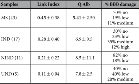
CFS/Serum Albumin ratio and Link index.
For all samples, Link index as a generic marker of intrath-ecal IgG synthesis, CSF/serum albumin ratio (Q Alb) as a marker of BBB integrity and percentage of samples with different type of BBB damage were evaluated and shown in Table 1. No statistically significant damage was observed in the BBB of the MS group compared to the other groups as evidenced by the Link index and the CFS/ albumin ratio.Elisa.
Abs against latent and lytic EBV proteins EBNA1 and BOLF1, MAP and Human homologues peptides were monitored in serum and CSF of MS patients and in IND, NIND, UND controls5. Abs against EBNA1400–413 were found in 26 out 43 (60%) MS patients whereas 3 out of 17 (18%) IND controls, 2 out of 11 (18%) NIND and only one UND were positive in serum (AUC = 0.74, p = 0.001) (Fig. 1A). In CSF were found positive 17 out of 43 (40%) MS, 3 out of 33 (10%) IND, none among 11 NIND and 5 UND patients (AUC = 0.80, p = 0.001) (Fig. 1B). Regarding MAP_106c121–132, 27 out of 43 (63%) MS patients, 4 out of 17 (24%) IND, 4 out of 11 (36%) NIND and one UND were positive (AUC = 0.73, p = 0.008) in sera (Fig. 1C), whereas 23 out of 43 (54%) MS patients, 3 out of 17 (18%) IND, 2 out of 11 (18%) NIND and none among UND controls reacted against MAP_106c121–132 in CSF (AUC = 0.71, p = 0.02) (Fig. 1D). Positivity against MBP85–98 was found in 27 out of 43 (63%) MS patients, in 5 out of 17 (29%) IND, 4 out of 11 (36%) NIND and only in one UND in serum (AUC = 0.71, p = 0.03) (Fig. 1E). In CSF among the MS subjects, we observed a similar situation where 33% MS, 27% of IND, 10% NIND and 0% of UND patients showed positivity against MBP85–98 (AUC = 0.7, p = 0.03) (Fig. 1F).
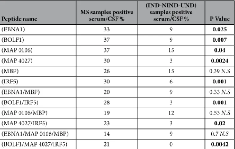
Regarding reactivity against the EBV lytic peptide BOLF1305–320 we found 30 out of 43 (69%) MS patients posi-tive, however only 4 out of 17 (23%) IND, 27% NIND and only one UND were positive (AUC = 0.8, p = 0.0002) in serum (Fig. 2A); In CSF, 21 out of 43 (48%) MS patients, 4 out of 17 (24%) IND, one UND and none NIND were positive for BOLF1305–320 (AUC = 0.65, p = 0.007). (Fig. 2B). About the homologous MAP_402718–32, 63% of MS patients, 35% IND, 36% NIND and 20% among the UND controls were positive in serum (AUC = 0.69, p = 0.03) whereas in CSF 46% of MS patients, 17% IND, none of NIND and UND controls were positive (AUC = 0.65, p = 0.01) (Fig. 2D). Finally, Ab positivity against IRF5424–434 was found in 27 out of 43 (63%) MS patients, 5 out of 17 (29%) IND, one NIND (9%) and in one UND control in serum (AUC = 0.67, p = 0.02) (Fig. 2F) whereas in CSF 19 out of 43 (44%) MS patients, 2 of 17 (11%) IND, one NIND resulted positive for IRF5424–434 (AUC = 0.67, p = 0.0002) (Fig. 2F). We considered also a double Abs positivity Serum/CSF in MS samples compared to that found in IND/NIND against the single peptides and their combination (Table 2). A double positivity for EBNA1 and BOLF1 peptides was found among the 33% and 37%, respectively in serum and CSF whereas only 9% of IND/NIND/UND controls was positive (p = 0.025 and 0.007 respectively) (Table 2). Same pattern was observed for the double positivity to MAP_106c121–132 and MAP_402718–32 in serum and CSF of MS (37% and 30%, respectively) against the 15% in serum and 4% in CSF of IND/NIND/UND subjects (p = 0.04 and 0.0024 respectively) (Table 2). A strong positive association was found for triple positivity (21%) both in serum and in CSF to BOLF1305–320, MAP_402718–32 and IRF5424–434 in MS patients in comparison to that (0%) of IND/NIND/ UND subjects (p = 0.0042) (Table 2). Moreover, CSF positivity to EBNA1400–413, MAP_106c121–132 and MBP85–98 was associated to MS patients with relapse compared to MS patients without relapse (Table 3).
Correlation analyses.
A correlation analyses was performed to establish association between the peptide positivity. We found a good correlation for EBNA1400–413 peptide with the homologous MAP_106c121–132 peptide in serum (R2 = 0.57; P < 0.0001) and a little less (R2 = 0.35; p < 0.0001) in CSF (Fig. 3A,B). Interestingly, the cor-relation analysis showed a stronger corcor-relation between MBP85–98 and EBNA1400–413 in CSF (R2 = 0.56; p < 0.0001)Samples Link Index Q Alb % BBB damage
MS (43) 0.45 ± 0.38 5.41 ± 2.50 19% low70% no 11% medium IND (17) 0.28 ± 0.40 6.9 ± 9.5 30% no 23% low 35% medium 12% high NIND (11) 0.21 ± 0.22 8.5 ± 11.1 18% low82% no UND (5) 0.11 ± 0.04 7.8 ± 2.5 40% low40% no 20% medium
Table 1. Link index as a generic marker of intrathecal IgG synthesis, CSF/serum albumin ratio (Q Alb) as a marker of BBB integrity and percentage of samples with different type of BBB damage are shown.
than in serum (R2 = 0.26; p = 0.0004) of MS patients (Fig. 2C,D). The same pattern was shown by the correlation between MBP85–98 and MAP121–132 in CSF (R2 = 0.4; p < 0.0001) in comparison to that found in serum (R2 = 0.24; p = 0.0007) in MS patients (Fig. 2E,F). These results indicate that the relation of EBNA1400–413 and MAP121–132 to MBP85–98 is stronger in CSF.
We found also a better correlation between Abs against BOLF1305–320 and MAP_402718–32 peptides in CSF (R2 = 0.54; p < 0.0001) in comparison with that found in serum (R2 = 0.24; p = 0.0008) (Fig. 4A,B). Same trend was found also for the correlation between BOLF1305–320 and its human homologue peptide IRF5424–434 in serum
Figure 1. Antibody OD measured by indirect ELISA. Forty-three patients, seventeen IND, eleven NIND and
5 UND were tested for their reactivity against plate-coated with EBNA1400–413, MAP_0106c121–132, MBP85–98, in serum (A,C,E) and in CSF (B,D,F). The horizontal black bars represent the mean value, while p values are indicated by two headed arrows drown on the top of each distribution. Cut-off values for positivity, calculated by ROC analysis, are indicated by dashed lines.
(R2 = 0.11; p = 0.11) and in CSF (R2 = 0.46; p < 0.0001) (Fig. 4C,D); However the opposite result was observed when we correlate the Ab positivity to MAP_402718–32 and its human homologue peptide IRF5424–434 in serum (R2 = 0.47; p < 0.0001) and in CSF (R2 = 0.40; p < 0.0001).
Competition assay.
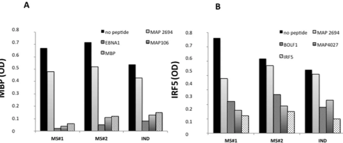
To demonstrate the cross-reactivity between MAP/EBV and self epitopes, we devel-oped two competition assays: one coating the MBP85–98 on plate and the other coating the IRF5424–434 on plate (Fig. 5A,B, respectively). Concerning MBP85–98 -coated competition assay (Fig. 5A), in all the CSF subjects (MS#1,Figure 2. Antibody OD measured by indirect ELISA. Forty-three patients, seventeen IND, eleven NIND and
5 UND were tested for their reactivity against plate-coated with BOLF1305–320, MAP_402718–32 and IRF5424–434 in serum (A,C,E) and in CSF (B,D,F). The horizontal black bars represent the mean value, while p values are indicated by two headed arrows drown on the top of each distribution. Cut-off values for positivity, calculated by ROC analysis, are indicated by dashed lines.
MS#2 and IND), both EBNA1 and MAP106 efficiently inhibited antibody binding to the MBP-coated peptide. In IRF5424–434-coated competition assay (Fig. 5B), both BOLF1 and MAP 4027 efficiently inhibited IRF5424–434-coated binding in all the CSF subjects (MS#1, MS#2 and IND), while the control peptides MAP 2694 did not cause any decrease in signal (Fig. 5A,B). Taken together, these results demonstrate, that Abs anti-EBNA1 and anti-MAP106 target the same conformational MBP epitope and also demonstrate that Abs anti-BOLF1 and anti-MAP 4027 targeting the same conformational IRF5 epitope are cross-reactive in CSF.
Peptide IgG-specific antibody index (AI) in MS patients.
Antibody Index (AI) for all MS patients was calculated with the following formula AI = QIgG[spec]/Qlim7. A high number of positive samples against the selected peptides in the MS group had an AI > 1.5 that demonstrate an intratecal IgG-specific antibody produc-tion against EBNA1400–413, MAP 106, MBP85–98 and BOLF1305–320, MAP_402718–32, IRF5424–434 peptides, as shown in Table 4.Discussion
MS aetiology remains unclear, and the current accepted theory for its pathogenesis assigns the main role to the destruction of the myelin-proteolipid shell of axons resulting in autoimmune reactions supposedly induced by a different viral or bacterial infection1,6,8. Although activation of the T-cell response has a crucial role in MS pathogenesis, the B-cell response is equally responsible for the development of the disease by the enhanced syn-thesis of immunoglobulins usually IgGs9. It has been shown that the CSF from MS patients contains oligoclonal immunoglobulins (IgG), which are synthesized within the central nervous system and presumably related to the immune dysfunction, a characteristic feature of MS. The aim of this study was to investigate the presence of a specific humoral response mounted against peptides derived from EBV and MAP antigens homologues to host proteins in serum and in CSF of MS compared to Inflammatory and No inflammatory neurological disease. We investigated different aspects of the association of CSF and serum EBV and MAP positivity in MS and con-trols. Our studies reported the ability of specific peptides from EBNA1, BOLF1, MAP and their human homo-logues to induce a strong humoral response in MS patients in comparison to healthy controls, and the existence of a cross-recognition between Abs recognizing these homologues peptides10–12. Also these works showed up that EBV and MAP are capable of inducing the production of autoantibodies targeting different MS correlated epitopes13. In this study we look for the presence of intrathecal immune response against EBV, MAP and the homologous human peptides. To check the BBB integrity, the Antibody Index (AI) as the marker of a specific intrathecal IgG synthesis against the selected peptides and CSF/serum albumin ratio (Q Alb) were evaluated. Results showed an high number of MS patients with intrathecal IgG synthesis against EBV and MAP with a AI > 1.5, this associated with a better BBB integrity in MS patients in comparison to that of IND, NIND and UND controls (Table 1) support a possible implication of MAP and EBV in MS development. ELISA analysis showed
Peptide name MS samples positive serum/CSF %
(IND-NIND-UND) samples positive serum/CSF % P Value (EBNA1) 33 9 0.025 (BOLF1) 37 9 0.007 (MAP 0106) 37 15 0.04 (MAP 4027) 30 3 0.0024 (MBP) 26 15 0.39 N.S (IRF5) 30 6 0.001 (EBNA1/MBP) 20 9 0.33 N.S (BOLF1/IRF5) 28 3 0.001 (MAP 0106/MBP) 19 12 0.53 N.S (MAP 4027/IRF5) 23 3 0.02 (EBNA1/MAP 0106/MBP) 14 9 0.7 N.S (BOLF1/MAP 4027/IRF5) 21 0 0.0042
Table 2. The table shows MS and IND/NIND/UND samples positive serum/CSF for the peptides and their homologous combination. P value of MS patients and IND/NIND/UND was calculated by Fisher’s exact test,
GraphPad Prism 6.0 software (San Diego, CA, USA).
Peptide name MS with relapse positive CSF % MS without relapse positive CSF % P Value
(MBP) 13/23 (57%) 3/20 (15%) 0.01
(EBNA1/MBP) 10/23 (43%) 3/20 (15%) 0.05
(MAP 0106/MBP) 10/23 (43%) 2/20 (10%) 0.02
Table 3. The table shows MS with and without relapse samples positive CSF for the EBNA1400–413,
MAP_0106c121–132 and MBP85–98 peptides and their combination. Fisher’s exact test was calculated by
that IgGs against peptides from EBV, MAP and human homologues peptides such as MBP85–98 and IRF5424–434 were higher in MS patients than in IND/NIND/UND controls in both CSF and serum. Interestingly, a higher correlation was found between the levels of IgGs against EBV, MAP and their homologous peptides in CSF rather than in serum5,10,12. Therefore, we set up a competition assay between the human peptides and their homologous from EBV and MAP in CSF. Our results highlighted that autoantibodies recognizing MBP and IRF5 were able to cross-react with the homologous peptides from EBV (EBNA1, BOLF1) and from MAP (MAP106, MAP4027), possibly by a molecular mimicry mechanism.
The soundness of the results was confirmed by the correlation results, the fact that a triple positivity was found, both in serum and in CSF, against BOLF1305–320, MAP_402718–32 and IRF5424–434 in MS patients in compar-ison to the absence in IND/NIND/UND subjects confirmed our hypothesis that MAP and EBV could ignite the
Figure 3. Scatter plot showing the correlations between EBNA1400–413 with MAP_0106c121–132 in serum (A) in CSF (B). EBNA1400–413 and MAP_0106c121–132 with MBP85–98 in serum (C,E) in CSF (D,F) in MS patients. Pearson’s correlation was calculated by Graph Pad 6.
autoimmune process in MS and positivity to different peptides (EBNA1400–413, MAP_106c121–132 and MBP85–98) in CSF could be related to MS patients with relapse compared with MS patients without relapse .
Sundström et al. also considered as a risk factor the interaction of antibodies against EBNA-1 specific domains and HLA DRB1*150114. In our hands we didn’t find any correlation between a specific HLA and the immuno-reactivity with our peptides (data not shown). Recently, another group showed that a productive EBV infection in the peripheral blood could facilitate entry of viral particles and/or newly infected B cells into the CNS15. This finding may support our result on the specific ABs against EBV peptides in CSF of MS patients. Our findings emphasize the significance of CSF and serum levels of anti-EBV and MAP IgG. Furthermore, the cross-reaction of MBP85–98 supports the possible role of IgG against myelin basic protein in MS disease. Therefore, it is not sur-prising that several studies report a statistically significant correlation of Abs titers of MBP in MS patients rather than in other neurological disease controls5. This data suggest that EBV and MAP may be capable of inducing the production of autoantibodies targeting different MS correlated epitopes expressed in CNS. In conclusion the
Figure 4. Scatter plot showing the correlations between BOLF1305–320 with MAP_402718–32 in serum (A) in CSF (B). BOLF1305–320, MAP_402718–32 with IRF5424–434 in serum (C,E) in CSF (D,F) in MS patients. Pearson’s correlation was calculated by Graph Pad 6.
detection of anti-EBV and anti-MAP IgG in MS patients provided additional evidence of the possible synergistic role of these microrganisms to induce the pathology. Thus, the detection of Abs against EBV and MAP in CSF of MS patients can be considered as an additional criterion (immunological parameter) crucial to better understand MS pathology.
Materials and Methods
Subjects.
The study protocol was approved by the ethic committee of the University of Cagliari, Italy. All the methods were carried out in “accordance” with the approved guidelines. All the participants provided written consent. CSF and serum samples prospectively collected from MS and Other Neurological Disease composed (OND) by Inflammatory Neurological Diseases (IND), Non Inflammatory Neurological Diseases (NIND) and Undetermined Neurological Diseases (UND) patients were obtained for diagnosis purposes and measured under equal conditions. CSF samples were obtained by a traumatic lumbar puncture performed for diagnosis purposes in the absence of contraindications. All MS samples derived from 43 patients (24 females and 19 males; mean age ± SD was 39 ± 14.0 years) that fulfilled the revised McDonald diagnostic criteria16. At the time of the study, 39 patients were diagnosed as relapsing remitting MS, 4 as secondary progressive MS. The duration of the disease was 6 ± 5.4 years, the average age for MS onset was 29.0 ± 10.5 and the expanded disability status scale (EDSS) values ranged from 0 to 7.0 with a 1.9 average. Other neurological diseases (OND) serum and CSF samples were collected from 33 patients (14 females and 19 males) so divided: 17 IND (5 females and 12 males, mean age ± SD was 39 ± 23.1 years ), 11 NIND (6 females and 5 males, mean age ± SD was 41 ± 27.1 years) and 5 UND (2 females and 3 males, mean age ± SD was 71 ± 11 years).Peptides.
Synthetic peptides derived from EBV antigens (BOLF1305–320, EBNA1400–413), MAP homologues antigens (MAP_402718–32, MAP_0106C121–132) and human homologues (MBP85–98, IRF5424–434) were included in the study3,4; peptides were synthesized commercially (LifeTein, South Plainfield, NJ 07080 USA) with a purity > 90% and kept frozen in single-use aliquots [10 mM] at − 80 °C.ELISA.
Indirect Enzyme-Linked Immunosorbent Assays (ELISA) was carried out to detect specific Abs for all the synthetic peptides (assayed at 10 μg/ml) included in the study as previously reported17,18 indirectFigure 5. Competition assay with MBP (A) and IRF5 (B) coated ELISA plates. (A) CSF from 2 MS patients
and 1 IND were pre-incubated overnight with saturating concentrations [10 μM] of MAP269438–46 (negative control), EBNA1400–413, MAP 106121–132 and MBP 85–98 (positive control), The first bar represents a regularly performed ELISA (1:2 CSF in PBS-T) peptide. (B) The same CSF were pre-incubated with MAP269438–46 (negative control), BOLF1305–320, MAP_402718–32, and IRF5424–434 (positive control). The first bar represents a regularly performed ELISA (1:2 CSF in PBS-T) peptide.
Peptide name MS samples AI > 1.5% MS samples positive serum/CSF AI > 1.5
(EBNA1) 33 14/14 (100%) (BOLF1) 31 13/16 (81%) (MAP 0106) 33 14/16 (88%) (MAP 4027) 29 12/13 (92%) (MBP) 24 10/11 (90%) (IRF5) 26 11/13 (85%)
Table 4. Antibody Index (AI) as specific marker of intrathecal IgG synthesis for each peptide. The table
shows the percentage of all MS patients with AI > 1.5 and the percentage of double positive (in serum and CSF) of MS patients with a AI > 1.5.
ELISA was set up to detect specific Abs for all peptides. Ninety-six-well Nunc immunoplates were coated over-night at 4 °C with 10 μg/ml of peptides diluted in 0.05 M carbonate–bicarbonate buffer, pH 9.5 (Sigma). Plates were then blocked for 1 h at room temperature with 5% non-fat dried milk (Sigma) and washed twice with phosphate-buffered saline (PBS) containing 0.05% Tween-20 (PBS-T). CSF samples were subsequently added at 1:2 dilution in PBS-T for 2 h at room temperature, while serum were added at 1:100 dilution. After 5 washes in PBS-T, 100 μl of alkaline phosphatase-conjugated goat anti-human immunoglobulin G polyclonal Ab (1:1000; Sigma) was added for 1 h at room temperature. Plates were washed again 5 times in PBS-T and para nitrophe-nylphosphate (Sigma) added as substrate for alkaline phosphatase. Plates were incubated at 37 °C in the dark for 3–6 min and the absorbance at 405 nm read on a VERSATunable Max microplate reader (Molecular Devices). Negative control wells were obtained by incubation of immobilized peptides with secondary Ab alone, and their mean values subtracted from all samples. Positive control sera were also included in all experiments. Results are expressed as means of triplicate 405 nm optical density (OD) values.
Competion assay.
Two MS and one IND CSF were subjected to ELISA on plates coated with MBP85–98 or IRF5424–434 respectively. Competitive assays were performed by pre-incubating the CSF overnight at 4 °C with saturating concentrations of peptides [10 μM] (MBP85–98; EBNA1400–413; MAP 106) and (IRF5424–434; BOLF1305–320, MAP_402718–32), sero-negative peptide (MAP 2694) was used as negative control [10 μM] and, CSF ELISA for, MBP85–98 or IRF5424–434 [10 μM] was a positive control.IgG -specific antibody index (AI).
Intratecal synthesis of EBNA1400–413, MAP 106, MBP85–98 and BOLF1305–320, MAP_402718–32 and IRF5424–434 IgG-specific antibodies were determined by the antibody index (AI)7,19,20. Data were normalized against a strongly positive control included in all experiments, the reactivity of which was set at 10,000 arbitrary units (AU)/ml. Antibody index by involving albumin concentrations in serum and CSF showed a normal blood/CSF barrier (QIgG[total] > Qlim)7. We applied the following formula: AI = QIgG[spec]/ Qlim because with QIgG[spec] = IgGspec[CSF]/IgGspec[serum] and Qlim21 = 0.93*√ (Qalb)2 + 62*10−6− 1.72 *10−3 7,21.Statistical analysis.
Statistical analysis was performed using GraphPad Prism 6.0 software (San Diego, CA, USA). P-values used to compare different categories in the univariate analysis were calculated by the Mann– Whitney U test. Differences with p < 0.05 were considered statistically significant. The diagnostic value of the indirect ELISA assays was evaluated by the receiver operating characteristic (ROC) curve. The optimal cut off val-ues was chosen according to ROC analysis, setting specificity at 90% (i.e., Ab + HCs ≤ 0.5%) for the MS patients. Pearson’s correlations test among continuous variables were calculated by GraphPad Prism 6.0.References
1. Ascherio, A. & Munger, K. L. Environmental risk factors for multiple sclerosis. Part I: the role of the infection. Ann Neurol 61, 288–299 (2007).
2. Mameli, G. et al. Epstein-Barr virus and Mycobacterium avium subsp. paratuberculosis peptides are cross recognized by anti-myelin basic protein antibodies in multiple sclerosis patients. J Neuroimmunol 270, 51–55 (2014).
3. Cossu, D. et al. Human interferon regulatory factor 5 homologous epitopes of Epstein-Barr virus and Mycobacterium avium subsp. paratuberculosisinduce a specific humoral and cellular immune response in multiple sclerosis patients. Multiple Sclerosis 21, 984–995 (2015).
4. Giovannoni, G. Multiple sclerosis cerebrospinal fluid biomarkers. Dis Markers 22, 187–196 (2006).
5. Belogurov Jr, A. et al. Recognition and degradation of myelin basic protein peptides by serum autoantibodies: novel biomarker for multiple sclerosis. J Immunol 180, 1258–1267 (2008).
6. Kakalacheva, K. & Lünemann, J. D. Environmental triggers of multiple sclerosis. FEBS Lett 585, 3724–3729 (2011).
7. Reiber, H. & Lange, P. Quantification of virus-specific antibodies in cerebrospinal fluid and serum: sensitive and specific detection of antibody synthesis in brain. Clin Chem 37(7), 1153–1160 (1991)
8. Westall, F. C. Molecular mimicry revisited: gut bacteria and multiple sclerosis. J Clin Microbiol 44, 2099–2104 (2006).
9. O’Connor, K. C., Bar-Or, A. & Hafler, D. A. The neuroimmunology of multiple sclerosis: possible roles of T and B lymphocytes in immunopathogenesis. J Clin Immunol 21, 81–92 (2001).
10. Cossu, D., Masala, S. & Sechi, L. A. A Sardinian map for multiple sclerosis. Future Microbiol 8, 223–232 (2013).
11. Cossu, D. et al. Anti Mycobacterium avium subsp. paratuberculosis heat shock protein 70 antibodies in the sera of Sardinian patients with multiple sclerosis. J Neurol Sci 335, 131–133 (2013).
12. Cossu, D. et al. Are Mycobacterium avium subsp. paratuberculosis and Epstein–Barr virus triggers of multiple sclerosis in Sardinia?
Mult Scler 18, 1181–1184 (2012).
13. Sechi, L. A. & Dow C. T. Mycobacterium avium ss. paratuberculosis Zoonosis - The Hundred Year War–Beyond Crohn’s Disease.
Front Immunol 6, 96 (2015).
14. Sundström, P., Nyström, L., Jidell, E. & Hallmans, G. EBNA-1 reactivity and HLA DRB1*1501 as statistically independent risk factors for multiple sclerosis: a case-control study. Mult Scler 14, 1120–1122 (2008).
15. Veroni, C. et al. Immune and Epstein-Barr virus gene expression in cerebrospinal fluid and peripheral bloodmononuclear cells from patients with relapsing-remitting multiple sclerosis. J Neuroinflammation 12, 132 (2015).
16. Polman, C. H. et al. Diagnostic criteria for multiple sclerosis: 2010 revisions to the McDonald criteria. Ann Neurol 69, 292–302 (2011).
17. Mameli, G. et al. EBNA-1 IgG titers in Sardinian multiple sclerosis patients and controls. J Neuroimmunol 15, 120–122 (2013). 18. Cossu, D. et al. Antigenic epitopes of MAP2694 homologous to T-cell receptor gamma-chain are highly recognize in multiple
sclerosis Sardinia patients. Mol Immunol 57, 138–140 (2014).
19. Reiber, H. Cerebrospinal fluid–physiology, analysis and interpretation of protein patterns for diagnosis of neurological diseases.
Mult Scler 4(3), 99–107 (1998).
20. Jacobi, C., Lange, P. & Reiber, H. Quantitation of intrathecal antibodies in cerebrospinal fluid of subacute sclerosing panencephalitis, herpes simplex encephalitis and multiple sclerosis: discrimination between microorganism-driven and polyspecific immune response. J Neuroimmunol 187, 139–46 (2007).
21. Castellazzi, M. et al. Epstein-Barr virus-specific intrathecal oligoclonal IgG production in relapsing-remitting multiple sclerosis is limited to a subset of patients and is composed of low-affinity antibodies. J Neuroinflammation 11, 188 (2014).
Acknowledgements
This work was supported by the Sardinian Region Grant LR7 2010 CRP 25160, FISM 2012 entitled: Geoepidemiology of Multiple Sclerosis: The Environment Risk Factors and FISM 2013/R/15.
Author Contributions
L.A.S. and E.C. conceived the study, L.A.S. and G.M. designed the study, G.M., L.A.S., M.G.M. and E.C. contributed in the acquisition and analysis of data, G.M., L.A.S., J.F. and E.C. drafted the manuscript.
Additional Information
Competing financial interests: The authors declare no competing financial interests.
How to cite this article: Mameli, G. et al. Epstein Barr Virus and Mycobacterium avium subsp. paratuberculosis
peptides are recognized in sera and cerebrospinal fluid of MS patients. Sci. Rep. 6, 22401; doi: 10.1038/srep22401 (2016).
This work is licensed under a Creative Commons Attribution 4.0 International License. The images or other third party material in this article are included in the article’s Creative Commons license, unless indicated otherwise in the credit line; if the material is not included under the Creative Commons license, users will need to obtain permission from the license holder to reproduce the material. To view a copy of this license, visit http://creativecommons.org/licenses/by/4.0/
![Azizi, UHF RFID Transponders Antenna Design for Metal and Wood Surfaces, IEEE International Conference on RFID, 2009 [4] C](data:image/gif;base64,R0lGODlhAQABAIAAAP///wAAACH5BAEAAAAALAAAAAABAAEAAAICRAEAOw==)