Università degli Studi di Sassari
Doctoral course in
Life Science and Biotechnologies
Director Prof. Sechi L.A.
Cycle XXIX
Biomedical applications of different nanomaterials:
Characterization and interaction with the immune system
Dr. Marco Orecchioni
Ph.D. Advisor Dr. Lucia Gemma Delogu
Università degli Studi di Sassari
Doctoral course in
Life Science and Biotechnologies
Director Prof. Sechi L.A.
Cycle XXIX
La presente tesi è stata prodotta durante la frequenza del corso di dottorato in Life Sciences and Biotechnologies dell’Università degli Studi di Sassari, a.a. 2016/2017 - XXIX ciclo, con il sostegno di una borsa di studio cofinanziata con le risorse del P.O.R. SARDEGNA F.S.E. 2007-2013 - Obiettivo competitività regionale e occupazione, Asse IV Capitale umano, Linea di Attività l.3.1 “Finanziamento di corsi di dottorato finalizzati alla formazione di capitale umano altamente specializzato, in particolare per i settori dell’ICT, delle nanotecnologie e delle biotecnologie, dell'energia e dello sviluppo sostenibile, dell'agroalimentare e dei materiali tradizionali”.
La tesi è stata prodotta, altresì, grazie al contributo della Fondazione di Sardegna.
In the field of nanotechnology, research is nowadays deeply focusing on the translational application of nanomaterials in medicine. Whereas findings from physics, genetics and immunology have already changed the everyday clinical practice in different fields. Nanotechnology is expanding its legacy by implementing approaches aimed to delivering therapeutics and developing new diagnostic and imaging tools. One of the most fascinating frontiers of nanotechnology is the development of nanomaterials for diagnostic and therapeutic purposes possibly at the same time. However, before any effective application of in medicine, a critical step to be done is represented by the assessment of their impact on the immune system, independently of their specific purpose. Following parenteral administration (e.g. intravenous, intramuscular, subcutaneous, etc.), nanomaterials immediately enter in contact with peripheral immune cells either in the blood or in the peripheral tissues. In this context, during my thesis, I focused on the immunological impact of some of the main promising nanomaterials for biomedical applications: Carbon based materials such as carbon nanotubes (CNTs) and graphene, lipid nanocapsules (NCs) and super paramagnetic iron oxide nanoparticles (SPIONs), in order to study their potential to be applied in therapy and diagnostic applications taking advantage from their intrinsic properties. Initially, thanks to the evidences suggested by the literature and previous works of my advisor, the potential immunostimulatory properties of functionalized-CNTs to restore the dysregulation of immune functions found in absence of gravity was evaluated. Furtherly I focused my attention on graphene, particularly on its oxidized form [called graphene oxide (GO)] investigating the effects of several types of thoroughly characterized GO sheets, different in their lateral dimension and functionalization, on human primary lymphomonocytes from healthy donors. Wide range of assays looking at cell viability, cell activation, and molecular interaction were done. Moreover, to better dissect the immunological effects of these nanomaterials on individual cells, we applied single-cell mass cytometry to evaluate the effect of functionalized GOs on 15 cellular populations corresponding to 200 nodes of distinct but logically interconnected cell sub-populations. We used whole-transcriptomic analysis (Illumina BeadArray) for functional and molecular characterization of graphenes on human T-cells and monocytes. Following part of the findings got during the immune characterization of different graphenes, we were able to apply a new few-layer graphene (FLG) dispersions prepared by a mechano-chemical approach employing melamine as the exfoliating agent of graphite. We found, indeed, an intrinsic specific impact of FLG on
explored the possible therapeutic application of FLG on neoplastic monocytes ex vivo, from acute myeloid leukemia and chronic myelomonocytic leukemia patients. We also performed related experiment on other nanomaterials, such as NCs and SPIONs, looking at their impact on immune cells and exploiting the possible ultrasound contrast properties of SPIONs. The different studies presented in this thesis explicitly demonstrate that the interactions between nanomaterials and the immune cells depend on many factors correlated to their physicochemical characteristics. A positive impact of nanomaterials on the immune system, able to trigger both immune suppression and immune activation, is a concept helpful in the development of new nanoscale platforms in medicine. These analyzed platforms can be investigated as immunotherapy tools, vaccine carriers, adjuvants, and drug delivery systems to target pathology or inflammatory and inflammation-associated disorders.
Chapter 1 Introduction
81.1 – Nanomedicine 8
1.2 - Nanomaterials and the immune system 9
1.3 - Nanomaterials for medical applications and interaction with
the immune system 10
1.3.1 - Carbon Based Materials 10
a) Carbon Nanotubes 11
b) Graphene 11
1.3.2 - Nanoemulsion the example of Lipid Nanocapsules 12
1.3.3 - Super Paramagnetic Iron Oxide Nanoparticles 13
1.4 - Nanomaterial biocompatibility and impact on immune cells 14
1.4.1 - Carbon nanotubes 14
1.4.2 – Graphene 16
1.4.3 - Lipid nanocapsules 18
1.4.4 - Super Paramagnetic Iron Oxide Nanoparticles 19
1.5 References 20
Chapter 2. Aim of the thesis
24Chapter 3. Brief summary of the presented results
26Chapter 4. Results
31 Paper I 32 Abstract 33 Manuscript 33 Supporting Informations 42 Paper II 48 Abstract 49 Manuscript 50 Supporting Informations 73 Paper III 79 Abstract 80 Manuscript 81 Supporting Informations 108 Paper IV 115 Abstract 116 Manuscript 117 Supporting Informations 127Manuscript 148 Supporting Informations 177 Paper VI 180 Abstract 181 Manuscript 182 Supporting Informations 206
Chapter 5. Conclusions
207Chapter 6. Acknowledgments
209Chapter 1
Introduction
The term “Nanotechnology” refers to the design, characterization and application of structures, devices and systems by controlling shape and size at atomic, molecular and supramolecular level [1]. Advances in nanotechnology have led to the development of new nanomaterials whose physico-chemical properties differ from those of their larger counterparts due to their higher surface-to-volume ratio. These novel properties make them excellent candidates for biomedical applications. Nanotechnology is a new discipline of science and engineering that has led to innovative approaches in many areas of medicine. Its applications in the screening, diagnosis, and treatment of diseases are collectively referred to as “nanomedicine”, an emerging field that has the potential to revolutionize individual and population-based health of this century [2]. In contrast to conventional therapies, where the basic approach is to remove diseased cells faster than healthy cells, nanomedicine attempts to use sophisticated approaches to either kill specific cells or repair them, one cell at a time [3]. It is now possible to provide therapy at a molecular level with the help of different nanomaterials, treating diseases and adding light to our understanding of their pathogenesis. Nanomedicine can be considered a refinement of molecular medicine, integrating innovations in genomics and proteomics on the road to a more personalized medicine, so as to allow improved treatment efficacies for many diseases.
1.1 Nanomedicine
The advent of nanomaterials was forecast as long ago as 1959 by Richard P. Feynman, “the man who dared to think small” [1]. Feynman proposed using new materials to make smaller machine tools and so on until the atomic level [4]. Nowadays, nearly sixty years later, nanomedicine is playing a growing part in pharmaceutical research and development, primarily in the form of nanomaterials/nanoparticle-based delivery systems for drugs, genes and imaging agents, connecting a broad range of disciplines form biology to engineering. This field is characterized by continuous increasing interest given by the scientific community and the public in this domain [5]. Leading specialists suggest that nanomedicine will revolutionize almost all the branches of medicine, starting from basic medical check-ups and early diagnosis to personalized therapies [6].
1.2 Nanomaterials and the immune system
For any translational applications of nanomaterials into medical tools, a critical step is represented by the assessment of nanomaterial impact on the immune system, independently of their specific purpose [7].
It is well known that the immune system is the guardian of our body with the aim to protects it from any foreign element. It senses and reacts to external and internal aggressions, from pathogens to malignancies through different types of materials it may be in contact with. Immune reactions bring into play different types of cells and soluble factors that, together, will induce the most adapted response in order to try to eliminate intruders. They are classically divided in two arms called respectively innate and adaptive immunity. Innate immunity is the first line of defense and is rapidly set up upon any threat encounter. It mainly relies on the activation of the complement system, a complex system of plasma proteins, as well as of phagocytic cells (e.g. macrophages, neutrophils) present in tissues and body fluids. The final purpose of innate immune reactions is to eliminate the detected foreign element but it also helps triggering specific long-lasting mechanisms, namely adaptive immune reactions. Adaptive immunity takes more time to proceed but it leads to highly specific immune responses thanks to T and B lymphocytes, which express antigen-specific receptors. Antigen recognition together with co-stimulatory signals leads to lymphocyte activation and differentiation into effector cells. CD8+ T cells become cytotoxic (CTL) while CD4+ T cells become helper T cells, (Th) harbor varied cytokine secretion profiles allowing to define several subsets: some of them are pro-inflammatory (Th1 and Th17 subsets) while others are rather anti-inflammatory (Th2 subset). This system is also specialized in supporting B cell differentiation into antibody-secreting cells (called plasma cells). Although usually protective, immune reactions can also become harmful when they are directed towards self-components, leading to autoimmune disorders, or when they are over-stimulated by persistent intruders, leading to chronic inflammation for example [8]. Some researchers have shown that nanomaterials can stimulate and/or suppress the immune responses, and that their compatibility with the immune system is largely determined by their surface chemistry and dimension [9-11]. Indeed, modifications of the surface chemistry can significantly reduce the immunotoxicity and change the immune impact of nanomaterials making them useful platforms for many applications in biomedicine
system. A contact between a nanoparticle and the immune system is considered desirable when it may lead to various beneficial medical applications, such as vaccines or therapeutics for inflammatory and autoimmune disorders [13]. However, the proper design of nanomaterials able to specifically modulate the immune system is still not well understood and needs to be clarified.
1.3 Nanomaterials for medical applications and interaction with the immune
system
In this part of the thesis, examples are provided for some of the most interesting nanomaterials platforms exploited in the last years for therapeutic purpose i.e. Carbon Based Nanomaterials (CBMs), Nanoemulsions such as lipid nanocapsuels and Super paramagnetic Iron Oxide Nanoparticles, currently developed for several kind applications. A brief discussion is provided for each nanomaterial highlighting also the knowledge already present regarding their interaction with the immune system.
1.3.1 Carbon Based Materials
Carbon exists in nature in three different allotropic forms: amorphous carbon, graphite and diamond. With the discovery of fullerene (C60) in 1985, a large family of others carbon based materials have been discovered and these new carbon nanostructures obtained great interest in the field of nanotechnology and nanomedicine. The most investigated new forms of carbon that I will take in consideration for the following discussion are carbon nanotubes and graphene (Figure 1).
a) Carbon Nanotubes
Carbon nanotubes (CNTs) have been described for the first time at the atomic level in 1991 by Iijima, but the first evidence of their existence appeared already in the ‘50-‘60, when they have been reported as carbon filament or carbon whiskers . CNTs are structures of 2D sheets of carbon (graphene) rolled up to form a 3D cylindrical structure with varying degrees of twist along their length, obtaining in this way a material with a variety of chiralities (Figure 1). The electronic properties of CNTs depend on the way to wrap the graphene layer. Depending to the number of wrap if they consist by a single shell of graphene or by multiple concentrical graphene sheets coaxially arranged around a central hollow core, CNTs can be classified in single-walled carbon nanotubes (SWCNTs) and multi-walled carbon nanotubes (MWCNTs). SWCNTs have a diameter from 0.7 to 3.0 nm and a length ranging between few nanometers to few micrometers. On the other hand, MWCNTs are larger and they can have a diameter up to 100 nm, with a length that could reach several millimetres.
CNTs, with their unique and extraordinary physicochemical properties, are currently being explored for biomedical and tissue engineering [14], imaging [9], biosensors [15] and drug delivery systems [16]. Interfacing CNTs with living systems has raised concerns about the toxicity of this material. This is an important issue that should render attentive the researchers when thinking about the potential use of CNTs in nanomedicine. It has been reported that pristine, (non-functionalized CNTs) are toxic. However, different in vitro and in vivo studies demonstrated that chemical functionalization of the surface of CNTs reduces their toxicity [7, 17, 18]. The effects of CNTs seem to depend on both the type of functionalization and the type of cells and/or organs analyzed [8, 19].
For example, Delogu LG et al discovered that functionalized water soluble CNTs increased the biocompatibility on primary cells isolated from human blood [20].
b) Graphene
Graphene is a single layer of carbon atoms arranged in a honeycomb lattice as shown in figure 1 It is the building block for other carbon-based materials where the rolling up graphene produces carbon nanotubes and buckyballs while stacking graphene produces graphite. The in-plane strength of the carbon bonds is much stronger than the c-axis bonds of graphite causing
[21]. Following the discovery of graphene in 2004 by Gaim and Novoselov at the University of Manchester came a huge wave of scientific research [21].
Graphene, is among other CBMs (fullerenes and CNTs) characterized by the most promising physicochemical properties [22]. The biological applications of graphene-based materials (GBMs) have significantly grown in the last years also in the field of medicine [23]. Graphene is a two-dimensional structure composed of carbon atoms with sp2 hybridization connected to form hexagons. It has impressive mechanical and physical properties such as over 100 times stronger than steel, and extremely high elasticity [24]. In addition, graphene presents high thermal durability and chemical resistance against water and organic solvents. Its extraordinary surface has the largest absorption capacity of chemical compounds including drugs, antibodies, proteins and nucleic acids. The last property is fundamental for medical applications ranging from drug delivery to biosensing. Its importance is given by the ability of graphene to be multifunctionalized, with drugs and also specific targeting molecules that can improve the specific delivery to the desired site [25].
1.3.2 Nanoemulsion the example of Lipid Nanocapsules
Nowadays, different colloidal systems such as nanoparticles and nanocapsules have been reported as potential carriers for drug delivery and also for different biomedical applications [26, 27]. Structured nanocapsules are generally described as colloidal systems with a core-shell structure, where the core acts as a liquid reservoir for several molecules or drugs, and the shell as a protective membrane. These nanosystems present promising applications as carriers of drugs, proteins, DNA chains and small interference RNA or in diagnosis applications as contrast agents [28, 29]. In particular, lipid nanocapsules (NCs), consisting of an oil-filled core with a surrounding polymer shell have special use for encapsulating and delivering hydrophobic drugs. The versatility of these NC for an efficient encapsulation in their oily core of several anti-cancer drugs has been demonstrated by several repots [30, 31]. Moreover, their useful properties include biocompatibility, biodegradability, low toxicity, controlled release of drugs and the ability to target specific tissues [32]. Current used drugs in pharmacotherapy, due to their hydrophobic character must use solubilizer agents for their intravenous administration [33, 34]. These agents being foreign substance has to be added in the blood stream and can interact with the immune system. This is why NCs offer a promising system and an excellent alternative to emulsions or microemulsions for pharmaceutical application of hydrophobic drugs [35].
Thanks to the drug protection and their controlled release on cancer cells, these kinds of nanoparticles provide an ideal solution, leading to selective cytotoxicity, minimizing the serious and unpleasant side effects of cancer drugs and preventing damage to healthy tissues [36, 37]. The shell of NCs can be formed by a wide variety of polymers/surfactants with hydrophilic segments such as polyethylene glycol (PEG), polyethylene oxide, poloxamers, poloxamines, polysorbates, chitosan, Pluronic and so on. [38-40]. These types of polymers, together with the enhancing of the intrinsic colloidal stability of the system, may help to avoid their recognition by the mononuclear phagocyte system, a major drawback that often arises after intravenous injection of drug carriers, causing a decrease in circulating NCs as well as undesirable accumulation of these colloids in different organs such as liver or spleen [28]. Therefore, the nature of the polymeric shell is crucial to minimize this action by repelling plasma proteins and achieving the so-called Stealth nanosystems [41]. This would increase the in vivo long-term stability of the nanoparticles, and it would also facilitate their ability to cross certain biological barriers. For example, nanoparticles coated with polysorbates or poloxamers have been reported to successfully pass the blood-brain-barrier and other physiological barriers [42].
1.3.3 Super Paramagnetic Iron Oxide Nanoparticles
Magnetic nanoparticles have attracted increasing interest as advanced nanomaterials for several types of applications, as well as for drug and gene delivery and/or imaging agents [43, 44]. In this context superparamagnetic iron oxide nanoparticles (SPIONs) have been investigated for biomedical applications such as tissue repair, immunoassays, detoxification of biological fluids, hyperthermia, drug delivery, probes, in vitro cell separation and to produce antibiotic-resistant biofilms. They are, indeed important candidate materials for new perspectives in nanomedicine [45]. SPIONs belong to the class of inorganic based particles having an iron oxide core coated by either inorganic materials (silica, gold) and organic materials (phospholipids, fatty acids, polysaccharides, peptides or other surfactants and polymers) [46, 47]. In contrast to other nanoparticles, their magnetic properties, based on their inducible magnetization, allow them to be directed to a defined location or heated in the presence of an externally applied magnetic field. These characteristics makes them attractive for many applications, ranging from various separation techniques and contrast enhancing agents for MRI to drug delivery systems, magnetic hyperthermia (local heat source in the case of tumor therapy), and magnetically
contrast. SPIONs coated with organic molecules showing an overall median diameter of less than 50 - 160 nm were available on the market as MRI contrast agents for detecting liver tumors or to differentiate metastatic from inflammatory lymph nodes. SPION functionalized with targeting peptides exhibit an additional potential for providing important information for a number of diseases for example due to the increased expression of cellular markers. Furthermore, they could help physicians to identify dangerous arteriosclerotic plaques by MRI [48]. The ongoing research is directed towards monitoring events on the physiological and molecular level, so that inflammatory diseases or tumors can be detected via the accumulation of SPIONs or markers expressed on the cell surface [49].
1.4 Nanomaterials biocompatibility and impact on immune cells
1.4.1 Carbon nanotubes
Carbon nanotubes have been investigated with great expectation by several research groups in many contexts, ranging from electronics to medicine. Through the years, however, their cytotoxic effects and their interaction with the immune system raised major concerns regarding the real possibility to apply carbon nanotubes in medicine as diagnostic or therapeutic applications. Pristine nanotubes in fact tend to form agglomerate, induce cellular necrosis/apoptosis and oxidative stress when ingested by phagocytes. In mice, they determine formation of granulomas, resembling in part the effect of asbestos [50, 51]. Moreover, carbon nanotubes can directly bind some plasma protein as fibrinogen and apolipoproteins and trigger activation of complement cascade [52]. Concerning the cytotoxicity, in general, higher is the dose higher is the toxic effect. The functionalization of pristine nanotubes is also the key for improving their biocompatibility [53]. Several studies have demonstrated that some f-CNTs bear immunostimulatory properties in absence of cytotoxicity [10, 20]. Importantly, most of the studies have shown lack of relevant toxicity of f-CNTs at relatively high concentrations both in vitro and in vivo [53, 54]. Following these aspects, several parameters as the number of walls, the diameter, the length, and, importantly, the type of functionalization of CNTs can influence the uptake, biocompatibility and/or activation also on the immune cells.
An example of the interaction of CNTs with immune cells is given by Dutta et al. that investigated the importance of plasma protein adsorption by oxidized SWCNTs in murine macrophages [55]. Intriguingly, the authors noticed that the complex between SWCNTs and
albumin inhibited the induction of cox-2 and suggested that the proteins adsorbed onto nanotubes may alter their immune modulatory and toxicity properties. Furthermore, other modifications (e.g. non anionic surfactant coating) could reduce albumin adsorption and decrease their anti-inflammatory properties. In a similar way also the uptake could be affected, indeed, Pantarotto et al. [56] had shown that f-CNTs were able to cross the cell membrane and accumulate into the cytoplasm or reach the nucleus of living cell, similarly Porter et al. observed that SWCNT can cross the membrane of macrophage and localize mostly into the lysosomes. It should be mentioned that different nanomaterial coating and functionalization are finalized not only to reach a good biocompatibility but also to elude phagocytic-mediated clearance. In this context a good example is given by the work performed by the group of Tasciotti [57], in which the authors showed how nanoporous silicon particles coated with cellular membranes can avoid opsonization, delay uptake by mononuclear phagocytes and elude lysosomal pathway. However, the elicitation of phagocytic cells (Monocytes/Macrophage) functions is desirable when the purpose is to enhance the immunogenicity of a certain molecule. Thus, monocytes and their differentiated progeny (macrophages and dendritic cells) play an important role in both the innate and adaptive immunity, by exerting immune-regulatory proprieties to the production of several modulatory cytokines [58]. During a potential injection of nanomaterials for diagnostic or therapeutic purposes, these cells will be the responsible population for a possible innate and thanks to a cross talk with lymphocytes, adaptive response of the immune system. Meunier et al. for example assessed the effect of pristine double-walled CNTs on monocytes, the authors observed an induced IL-1β secretion linked to caspase-1 and to Nlrp3 inflammasome activation in human monocytes, while no induction of the corresponding mRNA was observed. The authors also showed that similar increase of IL-1β was observed using oxidized Double walled CNTs. However, the researchers did not assess whether the IL-1β release mediated by oxidized CNTs was dissociated from the induction of caspase- mediated apoptotic stimuli. In absence of functionalization, in fact, CNTs are highly cytotoxic and can mediate the induction of several inflammatory cytokines through the activation of pathways associated to oxidative stress and caspase cascade [50]. As stated above during a normal immune response, Innate and adaptive immune systems often work together to induce an efficient protection against foreign intrusions and only ex vivo on different immune subpopulations and in vivo experiments allow to consider the complex molecular and cellular
(highly reproducible experiments, low cost and favorable culture conditions) are somehow in contrast with its neoplastic characteristics that may not always reflect the physiological condition of immune cell behavior. To avoid this concern, Delogu LG et al explored the impact of different functionalized MWCNTs on PBMCs from healthy donors [20]. MWCNTs oxidized and functionalized with ammonium group by 1,3-dipolar cycloaddition were found to not induce T cell expansion or activation (evaluated by CD25, and CD69 markers), while all of them were able to activate NK cells, which upregulated CD69, and CD161 activation markers following f-CNTs treatment. Moreover, f-MWCNTs induce the expression of the CD25 activation markers and release of IL-1β, IL-6, TNF, and IL-10 by mostly in human monocytes not accompanied by the activation of cytotoxic mechanisms [10, 20]. Following this aspect, through a genome-wide analysis on T cells and monocyte cells, Pescatori M et al showed that ammino-functionalized as well as oxidized MWCNTs with larger diameter provoke a profound modulation of immune-regulatory pathways not accompanied by the induction of apoptotic pathways. They demonstrated that these f-CNTs induce profound modulation of inflammatory molecules at the transcriptomic level. Molecular pathways activated by these nanotubes included toll-like receptor (TLR), IL-6, dendritic cell maturation, TNF, NFKB, and T helper 1 chemokine pathways (CXCR3 and CCR5 ligand pathways) [10]. Because of the critical role of these inflammatory pathways (especially the Th1 chemokine pathways) in controlling immune-mediated tumor rejection, their findings suggested the highly promising application of this type of f-CNTs as adjuvant molecules in the contest of cancer immunotherapy or other adjuvant applications [59]. These intrinsic CNT immunostimulatory properties, if well-controlled, could be exploited for vaccination or any therapeutic protocol that requires activation of the immune system. Following this way undesirable effects might become desirable.
1.4.2 Graphene
Graphene based materials seem to retrace the same road of scientific expectation of CNTs. Even for this materials a careful assessment of toxicity in vitro and in vivo in appropriate models is necessary before starting its testing in clinical trials [60]. The precise structure of graphene has been the subject of debate over the years since it varies greatly with the preparation methods and extent of oxidation [61]. Nevertheless, graphene can be rich in functional groups such as carboxylic and hydroxyl groups which facilitate its surface modifications increasing its biocompatibility. Very recently, graphene and graphene oxide (GO) have been investigated in a growing number of medical applications, such as drug delivery, diagnostics, tissue
engineering and gene transfection all with the final aim to use it as a therapy material [62, 63]. However, one of the main concerns of using graphene in nanomedicine is its biocompatibility. Similarly, to many other nanomaterials, it is necessary to carefully address its biodegradability in aqueous solutions. In addition, the dimensions of the flakes of graphene could be responsible of different impacts on cell viability [11]. On the other hand, specific toxic effects of graphene on cancer cells could represent a positive point. Indeed, many reports have shown that this function of graphene could be useful in possible future therapeutic applications [64, 65], for example as an inhibitor of cancer cell metastasis [66].
Risk to benefit ratio needs to be accurately evaluated before any medical application i.e. as delivery tool, immunotheraphy and for biomarker detection.
As discussed above graphene and its derivatives have been proposed for several attractive biomedical applications. Most of these applications demand intravenous injection of graphene, and hence the evaluation of its immune impact is an essential prerequisite. The majority of papers since now for immune interaction are performed with GO [25]. This is explained by the better immune compatibility of the oxidized form compared to pristine graphene. As shown by Sasidharan et al. the oxidation of graphene significantly reduces the toxicity in a macrophage cell line compared to pristine form with no reactive oxygen species (ROS)-mediated apoptosis [67]. Similarly Zhou et al. confirmed the effect of pristine graphene on macrophages, highlighting also that sub-cytotoxic concentrations of pristine graphene on both primary murine macrophages and immortalized macrophages significantly stimulates the secretion of Th1/Th2 cytokines including IL-1, IL-6, IL-10, TNF- and GM-CSF as well as chemokines such as MCP-1, MIP-1, MIP-1 and RANTES, evidencing a clear activation of macrophages [68]. Also the different size dimensions of GO are still under discussion in the context of the immune impact. Recent studies have shown that a lateral dimensions lower than 1 μm seems to trigger a higher activation of immune cells instead the higher lateral dimensions do not affect the immune activation or biocompatibility [11]. However, in other studies in macrophage cell lines GOs with an average dimension between 1 and 2 μm led to a Toll-like receptor 4 (TLR4)-dependent necrosis [69]. These contrasting findings highlight the need of new studies about the correlation of dimension and molecular immune interaction of graphene. In a similar way the chemical modifications of graphene can significantly affect their impact on the immune system. Indeed, is already illustrated by many reports how the functionalization can reduce the toxicity
was found to increase its stability in physiological conditions, minimizing the interactions with other biomolecules and reducing the risk of immunological response [70]. Feito et al. demonstrated a reduced activation of T- and B-lymphocytes and macrophages in the presence of GO-PEG in a dose-dependent manner. Moreover, no significant changes in cytokine secretion were found after treatment with different GO-PEGs [70]. On the other hand, alternative functionalizations of GO could improve the immune activation. Wu et al. for example demonstrated that both carboxylated and PEGylated GO was able to reduce the IgE production but at the same time modulate the humoral immunity and antigen-specific T-cell responses with no systemic toxicity [72]. With another functionalization strategy for the development of highly effective GO-based vaccine nano-adjuvants, Xu et al. proved the ability of GO functionalized with PEG andvarious types of polyethylenimine (PEI) to act as a positive modulator of the immune response promoting the maturation of dendritic cells (DCs) and enhancing their cytokine secretion through activation of multiple toll-like receptor (TLR) pathways while showing low toxicity [73]. The effects of different physicochemical parameters of graphene on immune cells are an open chapter and need to be fully understood. To date, understand how to control the different physicochemical parameters of graphene based on its biological action is mandatory to reduce possible immune toxicity effects and to take advantage of the intrinsic immune properties of this nanomaterial for possible biological applications.
1.4.3 Lipid nanocapsules
The appropriate carrier design and functionalization, particularly the composition and the surface properties, are essential to ensure high biocompatibility and to protect molecules of interest from degradation and premature elimination [41]. Biodegradable polymers and molecules have been extensively studied as loading molecules for NCs to improve their hydrophilicity in biological media, for new possible treatments of many diseases.
However, increasing the long term stability of NCs i.e. into the blood stream, could lead to augmented interactions with the immune system. It is already well known that emulsions and nanoemulsion are used in vaccines development. It has been reported that the nanoscale-sized emulsions such as NCs are able to permeate the nasal mucosa and carry the antigen to the antigen-presenting cells more efficiently than larger-sized emulsions. Nanoscale emulsion-based intranasal vaccines have been investigated for hepatitis B, HIV, influenza, and anthrax [13].
However, before any pre-clinical application in drug delivery, it is of fundamental importance to choose the most suitable coating for NCs able to avoid or to boost the interaction with the immune system. Indeed, the NCs immunocompatibility assessment is critical for any translation into clinical practice. As a matter of fact, the possible interactions between NCs with different coatings and the immune system, could result into an immunostimulation or an immunosuppression, which might either be applied in immunotherapy or may promote treatment in anti-inflammatory/autoimmune disorders [12, 13].
1.4.4 Super Paramagnetic Iron Oxide Nanoparticles
Regarding the SPION interaction with the immune cells there are still many question to be addressed to understand the mechanisms of their immune recognition. The composition of SPION comprise multiple magnetite-maghemite (Fe3O4 and -Fe2O3) crystals. The surface of the crystals is anionic. Biopolymer coating (dextran, carboxydextran, chitosan, etc.) is necessary to impart water solubility and colloidal stability to the particles. The main translational disadvantages of these nanoparticles are immune-related adverse effects and high nonspecific uptake by following injection. SPIONs show rapid clearance and uptake by liver and spleen macrophages [74] and leukocytes [75]. Several dextran-coated SPION formulations showed severe anaphylactic-like reactions in patients (this is the example of Feridex and Combidex recently withdrown from the market). Indeed, the SPION structure made by iron oxide crystal lattice exhibits a molecular pattern of periodical anionic charges, whereas dextran is the biopolymer composed of periodic sugar groups. Dextran maintains aqueous solubility of nanoparticles, but it does not cover the crystal surface and the anionic crystals are significantly exposed and can come in contact with the immune cells giving undesirable reactions [76]. This unsuccessful use of SPIONs can be further addressed to improve knowledge about mechanisms of interactions between these nanoparticles and the immune system, increasing and exploiting also new types of functionalizations that could overcame the undesired effect found with dextran-coated SPIONs.
1.5 References
[1] T. Appenzeller, Science, 254 (1991) 1300. [2] V. Dusastre, Nature, 451 (2008) 770-771.
[3] K.K. Wong, X.L. Liu, Pediatr Surg Int, 28 (2012) 943-951. [4] R.P. Feynman, Engineering and Science, 23 (1960) 22-36.
[5] G. Sechi, D. Bedognetti, F. Sgarrella, L. Van Eperen, F.M. Marincola, A. Bianco, L.G. Delogu, Nanomedicine (Lond), 9 (2014) 1475-1486.
[6] A. Tarangelo, S.J. Dixon, Nat Nanotechnol, 11 (2016) 921-922.
[7] M. Orecchioni, D. Bedognetti, F. Sgarrella, F.M. Marincola, A. Bianco, L.G. Delogu, J Transl Med, 12 (2014) 138.
[8] H. Dumortier, Adv Drug Deliv Rev, 65 (2013) 2120-2126.
[9] L.G. Delogu, G. Vidili, E. Venturelli, C. Menard-Moyon, M.A. Zoroddu, G. Pilo, P. Nicolussi, C. Ligios, D. Bedognetti, F. Sgarrella, R. Manetti, A. Bianco, Proc Natl Acad Sci U S A, 109 (2012) 16612-16617.
[10] M. Pescatori, D. Bedognetti, E. Venturelli, C. Menard-Moyon, C. Bernardini, E. Muresu, A. Piana, G. Maida, R. Manetti, F. Sgarrella, A. Bianco, L.G. Delogu, Biomaterials, 34 (2013) 4395-4403.
[11] J. Russier, E. Treossi, A. Scarsi, F. Perrozzi, H. Dumortier, L. Ottaviano, M. Meneghetti, V. Palermo, A. Bianco, Nanoscale, 5 (2013) 11234-11247.
[12] M.A. Dobrovolskaia, S.E. McNeil, Nat Nanotechnol, 2 (2007) 469-478.
[13] B.S. Zolnik, A. Gonzalez-Fernandez, N. Sadrieh, M.A. Dobrovolskaia, Endocrinology, 151 (2010) 458-465.
[14] S. Bosi, L. Ballerini, M. Prato, Top Curr Chem, 348 (2014) 181-204.
[15] R.J. Chen, S. Bangsaruntip, K.A. Drouvalakis, N.W. Kam, M. Shim, Y. Li, W. Kim, P.J. Utz, H. Dai, Proc Natl Acad Sci U S A, 100 (2003) 4984-4989.
[16] A. Bianco, K. Kostarelos, M. Prato, Curr Opin Chem Biol, 9 (2005) 674-679. [17] L.G. Delogu, S.M. Stanford, E. Santelli, A. Magrini, A. Bergamaschi, K.
Motamedchaboki, N. Rosato, T. Mustelin, N. Bottini, M. Bottini, J Nanosci Nanotechnol, 10 (2010) 5293-5301.
[18] C. Bussy, K. Kostarelos, Adv Drug Deliv Rev, 65 (2013) 2061-2062. [19] H. Ali-Boucetta, K. Kostarelos, Adv Drug Deliv Rev, 65 (2013) 1897-1898.
[20] L.G. Delogu, E. Venturelli, R. Manetti, G.A. Pinna, C. Carru, R. Madeddu, L. Murgia, F. Sgarrella, H. Dumortier, A. Bianco, Nanomedicine (Lond), 7 (2012) 231-243.
[21] K.S. Novoselov, A.K. Geim, S.V. Morozov, D. Jiang, Y. Zhang, S.V. Dubonos, I.V. Grigorieva, A.A. Firsov, Science, 306 (2004) 666-669.
[22] A.K. Geim, K.S. Novoselov, Nat Mater, 6 (2007) 183-191.
[23] M. Orecchioni, R. Cabizza, A. Bianco, L.G. Delogu, Theranostics, 5 (2015) 710-723. [24] Nat Mater, 6 (2007) 169.
[25] M. Orecchioni, C. Menard-Moyon, L.G. Delogu, A. Bianco, Adv Drug Deliv Rev, 105 (2016) 163-175.
[26] Y. Malam, M. Loizidou, A.M. Seifalian, Trends Pharmacol Sci, 30 (2009) 592-599. [27] D. Peer, J.M. Karp, S. Hong, O.C. Farokhzad, R. Margalit, R. Langer, Nat Nanotechnol, 2 (2007) 751-760.
[28] N.T. Huynh, C. Passirani, P. Saulnier, J.P. Benoit, Int J Pharm, 379 (2009) 201-209. [29] P. Sanchez-Moreno, J.L. Ortega-Vinuesa, A. Martin-Rodriguez, H. Boulaiz, J.A. Marchal-Corrales, J.M. Peula-Garcia, Int J Mol Sci, 13 (2012) 2405-2424.
[30] A. Beduneau, P. Saulnier, J.P. Benoit, Biomaterials, 28 (2007) 4947-4967.
[31] F. Lacoeuille, E. Garcion, J.P. Benoit, A. Lamprecht, J Nanosci Nanotechnol, 7 (2007) 4612-4617.
[32] M. Nasr, S. Abdel-Hamid, Curr Pharm Biotechnol, 16 (2015) 322-332. [33] K. Deepa, S. Singha, T. Panda, J Nanosci Nanotechnol, 14 (2014) 892-904.
[34] M.N. Khalid, P. Simard, D. Hoarau, A. Dragomir, J.C. Leroux, Pharm Res, 23 (2006) 752-758.
[35] G. Gaucher, R.H. Marchessault, J.C. Leroux, J Control Release, 143 (2010) 2-12. [36] R. Karim, C. Palazzo, B. Evrard, G. Piel, J Control Release, 227 (2016) 23-37. [37] B. Haley, E. Frenkel, Urol Oncol, 26 (2008) 57-64.
[38] N. Skandrani, A. Barras, D. Legrand, T. Gharbi, H. Boulahdour, R. Boukherroub, Nanoscale, 6 (2014) 7379-7390.
[39] J. Nicolas, S. Mura, D. Brambilla, N. Mackiewicz, P. Couvreur, Chem Soc Rev, 42 (2013) 1147-1235.
[40] P. Sanchez-Moreno, P. Buzon, H. Boulaiz, J.M. Peula-Garcia, J.L. Ortega-Vinuesa, I. Luque, A. Salvati, J.A. Marchal, Biomaterials, 61 (2015) 266-278.
[42] A. Ambruosi, S. Gelperina, A. Khalansky, S. Tanski, A. Theisen, J. Kreuter, J Microencapsul, 23 (2006) 582-592.
[43] P. Pang, C. Wu, M. Shen, F. Gong, K. Zhu, Z. Jiang, S. Guan, H. Shan, X. Shuai, PLoS One, 8 (2013) e76612.
[44] C. Wu, F. Gong, P. Pang, M. Shen, K. Zhu, D. Cheng, Z. Liu, H. Shan, PLoS One, 8 (2013) e66416.
[45] A.K. Gupta, M. Gupta, Biomaterials, 26 (2005) 3995-4021. [46] A.K. Gupta, A.S. Curtis, Biomaterials, 25 (2004) 3029-3040.
[47] M. Babic, D. Horak, M. Trchova, P. Jendelova, K. Glogarova, P. Lesny, V. Herynek, M. Hajek, E. Sykova, Bioconjug Chem, 19 (2008) 740-750.
[48] C. von Zur Muhlen, D. von Elverfeldt, N. Bassler, I. Neudorfer, B. Steitz, A. Petri-Fink, H. Hofmann, C. Bode, K. Peter, Atherosclerosis, 193 (2007) 102-111.
[49] D.L. Thorek, A.K. Chen, J. Czupryna, A. Tsourkas, Ann Biomed Eng, 34 (2006) 23-38. [50] C.C. Chou, H.Y. Hsiao, Q.S. Hong, C.H. Chen, Y.W. Peng, H.W. Chen, P.C. Yang, Nano Lett, 8 (2008) 437-445.
[51] C.A. Poland, R. Duffin, I. Kinloch, A. Maynard, W.A. Wallace, A. Seaton, V. Stone, S. Brown, W. Macnee, K. Donaldson, Nat Nanotechnol, 3 (2008) 423-428.
[52] C. Salvador-Morales, E. Flahaut, E. Sim, J. Sloan, M.L. Green, R.B. Sim, Mol Immunol, 43 (2006) 193-201.
[53] H. Ali-Boucetta, A. Nunes, R. Sainz, M.A. Herrero, B. Tian, M. Prato, A. Bianco, K. Kostarelos, Angew Chem Int Ed Engl, 52 (2013) 2274-2278.
[54] T. Chen, J. Zang, H. Wang, H. Nie, X. Wang, Z. Shen, S. Tang, J. Yang, G. Jia, J Nanosci Nanotechnol, 12 (2012) 8008-8016.
[55] D. Dutta, S.K. Sundaram, J.G. Teeguarden, B.J. Riley, L.S. Fifield, J.M. Jacobs, S.R. Addleman, G.A. Kaysen, B.M. Moudgil, T.J. Weber, Toxicol Sci, 100 (2007) 303-315. [56] D. Pantarotto, J.P. Briand, M. Prato, A. Bianco, Chem Commun (Camb), (2004) 16-17. [57] A. Parodi, N. Quattrocchi, A.L. van de Ven, C. Chiappini, M. Evangelopoulos, J.O. Martinez, B.S. Brown, S.Z. Khaled, I.K. Yazdi, M.V. Enzo, L. Isenhart, M. Ferrari, E. Tasciotti, Nat Nanotechnol, 8 (2013) 61-68.
[58] D.C. Dale, L. Boxer, W.C. Liles, Blood, 112 (2008) 935-945.
[59] J. Galon, H.K. Angell, D. Bedognetti, F.M. Marincola, Immunity, 39 (2013) 11-26. [60] A. Bianco, Angew Chem Int Ed Engl, 52 (2013) 4986-4997.
[61] K.V. Krishna, C. Menard-Moyon, S. Verma, A. Bianco, Nanomedicine (Lond), 8 (2013) 1669-1688.
[62] L. Zhang, J. Xia, Q. Zhao, L. Liu, Z. Zhang, Small, 6 (2010) 537-544.
[63] S. Dinescu, M. Ionita, A.M. Pandele, B. Galateanu, H. Iovu, A. Ardelean, M. Costache, A. Hermenean, Biomed Mater Eng, 24 (2014) 2249-2256.
[64] S. Jaworski, E. Sawosz, M. Grodzik, A. Winnicka, M. Prasek, M. Wierzbicki, A. Chwalibog, Int J Nanomedicine, 8 (2013) 413-420.
[65] S. Gurunathan, J. Han, J.H. Park, J.H. Kim, Int J Nanomedicine, 9 (2014) 1783-1797. [66] H. Zhou, B. Zhang, J. Zheng, M. Yu, T. Zhou, K. Zhao, Y. Jia, X. Gao, C. Chen, T. Wei, Biomaterials, 35 (2014) 1597-1607.
[67] A. Sasidharan, L.S. Panchakarla, A.R. Sadanandan, A. Ashokan, P. Chandran, C.M. Girish, D. Menon, S.V. Nair, C.N. Rao, M. Koyakutty, Small, 8 (2012) 1251-1263. [68] H. Zhou, K. Zhao, W. Li, N. Yang, Y. Liu, C. Chen, T. Wei, Biomaterials, 33 (2012) 6933-6942.
[69] G. Qu, S. Liu, S. Zhang, L. Wang, X. Wang, B. Sun, N. Yin, X. Gao, T. Xia, J.J. Chen, G.B. Jiang, ACS nano, 7 (2013) 5732-5745.
[70] M.J. Feito, M. Vila, M.C. Matesanz, J. Linares, G. Goncalves, P.A. Marques, M. Vallet-Regi, J.M. Rojo, M.T. Portoles, J Colloid Interface Sci, 432 (2014) 221-228.
[71] C.M. Girish, A. Sasidharan, G.S. Gowd, S. Nair, M. Koyakutty, Advanced healthcare materials, 2 (2013) 1489-1500.
[72] H.Y. Wu, K.J. Lin, P.Y. Wang, C.W. Lin, H.W. Yang, C.C. Ma, Y.J. Lu, T.R. Jan, International journal of nanomedicine, 9 (2014) 4257-4266.
[73] L. Xu, J. Xiang, Y. Liu, J. Xu, Y. Luo, L. Feng, Z. Liu, R. Peng, Nanoscale, 8 (2016) 3785-3795.
[74] D. Simberg, J.H. Park, P.P. Karmali, W.M. Zhang, S. Merkulov, K. McCrae, S.N. Bhatia, M. Sailor, E. Ruoslahti, Biomaterials, 30 (2009) 3926-3933.
[75] S. Inturi, G. Wang, F. Chen, N.K. Banda, V.M. Holers, L. Wu, S.M. Moghimi, D. Simberg, ACS Nano, 9 (2015) 10758-10768.
Chapter 2
2.1 Aim of the thesis
Despite the everyday progresses of medicine solutions for human health, the development of new technologies to cure and prevent diseases such as cancer is still one of the biggest challenges for humanity. As presented in the introduction section nanotechnology is one of the best promises to develop new therapies in medicine. In the recent years, numerous nanomaterials have been explored for potential medical applications thanks to their properties. Compared to traditional diagnostic agents or drugs, nanomaterials can be engineered to improve and integrate multiple functions in a single system also to give the control of drugs release, being of hope for the building of a next generation therapy and imaging tools. However, still limited amounts of results are available on the biological responses and the possible toxic effects of nanomaterials. Moreover, independently from any specific purpose, a critical step for any translational application in medicine is represented by the assessment of nanomaterials safety and impact on the immune system. Following any type of exposures, nanomaterials immediately contact the organism immune cells. Hence the investigation of the immune cell reactions induced by nanomaterials is a milestone for the safe exploitation of these materials for therapeutic and diagnostic applications.
The present thesis (as summarized in Scheme 1) aim to provide new insights on the safety and immune impact of several types of nanomaterials, such as carbon based materials (carbon nanotubes and graphene), lipid nanocapsules and super paramagnetic iron oxide nanoparticles, analyzing their molecular impact into the heterogeneity of the immune cells exploiting also their possible biological applications in cancer therapy and imaging.
The summarized main objectives are:
1. The assessment of the immuno-activity of the above mentioned nanomaterials on several immune cell subpopulations such as: T and B Lymphocytes, Natural Killer cells, Monocytes and Dendritic Cells.
2. The classification of nanomaterials by the immune cell response with their physico-chemical correlation using also OMICS technology, to characterize by high-throughput methods the immunological impact of the different nanomaterials analyzed.
3. The assessment of new therapy and diagnostic applications of characterized nanomaterials, applying their intrinsic immune modulatory properties.
Dr. Marco Orecchioni. Titolo “Biomedical applications of different nanomaterials: 26
Chapter 3
Brief summary of the presented results
In this thesis, the results are presented in six different papers according to the different nanomaterials used. A brief summary of the main results for each paper is presented below.
Paper I: In space, living organisms are confronted with two important factors: microgravity
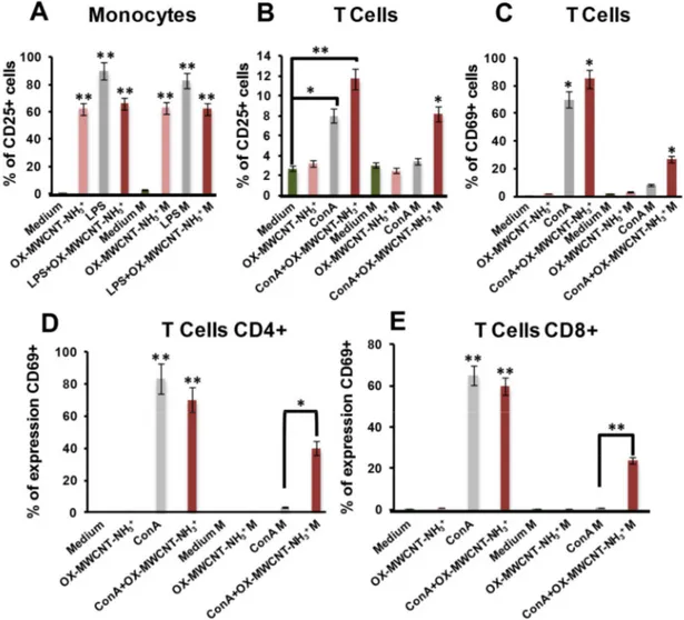
and cosmic radiation. A microgravity environment imparts to an object a lower acceleration compared to that produced by the Earth on its surface. It is well known that the constant influence of microgravity leads to several modifications of many physiological cellular processes, such as proliferation, differentiation, growth, signal transduction, cytoskeletal architecture, motility and gene expression. Moreover, immune cells are severely affected by microgravity, T lymphocyte functions were found altered in more than 50% of crewmembers in space. In a previous work my Ph.D. advisor and colleagues reported that functionalized multi-walled carbon nanotubes (f-MWCNTs) lead to an up-regulation of CD25 and CD69 marker expression in human primary immune cells, in particular in monocytes. In this study we wanted to evaluate the possibility of taking advantage of f-CNT immunostimulatory properties against spaceflight dysregulation of immune functions. Oxidation and functionalization of the nanotubes were performed with the ammonium groups and a fluorescent probe (FITC). First of all, we have seen with Transmission Electron Microscopy (TEM) that microgravity not affected the functionalization and the structure of f-CNTs. Moreover, flow cytometry and confocal microscopy analysis show a dose-dependent uptake of f-CNTs after 24h. Interestingly, we did not detect significant difference in the internalization under microgravity (0 x g). To evaluate the activating ability of f-CNTs to counteract spaceflight immune suppression, Peripheral Blood Mononuclear Cells (PBMC) were left untreated for 24 h or incubated with 50 µg/ml of f-CNTs in static controls and under microgravity conditions using a Random Positioning Machine (RPM). Lipopolysaccharides (LPS) and concanavalin A (ConA), due to their well-known activation properties, were used as positive controls for monocytes (CD14+) and T cells (CD3+) respectively. f-CNTs led to an increase expression of the activation marker CD25 on monocytes, both in static controls and under microgravity conditions. Instead, in microgravity we noticed a down-regulation of CD25 also in ConA treated samples due to the immune suppression as already reported. Interestingly, our results demonstrate that f-CNT together with
ConA counteract the down-regulation of CD25 due to the microgravity condition. We have seen indeed that nanotubes have a clear synergic effect with ConA both in static controls that in simulated microgravity. Cytokine analysis also confirm these data. Our experiments show that the effect of nanotubes appears to be particularly linked to an up-stimulation of the molecular effectors involved in the IL2 pathway. We assume that f-CNTs could promote the patching and capping of the ConA receptor. This action, together with IL2 pathway stimulation, can explain the synergic effect between f- CNTs and ConA in 0 x g. These preliminary findings open new perspectives on the capability of carbon nanotubes to act as immunomodulators proving this property by fighting immune function dysregulation under microgravity conditions, especially for T lymphocytes. We aim at reinforcing the concept that functionalized carbon based materials such as carbon nanotubes are able to stimulate immune cells having very interesting broad future applications in immunotherapy as vaccine adjuvants and, with data shown, as possible fighters to contrast spaceflight immune cell dysregulation.
Paper II: In the last few years, there has been enormous interest in graphene oxide (GO) for its
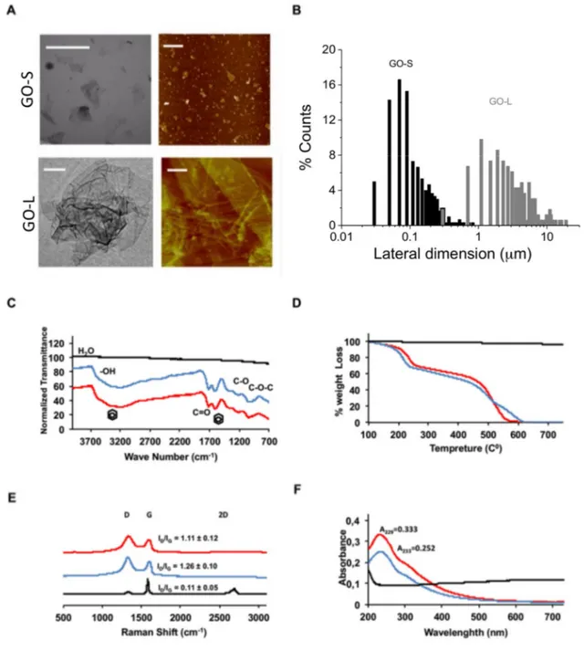
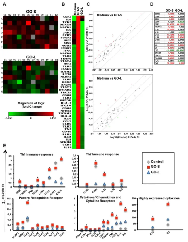
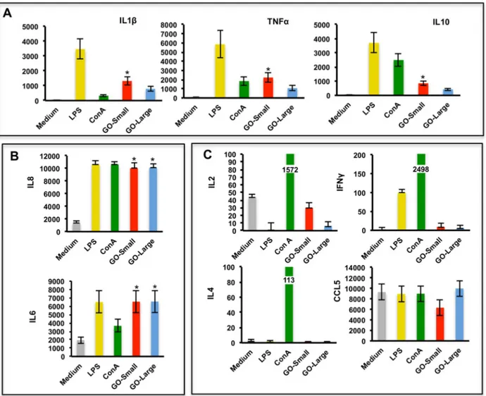
promises in medicine fated to change the known medical practice in many healthcare applications. To improve the knowledge about the immune impact of GO, this work look at the molecular effects of two types of thin graphene oxide sheets (GOs), different only in their lateral dimension, on primary human immune cell populations using a wide range of assays, including high throughput screenings. GOs were thoroughly characterized by TEM, AFM, Raman spectroscopy and several other techniques to determine the specific lateral dimensions, number of graphene layers, and surface properties of each GO used. PBMCs from healthy donors were used, being a pool of immune cells that provide a closer insight to real in vivo conditions in humans, better than isolated sub-populations. The extent of early and late apoptosis, necrosis, cell activation, cytokine release were determined following exposure to the GO materials. Moreover, the impact on 84 genes related to innate and adaptive immune responses were analyzed. Lastly, whole genome analysis was conducted on T lymphocytes (Jurkat cells) and monocytes (THP1 cells). Both GOs used at increasing concentration form 25 to 75g ml-1
generally did not show significant reduction in cell viability. However, exposure to small GO sheets (100-500 nm) was found to have a more significant impact on immune cells compared to the large sized GO (1- 10μm). The pilot expression analysis of 84 gene evidenced the higher genes induction of small GO compared to the large GO, confirmed in the secretion of many cytokine such as: IL1, IL1, TNF, and IL6. Microarray data confirmed the small GO impact on immune cells. Particularly the activation was underlined by the up regulation of genes such
CXCR3 receptor, that are commonly associated with inflammation, immune-mediated tumor rejection and pathogen clearance. Furthermore, also a significant switch on energy-dependent pathways was found. In this work we demonstrated that the distinct shape dimensions could regulate the nature of GO interactions with immune cells. These findings represent the first step of a comprehensive molecular-characterization of different sized GOs on immune cells impact giving crucial information for the design of graphene for the control of its biocompatibility; paving the foundations for future preclinical studies with well-designed GOs as i.e. immunotherapy tools or safety drug delivery platform.
Paper III: The rationale of this work was driven by the common hope of a nanotechnology
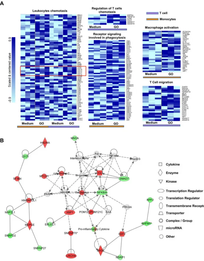
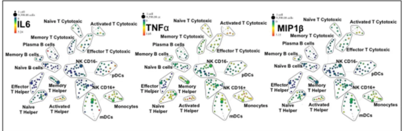
translation into the everyday clinical practice. However, such dream has to face something that can make the difference for almost any successful bio-application: the potential impact on the complex system of blood immune cells. The understanding of the interactions between nanoparticles and immune cells is hindered by the scant implementation of high throughput technologies in nanotechnology. Recently, a novel tool for flow cytometry analysis has been developed, gaining leverage with the precision of mass spectrometry. The use of the technique termed single-cell mass cytometry provides the measurement of more than 40 simultaneous cellular parameters at a single-cell resolution. We propose, for the first time in the context of nanotechnology, a new analytical strategy able to dissect the immunological impact of nanomaterials, at the single-cell level. The analytical pipeline here reported encompass the immunological characterization of the most studied nanomaterial in the last years: graphene. Mass cytometry enables us to describe the immune cell interactions of thin graphene oxide (GO) flakes and GO functionalized by amino groups (GONH2) on 15 cellular populations
corresponding to 200 nodes of distinct but logically interconnected cell sub-populations. Together we performed whole-transcriptomic analysis (Illumina BeadArray) for functional and molecular characterization on human T-cells and monocytes as a representative for the adaptive and innate response. Our results emphasize the importance of the functionalization on enhancing the biocompatibility of GO-based nanomaterials. Notably, only the functionalized GONH2 was able to induce a specific monocytoid dendritic cell and monocyte activation
skewed toward a T helper 1/M1 response. The positive impact of GONH2 on specific immune
cells could serve as a starting point for the development of new nanoscale platforms in medicine as novel immunotherapy, vaccine carrier, or nanoadjuvant tools.
Paper IV: Nanotechnology and specifically nanomedicine could provide new tools for an
effective treatment of several diseases such as cancer, not only by taking advantage of the ability to functionalize and target the nanomaterials but also of their possible intrinsic biological properties. In this work, we developed a new few-layer graphene (FLG) dispersions prepared
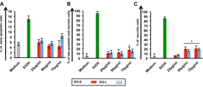
by a mechano-chemical approach employing melamine as the exfoliating agent of graphite. The biological impact of new FLG on multiple human immune primary cell population (T, B and Natural Killer lymphocyte, monocytes, macrophages and dendritic cells) looking at cell viability and cell activation using a wide range of analysis were explored. We found an intrinsic specific impact of FLG on cells belonging to the monocytic lineage (CD14 positive) showing neither toxic nor activation effects on the other immune cells. Thanks to this intrinsic biological property, we further explored the possible therapeutic application of FLG on neoplastic monocytes ex vivo, from acute myeloid leukemia and chronic myelomonocytic leukemia patients. Intriguingly we demonstrated the FLG unique ability to target and successfully boost the necrosis of monocytic cancer cells. Moreover, the comparison between FLG and a common chemotherapeutic drug (i.e. Etoposide) confirmed the specificity and higher toxicity of FLG on cancer cells, evidencing the absence of toxicity of other immune cell populations. The functionality in terms of viability and activation of the other immune cell populations was also not affected, suggesting the ability of our graphene to preserve the normal function of the immune system. We here showed, for the first time, the great intrinsic biological properties of graphene obtained by exfoliation from graphite and applied them in oncology research versus an aggressive form of blood cancer. As a further step, in the near future, we can imagine not only to use the specific effect of FLG to improve the cancer cell toxicity but also, thanks to its high surface area, to take advantage of the conjugation with other drugs to enhance cancer therapy.
Paper V: Following the necessity to understand the immunological fate of different
nanomaterials. In this manuscript we wanted to deeply investigate the action of three differently coated lipid nanocapsules (NCs) on human PBMCs. Considering the wide variety of NC applications in biomedicine a possible immune response to this type of nanomaterials is a crucial issue to be addressed. To the best of our knowledge this is the first comprehensive study on bio and immune-compatibility comparison analysis of differently functionalized NCs with chitosan, pluronic and PEG, fully characterized. We analyzed the impact of three different functionalizations of NCs looking first at the hemolytic impact. We then focused on human PBMCs and subpopulations (T, B, NK cells and monocytes) from healthy donors. Uptake assays were performed to assess whether a specific coating could be more suitable for the cell internalization. We explored the possible necrosis, apoptosis and proliferation of PBMCs and subpopulations of T cells and monocytes, as representative of the adaptive and innate immune response after NC treatment. The functionality effect was tested looking at the cell diameter modification, at the expression of the most critical activation markers (CD25 and CD69) and at the production of a wide variety of cytokines. In this work we demonstrated that the NCs impact
well internalized inside all immune cell populations studied. Intriguingly, Pluro-NCs were able to induce immunomodulation of innate immunity through a clear induction of monocyte activations. Chito-NCs instead, mediated the activation of monocytes and T helper response that give the whole enhancement of the immune response after treatment. All these actions are exclusive for Chito-NCs that in our screening resulted as good starting material for further studies in the context of vaccine delivery, immunotherapy and immunomodulation activities. On the contrary, PEG-NCs were completely inert for the population of immune cells analyzed opening their perspectives as inert drug carriers.
Paper VI: In this manuscript we deeply investigate the immune impact of novel cystine
functionalized SPIONs (Cy-SPIONs) exploring also their potential as ultrasound contrast agents. SPIONs are deeply investigated for many biomedical applications such as imaging for diagnostic and therapeutic purposes. A good functionalization that can combine the imaging goals together with a good biocompatibility remains one of the challenge for particles translation into medical practice. In this work, we focused on a novel functionalization of SPION with cystine (Cy-SPIONs); cystine, indeed, is able to make SPION stable and dispersible in culture cell media being an aminoacid easily biosynthesized in humans. To prove their potential as biomedical tools, we first gave new insights into the biological and immune effects of Cy-SPIONs with a wide variety of standard and molecular assays to evaluate cytotoxicity, cell activation, cytokine release and the expression of 84 genes related to the immune response. A good immune biocompatibility of Cy-SPIONs on ex vivo primary immune cells as well as in vitro cell lines was found. Moreover, an interesting potential of Cy-SPIONs for in vivo studies was pointed out where the preferential route of administration is by intravenous injections, thus in contact with immune cells. Currently, many studies focus on the use of SPION on Magnetic Resonance Imaging (MRI), instead our study focused on ultrasonography, which is a safer, less expensive and common imaging technology. The good echogenic properties of Cy-SPIONs in water and in whole blood were shown both in a phantom vein and in a microfluidic device for bloodstream simulations. Moreover, we report no cell toxicity under Cy-SPION treatment together with ultrasound irradiation, giving new insight on the use of Cy-SPIONs as new ultrasound contrast agents.
Paper I: Immunomodulatory properties of carbon nanotubes are
able to compensate immune function dysregulation caused by
microgravity conditions
Claudia Crescioa, Marco Orecchionib, Cécilia Ménard-Moyonc, Francesco Sgarrellab, Proto
Pippiaa, Roberto Manettid, Alberto Biancoc* and Lucia Gemma Delogub*
aDipartimento di Scienze Biomediche, Università degli studi di Sassari, 07100 Sassari, Italy. bDipartimento di Chimica e Farmacia, Università degli Studi di Sassari, 07100 Sassari, Italy. cCNRS, Institut de Biologie Moléculaire et Cellulaire, Laboratoire d’Immunopathologie et
Chimie Thérapeutique, 67000 Strasbourg, France.
dDipartimento di Medicina Clinica, Sperimentale e Oncologica Università degli Studi di
Sassari, 07100 Sassari, Italy.
Published in:
Abstract
Spaceflights lead to dysregulation of the immune cell functionality affecting the expression of activation markers and cytokine production. Short oxidized multi-walled carbon nanotubes functionalized by 1,3-dipolar cycloaddition have been reported to activate immune cells. In this Communication we have performed surface marker assays and multiplex ELISA on primary monocytes and T cells under microgravity. We have discovered that carbon nanotubes, through their immunostimulatory properties, are able to fight spaceflight immune system dysregulations.
Manuscript
Gravity is the force of attraction by which terrestrial bodies tend to fall toward the Earth. In space, living organisms are confronted with two important factors: microgravity and cosmic radiation. A microgravity environment imparts to an object a lower acceleration compared to that produced by Earth at its surface. Experiments conducted by American, Russian, and European investigators, in dedicated space missions as well as in simulations on Earth, have shown that mammalian cells are sensitive to gravitational changes.1-8 It is well known that the
constant influence of weightlessness leads to several modifications of many physiological cellular processes, such as proliferation, differentiation, growth, signal transduction, cytoskeletal architecture, motility and gene expression.1-4 Moreover, immune cells are severely
affected by microgravity.5 T lymphocyte functions were found altered in more than 50% of
crew members in space.6 A severe inhibition of T cell activation in real and simulated
microgravity conditions has been extensively demonstrated. Hashemi et al. reported a down regulation of CD25 and CD69 cell membrane activation markers in T cells after 24 hours of microgravity condition.7 These findings were recently confirmed on US Astronauts onboard
the Space Shuttle.8 CD25 and CD69 are important markers in the T Cells mediate immune
response. CD25 (alpha chain of the IL-2 receptor) is a late activation antigen. Activation mediated by the T-cell receptor (TCR) and costimulatory molecules induce an up-regulation of CD25 in T cells making them highly sensitive to IL-2. Whereas, CD69, member of the C-type lectin superfamily (Leu-23), is one of the earliest cell surface antigens expressed by T cells following activation.
We reported that functionalized multi-walled carbon nanotubes (f-MWCNTs) lead to an up-regulation of CD25 and CD69 markers expression in human primary immune cells, in particular in monocytes.9 Very recently, through a whole genome wide study we proposed functionalized
CNTs (f-CNTs) as immunomodulator systems showing their potential as immune activators 10.
We would like to highlight that in contrast to other types of CNTs, different in functionalization and shape investigated in immune cells,9-12 we found that oxidized MWCNTs, further
functionalized by 1,3-dipolar cycloaddition, can act as immunomodulators.9 No data are present
in literature regarding the interaction of CNTs and immune cells under microgravity conditions. Encouraged by our recent results, we wanted to evaluate the possibility of taking advantage of f-CNT immunostimulatory properties against spaceflight dysregulation of immune functions. The high cost of experiments on board of spacecraft and space station facilities and the limited number of doubling experiments do not allow scientists to give continuity to the studies in real microgravity outside Earth. Currently, thanks to different and advanced facilities, it is possible to carry out studies in microgravity simulating in part the spaceflight conditions. In this work, a tridimensional clinostat or Random Positioning Machine (RPM) was used to simulate microgravity (M; 0xg) to evaluate if f-CNTs are able to compensate immune microgravity induced dysregulation. Static control cell cultures were installed in the basement of the RPM. We first assessed the possible impact of microgravity on the functionalization of CNTs by transmission electron microscopy (TEM) and Kaiser test. To assess whether microgravity affect CNT uptake on primary human T cells and monocytes, we studied their internalization in microgravity by flow cytometry and confocal microscopy. Taking into consideration the results reported on activation markers by Hashemi et al.,7 we then focused on CD25 expression on
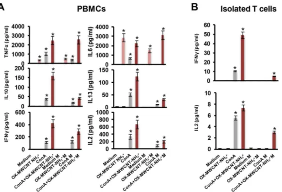
monocytes and T cells through peripheral blood mononuclear cells (PBMCs) analysis and CD69, both on T cells present on PBMCs and isolated T lymphocytes, T-helper cells (CD4+) and cytotoxic T cells (CD8+). To gain a larger picture about CNT effect on immune microgravity dysregulation, we also performed a multiplex cytokine assay on PBMCs (TNFα, IL6, IL10, IL13, IFNγ, IL2) and on isolated T lymphocytes (IFNγ, IL2).
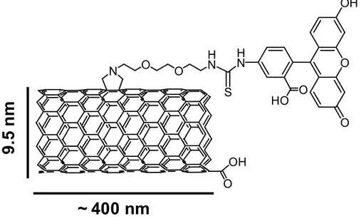
By screening a series of functionalized carbon nanotubes, we previously obtained the major effect on immune activation treating human primary cells with oxidized MWCNTs subsequently functionalized by 1,3-dipolar cycloaddition of azomethine ylides.9, 10 The present
study was carried out with the same type of nanotubes in terms of functionalization and shape (Fig. S1). Characterization and functionalization methods of the MWCNTs has been previously
described by our group.13 Kawanami et al. focused on the effect of microgravity on CNT
synthesis,14 but no data are reported in the literature about the possible impact of microgravity
on f-CNTs. We first assessed by TEM and Kaiser test that MWCNTs (OX-MWCNT-NH3+)
were not affected in their functionalization in microgravity. TEM images do not display differences in the morphology of the tubes under the microgravity (Fig. 1), while Kaiser test gave approximately the same values (within the uncertainty of this type of measurement) in term of amount of ammonium groups before (~40 µmol/g) and after (~50 µmol/g) treatment under microgravity. We previously showed a clear uptake on monocytes and T cells by the same type of nanotubes.9
Figure 1. TEM images of OX-MWCNT-NH3+ before (A) and after microgravity treatment (B).
To make sure that the equivalent internalization can be possible under microgravity conditions, we used the same fluorescently labeled nanotubes (Fig. S2). As expected, data from flow cytometry showed a dose-dependent uptake of f-CNTs after 24 h. Interestingly, we did not detect significant difference in the internalization at 0xg (Fig. S3A). We confirmed our data at 100 µg/ml concentration for both the samples treated in static control or in microgravity by confocal microscopy (Fig. S3B). The absence of difference in cell uptake prompted us to go ahead in understanding the potential of carbon nanotubes to modulate the immune spaceflight effects. Cellular uptake results led us to choose a working concentration of 100 µg/ml of f-CNTs. We fixed the time of incubation at 24 h, the best time point to assess a possible immune response on primary human cells as used in the space mission and microgravity experiment on activation markers.7
To evaluate the ability of activator MWCNTs to counteract spaceflight immune suppression,9 PBMCs were left untreated for 24 h or incubated with 100 μg/ml of OX-MWCNT-NH3+ in static controls and under microgravity conditions. Lipopolysaccharides (LPS) and concanavalin