Review
Ellagitannins in Cancer Chemoprevention
and Therapy
Tariq Ismail1, Cinzia Calcabrini2,3, Anna Rita Diaz2, Carmela Fimognari3, Eleonora Turrini3, Elena Catanzaro3, Saeed Akhtar1and Piero Sestili2,*
1 Institute of Food Science & Nutrition, Faculty of Agricultural Sciences and Technology, Bahauddin Zakariya University, Bosan Road, Multan 60800, Punjab, Pakistan;
[email protected] (T.I.); [email protected] (S.A.)
2 Department of Biomolecular Sciences, University of Urbino Carlo Bo, Via I Maggetti 26, 61029 Urbino (PU), Italy; [email protected]
3 Department for Life Quality Studies, Alma Mater Studiorum-University of Bologna, Corso d'Augusto 237, 47921 Rimini (RN), Italy; [email protected] (C.C.); [email protected] (C.F.);
[email protected] (E.T.); [email protected] (E.C.) * Correspondence: [email protected]; Tel.: +39-(0)-722-303-414 Academic Editor: Jia-You Fang
Received: 31 March 2016; Accepted: 9 May 2016; Published: 13 May 2016
Abstract: It is universally accepted that diets rich in fruit and vegetables lead to reduction in the risk of common forms of cancer and are useful in cancer prevention. Indeed edible vegetables and fruits contain a wide variety of phytochemicals with proven antioxidant, anti-carcinogenic, and chemopreventive activity; moreover, some of these phytochemicals also display direct antiproliferative activity towards tumor cells, with the additional advantage of high tolerability and low toxicity. The most important dietary phytochemicals are isothiocyanates, ellagitannins (ET), polyphenols, indoles, flavonoids, retinoids, tocopherols. Among this very wide panel of compounds, ET represent an important class of phytochemicals which are being increasingly investigated for their chemopreventive and anticancer activities. This article reviews the chemistry, the dietary sources, the pharmacokinetics, the evidence on chemopreventive efficacy and the anticancer activity of ET with regard to the most sensitive tumors, as well as the mechanisms underlying their clinically-valuable properties.
Keywords:ellagitannins; phytochemicals; cancer; chemoprevention; cancer therapy; safety
1. Introduction
Despite the enormous efforts of the scientific and medical community, cancer still represents the second leading cause of death and is nearly becoming the leading one in the elderly [1]. It is estimated that, due to demographic changes alone, in the next 15 years the number of new cancer cases will increase by 70% worldwide [2].
The lack of effective diagnostic tools for early detection of several tumors, the limited treatment options for patients with advanced stages of cancer, and the onset of multiple drug resistance favor poor prognosis and high mortality rates. The significant, but still unsatisfactory, improvement of survival, the severe toxicity profile, and the high costs characterizing many current anticancer therapies clearly show that a threshold in terms of clinical benefit and patients’ tolerance has been reached. Thus, the identification and development of innovative, preventive as well as therapeutic strategies to contrast cancer-associated morbidity and mortality are urgently needed.
Epidemiological, preclinical, and clinical studies have generally concluded that a diet rich in phytochemicals can reduce the risk of cancer [2,3]. Due to their pleiotropism which includes
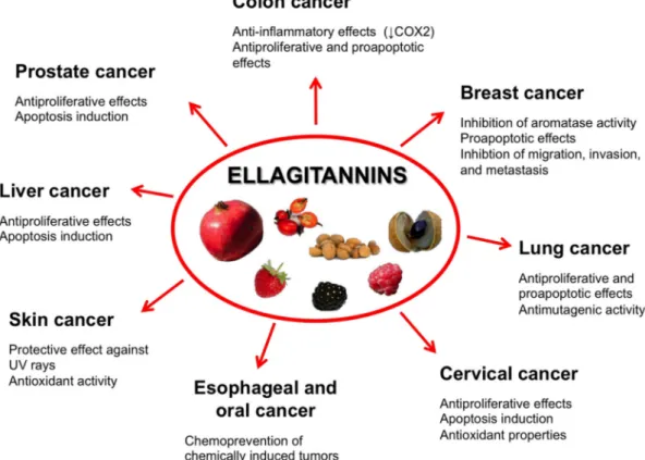
antioxidant, anti-inflammatory, and antiproliferative activities as well as modulatory effects on subcellular signaling pathways, phytochemicals from edible fruits and vegetables are recognized as an effective option to counteract cancer incidence and mortality [3–5]. Plants constitute a primary and large source of various chemical compounds including alkaloids, flavonoids, phenolics, tannins, tocopherols, triterpenes, and isothiocyanates. Ellagitannins (ET) are an important class of phytochemicals contained in a number of edible plants and fruits recommended by the traditional medicine of a variety of cultures, both in the developing and developed countries, to treat common health problems. ET biological and nutraceutical potential has received increasing attention over the last several decades. ET exert multiple and clinically-valuable activities [4], and among them the chemopreventive, anticarcinogenic, and antiproliferative activities are being receiving growing interest and attention (Figure1).
subcellular signaling pathways, phytochemicals from edible fruits and vegetables are recognized as an effective option to counteract cancer incidence and mortality [3–5]. Plants constitute a primary and large source of various chemical compounds including alkaloids, flavonoids, phenolics, tannins, tocopherols, triterpenes, and isothiocyanates. Ellagitannins (ET) are an important class of phytochemicals contained in a number of edible plants and fruits recommended by the traditional medicine of a variety of cultures, both in the developing and developed countries, to treat common health problems. ET biological and nutraceutical potential has received increasing attention over the last several decades. ET exert multiple and clinically‐valuable activities [4], and among them the chemopreventive, anticarcinogenic, and antiproliferative activities are being receiving growing interest and attention (Figure 1).
Figure 1. Fruits containing ET with chemopreventive, anticarcinogenic, and antiproliferative activities.
2. Dietary Sources, Types, and Occurrence
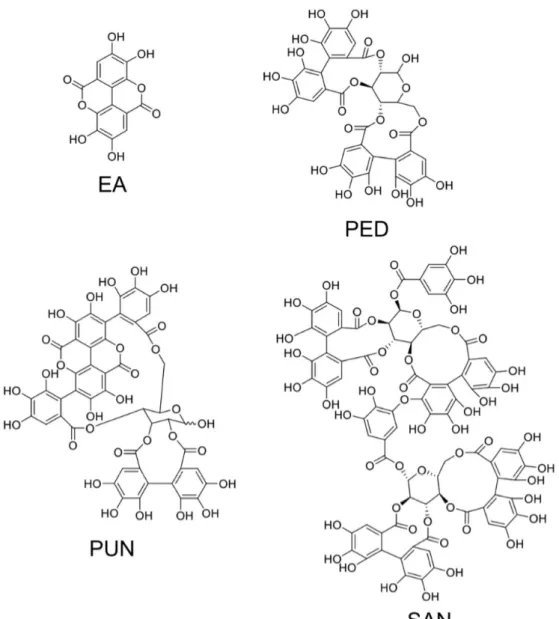
ET and their derivatives are noticeably contained in edible seeds, nuts, and various fruits of nutritional interests. The structures of relevant ETs and of ellagic acid are shown in Figure 2. A wide variety of fresh fruits including berries, like raspberries, black raspberries, strawberries, pomegranate, longan, and dried nuts, are renowned for their ample polyphenols concentration in the form of ET [5]. Five species of berries including raspberry, strawberry, cloudberry, rose hip, and sea buckthorn were identified by Koponen et al., [6] as significant carrier of ET in a range of 1–330 mg per 100 g of fruit. Sanguiin H‐6 and lambertianin C were reported from Glen Ample raspberries and Scottish‐grown red raspberries, along with some trace levels of ellagic acid [7,8]. Blackberries (fruit and seeds) have been reported for a range of ET including pedunculagin, casuarictin, sanguiin H‐6 (lambertianin A), and lambertianin (C and D) [9–11]. Pomegranate and various fractions of the fruit are known for their cancer chemopreventive properties owing to their unique phenolics composition in the form of ET, which include punicalagin, punicalin, granatin A, granatin B, tellimagrandin I, pedunculagin, corilagin, gallagic acid, ellagic acid, and casuarinin [12].
Figure 1.Fruits containing ET with chemopreventive, anticarcinogenic, and antiproliferative activities. 2. Dietary Sources, Types, and Occurrence
ET and their derivatives are noticeably contained in edible seeds, nuts, and various fruits of nutritional interests. The structures of relevant ETs and of ellagic acid are shown in Figure2. A wide variety of fresh fruits including berries, like raspberries, black raspberries, strawberries, pomegranate, longan, and dried nuts, are renowned for their ample polyphenols concentration in the form of ET [5]. Five species of berries including raspberry, strawberry, cloudberry, rose hip, and sea buckthorn were identified by Koponen et al., [6] as significant carrier of ET in a range of 1–330 mg per 100 g of fruit. Sanguiin H-6 and lambertianin C were reported from Glen Ample raspberries and Scottish-grown red raspberries, along with some trace levels of ellagic acid [7,8]. Blackberries (fruit and seeds) have been reported for a range of ET including pedunculagin, casuarictin, sanguiin H-6 (lambertianin A), and lambertianin (C and D) [9–11]. Pomegranate and various fractions of the fruit are known for their cancer chemopreventive properties owing to their unique phenolics composition in the form of ET, which include punicalagin, punicalin, granatin A, granatin B, tellimagrandin I, pedunculagin, corilagin, gallagic acid, ellagic acid, and casuarinin [12].
Figure 2. Structures of ellagic acid and of three major and representative ellagitannins. EA, ellagic acid; PED, pedunculagin; PUN, punicalagin; SAN, sanguiin H‐6.
ET, predominately those isolated from pomegranate (e.g., punicalagin), have gained a wide popularity as preventive and therapeutic ethnopharmacological approaches for cancer treatment. However, a lot more has been added to this class of compounds from fruits other than pomegranate, including raspberries, blueberries, strawberries, muscadine grapes, and longan [7,13–18]. Major phenolic fractions recovered from longan include gallic acid, ellagic acid, and corilagin, much more concentrated in the seed segment as compared to the fruit pulp and peel [17]. Good essential fatty acid composition of nuts and fairly high concentrations of ET and their derived fractions, such as ellagic acid and its glycosidic derivatives have been associated with the potential cardioprotective properties of nuts. Ellagic acid (free and total) has been reported in a range of 0.37–823 mg per 100 g of dried nuts [19]. High concentrations of a variety of ET (ellagic acid, sanguiin H2 and 6, lambertianin C, castalagin/vescalagin, galloyl‐bis‐HHDP glucose, pedunculagin) can be found in blackberries (Rubus sp.) [20]. Shi et al., [21] identified agrimoniin as the second highest phenolic compound of strawberries.
Irrespective of the edible fractions of fruiting plants, some inedible fractions like fruit peels, bark and foliage have also been reported as good source of hydrolysable tannins including bioactive ET [4,22]. Leaves extracts of Shepherdia argentea—a deciduous shrub commonly known as silver buffaloberry—were reported as a good reserve of gluconic acid core carrying the potential anti‐HIV novel ET, such as hippophaenin A, shephagenin A and shephagenin B [23].
Figure 2.Structures of ellagic acid and of three major and representative ellagitannins. EA, ellagic acid; PED, pedunculagin; PUN, punicalagin; SAN, sanguiin H-6.
ET, predominately those isolated from pomegranate (e.g., punicalagin), have gained a wide popularity as preventive and therapeutic ethnopharmacological approaches for cancer treatment. However, a lot more has been added to this class of compounds from fruits other than pomegranate, including raspberries, blueberries, strawberries, muscadine grapes, and longan [7,13–18]. Major phenolic fractions recovered from longan include gallic acid, ellagic acid, and corilagin, much more concentrated in the seed segment as compared to the fruit pulp and peel [17]. Good essential fatty acid composition of nuts and fairly high concentrations of ET and their derived fractions, such as ellagic acid and its glycosidic derivatives have been associated with the potential cardioprotective properties of nuts. Ellagic acid (free and total) has been reported in a range of 0.37–823 mg per 100 g of dried nuts [19]. High concentrations of a variety of ET (ellagic acid, sanguiin H2 and 6, lambertianin C, castalagin/vescalagin, galloyl-bis-HHDP glucose, pedunculagin) can be found in blackberries (Rubus sp.) [20]. Shi et al., [21] identified agrimoniin as the second highest phenolic compound of strawberries.
Irrespective of the edible fractions of fruiting plants, some inedible fractions like fruit peels, bark and foliage have also been reported as good source of hydrolysable tannins including bioactive ET [4,22]. Leaves extracts of Shepherdia argentea—a deciduous shrub commonly known as silver
buffaloberry—were reported as a good reserve of gluconic acid core carrying the potential anti-HIV novel ET, such as hippophaenin A, shephagenin A and shephagenin B [23].
3. Ellagitannins—Classification and Chemistry
Tannins are unique secondary metabolites of plant phenolics with relatively higher molecular weight (300–30,000 Da) and bear the ability to generate complexes with some macromolecules, like proteins and carbohydrates [24]. Chemistry and nomenclature of the tannins is complicated by virtue of the frequent changes which parallel the advancement in this very specific field [25]. Taking into account different definitions of tannins [26,27], these compounds may be referred as either galloyl esters and their derivatives (ET, gallotannins, and complex tannins), or the oligomeric and polymeric proanthocyanidins (condensed tannins). In a broader perspective, tannins may be classified most satisfactorily and unambiguously on the basis of structural configuration and/or solubility [28]. C–C coupling of galloyl units in absence of glycosidically-linked catechin make ETs structurally different from the condensed tannins that are characterized by monomeric catechin linkages (C4–C8 or C4–C6) to generate oligomeric likewise polymeric proanthocyanidins [27]. Gallotannins and ETs constitute a major group of tannins i.e., hydrolysable tannins that are well known for their properties to hydrolyze into hexahydroxydiphenol (HHDP) or gallic acid moieties. Gallotannins are the gallic acid derivatives carrying ě six gallyol groups and might further be characterized on account of one or more than one digalloyl group [29].
ETs (hydrolysable tannins) on their hydrolysis yield gallic acid and ellagic acid from the compounds carrying gallyol groups and HHDP groups, respectively [28]. In vitro digestion models declare ETs to remain stable under the normal physiological condition of the stomach [30]. However, ETs hydrolysis to free ellagic acid or their degradation may proceed in the small intestine at neutral to alkaline pH [31]. Biologically, condensed tannins and gallotannins are thought to deliver relatively higher protein precipitation properties as compare to the ETs and hence are considered potential antinutritional compounds from the class of plants polyphenolics [32]. Gallotannins and condensed tannins have also been reported as oxidatively least active tannins as compared to the ETs and on the same time gallotannins and condensed tannins have also been found to reduce pro-oxidant properties of ETs [33,34].
3.1. Simple Ellagitannins
ET (M.W. 300–20,000 Da) are non-nitrogenous compounds with at least two C–C coupled galloyl units with no glycosidically-bonded catechin unit [3,35]. ET are derivatives of 1,2,3,4,6-penta-O-galloyl-β-D-glucopyranose (PGG). Structurally, ET are esters of carbohydrates and or cyclitols and also include metabolic compounds derived from oxidative cleavage of either condensed or hydrolysable tannins [27,35,36]. The presence of hexahydroxydiphenol (HHDP) in a glucopyranose ring in addition to acyl units and certain HHDP metabolites such as dehydrohexahydroxydiphenol (DHHDP), valoneoyl and chebuloyl groups constitute simple ET. Tellimagrandin I and II, pedunulagin, casuarictin, and chebulagic acid originate from the specific orientation and number of acyl groups on glucose units. Variation in HHDP group originates by linking (C–C or C–O) one or more galloyl groups to HHDP unit.
Structural diversity of ET has been reported to correlate with their carrier-plants’ taxonomy and evolutionary hierarchy [37]. More often, monomeric ET or oligomeric ET constitute the major tannic component of plant species. The monomeric compounds of the group include tellimagrandins I and II, pedunculagin, casuarictin, and potentillin. Type I hydrolysable tannins (i.e., gallotannins) carrying HHDP in stable conformation at either the 2,3 or 4,6 position on aD-glucopyranose may be referred to as a simple ET [38–40]. Geraniin, a type III ET, is another example of monomeric simple ET carrying a DHHDP unit linked toD-gluopyranose of HHDP unit via1C4conformation. Dimers of ET are generated by intermolecular oxidative coupling/condensation of simple ET.
3.2. Glycosidic Ellagitannins
Chemically, the C-glycosidic linkage of ET is established via intermolecular bonds between two monomeric units, one carrying anomeric carbon while the second one galloyl or HHDP group [3,35,41]. Most recently C-glycosidic ET including granadinin, vescalagin, methylvescalagin, castalagin, stachyurin, and casuarinin have been reported from the peel and seed fraction of camu-camu, a fruiting tree of Amazon rainforest [42]. Woody fractions of various fruits, particularly the nuts and berries, have also been observed to hold novel C-glycosidic ET (e.g., castalagin and vescalagin). Castacrenins D and F are two other forms of C-glycosidic ET isolated from the woody fraction of Japanese chestnut and carry gallic acid/ellagic acid moieties [43]. Treating vescalagin with Lentinula edodes generates quercusnins A and B that may be referred as fungal metabolites of C-glycosidic ET [44]. Castacrenins D and F isolated from chestnut wood may generate oxidative metabolites, namely castacrenins E and G, by replacing pyrogallol rings of C-glycosidic ET with cyclopentone rings [43]. Rhoipteleanins H, I, and J were reported as novel C-glycosidic ET isolated from the fruit and bark fractions of Rhoiptelea chiliantha. Structural configuration of rhoipteleanins H revealed the presence of cyclopentenone carboxy moieties that are generated by oxidation and rearrangement of C-glycosidic ET aromatic ring [45].
Condensate of C-glycosidic ET is another subclass of hydrolysable tannins, which includes rhoipteleanin J produced by the intermolecular condensation (C–C or C–O) of monomeric C-glycosidic ET followed by oxidation of aromatic rings of ET [45]. Wine aged in oak wood barrels is often reported to carry oak ET, particularly the condensation products of monomeric C-glycosidic ET. The studies infer C-glycosidic ET to play a significant role in modulation of organoleptic features of wine aged in oak wood barrels [46].
4. Ellagitannins Pharmacokinetics
A precise knowledge of phytochemicals’ pharmacokinetics is very important to exploit their health benefits, as well as the effects of their metabolites [47]. In vivo, ET, instead of being absorbed directly into the blood stream, are physiologically hydrolyzed to ellagic acid, which is further metabolized to biologically-active and bioavailable derivatives, i.e., urolithins, by the activity of microbiota in gastrointestinal (GI) tract [5,48]. The biological properties of ET, such as free radical scavenging, further depend on their metabolic transformation inside gut. ET recovered from pomegranate juice may be metabolically converted by gut microbiota to urolithin A, B, C, D, 8-O-methylurolithin A, 8,9-di-O-methylurolithin C, and 8,9-di-O-methylurolithin D, and some of these metabolites display higher antioxidant activity than the parental tannins themselves. For instance, urolithin C and D show an antioxidant capacity—as determined in a cell-based assay—which is 10- to 50-fold higher as compared to punicalagin, punicalin, ellagic acid, and gallic acid [49]. This finding suggests that intestinal transformation products of ET are likely to play a central role for the antioxidant properties at least inside the GI tract. Significant differences in urolithins’ profiles in individual human subjects feed on raspberries—a renowned source of ET—have been attributed to gut microflora, whose variations on an inter-individual basis affect their capacity of hydrolyzing ET and subsequent metabolite synthesis [48,50]. The interaction of gut microbiota composition and the host endogenous excretory system is also likely to play a further role in the observed inter-individual variability [51]. ET are highly stable under the acidic environment of stomach, and retain their composition without being hydrolyzed to simpler compounds when exposed to various gastric enzymes. Consequently the complex structure of ET impedes their gastric absorption: however, the stomach might serve as the first site of absorption of free ellagic acid and pre-hydrolyzed forms of ET.
Contrary to stomach, the neutral or alkaline environments of duodenum and small intestines, characterized by pH values ranging from 7.1 to 8.4, allow ET hydrolyzation [31,41]. In humans, ET are rapidly absorbed and metabolized, as documented by [18,52]: following ingestion of pomegranate juice (at a dose containing 25 mg of ellagic acid and 318 mg of ET), ellagic acid can be found in plasma for up to 4 h while, at later times, it is no more detectable. In contrast, another study reported that no ellagic
acid could be detected in plasma during the 4 h following the juice intake [53], a discrepancy which has been attributed to inter-individual variability [54]. Ellagic acid is converted by catechol-O-methyl transferase to dimethylellagic acid, which is then glucuronidated and excreted [52].
Finally, the microbiologically metabolized fraction of ET, i.e., urolithins, is further incorporated to enterohepatic circulation system [18,53,55,56].
5. Ellagitannins for Tumor Chemoprevention and Therapy
The development of novel mechanism-based chemopreventive and antitumor approaches to fight cancer through the use of dietary substances which humans can easily accept has become an important goal. Along this line, ET have received increasing attention over the last two decades.
Similarly to other anticancer phytochemicals, ET display chemopreventive and chemotherapeutic activities [3]. The chemopreventive activity of ET and derivatives, such as ellagic acid has been primarily associated with their antioxidant capacity, that varies with the degree of hydroxylation [5,57] and depends from both a direct radical scavenging and iron chelation activity.
The well-known anti-inflammatory capacity represents another important feature of ET chemopreventive and antitumor activity [55], that being persisting inflammation involved both as a causative and a facilitating factor in carcinogenesis and cancer development [58]. For example, pomegranate ET inhibit pro-inflammatory pathways including, but not limited to, the NF-κB pathway, whose activation leads to immune reactions, inflammation, and the transcription of genes involved in cell survival, such as Bclx and inhibitors of apoptosis. Constitutive activation of NF-κB has been observed in prostate cancers, where it sustains chronic inflammation and promotes the development of high-grade prostate cancer. With respect to inflammation, it is worth noting that, similarly to many polyphenols, the antioxidant activity of ET participates to an “anti-inflammatory loop” with other mechanisms, since it lowers the levels of radicals which otherwise would act as pro-inflammatory stimuli.
The direct antiproliferative effects of ET have been attributed to multiple mechanisms (see the next subchapters) including the cell cycle arrest capacity and the properties enabling cancer cells to follow apoptosis through the mitochondrial route and self-destruction after replication [59–62]. In addition to directly targeting tumor cell survival, the cytotoxic/cytostatic activities of ET might also concur with the chemopreventive potential, since they prevent tumor cells from converting into more malignant phenotypes and from replicating.
A study on 1,3-di-O-galloyl-4,6-(s)-HHDP-β-D-glucopyranose (an ET from Balanophora japonica MAKINO) points to the complexity and multiplicity of the mechanisms contributing to the anticancer activity of ET, i.e., the same complexity and multiplicity characterizing also other classes of phytochemicals. Indeed, the antiproliferative activity of 1,3-di-O-galloyl-4,6-(s)-HHDP-β-D-glucopyranose in human Hep-G2 liver cancer cells was also associated to an altered regulation of 25 miRNAs including the let-7 family members miR-370, miR-373, and miR-526b, identified as likely targets with roles in cell proliferation and differentiation [63]. The fact that in cell culture systems combinations of ET or of ET and other phytochemicals present in plant or fruit extracts are more cytotoxic than any single ET [64], is suggestive of the multifactorial effects, chemical synergy, and multiplicity of the mechanisms behind their antitumor activity. To this regard, the capacity of some ET to inhibit angiogenesis, a fundamental event accompanying tumor growth, both in in vitro and in vivo prostate cancer models [65], and to reduce endothelial cell growth through binding to vascular endothelial growth factor receptors [66] represents a further and significant antitumor mechanism.
In analogy to other polyphenols, ET could also be utilized to increase the sensitivity of tumor cells to standard chemotherapeutic drugs [67], with the aim of obtaining an increase of their antitumor efficacy along with a reduction of their doses and, consequently, of their severe adverse effects which often represent a limiting factor for the prosecution of the therapeutic regimens.
As a premise to the literature data discussed in the next paragraphs, it is important noting that, since ET are not absorbed systemically after oral administration as such [48], the studies where ET
extracts were given to cultured cancer cells are unlikely to be predictive of the effects which could be attained after oral ingestion in vivo. Rather these data could be representative of intravenously administered ET, but the toxicology of this administration route is not known.
The next sections of the review will discuss more in depth the ET anticancer mechanisms and properties emerging from in vivo and in vitro studies on a panel of tumors or tumor cells which appear as potentially sensitive targets for these phytochemicals.
5.1. Prostate Cancer
Prostate cancer is the second leading cancer-associated death risk factor among U.S. males [68]. Phytochemicals originating from various food sources slow down the progression of prostate cancer, whereas a majority of other nutrients are reported to be non-effective in either preventing or curing prostate cancer [69]. Evidence-based findings support the consolidated role of fruits, vegetables, and various culinary herbs of different cultures in averting various forms of cancers, but relatively weak and inconsistent relationships have been presented so far for prostate cancer [70,71]. Somehow more promising seem to be the edible fruits containing high amounts of ET, which have been extensively tested in vivo for their prostate cancer inhibitory properties. As it has been shown in animal models, higher concentrations of ET are recorded in prostate and colon tissues as compared to the others [72]. Pomegranate holds one of the highest concentration of ET [55]. Antitumor activities of pomegranate fruit juice, peel extracts, and seed oil have been reported against prostate cancer cells [73]. Dose-dependent anti-proliferative and pro-apoptotic effects of pomegranate fruit extracts (10–100 µg/mL) have been documented against aggressive human prostate cancer cells (PC3) [74]: induction of pro-apoptotic mediators (Bax and Bak), downregulation of Bcl-2 and Bcl-XL, and reduced expression of cyclin-dependent kinases 2, 4, 6, and cyclins D1, D2, and E have been identified as the mechanisms responsible for these effects.
Pomegranate extract inhibited proliferation of endothelial (HUVEC) and prostate (LNCaP) cancer cells; the extract also reduced LNCaP prostate cancer xenograft size, tumor vessel density, VEGF peptide levels and HIF-α expression after four weeks of treatment in severe combined immunodeficient mice [65].
Oenothein B, a macrocyclic ET, and quercetin-3-O-glucuronide from Epilobium sp. herbs—used in traditional medicine to treat benign prostatic hyperplasia and prostatic adenoma—have been proven to strongly inhibit the proliferation of human prostate cancer cells [75]. Hibiscus sabdariffa leaf extracts, which contain high amounts of ellagic acid, have been reported to inhibit the growth [76] and the expressions of metastasis-related molecular proteins [75] of LNCaP cells via activation of the mitochondrial pathway and suppression of the Akt/NF-kB signaling pathway, respectively. Terminalia chebula—a common ayurvedic ethnic drug of the Indian subcontinent—has been recognized for its potential biological and pharmacological properties [77]. Chebulinic acid is the predominant and more characteristic ET among the various constituents of chebula fruit (T. chebula). Methanolic extract (70%) of T. chebula fruits was shown to inhibit proliferation and induce cell death in PC3 prostate cancer cells as well as in PNT1A non-tumorigenic human prostate cells in a dose-dependent manner [78]. At low concentrations, the extract promoted initiation of apoptotic cell death, while at higher doses necrosis was the predominant type of cell demise; chebulinic acid, tannic acid, and ellagic acid were the most cytotoxic phenolics, and are likely responsible for the antitumor activity of T. chebula fruit extracts [78].
5.2. Colon Cancer
Cancer statistics, as reported from Centers for Disease Control and Prevention, rate colon cancer as the fourth highest death factor in USA [68]. It is widely accepted that herbal sources may provide therapeutically relevant compounds for the management of colorectal cancers. In this regard, it is worth noting that World Health Organization estimates over 80% of the entire world population rely on biomolecules with broad ethnopharmacological properties as a primary health care solution [79].
The strict correlation between chronic inflammation, malignant transformation and development of colorectal cancer is widely recognized [80]. Indeed, non-steroidal anti-inflammatory drugs have proven to be effective in preventing the formation of colorectal tumors and their malignant transformation in both preclinical and clinical studies [81]. However, unwanted, sometimes severe or even fatal, side effects (ulceration, renal toxicity, gastric bleeding) represent a major limitation for the use of these synthetic drugs: in search of alternative therapeutic options, exploration and utilization of natural biomolecules as anti-inflammatory formulations are in progress [82]. Various phytochemicals modulate inflammatory cell signaling in colon cancer: among them, pomegranate ET (i.e., punicalagin and ellagic acid) have been shown to suppress cyclooxygenase-2 (COX-2) protein expression in human colon cancer (HT-29) cells [83]. Exposing HT-29 cells to 50 mg/L of powdered pomegranate juice, total pomegranate ET, or punicalagin reduces the expression of COX-2 protein by 79%, 55%, and 48%, respectively, and inhibits production of pro-inflammatory prostaglandins [83]. Another study conducted by Kasimsetty et al., [84] reported that pomegranate ET and their metabolites, i.e., urolithins A and C, inhibit HT-29 cells proliferation via G0/G1 and G2/M arrest, followed by induction of apoptosis. Interestingly, urolithins display advantageous pharmacokinetics over other agents, in that they tend to persist in the colon through enterohepatic circulation. Scarce information is available on the mechanistic role of ET and their metabolites, mainly urolithins, in colon cancer chemoprevention. Sharma et al., [85] showed that fruit ET and their metabolites inhibit canonical Wnt signaling pathway, which is involved in the development of the majority (~90%) of colon cancers. In this light, ET and their colon-derived metabolites may be most relevant in relation to cancer prevention rather than treatment. To this regard, it is important noting that the concentrations of ET and their metabolites such as ellagic acid or urolithin A resulting in a 50% inhibition of Wnt signaling in 293T human colon cancer cells are comparable with those nutritionally attainable after regular consumption of ET-rich fruits or beverages [85].
5.3. Breast Cancer
Breast cancer is the most prevalent, spontaneous hormone-associated malignancy in women and is the most common gender-related cause of death around the globe [86,87]. Estrogen is the major stimulating factor of breast cancer cells’ proliferation and tumor cells’ growth. Upregulation of growth hormone receptors in breast malignant cells, as compared to the normal breast tissue, points to the key role of the pituitary, as well as the growth hormones, in the development of breast cancer in humans [88].
Complementary and alternative medicines in the form of bioactive fractions and raw decoctions of herbs, edible and inedible segments of various fruits and vegetables, are under assessment for their potential in treating breast cancer [89]. Pomegranate, its juice, and other fractions of the fruit are the richest source of high-molecular-weight ET, in particular punicalagin, as compared to any other known and commonly-consumed fruit [55]. Estrogen-induced expression of peptides growth factors is the major concern in the development and growth of estrogen-responsive mammary cancer: inhibition of this circuitry is the rationale for the use of antiestrogens and aromatase inhibitors to treat these types of breast cancer [90,91]. Pomegranate ET-derived compounds have been shown to block endogenous estrogen synthesis by inhibiting aromatase activity. Polyphenol-rich fractions derived from fermented juice, aqueous pericarp extract and cold-pressed or supercritical CO2-extracted seed oil of pomegranate (Wonderful cultivar) have been reported to inhibit aromatase and 17-beta-hydroxysteroid dehydrogenase type 1 (a key determinant of the increase in estradiol/estrone ratio) activities [92]. The same authors found that the polyphenol-rich fractions from fermented juice and pericarp inhibited the viability of MCF-7 estrogen-dependent tumor cells to a higher extent as compared to estrogen-independent MB-MDA-231 cells; interestingly, normal human breast epithelial cells (MCF-10A) were far less sensitive to the inhibitory effect of polyphenol-rich fractions. Among some other fruits, the ripened fruit and seeds of Syzygium cumini (commonly known as jamun in Indian subcontinent culture) and Eugenia jambolana have also been reported as good reservoir of ellagic
acid/ET which, in addition to anthocyanins, can exhibit anti-proliferative properties against various cancer cells [93]. Accordingly, and in strict analogy with the study by Kim et al., [92], Jamun fruit extracts have been shown to inhibit over-expressing aromatase and estrogen-dependent MCF-7aro cell proliferation (IC50 27 µg/mL) more effectively as compared to estrogen receptor-negative MDA-MB-231 (IC50 40 µg/mL) breast cancer cells [94]. Pro-apoptotic effects were observed (200 µg/mL) against both MCF-7aro and MDA-MB-231 breast cancer cells, but not toward the normal MCF-10A breast cells.
Upregulation of the phosphoinositide-3 kinase (PI3K)/Akt signaling pathway is a common feature in most human cancers, including breast cancer. Targeting the PI3K pathway with small molecule inhibitors has been studied for therapeutic purposes, and inhibitors such as GDC-0941 or GDC-0980 have entered preclinical trials [95].
Cistaceae family—rock rose family—has been traditionally used in Mediterranean cultures since ancient times. Aqueous extracts recovered from the leaves of C. populifolius, which contain high amounts of punicalagin and other ET, have been shown to be cytotoxic against HER 2-dependent (MCF 7/HER2) and -independent (JIMT-1) human breast cancer cells [96]. Since JIMT-1 cells are representative of trastuzumab-resistant cells, C. populifolius extracts may be important in the treatment of breast tumors insensitive to this targeted drug.
Finally, oenothein B has proven to exert in vitro inhibitory properties against mammary ascites tumors (MM2) cells and Meth-2 solid tumors by releasing interlukin-1 and interlukin-1β-like cytokines [97].
5.4. Oral, Esophageal, and Gastric Cancers
Enzinger and Mayer [98] in their report published in the New England Journal of Medicine indicated esophageal cancer as the deadliest and least-studied type of cancer, with relatively small advancements in diagnosis and treatment over a three decades period. Among other etiological factors of esophageal cancer, inhalation of cigarette smoke is the most obnoxious one in exposing esophageal mucosa to potential carcinogens (i.e., nitrosamines) [99]. Fruits, particularly berries, are a good source of antioxidant including vitamins, anthocyanins, ET, and a wide range of phenolic acids [100]. Consumption of fruits and vegetables has been linked with lower risks of gastrointestinal tract cancer development. This is one of the reasons that prompted researchers to exploit the nutraceutical potential of berries and their biomolecules as chemopreventive food and dietary supplements [101].
As demonstrated by Yoshida et al., [23], high molecular weight oligomeric ET (eucarpanins and elaeagnatins) and macrocyclic dimers including camelliin B, oentothein B, and woodfordin C have cytotoxic properties and induce apoptosis through a pro-oxidant mechanism in tumor cells of oral squamous cell carcinoma (HSC-2, HSG) to a higher extent as compared to normal fibroblasts. These ET are contained in high amounts in flowering plants of Myrtaceae and Elaeagnaceae family. Black raspberries possess conspicuous quantities of anthocyanins and ET that make them rational candidates for a preventive and therapeutic approach against certain GI tract cancers [102]. Previous studies by Mandal and Stoner [103] and Daniel and Stoner [104] demonstrated that ellagic acid (4 g/kg b.w.) significantly decreased (~60%) the number of N-nitrosomethylbenzylamine (NBMA)-induced esophageal tumors in rats. Latter work by Stoner and Morse [105] confirmed the potent anti-tumorigenic property of ellagic acid in rats exposed to NMBA and tobacco nitrosamines through the inhibition of cytochrome P450, which is responsible for the metabolic activation of these carcinogens. Another study by Stoner et al., [100] showed that a lyophilized mix of berries (black raspberries, blackberries, and strawberries) inhibits tumor initiation and progression via downregulation of COX-2 and inducible nitric oxide synthase, events leading to reduced prostaglandin production and nitrate/nitrite levels in the esophagus, respectively.
In a more recent study [106], NBMA-treated rats fed 5%–10% freeze-dried black raspberries showed fewer hyperplastic and dysplastic esophageal lesions, reduced tumor incidence (~54%), multiplicity (~62%), and proliferation as compared to NBMA control rats; more interestingly, it was
shown that black raspberries modulate the expression of a panel of genes and proteins involved in the late stages of NMBA-induced rat esophageal tumorigenesis, such as genes involved in carbohydrate and lipid metabolism, cell proliferation and death, inflammation, and proteins involved in cell-cell adhesion, cell proliferation, apoptosis, inflammation, angiogenesis, and both COX and lipoxygenase pathways of arachidonic acid metabolism.
However, the question of which is the relative contribution of ET and anthocyanins to the above chemopreventive activity of berries in esophageal cancer is still open. Indeed, a study by Wang et al., [107] reported that different berries suppress NMBA-induced tumorigenesis irrespective of their ET and anthocyanin content. This finding suggests that also other components of the active preparations of berries, such as lignans and fibers, contribute to the whole chemopreventive capacity, which does not necessarily coincide with the simple sum of the intrinsic activity of each active constituent, but rather depends on positive (or negative) interactions occurring at specific proportions.
Gemin A and B, two ET from Geum japonicum Thunb., were found to exert mild cytotoxic effects on human BGC-823 gastric cancer cells [108].
As to oral cancer, Zhang et al., reported that strawberry crude extracts or their isolated components including ellagic acid were toxic toward human oral CAL-27 and KB tumor cells [109]; ellagic acid alone (50–200 µM) exhibited selective cytotoxicity against HSC-2 oral carcinoma cells [110].
Lyophilized strawberries (LS), which carry 42.9% ET and their derivatives and 48.8% anthocyanins, have been referred as an effective option to prevent oral carcinogenesis: indeed a diet containing 5% LS reduced the number of 7,12-dimethylbenz[a]anthracene (DMBA)-induced cheek pouch tumors in hamsters inhibiting Ras/Raf/ERK-dependent cell proliferation, VEGF-dependent angiogenesis, 5-LOX/LTB4 pathway, and prevented oxidative damage [111]; LS was also found to modulate the genetic signature related to DMBA-induced tumor development, such as p13Arf, p16, p53, and Bcl-2 [112].
In the same experimental model of hamster buccal pouch carcinoma, it was demonstrated that dietary supplementation of ellagic acid (up to 0.4%) modulated the expression profiles of 37 genes involved in DMBA-induced oral carcinogenesis [113], blocked the development of carcinomas by suppression of Wnt/β-catenin signaling associated with the inactivation of NF-κB and modulation of key components of the mitochondrial apoptotic network [114], and prevented angiogenesis by abrogating hypoxia-driven PI3K/Akt/mTOR, MAPK, and VEGF/VEGFR2 signaling pathways. These effects were mediated by the suppression of histone deacetylase 6 and HIF-1α responses [115]. By virtue of these properties, LS and its major component ellagic acid are considered among the most important and attractive nutraceutical tools for the prevention of oral cancer [116].
5.5. Liver Cancer
Primary liver cancer is, globally, the sixth most frequent cancer, and the second leading cause of cancer death, with a 17% five year survival rate; the leading cause of liver cancer is cirrhosis due to either hepatitis B, hepatitis C, or alcohol [117].
PGG, a major component of Paeonia suffruticosa ANDREWS and from Rhus chinensis Mill, was found to exhibit in vitro antiproliferative activity on human SK-HEP-1 hepatocellular carcinoma cells [118]. The growth-inhibitory effect was related to the ability to cause a G0/G1-phase arrest and to suppress the activation of NF-κB, likely via an IκB-mediated mechanism. PGG was also shown to induce atypical senescence-like S-phase arrest in HepG2 and Huh-7 human hepatocarcinoma cells at sub-lethal doses, increased senescence-associated β-galactosidase activity, and loss of proliferative capacity, through a mechanism involving intracellular generation of oxygen free radicals [119]. No evidence of necrosis or apoptosis was noticed in this study. Interestingly, a more recent report from the same group showed that autophagy was involved in the PGG-induced senescence-like growth arrest, and that activation of MAPK8/9/10 (mitogen-activated protein kinase 8/9/10/c-Jun N-terminal kinases) was an essential upstream signal for autophagy to occur [120]; interestingly, these in vitro results were also validated in vivo in a xenograft mouse model of human HepG2 liver cancer.
Intraperitoneal administration of corilagin from Phyllanthus urinaria was found to significantly reduce the in vivo growth of xenografted Hep3B hepatocellular carcinoma cells in athymic nude mice with no adverse effects on liver [121]. Corilagin inhibited the growth of normal or tumor hepatic cells with remarkably different IC50s: indeed the values for normal Chang-liver cells vs. the hepatocarcinoma cell lines Bel7402 and SMMC7721 were 131.4 vs. 24.5 and 23.4 µM, respectively [122]. The antiproliferative effect in SMMC7721 cells was causally associated with arrest at the G2/M phase by the activation of phospho-p53-p21(Cip1)-cdc2/cyclin. Furthermore, a 47.3% growth inhibition was recorded in hepatocarcinoma MHCC97-H cells xenografted in Balb/c mice intraperitoneally treated with 30 mg/kg b.w. corilagin for five weeks.
In a parallel, but different direction, corilagin was found to enhance the cytotoxicity of the reference antitumor drugs cisplatin and doxorubicin on Hep3B hepatoma cells at nutritionally-attainable concentrations [67]. The association of corilagin with low dosages of standard anticancer drugs such as cisplatin or doxorubicin could increment their anticancer effect, enhance their cytotoxic activity toward multi-drug resistant cells, and reduce their toxicity.
Thonningianin A from Thonningia sanguinea inhibited the proliferation of HepG-2 human hepatocellular carcinoma cells [123]. Thonningianin A induced caspase-dependent apoptotic cell death, accompanied by an increase in the sub-G1 cell population and DNA fragmentation. Several mechanisms contributing to the antitumor effects were identified: thonningianin A disrupted the mitochondrial membrane potential promoting an increased generation of reactive oxygen species, downregulated the Bcl-xL mRNA expression, induced cell-cycle arrest by changing the cyclin D1 and CDK4 mRNA expression levels. Furthermore, thonningianin A significantly downregulated the NF-κB cell survival pathway concomitantly with the upregulation of the expression level of phosphorylated P38 and downregulation of the expression level of phosphorylated ERK.
5.6. Cervical Cancer
Cervical cancer has long remained a leading cause of malignancies-related death in women from United States of America. However, the number of cervical cancer patients and associated death toll has significantly decreased since last few decades, probably due to the regular Human Papilloma Virus (HPV) screening [124]. Apart from other risk factors, strong association exists between cervical cancers and HPV infection, and HPVs are indicated as central etiological factor in incidents of cervical cancer, globally [125]. Ramasamy et al., [126] found that Phyllanthus watsonii extract induced apoptosis in HPV-transformed CaSki epidermoid cervical carcinoma cells, and attributed to the high ellagic content its cytotoxic effect. Raspberry extracts naturally enriched with ET inhibit proliferation of cervical cancer cells (HeLa) in a dose-dependent manner [127]. The study further reported the bound ET-enriched fraction of raspberry extracts as more effective (IC50 = 13 µg/mL) than the unbound anthocyanin-enriched fraction (IC50 = 67 µg/mL).
Hydrolysable tannins improve dysfunctional gap junctions communication, which are involved in carcinogenesis. Tellimagrandin I and chebulinic acid restore dysfunctional gap junctions in HeLa cells. In vitro exposure of HeLa cells to tellimagrandin I inhibits their proliferation as well as their substrate-independent growth [128].
Camelliin B, the hydrolysable tannin isolated from a non-edible plant (i.e., Gordonia axillaris or fried eggplant), is another example of phytochemical useful for cervical cancer treatment. Camelliin B isolated from G. axillaris inhibited the growth of HeLa cells with an IC50 of 46.3 µg/mL as compared to the IC50 of 108.0 µg/mL observed in normal cervical fibroblasts [129]. The study showed that camelliin B induces chromatin condensation, a hallmark of apoptosis. Furthermore, camelliin B also exhibited DNA fragmentation properties and inhibited the DNA repair-associated enzyme poly (ADP-ribose) polymerase in HeLa cells. Walnut extracts rich in tellimagrandin I and II induce cytotoxic effects in human HeLa cancer cells by reducing mitochondrial respiration and promoting apoptosis [130]. Ellagic acid was shown to induce G1 arrest via induction of p21 and apoptosis in CaSki human cervix carcinoma cells [59].
The elevated risk of cervical cancer in cigarette smokers is thought to depend on the increased mutations in cervical cells caused by the persistence of smoke habit-associated DNA damage in the presence of HPV infection. Importantly, ellagic acid significantly attenuates cigarette smoke-induced DNA damage in HPV16-transformed human ECT1/E6 E7 ectocervical cells [131], an effect which is likely to derive from ellagic acid antioxidant and free-radical scavenging activity and that further support its chemopreventive potential.
5.7. Lung Cancer
Lung cancer is the most prevalent cancer worldwide [68]. The prognosis of lung cancer patients is still poor, and while it is not the most frequently diagnosed cancer in the United States, it is by far the leading cause of cancer-related deaths in the US and also worldwide. Therefore, advances in the treatment of lung cancer are urgently needed.
Although the relative importance of its major constituents, ET and anthocyanins, was not addressed, pomegranate extracts have been found to exert antiproliferative and chemopreventive activities against lung cancer in vitro and in animal models [132,133]. Other reports suggest a specific and important role for ET in the pomegranate extract activity against this type of malignancy, in both in vitro and animal experimental settings. In a study focusing on purified ellagic acid and punicalagin, Zahin et al., [134] demonstrated that these two compounds were antimutagenic, prevented the formation of benzo[a]pyrene-induced DNA adducts, and were antiproliferative in non-small cell lung cancer A549 and H1299 lung cancer cells. It is worth noting that punicalagin, using the same toxicity tetrazolium assay, had been shown to be far less antiproliferative toward the same A549 cell line [135] as compared to the data reported by Zahin et al, [134]. This apparent discrepancy, which points to the importance of standardizing the experimental settings in this kind of studies, is likely to depend on the post-treatment incubation times before determining cell viability: in the first study, cell viability was determined at 24 h [121], while in the second one at 48 h [122], a time which allows a more accurate estimate of the growth inhibitory activity. Kuo et al., [136] found that the ET casuarinin from the bark of Terminalia arjuna induced apoptosis in human breast adenocarcinoma MCF-7 cells and in A549 cells by blocking cell-cycle progression in the G0/G1 phase.
Similarly, jamun (Syzygium cumini L.) seeds and pulp hydrolyzed extracts have been reported to exert antiproliferative activity in A549 cells, which has been associated with the presence of ellagic acid [93].
5.8. Skin Cancer
Prolonged exposure of skin to UV radiation is causally linked to several pathological conditions, including photo-aging and photocarcinogenesis. UV damage is partly attributable to increased skin reactive oxygen species generation. Pomegranate fruit extract, which contains very high amounts of ET, has been shown to exert a significant protective effect against UV rays insult and pathological consequences. Orally-administered pomegranate extract containing 90% ellagic acid, by virtue of its antioxidant activity, has been shown to inhibit skin pigmentation induced by exposure to UV radiation in brown guinea pigs [137]; under the same conditions, the extract decreased melanocyte proliferation and melanin synthesis via inhibition of tyrosinase activity to a degree comparable to that of arbutin, an established tyrosinase inhibitor.
Several studies have confirmed the ability of standardized pomegranate extract and pomegranate ET (500–10,000 mg/L) to inhibit free radical generation in UVA- and UVB-irradiated human skin, thus protecting it from DNA fragmentation, skin burns, and pigmentation, and finally decreasing the risk of malignant transformation [4]. Various mechanisms involved include reduction of DNA damage, prevention of UVB-caused matrix metalloproteinases induction, inhibition of matrix metalloproteinases 2 and 9 activity, and decrease in UVB-induced c-Fos protein expression and c-Jun phosphorylation [138].
Animal studies further confirmed the chemopreventive and anticancer activity of ET-rich pomegranate extract: in a UVB initiation-promotion protocol, SKH-1 hairless mice receiving oral pomegranate extract supplementation showed reduced tumor incidence, prolonged latency periods of tumor appearance, and lower tumor body burden compared to that of unsupplemented UVB-irradiated control animals [139].
6. Risks and Safe Consumption Levels
In contrast with the widely accepted notion that ET, similarly to other phytochemicals, are health-promoting, chemopreventive, and therapeutically-valuable compounds, data emerged from some studies raised the question of the safety of their consumption [140]. In general, tannins may be toxic to cells and tissues because of their protein precipitation, enzymes inhibition, and mineral binding properties [140,141]. Furthermore, it was reported that pomegranate hydroalcoholic extract exerts mutagenic, genotoxic and clastogenic effects in a panel of in vitro and in vivo assays [142]. In Chinese hamster B14 cells, ellagic acid and gallic acid caused the production of DNA single-strand breaks with no relation to the concentration used, cytotoxic effects and increased lipid bilayer fluidity, an event which the authors suggested as contributing to DNA single-strand breakage [143]. However, these results are controversial and contradicted by studies demonstrating the lack of mutagenicity of ellagic acid in similar experimental settings [144] and by the hundreds of reports on the DNA protective activity of polyphenols, including ET, against established genotoxic agents.
A study conducted by Filippich et al., [145] linked the generation of lesions on mice liver, early and severe liver necrosis, to punicalagin. However, an update on punicalagin risk assessment revealed neither hepatotoxic nor nephrotoxic effects following sub-chronic oral exposure (6% daily) to Sprague–Dawley rats [146].
ET have been reported to act as α-glucosidase inhibitors and, thus, proposed as adjunctive agents in type-2 diabetes management [147]: a caveat has been associated with this property since the dietary intake of any α-glucosidase inhibitor in normal circumstances might generate risks of carbohydrate malabsorption, gastrointestinal discomfort, flatulence, and diarrhea, such as for acarbose [148,149]. However, to the best of our knowledge, there is no report of such side effects causally linked to ingestion of ET-rich food and fruits.
ET, alongside the condensed tannins, could be considered as antinutritional in animal diets due to their ability of interacting with protein and inhibiting certain enzymes. Antinutritional effects have been reported in animal models, where diet carrying tannins at dosages higher than 10 g/kg b.w. affected animal growth and digestive capacity [150]. However, levels ě10 g/kg b.w. are unlikely to be attained using standard nutritional regimens; furthermore, a study conducted for risk assessment of chestnut hydrolysable tannins included in lamb diet revealed the lack of any toxic response in terms of weight gain, protein conversion efficiency, and histopathological features [151].
To date, incomplete information is available on toxicity and risk assessment of individual ET. However, the no observed effect levels (NOEL) and no observed adverse effect levels (NOAEL) as determined in some reports are unlikely to portray dietary consumption-associated toxicity. For example, a 90-day sub-chronic toxicity study performed in F344 rats showed that ellagic acid NOEL was 3011 mg/kg b.w./day for males and the NOAEL and NOEL in females were 3254 mg/kg b.w./day and ď778 mg/kg b.w./day, respectively, and there were no obvious histopathological changes in any of the groups [152]. A 90-day sub-chronic study showed that the LD50 of a standardized pomegranate fruit extract containing 30% punicalagin in Wistar rats was >5 g/kg b.w., with no visible sign of toxicity in terms of feed consumption, weight gain, ophthalmic, and pathological evaluation [153].
Dietary intake of ET varies among cultures, communities and region as has been evidently documented in studies from different countries [6,154]. A global report on the dietary consumption of phytonutrients reveals that peoples from Western Europe have maximum ellagic acid consumption trends in both genders (7.6 mg/day in males and 7.9 mg/day in females). Berries account approximately for 90% of the daily ellagic acid intake [154]. A few reports on the nutritional habits of
German and Finnish communities indicate that consumption of berries provides up to 5 mg and 12 mg ET per day, respectively [6,155,156]. Correlating ET consumption trends from various dietary sources with the so far identified NOEL or NOAEL for these biomolecules undoubtedly indicate that ET pose negligible threats to the safety and health security of the consumers, consolidating the notion that ET, either in individual or composite form, can potentially be exploited as health-promoting and potential chemopreventive phytonutrients.
As a final consideration, it could be speculated that an increasing use of ET as anticancer agents could pave the way to the adoption of administration routes different from oral one, such as the intravenous administration: such a route, however, would need to be characterized from the toxicological point of view since this kind of data is still lacking.
7. Concluding Remarks
The increasing awareness and knowledge of the capacity of plant-derived compounds to modify cell transformation and cancer cell growth suggest that they could serve as new tools for either preventive and therapeutic interventions. Today, ET are recognized as a class of phytochemicals characterized by a strong potential for development as chemopreventive, and possibly as therapeutic, agents against various human cancers. This could have a direct clinical and translational relevance to cancer patients if consumption of ET-rich fruits and vegetables will unequivocally prove to contrast the process of carcinogenesis and tumor growth, with positive outcomes in terms of survival and quality of life of the patient. To this end, future research should be addressed to define the actual clinical potential of ET through specific studies such as the determination of the systemic bioavailability from either food sources or concentrated formulations, the optimal period of administration and dosing, the toxicity and side effects (if any), the anticancer activity. The effects of single ET and of rational combinations of different ET should also be addressed. A multidisciplinary and coordinated approach will be needed and will involve basic research investigations, epidemiological and preclinical studies including the effect of combining ET with conventional antineoplastic drugs.
Author Contributions:All the authors contributed to the writing and revision of this review article. Conflicts of Interest:The authors declare no conflict of interest.
References
1. Siegel, R.; Ward, E.; Brawley, O.; Jemal, A. The impact of eliminating socioeconomic and racial disparities on premature cancer deaths. CA Cancer J. Clin. 2011, 61, 212–236.
2. Durko, L.; Malecka-Panas, E. Lifestyle modifications and colorectal cancer. Curr. Colorectal. Cancer Rep. 2014, 10, 45–54. [CrossRef] [PubMed]
3. Quideau, S.; Deffieux, D.; Douat-Casassus, C.; Pouysegu, L. Plant polyphenols: Chemical properties, biological activities, and synthesis. Angew. Chem. Int. Ed. Engl. 2011, 50, 586–621. [CrossRef] [PubMed] 4. Ismail, T.; Sestili, P.; Akhtar, S. Pomegranate peel and fruit extracts: A review of potential anti-inflammatory
and anti-infective effects. J. Ethnopharmacol. 2012, 143, 397–405. [CrossRef] [PubMed]
5. Landete, J. Ellagitannins, ellagic acid and their derived metabolites: A review about source, metabolism, functions and health. Food Res. Int. 2011, 44, 1150–1160. [CrossRef]
6. Koponen, J.M.; Happonen, A.M.; Mattila, P.H.; Torronen, A.R. Contents of anthocyanins and ellagitannins in selected foods consumed in Finland. J. Agric. Food Chem. 2007, 55, 1612–1619. [CrossRef] [PubMed] 7. Mullen, W.; McGinn, J.; Lean, M.E.; MacLean, M.R.; Gardner, P.; Duthie, G.G.; Yokota, T.; Crozier, A.
Ellagitannins, flavonoids, and other phenolics in red raspberries and their contribution to antioxidant capacity and vasorelaxation properties. J. Agric. Food Chem. 2002, 50, 5191–5196. [CrossRef] [PubMed] 8. Mullen, W.; Stewart, A.J.; Lean, M.E.; Gardner, P.; Duthie, G.G.; Crozier, A. Effect of freezing and storage
on the phenolics, ellagitannins, flavonoids, and antioxidant capacity of red raspberries. J. Agric. Food Chem. 2002, 50, 5197–5201. [CrossRef] [PubMed]
9. Gancel, A.L.; Feneuil, A.; Acosta, O.; Perez, A.M.; Vaillant, F. Impact of industrial processing and storage on major polyphenols and the antioxidant capacity of tropical highland blackberry (Rubus adenotrichus). Food Res. Int. 2011, 44, 2243–2251. [CrossRef]
10. Hager, T.J.; Howard, L.R.; Liyanage, R.; Lay, J.O.; Prior, R.L. Ellagitannin composition of blackberry as determined by HPLC-ESI-MS and MALDI-TOF-MS. J. Agric. Food Chem. 2008, 56, 661–669. [CrossRef] [PubMed]
11. Mertz, C.; Cheynier, V.; Gunata, Z.; Brat, P. Analysis of phenolic compounds in two blackberry species (Rubus glaucus and Rubus adenotrichus) by high-performance liquid chromatography with diode array detection and electrospray ion trap mass spectrometry. J. Agric. Food Chem. 2007, 55, 8616–8624. [CrossRef] [PubMed]
12. Lansky, E.P.; Newman, R.A. Punica granatum (pomegranate) and its potential for prevention and treatment of inflammation and cancer. J. Ethnopharmacol. 2007, 109, 177–206. [CrossRef] [PubMed]
13. Beekwilder, J.; Jonker, H.; Meesters, P.; Hall, R.D.; van der Meer, I.M.; de Vos, C.H.R. Antioxidants in raspberry: On-line analysis links antioxidant activity to a diversity of individual metabolites. J. Agric. Food Chem. 2005, 53, 3313–3320. [CrossRef] [PubMed]
14. Lee, J.H.; Johnson, J.V.; Talcott, S.T. Identification of ellagic acid conjugates and other polyphenolics in muscadine grapes by HPLC-ESI-MS. J. Agric. Food Chem. 2005, 53, 6003–6010. [CrossRef] [PubMed] 15. Maatta-Riihinen, K.R.; Kamal-Eldin, A.; Torronen, A.R. Identification and quantification of phenolic
compounds in berries of Fragaria and Rubus species (family Rosaceae). J. Agric. Food Chem. 2004, 52, 6178–6187. [CrossRef] [PubMed]
16. Mullen, W.; Yokota, T.; Lean, M.E.; Crozier, A. Analysis of ellagitannins and conjugates of ellagic acid and quercetin in raspberry fruits by LC-MSn. Phytochemistry 2003, 64, 617–624. [CrossRef]
17. Rangkadilok, N.; Worasuttayangkurn, L.; Bennett, R.N.; Satayavivad, J. Identification and quantification of polyphenolic compounds in longan (Euphoria longana Lam.) fruit. J. Agric. Food Chem. 2005, 53, 1387–1392. [CrossRef] [PubMed]
18. Seeram, N.P.; Lee, R.; Heber, D. Bioavailability of ellagic acid in human plasma after consumption of ellagitannins from pomegranate (Punica granatum L.) juice. Clin. Chim. Acta 2004, 348, 63–68. [CrossRef] [PubMed]
19. Abe, L.T.; Lajolo, F.M.; Genovese, M.I. Comparison of phenol content and antioxidant capacity of nuts. Food Sci. Technol. (Camp.) 2010, 30, 254–259. [CrossRef]
20. Kaume, L.; Howard, L.R.; Devareddy, L. The blackberry fruit: A review on its composition and chemistry, metabolism and bioavailability, and health benefits. J. Agric. Food Chem. 2012, 60, 5716–5727. [CrossRef] [PubMed]
21. Shi, N.; Clinton, S.K.; Liu, Z.; Wang, Y.; Riedl, K.M.; Schwartz, S.J.; Zhang, X.; Pan, Z.; Chen, T. Strawberry phytochemicals inhibit azoxymethane/dextran sodium sulfate-induced colorectal carcinogenesis in Crj: CD-1 mice. Nutrients 2015, 7, 1696–1715. [CrossRef] [PubMed]
22. Yoshida, T.; Ito, H.; Hatano, T.; Kurata, M.; Nakanishi, T.; Inada, A.; Murata, H.; Inatomi, Y.; Matsuura, N.; Ono, K.; et al. New hydrolyzable tannins, Shephagenins A and B, from shepherdia argentea as HIV-1 reverse transcriptase inhibitors. Chem. Pharm. Bull. 1996, 44, 1436–1439. [CrossRef] [PubMed]
23. Yoshida, T.; Hatano, T.; Ito, H. Chemistry and function of vegetable polyphenols with high molecular weights. BioFactors 2000, 13, 121–125. [CrossRef] [PubMed]
24. Harborne, J.B. Plant Phenolics; Academic Press Ltd.: Chicago, IL, USA, 1983; Volume 1. 25. Haslam, E. Plant Polyphenols: Vegetable Tannins Revisited; CUP Archive: Cambridge, UK, 1989.
26. Haslam, E.; Cai, Y. Plant polyphenols (vegetable tannins): Gallic acid metabolism. Nat. Prod. Rep. 1994, 11, 41–66. [CrossRef] [PubMed]
27. Khanbabaee, K.; van Ree, T. Tannins: Classification and definition. Nat. Prod. Rep. 2001, 18, 641–649. 28. Salminen, J.P.; Karonen, M. Chemical ecology of tannins and other phenolics: We need a change in approach.
Funct. Ecol. 2011, 25, 325–338. [CrossRef]
29. Gross, G.G. From lignins to tannins: Forty years of enzyme studies on the biosynthesis of phenolic compounds. Phytochemistry 2008, 69, 3018–3031. [CrossRef] [PubMed]
30. Larrosa, M.; García-Conesa, M.T.; Espín, J.C.; Tomás-Barberán, F.A. Bioavailability and Metabolism of Ellagic Acid and Ellagitannins; CRC Press: Boca Raton, FL, USA, 2012.
31. Larrosa, M.; Tomas-Barberan, F.A.; Espin, J.C. The dietary hydrolysable tannin punicalagin releases ellagic acid that induces apoptosis in human colon adenocarcinoma Caco-2 cells by using the mitochondrial pathway. J. Nutr. Biochem. 2006, 17, 611–625. [CrossRef] [PubMed]
32. Kilkowski, W.J.; Gross, G.G. Color reaction of hydrolyzable tannins with bradford reagent, coomassie brilliant blue. Phytochemistry 1999, 51, 363–366. [CrossRef]
33. Barbehenn, R.V.; Jones, C.P.; Hagerman, A.E.; Karonen, M.; Salminen, J.P. Ellagitannins have greater oxidative activities than condensed tannins and galloyl glucoses at high pH: Potential impact on caterpillars. J. Chem. Ecol. 2006, 32, 2253–2267. [CrossRef] [PubMed]
34. Barbehenn, R.V.; Jones, C.P.; Karonen, M.; Salminen, J.P. Tannin composition affects the oxidative activities of tree leaves. J. Chem. Ecol. 2006, 32, 2235–2251. [CrossRef] [PubMed]
35. Quideau, S. Chemistry and Biology of Ellagitannins: An Underestimated Class of Bioactive Plant Polyphenols; World Scientific: Hackensack, NJ, USA, 2009.
36. Okuda, T.; Yoshida, T.; Hatano, T. Hydrolyzable tannins and related polyphenols. In Fortschritte der Chemie Organischer Naturstoffe/Progress in the Chemistry of Organic Natural Products; Springer: Vienna, Austria, 1995; pp. 1–117.
37. Okuda, T.; Yoshida, T.; Hatano, T. Correlation of oxidative transformations of hydrolyzable tannins and plant evolution. Phytochemistry 2000, 55, 513–529. [CrossRef]
38. Okuda, T.; Yoshida, T.; Ashida, M.; Yazaki, K. Tannis of Casuarina and Stachyurus species. Part 1. Structures of pendunculagin, casuarictin, strictinin, casuarinin, casuariin, and stachyurin. J. Chem. Soc. Perkin Trans. 1983, 1, 1765–1772. [CrossRef]
39. Okuda, T.; Yoshida, T.; Kuwahara, M.; Memon, M.U.; Shingu, T. Agrimoniin and potentillin, an ellagitannin dimer and monomer having an α-glucose core. J. Chem. Soc. Chem. Commun. 1982, 163–164. [CrossRef] 40. Okuda, T.; Yoshida, T.; Kuwahara, M.; Memon, M.U.; Shingu, T. Tannins of rosaceous medicinal plants. I.
Structures of potentillin, agrimonic acids A and B, and agrimoniin, a dimeric ellagitannin. Chem. Pharm. Bull. 1984, 32, 2165–2173. [CrossRef]
41. Lipi ´nska, L.; Klewicka, E.; Sójka, M. Structure, occurrence and biological activity of ellagitannins: A general review. Acta Sci. Pol. Technol. Aliment. 2014, 13, 289–299. [CrossRef] [PubMed]
42. Kaneshima, T.; Myoda, T.; Nakata, M.; Fujimori, T.; Toeda, K.; Nishizawa, M. Antioxidant activity of C-glycosidic ellagitannins from the seeds and peel of camu-camu (Myrciaria dubia). LWT Food Sci. Technol. 2016, 69, 76–81. [CrossRef]
43. Tanaka, T.; Ueda, N.; Shinohara, H.; Nonaka, G.-I.; Kouno, I. Four new C-glycosidic ellagitannins, castacrenins DG, from Japanese chestnut wood (castanea crenata SIEB. Et ZUCC.). Chem. Pharm. Bull. 1997, 45, 1751–1755. [CrossRef]
44. Omar, M.; Matsuo, Y.; Maeda, H.; Saito, Y.; Tanaka, T. New metabolites of C-glycosidic ellagitannin from Japanese oak sapwood. Org Lett. 2014, 16, 1378–1381. [CrossRef] [PubMed]
45. Jiang, Z.-H.; Tanaka, T.; Kouno, I. Three novel C-glycosidic ellagitannins, Rhoipteleanins H, I, and J, from Rhoiptelea c hiliantha. J. Nat. Prod. 1999, 62, 425–429. [CrossRef] [PubMed]
46. Quideau, S.; Jourdes, M.; Lefeuvre, D.; Montaudon, D.; Saucier, C.; Glories, Y.; Pardon, P.; Pourquier, P. The chemistry of wine polyphenolic C-glycosidic ellagitannins targeting human topoisomerase II. Chemistry 2005, 11, 6503–6513. [CrossRef] [PubMed]
47. Clifford, M.N.; Scalbert, A. Ellagitannins—Nature, occurrence and dietary burden. J. Sci. Food Agric. 2000, 80, 1118–1125. [CrossRef]
48. Garcia-Munoz, C.; Vaillant, F. Metabolic fate of ellagitannins: Implications for health, and research perspectives for innovative functional foods. Crit. Rev. Food Sci. Nutr. 2014, 54, 1584–1598. [CrossRef] [PubMed]
49. Bialonska, D.; Kasimsetty, S.G.; Khan, S.I.; Ferreira, D. Urolithins, intestinal microbial metabolites of pomegranate ellagitannins, exhibit potent antioxidant activity in a cell-based assay. J. Agric. Food Chem. 2009, 57, 10181–10186. [CrossRef] [PubMed]
50. González-Barrio, R.O.; Borges, G.; Mullen, W.; Crozier, A. Bioavailability of anthocyanins and ellagitannins following consumption of raspberries by healthy humans and subjects with an ileostomy. J. Agric. Food Chem. 2010, 58, 3933–3939. [CrossRef] [PubMed]
51. Garcia-Munoz, C.; Hernàndez, L.; Pèrez, A.; Vaillant, F. Diversity of urinary excretion patterns of main ellagitannins’ colonic metabolites after ingestion of tropical highland blackberry (Rubus adenotrichus) juice. Food Res. Int. 2014, 55, 161–169. [CrossRef]
52. Seeram, N.P.; Lee, R.; Scheuller, H.S.; Heber, D. Identification of phenolic compounds in strawberries by liquid chromatography electrospray ionization mass spectroscopy. Food Chem. 2006, 97, 1–11. [CrossRef] 53. Cerdá, B.; Espín, J.C.; Parra, S.; Martínez, P.; Tomás-Barberán, F.A. The potent in vitro antioxidant
ellagitannins from pomegranate juice are metabolised into bioavailable but poor antioxidant hydroxy-6H-dibenzopyran-6-one derivatives by the colonic microflora of healthy humans. Eur. J. Nutr. 2004, 43, 205–220. [CrossRef] [PubMed]
54. Larrosa, M.; Garcia-Conesa, M.T.; Espin, J.C.; Tomas-Barberan, F.A. Ellagitannins, ellagic acid and vascular health. Mol. Asp. Med. 2010, 31, 513–539. [CrossRef] [PubMed]
55. Heber, D. Multitargeted therapy of cancer by ellagitannins. Cancer Lett. 2008, 269, 262–268. [CrossRef] [PubMed]
56. Tomás-Barberán, F.A.; García-Villalba, R.; González-Sarrías, A.; Selma, M.V.; Espín, J.C. Ellagic acid metabolism by human gut microbiota: Consistent observation of three urolithin phenotypes in intervention trials, independent of food source, age, and health status. J. Agric. Food Chem. 2014, 62, 6535–6538. [CrossRef] [PubMed]
57. Nicoli, M.; Anese, M.; Parpinel, M. Influence of processing on the antioxidant properties of fruit and vegetables. Trends Food Sci. Technol. 1999, 10, 94–100. [CrossRef]
58. Balkwill, F.; Coussens, L.M. Cancer: An inflammatory link. Nature 2004, 431, 405–406. [CrossRef] [PubMed] 59. Narayanan, B.A.; Geoffroy, O.; Willingham, M.C.; Re, G.G.; Nixon, D.W. p53/p21(WAF1/CIP1) expression and its possible role in G1 arrest and apoptosis in ellagic acid treated cancer cells. Cancer Lett. 1999, 136, 215–221. [CrossRef]
60. Vanella, L.; di Giacomo, C.; Acquaviva, R.; Barbagallo, I.; Cardile, V.; Kim, D.H.; Abraham, N.G.; Sorrenti, V. Apoptotic markers in a prostate cancer cell line: Effect of ellagic acid. Oncol. Rep. 2013, 30, 2804–2810. [PubMed]
61. Vicinanza, R.; Zhang, Y.; Henning, S.M.; Heber, D. Pomegranate juice metabolites, ellagic acid and urolithin a, synergistically inhibit androgen-independent prostate cancer cell growth via distinct effects on cell cycle control and apoptosis. Evid. Based Complement. Altern. Med. 2013, 2013, 1–12. [CrossRef] [PubMed] 62. Chen, H.-S.; Bai, M.-H.; Zhang, T.; Li, G.-D.; Liu, M. Ellagic acid induces cell cycle arrest and apoptosis
through TGF-β/Smad3 signaling pathway in human breast cancer MCF-7 cells. Int. J. Oncol. 2015, 46, 1730–1738. [CrossRef]
63. Wen, X.Y.; Wu, S.Y.; Li, Z.Q.; Liu, Z.Q.; Zhang, J.J.; Wang, G.F.; Jiang, Z.H.; Wu, S.G. Ellagitannin (BJA3121), an anti-proliferative natural polyphenol compound, can regulate the expression of miRNAs in HepG2 cancer cells. Phytother. Res. 2009, 23, 778–784. [CrossRef] [PubMed]
64. Seeram, N.P.; Adams, L.S.; Henning, S.M.; Niu, Y.; Zhang, Y.; Nair, M.G.; Heber, D. In vitro antiproliferative, apoptotic and antioxidant activities of punicalagin, ellagic acid and a total pomegranate tannin extract are enhanced in combination with other polyphenols as found in pomegranate juice. J. Nutr. Biochem. 2005, 16, 360–367. [CrossRef] [PubMed]
65. Sartippour, M.R.; Seeram, N.P.; Rao, J.Y.; Moro, A.; Harris, D.M.; Henning, S.M.; Firouzi, A.; Rettig, M.B.; Aronson, W.J.; Pantuck, A.J. Ellagitannin-rich pomegranate extract inhibits angiogenesis in prostate cancer in vitro and in vivo. Int. J. Oncol. 2008, 32, 475–480. [CrossRef] [PubMed]
66. Lee, S.-J.; Lee, H.-K. Sanguiin H-6 blocks endothelial cell growth through inhibition of VEGF binding to VEGF receptor. Arch. Pharmacal. Res. 2005, 28, 1270–1274. [CrossRef]
67. Gambari, R.; Hau, D.K.P.; Wong, W.Y.; Chui, C.H. Sensitization of Hep3B hepatoma cells to cisplatin and doxorubicin by corilagin. Phytotherapy Res. 2014, 28, 781–783. [CrossRef] [PubMed]
68. CDC. 2012 Top Ten Cancers. Available online: https://nccd.cdc.gov/uscs/toptencancers.aspx (accessed on 29 January 2016).
69. Masko, E.M.; Allott, E.H.; Freedland, S.J. The relationship between nutrition and prostate cancer: Is more always better? Eur. Urol. 2013, 63, 810–820. [CrossRef] [PubMed]
70. Cohen, J.H.; Kristal, A.R.; Stanford, J.L. Fruit and vegetable intakes and prostate cancer risk. J. Natl. Cancer Inst. 2000, 92, 61–68. [CrossRef] [PubMed]