Butyrylcholinesterase
and Acetylcholinesterase
polymorphisms in Multiple Sclerosis
patients: implication in peripheral
inflammation
Marcella Reale
1, Erica Costantini
1, Marta Di Nicola
2, Chiara D’Angelo
1, Sara Franchi
3,4,
Marco D’Aurora
3,4, Maria Di Bari
5, Viviana Orlando
5, Sabrina Galizia
5, Serena Ruggieri
6,
Liborio Stuppia
3,4, Claudio Gasperini
6, Ada Maria Tata
5& Valentina Gatta
3,4Multiple Sclerosis (MS) is an autoimmune disease, having not fully understood aetiology, and both genetic and environmental factors contribute to the pathogenesis of the disease. The cholinergic system has been indicated as a mediator of neuro-immune interactions, as well as an internal regulator of immune responses. The aim of the present research was to assess the associations between BChE and AChE genetic variations and serum cholinergic and inflammatory profiles in 102 Relapsing Remitting-MS patients and 117 healthy controls. An increased frequency of the BChE K-allele in MS patients as compared to controls was found. In addition, data showed that patients had higher BChE enzymatic activity, which is increased by the presence of the polymorphic allele and reduced amounts of circulating ACh. AChE polymorphism was significantly associated to reduced activity in both patients and controls. We propose that serum BChE and AChE activity may be used as a secondary markers to assess the role of non-neuronal cholinergic system in regulating peripheral inflammation via ACh regulation. This pilot study shed light on the role of the non-neuronal cholinergic system in immune cells to better understand MS pathogenesis. The cross-talk between the periphery and the CNS could have a new undescribed crucial role for MS, regarded as a systemic disease.
Multiple Sclerosis (MS) is an autoimmune disease, and even if its aetiology is not yet fully understood, both genetic and environmental factors may contribute to the pathogenesis of the disease1–3. MS is considered a
chronic autoimmune disease resulting in inflammation and demyelination of the central nervous system (CNS). Its inflammatory phase is characterized by the breaking down of immunological self-tolerance, by episodes of neurological disturbance (relapses) with complete or incomplete remissions, eventually leading to permanent impairment.
The cholinergic neurotransmission is involved in the regulation of the immune response during inflamma-tion. Albeit other autoimmune or neurodegenerative processes could show a similar peripheral immune modi-fication, and peripheral blood markers may not be specific for MS4, the blood remains the only readily available
biological sample in patients and may help to identify new possible factors involved in the MS pathogenesis5.
In addition, a crucial role for the cross-talk between the immune and nervous systems has been described. The
1Unit of Immunodiagnostic and Molecular Pathology, Department of Medical, Oral and Biotechnological Sciences,
“G.d’Annunzio” University, Via Dei Vestini 31, 66100, Chieti, Italy. 2Department of Medical, Oral and Biotechnological
Science, “G.d’Annunzio” University, Via Dei Vestini 31, 66100, Chieti, Italy. 3Department of Psychological, Health and
Territorial Sciences, School of Medicine and Health Sciences, “G.d’Annunzio” University, Via Dei Vestini 31, 66100, Chieti, Italy. 4Molecular Genetics, Unit, CeSI-Met, Via Luigi Polacchi 1, 66100, Chieti, Italy. 5Department of Biology
and Biotechnologies Charles Darwin, Research, Center of Neurobiology Daniel Bovet, Sapienza University of Rome, 00185, Rome, Italy. 6Department of Neurosciences, San Camillo Forlanini Hospital, Rome, Italy. Correspondence and
requests for materials should be addressed to M.R. (email: [email protected]) Received: 23 August 2017
Accepted: 29 December 2017 Published: xx xx xxxx
cholinergic system has been suggested as a mediator of neuro-immune interactions, as well as an internal regu-lator of immune responses6,7.
Previous studies have demonstrated that acetylcholine (ACh) can modify immune responses. Thus, stim-ulation of the α−7 nAChR (nicotinic cholinergic receptor) by neuronal or non-neuronal ACh, inhibits the release of pro-inflammatory mediators from immune cells by a cholinergic pathway called “anti-inflammatory non-neuronal cholinergic pathway”8,9. A comparable regulatory pathway has been described for microglia10.
Many studies have indicated that, besides neuronal ACh, non-neuronal cell types such as lymphocytes, mac-rophages, dendritic cells, adipocytes, keratinocytes, endothelial cells, and epithelial cells, can produce and release ACh and express the ACh-hydrolysing enzymes acetyl-cholinesterase (AChE) and butyrylcholinesterase (BChE), the choline acetyltransferase (ChAT), and acetylcholine receptors of muscarinic and nicotinic type (AChRs)11–13.
AChE and BChE share approximately 54% amino acid sequence identity. AChE rapidly hydrolyses ACh into acetic acid and choline at extremely fast turnover rates14. Alternative functions for AChE have been
demon-strated in neuronal and hematopoietic cells such as cell adhesion and proliferation of different types of neurons or regulation of proliferation and apoptosis of multipotent stem cells15–17. On the other hand, most tissues and
body fluids contain another cholinesterase (ChE) named BChE. Despite the lower catalytic efficiency of BChE than AChE in hydrolysing ACh, BChE contributes to ACh homeostasis as judged by its role in AChE-null mice18.
The catalytic action of AChE and BChE ensures rapid withdrawal of ACh, which, otherwise, may lead to cholin-ergic over-activation. Butyrylcholinesterase (BChE) in normal brain and in Alzheimer’s disease (AD) regulates cognitive and behavioural functions19. However, the ability of BChE to hydrolyse ACh links this enzyme also to
cholinergic neurotransmission and immune modulation. Changes in BChE activity in MS white matter lesions have been described20. Myelin, which is composed of a lipid bilayer membrane wraping the axons, is critical for
neural signalling and transmission. BChE may be involved in MS in demyelination, due to its “lipolytic” activity, and in neuro-inflammation through acetylcholine hydrolysis. An increase in BChE activity results in reduced ace-tylcholine levels and absent cholinergic anti-inflammatory responses, that may amplify systemic inflammation21.
However, it is difficult to conceive how hyperactivation of the two enzymes with high intrinsic ACh-hydrolysing capacity may have a meaningful pathophysiological impact on ACh levels. In order to balance the actions of highly abundant and efficient cholinesterases, the ACh-synthesizing machinery is however present in peripheral blood cells, whose function is to uphold steady-state equilibrium of ACh levels.
The genetic variations in BChE and AChE genes have been investigated in a number of low-grade systemic inflammation pathologies and in relation to the onset of Alzheimer’s disease22–26. In particular, two specific
pol-ymorphisms within these genes have been studied, namely rs1803274 for BChE and rs2571598 for AChE. These single nucleotide substitutions have been reported to reduce the enzymes activity of about 30–50% at least for BChE17,27. The BuChE rs1803274 is characterized by a G/A substitution inducing the Ala/Thr change at the codon
539. This variation produces the so called K-allele which causes the reduction of 30%-60% of the ACh hydrolys-ing activity28 and 30% of capacity of hydrolysing butyrylthiocholine29. The lowered hydrolytic activity of BChE
K-allele predicts that BChE K-carriers would potentially sustain improved cholinergic activity. The presence of the K variant of BChE is controversially considered a risk factor for Alzheimer disease (AD). BChE K-carriers are refractory to cholinesterase inhibitor therapy, the current leading treatment for AD30.
The AChE rs2571598 is an intronic point mutation characterized by a C/T substitution which affects enzyme activity31. Discordant results have been reported about the association between AChE rs2571598 genotype and
response of AD patients to AChE inhibitors treatment31,32.
We previously reported that serum BChE, AChE and ACh levels differs between MS and healty subjects33.
The aim of the present research has been to analyse the associations between BChE and AChE genetic variations and altered serum cholinergic and inflammatory profiles. These data could be useful to shed in light the role of cholinergic system in modulating MS inflammation.
Results
AChE rs2571598 and BChE rs1803274 genotype and allele frequencies distribution in RR-MS
patients and HD.
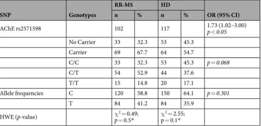
Table 1 compares the genotype and allele frequencies of the AChE rs2571598 in RR-MSSNP Genotypes RR-MS HD OR (95% CI) n % n % AChE rs2571598 102 117 1.73 (1.02–3.00) p < 0.05 No Carrier 33 32.3 53 45.3 Carrier 69 67.7 64 54.7 C/C 33 32.3 53 45.3 p = 0.068 C/T 54 52.9 44 37.6 T/T 15 14.8 20 17.1 Allele frequencies C 120 58.8 150 64.1 p = 0.301 T 84 41.2 84 35.9 HWE (p-value) χp = 0.52 = 0.49; * χp = 0.1*2 = 2.55;
patients versus HD controls. Genotype frequencies is according to the Hardy-Weinberg equilibrium, and no sta-tistical significant differences were found in either genotype or allele frequencies distributions.
However, carrying C/T or T/T genotypes were more frequent in RR-MS patients than in HD controls (67.7% vs 54.7%; p < 0.05). The crude OR was 1.73 (95% CI: 1.02–3.00).
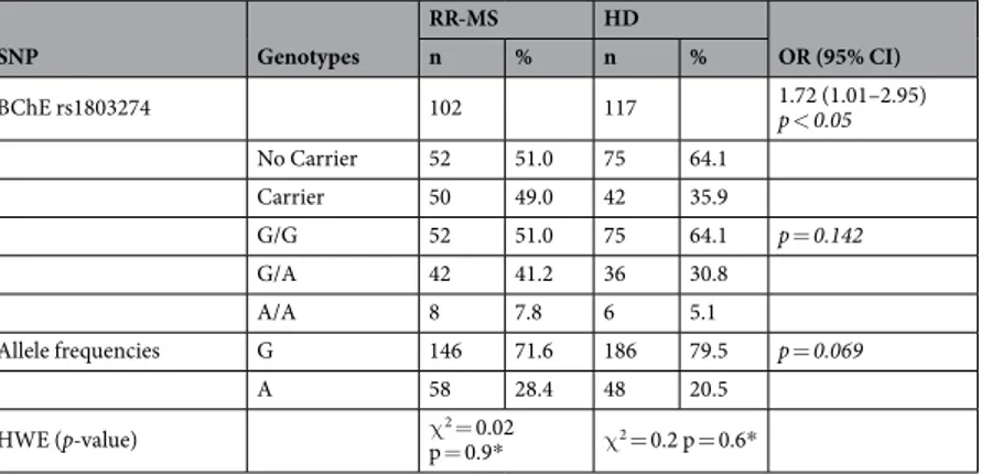
Similarly, when comparing the genotype and allele frequencies of the BChE rs1803274 (the so called K-variant) in RR-MS patients versus HD controls, the genotype frequencies is according to the Hardy-Weinberg equilibrium, and no statistical significant differences were found in either genotype or allele frequencies distri-butions. Carrying G/A or A/A genotypes were more frequent in RR-MS patients than in HD control (49.0% vs 35.9%; p < 0.05). The crude OR was 1.72 (95% CI: 1.01–2.95), (Table 2).
Peripheral cholinergic profiles in BChE K-carrier and no-carrier RR-MS patients and HD
sub-jects.
Accordingly with our previous data34, BChE activity in serum was different between RR-MS patientsand HD subjects, being higher in patients than in control (2510.2 ± 227.4 vs 2234.5 ± 192.9; p < 0.001).
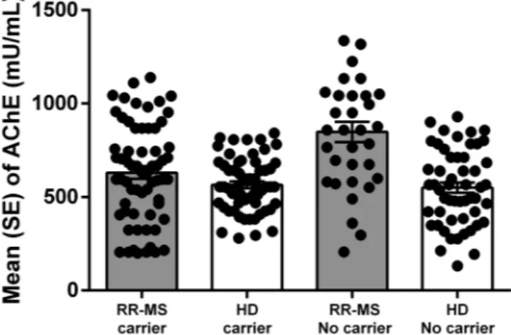
In K-carriers HD groups we observed a reduction of serum BChE activity, while in K-carriers RR-MS patients an increase of serum BChE activity was detected (Fig. 1). Table 3 shows that in HD, BChE activity was about
SNP Genotypes RR-MS HD OR (95% CI) n % n % BChE rs1803274 102 117 1.72 (1.01–2.95) p < 0.05 No Carrier 52 51.0 75 64.1 Carrier 50 49.0 42 35.9 G/G 52 51.0 75 64.1 p = 0.142 G/A 42 41.2 36 30.8 A/A 8 7.8 6 5.1 Allele frequencies G 146 71.6 186 79.5 p = 0.069 A 58 28.4 48 20.5 HWE (p-value) χp = 0.9*2 = 0.02 χ2 = 0.2 p = 0.6*
Table 2. BChE rs1803274 genotype and Allele Frequencies in RR-MS patients and HD subjects.
Figure 1. Levels of BChE enzymatic activity in RR-MS patients and HD stratified for presence of BChE K-polymorphism (carrier) and no-carrier for both polymorphism. (Mean ± Standard error; SE).
BChE rs1803274
RR-MS HD ANOVA p-value
Carrier No Carrier Carrier No Carrier RR-MS vs HD Carrier vs No carrier Interaction BChE (mU/mL) 2757.3 ± 262.6 2418.6 ± 184.1 2121.3 ± 162.2 2348.1 ± 290.7 0.035 0.365 0.089 AChE (mU/mL) 1066.5 ± 843.8 858.2 ± 189.1 420.6 ± 41.6 567.2 ± 145.7 0.012 0.871 0.571 ACh (pmol/mL) 67.7 ± 56.7 98.1 ± 60.1 681.7 ± 418.8 514.0 ± 144.4 <0.001 0.623 0.397 Table 3. Levels of cholinergic markers in BChE K-carrier and no-carrier RR-MS patients and HD subjects. Data are expressed as mean and standard deviation.
10% lower in K-carriers than in no-carriers. In addition, in the HD group, K-carriers had higher levels of ACh (about + 25%), and reduced AChE levels that may be responsible for higher ACh levels observed.
In HD the ratio BChE/ACh was respectively 3.1 in K-carriers, and 4.9 in non-carriers. In these subjects, the ratio AChE/ACh was lower, while the ratio BChE/AChE was not significantly different, when compared to non-carrier subjects. Thus, our observation suggest that in HD, the BChE-K allele might have an impact on the AChE activity and ACh levels.
In RR-MS patients BChE activity was about 14% higher in K-carriers than in non-carriers, while AChE activ-ity is higher in K-carriers than in non-carriers. The K-carrier RR-MS patients had lower levels of ACh and the ratio BChE/ACh was 32.5 and 23.7 in K-carrier and non-carrier patients, respectively.
The ratio BChE/ACh may be indicative of the extra-synaptic ACh equilibrium status, since the higher BChE/ ACh ratio observed in K-carriers compared to non-carrier RR-MS patients plausibly will lead to less degradative activity of BChE, which is however higher compared to HD K-carrier subjects (Table 4).
BChE genotype and cytokine levels.
Since BChE K-variant is associated with reduced ACh-hydrolysing capacity of the enzyme, and considering the increasing evidence of the immune-regulatory role of ACh, we investigated if genetic heterogeneity in BChE may affect pro-inflammatory cytokines. Particularly, we investi-gated whether in RR-MS patients and HD subjects, the K allele was related to serum levels of pro-inflammatory cytokines TNFα, IL-17, IL-18 and IL-12/p40.In HD K-carriers, in accord with lower BChE-hydrolysing capacity and higher ACh levels, we observed lower TNFα, IL-17, IL-18, and IL-12p40 levels when compared to non-carriers HD (Table 5). Thus in HD, BChE geno-type exerts regulatory effects on pro-inflammatory cytokines, possibly via regulation of extracellular ACh levels. This finding lends to further support the immune-regulatory function of ACh, that may act as possible negative regulator of inflammation caused by the aberrant T-cell activation.
In RR-MS K-carrier patients we observed not significantly differences in inflammatory cytokine levels, prob-ably dependent on the lower ACh levels, as a result of higher levels of both ChE enzymes.
Thus, while BChE K-genotype in HD donors causes the reduction of BChE hydrolysing activity with expected increase of ACh levels and reduced inflammatory environment in the serum, in RR-MS K-carriers the increase of BChE and ACh was associated to an increase of inflammatory cytokines levels.
Peripheral cholinergic profiles in AChE rs2571598 genotype carrier and non-carrier RR-MS
patients and HD subjects.
AChE activity in serum is different between RR-SM patients and HD, showing higher activity in patients (793.5 ± 223.5 vs 561.3 ± 100.2; p < 0.001). In both RR-MS patient and HD groups we observed a reduction of AChE activity in serum of AChE rs2571598 genotype carriers (Fig. 2).In HD carriers of the AChE rs2571598 genotype, ACh levels were 40% higher than in non-carriers. In RR-MS carriers of the AChE rs2571598 genotype, AChE activity was about 30% lower, while ACh levels were significantly higher, than in non-carriers (p = 0.047). The lower levels of ACh, in non-carrier RR-MS patients, probably are modulated by higher AChE and BChE hydrolysing activity (Table 6).
In HD rs2571598 carriers the ratio AChE/ACh is 1.1 and 1.5 in carriers and non-carriers HD, respectively. In RR-MS patients the AChE/ACh ratio is 3.1 and 7.7 in carriers and non-carriers, respectively (Table 7).
AChE genotype and cytokine levels.
In HD rs2571598 carriers, in accord with higher ACh levels, we observed lower TNFα, IL-17, IL-18 and IL-12/p40 levels compared to non-carriers. In addition, RR-MS rs2571598 carriers show lower levels of inflammatory cytokine TNFα and IL-17, and no differences for IL-18BChE rs1803274
RR-MS HD
Carrier No Carrier Carrier No Carrier AChE/ACh 15.8 ± 8.2 7.8 ± 3.0 0.7 ± 2.7 1.2 ± 0.8 BChE/ACh 32.5 ± 16.2 23.7 ± 10.9 3.1 ± 1.5 4.9 ± 2.9 BChE/AChE 2.7 ± 0.9 3.5 ± 0.3 5.0 ± 0.2 4.2 ± 0.3 AChE + BChE/ACh 43.7 ± 27.1 24.2 ± 15.7 3.9 ± 2.7 5.8 ± 2.7
Table 4. Ratio between cholinergic enzymes activity and ACh levels BChE K-carrier and no-carrier RR-MS patients and HD subjects. Data are expressed as mean and standard error.
BChE rs1803274
RR-MS HD ANOVA p-value
Carrier No Carrier Carrier No Carrier RR-MS vs HD Carrier vs no carrier Interaction TNFα (pg/mL) 19.4 ± 2.1 16.9 ± 5.6 2.4 ± 0.9 4.8 ± 3.1 <0.001 0.065 0.564 IL-17 (pg/mL) 41.8 ± 13.9 40.4 ± 22.0 10.4 ± 5.2 16.8 ± 3.8 0.027 0.467 0.768 IL-18 (pg/mL) 357.0 ± 63.5 286.7 ± 41.8 168.2 ± 17.1 205.1 ± 22.0 <0.001 0.787 0.642 IL12/p40 (pg/mL) 329.3 ± 41.2 274.4 ± 15.9 76.7 ± 43.2 109.6 ± 36.9 <0.001 0.678 0.498 Table 5. Levels of cytokines in serum in BChE K-carrier and no-carrier RR-MS patients and HD subjects. Data are expressed as mean and standard deviation.
and IL-12/p40 levels. Thus, AChE genotype exerts regulatory effects on pro-inflammatory cytokines, possibly via regulation of ACh levels in both HD and RR-MS. In RR-MS patients carrying rs2571598 both AChE and BChE hydrolysing activities were reduced, parallel to higher ACh levels. In HD rs2571598 carriers, a not significantly reduction of BChE and AChE activity was responsible of a significant increase of ACh levels that may drive the observed reduction of inflammatory cytokines (Table 8).
Figure 2. Levels of AChE enzymatic activity in in RR-MS patients and HD stratified for presence of AChE rs2571598 (carrier) and no-carrier for both polymorphism. (Mean ± Standard error; SE).
AChE rs2571598
RR-MS HD ANOVA p-value
Carrier No Carrier Carrier No Carrier RR-MS vs HD Carrier vs no carrier Interaction BChE (mU/mL) 2325.9 ± 998.1 2418.6 ± 184.1 2271.7 ± 252.9 2348.1 ± 290.7 0.227 0.654 0.943 AChE (mU/mL) 596.8 ± 543.9 858.2 ± 189.1 556.0 ± 87.2 567.2 ± 145.7 0.015 0.763 0.781 ACh (pmole/mL) 237.1 ± 354.2 98.1 ± 60.1 701.2 ± 345.7 514.0 ± 144.4 0.004 0.047 0.652 Table 6. Levels of cholinergic markers in AChE rs2571598 genotype carrier and no-carrier RR-MS patients and HD subjects. Data are expressed as mean and standard deviation.
AChE rs2571598
RR-MS HD
Carrier No Carrier Carrier No Carrier AChE/ACh 3.1 ± 3.7 7.7 ± 2.4 1.1 ± 1.7 1.5 ± 0.8 BChE/ACh 9.9 ± 8.7 25.2 ± 14.1 3.5 ± 2.2 4.5 ± 3.8 BChE/AChE 3.8 ± 0.5 3.1 ± 0.8 4.1 ± 0.2 4.2 ± 0.1 AChE + BChE/
ACh 13.1 ± 8.7 32.8 ± 12.7 4.8 ± 0.7 5.5 ± 3.7
Table 7. Ratio between cholinergic enzymes activity and ACh levels in AChE rs2571598 genotype RR-MS patients and HD subjects. Data are expressed as mean and standard error.
AChE rs2571598
RR-MS HD ANOVA p-value
Carrier No Carrier Carrier No Carrier RR-MS vs HD Carrier vs no carrier Interaction TNFα (pg/mL) 14.3 ± 3.6 16.9 ± 5.6 1.6 ± 1.2 4.8 ± 3.1 <0.001 0.051 0.617 IL-17 (pg/mL) 37.2 ± 18.2 40.4 ± 22.0 9.7 ± 4.8 16.8 ± 3.8 0.009 0.047 0.987 IL-18 (pg/mL) 283.6 ± 52.0 286.7 ± 41.8 168.9 ± 12.9 205.1 ± 22.0 <0.001 0.465 0.632 IL-12/p40(pg/mL) 276.4 ± 33.2 274.4 ± 15.9 80.4 ± 47.6 109.6 ± 36.9 <0.001 0.871 0.596 Table 8. Cytokines levels in AChE rs2571598 genotype carrier and no-carrier RR-MS patients and HD subjects. Data are expressed as mean and standard deviation.
Discussion
Increasing evidences suggested a role for the cholinergic system in the regulation of the inflammatory pathway. One of the consequences of changes in the ACh equilibrium concerns the immune-regulatory function of cho-linergic signalling through the action of ACh on cells involved in native and adoptive immune responses33–35.
Neuroinflammation in MS brain, initially described as an accumulation of leukocytes, is based upon and is regu-lated by bidirectional communication pathways involving cytokines that connect the CNS and immune system. Several studies have reported high levels of inflammatory cytokines in MS and have hypothesized the influences of circulating cholinesterases and ACh levels in the serum34,36,37. Thus, we have investigated, for the first time,
the relationships between the over-production of circulating cholinesterases, the genetic variations of BChE and AChE genes and the levels of circulating cytokines in MS patients.
Our results showed that RR-MS patients have higher serum levels of BChE and AChE compared to HD sub-jects, and this could explain the reduced amount of circulating ACh observed in patients. We investigated if lower ACh concentration observed in RR-MS compared to HD may be related to the ability to regulate the extracellular ACh, when needed, or to the variation of AChE or BChE activity.
Data showed that AChE rs2571598 and BChE rs1803274 genotype were more frequent in RR-MS patients than in HD control. Results highlighted that in RR-MS patients carrying the K-allele, higher BChE levels were observed, albeit it is in contrast with the reported role of this SNP in BChE gene, and that this over-load of circu-lating BChE remove ACh, but not sufficiently to reduce efficiently levels of all examined cytokines. Probably, other mechanisms, such as increased transduction of BChE, microRNAs or other genetic factors acting in conjunction with BChE-K, could be involved in the balance of the BChE. The reduced peripheral cholinergic activity, detected in K-carrier HD subjects, may be responsible for the higher ACh levels and for reduction of pro-inflammatory cytokines analysed. The differential levels of ACh detected in K-carriers subjects seem to reflect a physiological adaptation to the ACh-hydrolysing status. Thus, as a relative index for the extracellular ACh equilibrium state, we calculated the ratio of AChE or BChE activity to ACh in the serum of MS K-carrier or no-carrier patients, com-pared to age-matched controls. An higher BChE + AChE/ACh ratio should indicate a high ACh-hydrolysing sta-tus and/or a low ACh-synthesizing stasta-tus. We found that this ratio is higher in the BChE K-carriers RR-MS group than in non-carriers RR-MS group. In the same group we observed higher levels of the inflammatory cytokines, related to reduced ACh levels. In HD group expressing BChE K-variant, the BChE + AChE/ACh ratio is lower than in non-carriers HD group. When comparing the BChE + AChE/ACh ratio in both RR-MS and HD BChE K-carriers, the ratio resulted higher in patients versus control, in accord with higher inflammatory cytokines levels. This observation strengthen the role of ACh in the control of inflammation21,36,38.
In addition our data showed that the BChE K-allele is associated with an increase of circulating AChE in RR-MS, confirming a close relation between the expression of BChE and that of AChE which has long been known39. HD show, as expected, increased levels of ACh when the serum levels of BChE and AChE are lower.
Albeit AChE rs2571598 genotype results at least associated with reduced serum AChE levels, more evident in RR-MS patients. The single nucleotide substitution may alter hydrolizing properties, possible for misfolded con-formation and con-formation of a unstable concon-formation23. We found that in both RR-MS patients and HD groups,
ACh levels were higher in subjects carrying the polymorphic allele, while no significantly differences in BChE lev-els were observed. This suggests that AChE rs2571598 genotype may be involved in the lower levlev-els of cytokines detected in serum of AChE rs2571598 carrier subjects, accordingly to lower Ach degradation. This could indicate that AChE genotype may affects the production of inflammatory mediators via regulation of the extracellular levels of ACh. Taken together these data demonstrate that in RR-MS patients the pro-inflammatory immune responses through hydrolysis of acetylcholine could be related to alterations in ChE activity, depending on spe-cific genetic endophenpotypes.
In conclusion, we confirmed that total plasma BChE and AChE activity are higher in RR-MS patients than in healthy controls, an increased frequency of the AChE rs2571598 and BChE rs1803274 in MS patients as com-pared to controls was found, the plasma ChE activity is associated with ACh levels and with inflammatory status. Cholinergic system modifications, observed in the periphery could reflect what happens in the CNS, being relevant for immune activation/inflammation, underlining that MS may be considered a systemic disease, where a cross-talk between the periphery and the CNS could have an undescribed crucial role. Furthermore, we propose that serum BChE and AChE activity may be used as a secondary markers to assess the role of non-neuronal cho-linergic system in regulating peripheral inflammation via ACh regulation. However, studies on larger populations are needed to confirm the role of non-neuronal cholinergic system in immune cells to better understand MS aetiology and progression and to develop new disease-modifying therapies for MS, with ChE and ACh as targets.
Methods
Subjects.
Relapsing Remitting-MS (RR-MS) patients were enrolled and followed at Department of Neuroscience of S. Camillo-Forlanini Hospital (Rome, Italy). The diagnosis of RR-MS was confirmed according to revised Mc Donald Diagnostic Criteria40 and RR-MS course was established by clinical parameters41 inagree-ment with recent classification42. Enrolled patients haven’t been exposed to prior immunomodulatory therapies,
such as interferon beta preparations, glatiramer acetate and monthly pulses of intravenously-administered sol-umedrol; however, they were off these therapies for a minimum of 6 months before the blood collection.
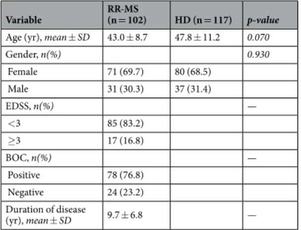
Healthy donors (HD) were enrolled from the Transfusion Blood Bank Services of Chieti (Italy) and were frequency matched for age and gender. Mean age, mean disease duration and EDSS distribution are shown in Table 9. All subjects had not suffered, in the previous month, from inflammatory diseases that might be asso-ciated with modulation of cytokines, and had not received corticosteroids or immunosuppressive drugs. All RR-MS patients and HD signed an informed consent. The study was approved by Ethical Committee of San Camillo-Formanini Hospital (prot. n 1457/2016).
Determination of ACh levels, AChE and BChE Activity.
ACh was measured by commercial fluori-metric kit (Abcam, Cambridge, UK), using the Glomax Multi Detection System (Promega, Mi, Italy) at λ Ex/Em 535/587 nm.Cholinesterases activity were measured in sera of RR-MS patients and HD by Ellman assay43, using 1 mM final
concentration of acetyl-thiocholine iodide as substrate. In order to evaluate the contribution of AChE and BChE to the total cholinesterase activity, 1.4 × 10⁻⁵ M BW284c51 or 1.4 × 10⁻⁵ M lysivane were respectively added as
appropriate inhibitors, in the reaction mixture containing 0.33 mM DTNB (di-nitro-thiocyanobenzene) in 0.1 M phosphate buffer, pH 7. Enzyme activity was expressed as mU; 1 mU corresponding to 1 nmole of substrate hydrolysed/min at 30 °C.
Genotyping.
Genomic DNA was extracted from whole blood using the NucleoSpin Tissue kit (Macherey-Nagel, Bethlehem, PA, USA). Genotyping was performed with being blind to case control status for the specific polymorphisms reported in Table 10. The amplification reaction conditions were as follows: denatur-ation at 94 °C for 10 min followed by 30 cycles at 94 °C for 30 sec, 30 sec at the annealing temperature optimum for each pair of primers (Table 10), 72 °C for 30 sec, with a final elongation step at 72 °C for 10 min.The specificity of the PCR amplified products was evaluated by 2% agarose gel electrophoresis. The gene pol-ymorphisms were observed by direct sequencing on ABI 3130xl genetic analyzer (Applied Biosystems), on both strands.
Hardy-Weinberg equilibrium (HWE) deviations in the genotype frequency distributions were calculated using the χ square analysis. Hardy-Weinberg-equilibrium-calculator software was used to check for Haplotype association analysis (http://www.had2know.com/academics/hardy-weinberg-equilibrium-calculator-2-alleles. html).
Cytokine measurements.
ELISA assay was conducted with commercial kits (R&D System) according to the manufacturer’s instructions to quantify human cytokine levels in serum. The plates were read at 450 nm and the absorbance was converted in pg/ml, using calibration curves prepared with cytokine standards. The intra- and inter-assay reproducibility were > 90%.Statistical analysis.
The quantitative variables were summarized as mean and standard deviation (SD) or median and interquartile range (IQR), according to their distribution. Qualitative variables were summarized as frequency and percentage. A Shapiro-Wilk’s test was performed to evaluate the departures from normality distribution for each variable.The difference in frequencies between patients and controls were assessed by a chi-square test or Fischer exact test when appropriate.
The association between AChE and BChE polymorphisms with SM was assessed by univariate logistic regres-sion analyses. Logistic regresregres-sion model was applied to estimate the adjusted Odds Ratio (OR) and relative 95% confidence intervals.
The analyses of relationship between genotypes and enzymes activity was performed excluding patients carrying both polymorphisms. Patients with only one polymorphism (carrier group) or without both variants (no-carrier group) were analysed. Two-way analysis of variance (ANOVA) was performed to test the effect of
Variable RR-MS (n = 102) HD (n = 117) p-value Age (yr), mean ± SD 43.0 ± 8.7 47.8 ± 11.2 0.070
Gender, n(%) 0.930 Female 71 (69.7) 80 (68.5) Male 31 (30.3) 37 (31.4) EDSS, n(%) — <3 85 (83.2) ≥3 17 (16.8) BOC, n(%) — Positive 78 (76.8) Negative 24 (23.2) Duration of disease (yr), mean ± SD 9.7 ± 6.8 —
Table 9. Characteristics of RR-MS patients and HD subjects.
Polymorphism Primer Forward Primer Reverse Annealing Temperature
BChE rs1803274 (G > A) ATTAGAGACCCACACAACTT ATATTTTACAGGAAATATTGATGAA 55 AChE rs2571598 (C > T) AGAGTCGGGGTCTTGTTATGT AAAGTGAGGAGGAGACGAGG 60 Table 10. Sequences of the primers for BChE rs1803274 and AChE rs2571598.
different AChE and BChE genotypes (carrier or no-carrier), different group (RR-MS or HD) and their interaction on levels of BChE, AChE, ACh and cytokines with group as a fixed effect and genotype as a random effect.
The level of statistical significance was set at p < 0.05. Statistical analysis was performed using IBM
®
SPSS Statistics v 20.0 software (SPSS Inc, Chicago, Illinois, USA).References
1. Olsson, T., Barcellos, L. F. & Alfredsson, L. Interactions between genetic, lifestyle and environmental risk factors for multiple sclerosis. Nat. Rev. Neurol. 13, 25–36 (2017).
2. O’Gorman, C., Lucas, R. & Taylor, B. Environmental risk factors for multiple sclerosis: a review with a focus on molecular mechanisms. Int. J. Mol. Sci. 13, 11718–11752 (2012).
3. Morandi, E., Tarlinton, R. E. & Gran, B. Multiple Sclerosis between Genetics and Infections: Human Endogenous Retroviruses in Monocytes and Macrophages. Front. Immunol. 6, 647 (2015).
4. Kunz, M. & Ibrahim, S. M. Cytokines and cytokine profiles in human autoimmune diseases and animal models of autoimmunity.
Mediators. Inflamm. 2009, 979258 (2009).
5. Harris, V. K. & Sadiq, S. A. Disease biomarkers in multiple sclerosis: potential for use in therapeutic decision making. Mol. Diagn.
Ther. 13, 225–244 (2009).
6. Nizri, E., Hamra-Amitay, Y., Sicsic, C., Lavon, I. & Brenner, T. Anti-inflammatory properties of cholinergic up-regulation: A new role for acetylcholinesterase inhibitors. Neuropharmacology. 50, 540–547 (2006).
7. Pavlov, V. A. et al. Brain acetylcholinesterase activity controls systemic cytokine levels through the cholinergic anti-inflammatory pathway. Brain. Behav. Immun. 23, 41–45 (2009).
8. Borovikova, L. V. et al. Vagus nerve stimulation attenuates the systemic inflammatory response to endotoxin. Nature. 405, 458–462 (2000).
9. Pavlov, V. A. & Tracey, K. J. The cholinergic anti-inflammatory pathway. Brain. Behav. Immun. 19, 493–499 (2005).
10. Pavlov, V. A., Wang, H., Czura, C. J., Friedman, S. G. & Tracey, K. J. The cholinergic anti-inflammatory pathway: a missing link in neuroimmunomodulation. Mol. Med. 9, 125–134 (2003).
11. Kawashima, K. & Fujii, T. The lymphocytic cholinergic system and its contribution to the regulation of immune activity. Life. Sci. 74, 675–696 (2003).
12. Grando, S. A., Kawashima, K. & Wessler, I. Introduction: the non-neuronal cholinergic system in humans. Life. Sci. 72, 2009–2012 (2003).
13. Wessler, I. & Kirkpatrick, C. J. Acetylcholine beyond neurons: the non-neuronal cholinergic system in humans. Br. J. Pharmacol.
154, 1558–1571 (2008).
14. Taylor, P. & Radić, Z. The cholinesterases: from genes to proteins. Annu. Rev. Pharmacol. Toxicol. 34, 281–320 (1994).
15. Grisaru, D., Sternfeld, M., Eldor, A., Glick, D. & Soreq, H. Structural roles of acetylcholinesterase variants in biology and pathology.
Eur. J. Biochem. 264, 672–686 (1999).
16. Soreq, H. & Seidman, S. Acetylcholinesterase-new roles for an old actor. Nat. Rev. Neurosci. 2, 294–302 (2001). 17. Darvesh, S., Hopkins, D. A. & Geula, C. Neurobiology of butyrylcholinesterase. Nat. Rev. Neurosci. 4, 131–138 (2003).
18. Camp, S. et al. Contribution of selective knockout studies to understanding cholinesterase deposition and function. Chem. Bioch.
Int. 187, 72–77 (2010).
19. Ballard, C. G., Greig, N. H., Guillozet-Bongaarts, A. L., Enz, A. & Darvesh, S. Cholinesterases: roles in the brain during health and disease. Curr. Alzheimer. Res. 2, 307–318 (2005).
20. Darvesh, S. et al. Butyrylcholinesterase activity in multiple sclerosis neuropathology. Chem. Biol. Interact. 187, 425–431 (2010). 21. Das, U. N. Acetylcholinesterase and butyrylcholinesterase as possible markers of low-grade systemic inflammation. Med. Sci. Monit.
13, RA214–221 (2007).
22. Darreh-Shori, T. et al. Functional variability in butyrylcholinesterase activity regulates intrathecal cytokine and astroglial biomarker profiles in patients with Alzheimer’s disease. Neurobiol. Aging. 34, 2465–2481 (2013).
23. Scacchi, R., Ruggeri, M. & Corbo, R. M. Variation of the butyrylcholinesterase (BChE) and acetylcholinesterase (AChE) genes in coronary artery disease. Clin. Chim. Acta. 412, 1341–1344 (2011).
24. Lehmann, D. J., Johnston, C. & Smith, A. D. Synergy between the genes for butyrylcholinesterase K variant and apolipoprotein E4 in late-onset confirmed Alzheimer’s disease. Hum. Mol. Genet. 6, 1933–1936 (1997).
25. Lehmann, D. J., Nagy, Z., Litchfield, S., Borja, M. C. & Smith, A. D. Association of butyrylcholinesterase K variant with cholinesterase-positive neuritic plaques in the temporal cortex in late-onset Alzheimer’s disease. Hum. Genet. 106, 447–452 (2000). 26. Darreh-Shori, T. et al. Long-lasting acetylcholinesterase splice variations in anticholinesterase-treated Alzheimer’s disease patients.
J. Neurochem. 88, 1102–1113 (2004).
27. Darreh-Shori, T., Siawesh, M., Mousavi, M., Andreasen, N. & Nordberg, A. Apolipoprotein ε4 modulates phenotype of butyrylcholinesterase in CSF of patients with Alzheimer’s disease. J. Alzheimers. Dis. 28, 443–458 (2012).
28. Rubinstein, H. M., Dietz, A. A. & Lubrano, T. E1k, another quantitative variant at cholinesterase locus 1. J. Med. Genet. 15, 27–29 (1978).
29. Bartels, C. F. et al. DNA mutation associated with the human butyrylcholinesterase K-variant and its linkage to the atypical variant mutation and other polymorphic sites. Am. J. Hum. Genet. 50, 1086–1103 (1992).
30. Lopez, O. L. et al. Cholinesterase inhibitor treatment alters the natural history of Alzheimer’s disease. J. Neurol. Neurosurg. Psychiatry.
72, 310–314 (2002).
31. Yoon, H. et al. Association of the choline acetyltransferase gene with responsiveness to acetylcholinesterase inhibitors in Alzheimer’s disease. Pharmacopsychiatry. 48, 111–117 (2015).
32. Scacchi, R., Gambina, G., Moretto, G. & Corbo, R. M. Variability of AChE, BChE, and ChAT genes in the late-onset form of Alzheimer’s disease and relationships with response to treatment with Donepezil and Rivastigmine. Am. J. Med. Genet. B.
Neuropsychiatr. Genet. 150B, 502–507 (2009).
33. Reale, M. et al. Nicotinic receptor activation negatively modulates pro-inflammatory cytokine production in multiple sclerosis patients. Int. Immunopharmacol. 29, 152–157 (2015).
34. Di Bari, M. et al. Dysregulated Homeostasis of Acetylcholine Levels in Immune Cells of RR-Multiple Sclerosis Patients. Int. J. Mol.
Sci. 17, pii: E2009 (2016).
35. Hosoi, T. & Nomura, Y. Functional role of acetylcholine in the immune system. Front. Biosci. 9, 2414–2419 (2004).
36. Reale, M. et al. Relation between pro-inflammatory cytokines and acetylcholine levels in relapsing-remitting multiple sclerosis patients. Int. J. Mol. Sci. 13, 12656–12664 (2012).
37. Nizri, E. et al. Activation of the cholinergic anti-inflammatory system by nicotine attenuates neuroinflammation via suppression of Th1 and Th17 responses. J. Immunol. 183, 6681–6688 (2009).
38. Jiang, W. et al. Acetylcholine-producing NK cells attenuate CNS inflammation via modulation of infiltrating monocytes/ macrophages. Proc. Natl. Acad. Sci. USA pii: 201705491 [Epub ahead of print] (2017).
39. Robitzki, A., Mack, A., Hoppe, U., Chatonnet, A. & Layer, P. G. Regulation of cholinesterase gene expression affects neuronal differentiation as revealed by transfection studies on reaggregating embryonic chicken retinal cells. Eur J Neurosci. 9, 2394–2405 (1997).
40. Polman, C. H. et al. Diagnostic criteria for multiple sclerosis: 2010 revisions to the McDonald criteria. Ann. Neurol. 69, 292–302 (2011).
41. Miller, D. H. et al. Differential diagnosis of suspected multiple sclerosis: a consensus approach. Mult. Scler. 14, 1157–1174 (2008). 42. Lennon, V. A. et al. A serum autoantibody marker of neuromyelitis optica: distinction from multiple sclerosis. Lancet. 364,
2106–2112 (2004).
43. Ellman, G. L., Courtney, K. D., Andres, V. Jr. & Feather-Stone, R. M. A new and rapid colorimetric determination of acetylcholinesterase activity. Biochem. Pharmacol. 7, 88–95 (1961).
Acknowledgements
This study was supported by grants from FISM—Fondazione Italiana Sclerosi Multipla—Cod. 2013/R/25.
Author Contributions
M.R. conceived and designed the study, interpreted the data and wrote the manuscript; E.C. and C.D.A. performed enzymes and cytokines measurements; M.D.N. performed statistical analyses; S.F. participated in DNA extraction and genotyping; M.D.A. participated in genotyping and data analysis and draft the manuscript; M.D.B.; S.G. and V.O. measured the cholinergic markers; S.R. and C.G. collected the blood and serum from M.S. patients; L.S. revised the manuscript for important intellectual content; A.M.T. participated in study design and revising the manuscript critically for important intellectual content; V.G. participated in study design and coordination, data analysis and interpretation and wrote the manuscript. All authors read and approved the final manuscript.
Additional Information
Competing Interests: The authors declare that they have no competing interests.
Publisher's note: Springer Nature remains neutral with regard to jurisdictional claims in published maps and institutional affiliations.
Open Access This article is licensed under a Creative Commons Attribution 4.0 International License, which permits use, sharing, adaptation, distribution and reproduction in any medium or format, as long as you give appropriate credit to the original author(s) and the source, provide a link to the Cre-ative Commons license, and indicate if changes were made. The images or other third party material in this article are included in the article’s Creative Commons license, unless indicated otherwise in a credit line to the material. If material is not included in the article’s Creative Commons license and your intended use is not per-mitted by statutory regulation or exceeds the perper-mitted use, you will need to obtain permission directly from the copyright holder. To view a copy of this license, visit http://creativecommons.org/licenses/by/4.0/.