Università degli Studi di Ferrara
DOTTORATO DI RICERCA IN
BIOCHIMICA, BIOLOGIA MOLECOLARE E BIOTECNOLOGIE
CICLO XXIVCOORDINATORE Prof. BERNARDI FRANCESCO
Biological activity of anti-miR-221 Peptide
Nucleic Acids and relative building blocks
Settore Scientifico Disciplinare BIO/10
Dottoranda Tutore
Preface
The study of new molecules able to selectively and stably interact with DNA and RNA is a field of great interest in consideration all the possible applications in medicine. In the last years, many studies have been conducted aimed to the explanation of epigenetic modifications (DNA methylation, histone modification and expression of non-coding RNA molecules, as microRNA) important in the mechanisms of gene regulation; in fact this is one of the main steps for understanding the biology in both human conditions, normal and pathological, and for the development of new bioactive molecules. RNA has been the preferred target of long-standing studies aimed at the discovery of molecules able to block gene expression in a sequence selective way.
During the past few years, molecular biologists have discovered hundreds of gene sequences that encode small RNA molecules, the microRNAs (miRNAs), 21 to 25 nucleotides in length, which are involved in the post-transcriptional regulation of gene expression. Indeed, there are hints that the level of some miRNAs is altered in cancer and in other pathologies; for instance, miRNAs regulate cancer-promoting genes, playing a pivotal role in cancer onset and progression. Briefly, microRNAs recognizing a target sequence in the 3'UTR of the mRNA target, and based on the partial or total complementarity of the sequences, they bind to mRNA leading to the inhibition of translation or degradation of the mRNA, respectively. Furthermore, in the pathogenesis of tumors, miRs play a double role; in fact, if it interferes with an mRNA coding for a tumor-suppressor protein, it become "oncogenes", applying itself as a target for anticancer therapy.
Synthetic oligonucleotides or analogues acting as competitors by binding to miRNA have been proposed as novel potential drugs. Among the most efficient molecules proposed for these applications are the PNAs. Peptide nucleic acids (PNAs) are oligonucleotide analogues with a polyamide backbone and are very promising tools for binding RNA, since they have a higher affinity for RNA than DNA, are stable to nucleases and are very specific. Their use as therapeutic agents have been proposed since early studies and recent advancements in cellular delivery systems; anti-gene and antisense strategies make them good candidates for drug development.
In this contest, we have selected the miR-221 as target for the ours novel engineered PNAs; miR-221 have been shown to be overexpressed in many different types of tumor, as glioblastoma, hepatocarcinoma, prostate and breast cancer; moreover, it had been shown to be associated to the suppressor of p27kip1, a cell cycle inhibitor and tumor suppressor protein. In fact, it had been found that in the 3’UTR of p27kip1 mRNA there are at least two binding sites for miR-221.
The major limit of the PNAs technology is the low cellular uptake, in particular on eukaryotic cells; in order to solve this problem, several approaches have been proposed; our PNAs presents modifications of the backbone with positive charged groups; in fact it has been demonstrated that these modifications are able to enhance cellular uptake and consequently PNA efficiency. Another modification that we have insert in the PNAs employed in our experiments is to link to PNAs a polyarginine (R) tails, based on the observation that this cell-membrane penetrating oligopeptides are able to facilitate uptake of conjugated molecules.
The anti-miR-221 PNAs (PNA-a221 and Rpep-PNA-a221) proposed in this PhD thesis were synthetized by Prof. Corradini, Prof. Marchelli and collaborators at University of Parma, Department of Organic and Industrial Chemistry. They first designed the synthesis of new class of uracil dimers by substitution of amide linker on C(5) with more flexible methylamine linker, obtaining the 5-methylamino uracil. This reaction intermediate had been used for the synthesis of the 5-methylazidouracil PNA monomer. The R8 peptide was linked to the N-term of PNA, following the synthesis on solid phase.
The results presented in this PhD Thesis clearly show that: (a) the polyarginine-PNA conjugated anti-miR-221 is efficient internalized in the target cell lines without any transfection reagents; (b) targeting miR-221 by PNA resulted in a lowering of level of miR-221 and up-regulation of p27kip1 mRNA and protein in breast cancer MDA-MB-231 cell line. On the other hand, protocols of medicinal chemistry finalized to the design and the production of the final form of bioactive molecules, generate intermediates during chemical synthesis that, at least in theory, might retain effects on biological function. Therefore, in order to maximize the production of bioactive agents, all the synthetic intermediates deserve attention at least in a first screening.
In our case, the uracil dimers derivatives, employed during the synthesis of PNA-anti-miR-221 were considered; in addition we also considered the C(5) uracil
modified monomers, used as starting molecules for the synthesis of dimers. As a first explorative investigation, we analyzed the possible antitumor activity of their analogs, possibly associated, as found in several other antitumor agents, with activation of terminal erythroid differentiation. We have first evaluated the antiproliferative and induction of erythroid differentiation activities of dimers; our results are published in Accetta et al. (2009) and briefly described in the Introduction Part 1 of this PhD Thesis. We have demonstrated for the first time that this kind of uracil derivatives can be considered to be a new class of erythroid differentiation inducers.
Following this initial results, we have analyzed also the C(5) uracil derivative monomers; we present the Results Part 1 of this thesis. We have focused our attention in particular on Compound 9, which between all, has proved to be a potent erythroid inducer on K562 cell line. In this respect, it’s widely demonstrated that inducers of K562 erythroid differentiation are often in erythroid cell isolated from beta- thalassemia patients.
Contents Part 1
1. INTRODUCTION
1.1 The pyrimidine ... 3
1.1.1 Biological Activity of pyrimidine derivatives ... 3
1.1.2 C(5)-substituted uracil derivatives ... 4
1.1.3 Synthesis of 5-carboxamido uracil dimmers... 5
1.2 Terminal differentiation therapy of human cancer ... 7
1.2.1 Cell differentiation ... 9
1.2.2 The differentiation therapy ... 10
1.2.3 Antitumor agents acting through induction of terminal erythroid differentiation ... 14
1.2.3.1 Cytotoxic agents... 14
1.2.3.2 Hydroxyurea... 15
1.2.3.3 Histone deacetylases inhibitors ... 16
1.2.3.4 DNA-binding drugs ... 16
1.2.3.5 HbF inducers from natural world ... 17
1.2.4 K562 cell line as useful experimental model for erythroid differentiation ... 20
1.3 New uracil dimmers showing erythroid differentiation inducing activities ... 21
1.3.1 Screening of anti-proliferative and differentiation activity... 21
1.3.2 Conclusions... 22
1.4 C(5) modified uracil derivatives showing antiproliferative and erythroid differentiation inducing activities on human chronic myelogenous leukemia K562 cells ... 23
1.4.1 Chemical synthesis of uracil monomers ... 23
1.5 Aim and thesis outlook ... 26
2. MATHERIALS AND METHODS 2.1 Cell culture: human erythroleukemic K562 cell line... 27
2.2 C(5) modified uracil monomers... 27
2.3 Antiproliferative activity ... 28
2.4 Benzidine assay ... 28
2.5 Transfection of K562 cells with fluorescence protein genes under the γ-globin and the β-globin gene promoters ... 28
2.6 RNA isolation ... 29
2.6.2 RNA electrophoresis on agarose gel ... 30
2.7 Reverse transcription reaction- Random Hexamer ... 30
2.8 Real-Time Quantitative Polymerase Chain Reaction ... 30
2.9 High Performance Liquid Chromatography ... 31
2.10 FACS analysis ... 32
2.11 Measurement of apoptosis ... 32
2.11.1 DeadEndTM Colorimetric TUNEL System ... 32
2.11.2 Annexin V/PI release assay ... 33
2.11.3 Cell cycle analysis by FACS ... 34
3. RESULTS 3.1 Antiproliferative and erythroid differentiating activities of the uracil monomers ... 35
3.2 Effects of compound 9 on proliferation and erythroid differentiation of K562 cells ... 36
3.3 Effects of compound 9 on the transcriptional activity of the γ-globin and the β-globin gene promoters of K562 cells ... 39
3.4 Effects of compound 9 on biochemical parameters associated to the activation of the K562 erythroid phenotype ... 40
3.5 Effect of compound 9 on hemoglobin accumulation measured by HPLC ... 42
3.6 The induction of erythroid differentiation of compound 9 is not associated with the activation of the apoptotic pathway... 42
4. DISCUSSION AND CONCLUSIONS (1) ... 45
5. REFERENCES... 47
Contents Part 2
1. INTRODUCTION 1.1 Peptide Nucleic Acids... 551.1.1 PNAs: structure and properties... 56
1.1.2 Strand invasion and PNAs biological applications ... 57
1.1.3 PNAs as bioactive molecules for gene expression control ... 58
1.1.4 Modified PNAs and PNAs delivery ... 60
1.1.5 Synthesis of modified peptide nucleic acids monomers... 63
1.2 Targeting microRNA involved in human disease
1.2.1 MicroRNA ... 67
1.2.2 microRNA biological relevance: involvement in the gene expression control ... 68
1.2.3 Biogenesis of microRNAs ... 68
1.2.4 MicroRNA and gene regulation... 69
1.2.5 microRNA and cancer ... 71
1.2.5.1 microRNA in breast cancer ... 76
1.2.5.2 Tumor suppressor miRNA in breast cancer ... 76
1.2.5.3 Oncogenic miRNA in breast cancer ... 77
1.2.6 Antisense strategy and miR targeting... 78
1.2.6.1 MiR targeting by PNAs... 80
1.3 Cell cycle deregulation in human breast cancer 1.3.1 Cell cycle ... 81
1.3.2 Cyclins and CDKs ... 82
1.3.2.1 Cyclin D1-CDK4/6... 84
1.3.2.2 Cyclin E-CDK2 ... 85
1.3.3 Cyclin Kinase Inhibitors... 86
1.3.4 Cyclin Kinase Inhibitor p27kip1 ... 87
1.3.4.1 Regulation of p27 kip1 ... 89
1.3.4.2 p27 kip1 deregulation in breast cancer ... 90
1.3.5 Regulation of cell cycle factors by microRNAs in human cancer... 92
1.4 Human breast cancer MDA-MB-231 and MCF-7 cell lines as experimental model to study the miR-221 and p27kip1 modulation ... 95
2. MATHERIALS AND METHODS 2.1 Human breast cancer cell lines... 98
2.2 Anti-miR-221 PNAs ... 98
2.3 PNAs antiproliferative activity on MDA-MB-231 and MCF-7 cell lines... 99
2.4 Cellular uptake of PNAs ... 99
2.4.1 Fluorescence-activated cell sorting... 99
2.4.2 BioStation IM technology ... 100
2.5 RNA isolation ... 101
2.6 Reverse transcription reaction ... 101
2.6.1 RT with Random Primers ... 101
2.6.2 RT for microRNA ... 101
2.8 Nuclear and cytoplasmic proteins extraction ... 102
2.8.1 Protein extracts quantification: Bradford Assay ... 103
2.9 Western Blot Assay ... 103
3. RESULTS 3.1 Uptake and biostability of PNAs in breast cancer cell lines ... 105
3.1.1 Treatment with trypsin... 105
3.1.2 Culture w/ or w/o FBS ... 106
3.1.3 Cellular uptake in MDA-MB-231 and MCF-7 cell lines ... 107
3.1.4 Cellular uptake by BioStation IM ... 109
3.2 Antiproliferative activity of PNA-a221 and Rpep-PNA-a221 ... 110
3.3 Rpep-PNA inhibitory effects on miR-221 analyzed by qPCR assay ... 111
3.4 p27kip1 regulation by PNA anti-miR-221 ... 112
3.4.1 Analysis of accumulation of p27kip1 mRNA... 113
3.4.2 Western Blot analysis of p27kip1 ... 114
4. DISCUSSION AND CONCLUSIONS (2) ... 115
5. REFERENCES... 117
GENERAL CONCLUSIONS ... 126
PART 1
URACIL DERIVATIVED SHOWING
ANTIPROLIFERATIVE AND ERYTHROID DIFFERENTIATION
- INTRODUCTION -
1.1 Pyrimidine
Pyrimidine is a heterocyclic aromatic organic compound similar to benzene and pyridine, containing two nitrogen atoms at positions 1 and 3 of the six-member ring, as shows in Figure 1 (Gilchrist et al., 1997). Three nucleobases found in nucleic acids, cytosine (C), thymine (T), and uracil (U), are pyrimidine derivatives. In DNA and RNA, these bases form hydrogen bonds with their complementary purines. Thus, in DNA, the purines adenine (A) and guanine (G) pair up with the pyrimidines thymine (T) and cytosine (C), respectively.
Figure 1. Chemical structure of natural pyrimidine
The pyrimidine system is a very important pharmacophor core of naturally occurring and synthetic bioactive compounds (Lagoja et al., 2005); the potential of pyrimidine compounds is linked to the possibility of being used as antagonists in the biosynthetic pathways of pyrimidine nucleobases or in other important processes, by competing for the same binding sites of naturally occurring compounds.
1.1.1 Biological activity of pyrimidine derivatives
A series of modified pyrimidines were and are currently used as drugs. For example, fluoropyrimidine drugs such as 5-fluorouracil (5-FU) and capecitabine are a mainstay in the treatment of numerous solid tumors, alone or as part of combination
therapies. Despite the high interpatient pharmacokinetic variability and the cytotoxic effect, 5-FU is currently used in cancer therapeutics, in particular for colorectal and gastric cancer (Mercier et al., 2006).
Azidothymidine (AZT) was the first applied drug for HIV treatment. AZT is phosphorylated by intracellular kinases to AZT-5'- triphosphate (AZT-5’-3P), which is the active metabolite. AZT-5’-3P is able to interferes with the RNA-dependent-DNA- polymerase (reverse transcriptase), acts as viral inhibitor. It can also be incorporated into the growing viral DNA, acting as a terminator signal.
Another example, bacimethrin (4-amino-5-hydroxymethyl-2- methoxypyrimidine) is known as the simplest pyrimidine antibiotics, acting as antagonist on bacterial thiamine biosynthesis (Reddick et al., 2001).
In literature it is also reported that uracils derivatives are potentially biologically active compounds. For examples, oxime libraries based on dimeric uracil derivatives have been proposed for the development of uracil DNA glycosylase (UNG) inhibitors (Jiang et al., 2005). As described by Maruyama et al., 1,3-disubstituted uracil derivatives have shown an antiviral activity against HIV-1 and human cytomegalovirus (HCMV), probably interacting with the amino acid residues of HIV-1 reverse transcriptase (Maruyama et al., 2007).
Another class of molecules, obtained by Isobe et al., the N(1)- and N(3)-uracil derivatives, have shown an anti-inflammatory activities via inhibition of the picryl chloride-induced contact hypersensitivity reaction (CHR) in mice. These activities were essentially equipotent with that of tacrolimus, a strong immunosuppressant. (Isobe et al., 2003). Another pyrimidine analogs, 1-Benzyl derivatives of 5- (arylamino)-uracil, exhibit promising inhibitory activity against HIV-1 in CEM-SS culture, and activity anti-EBV in АKАТА cell culture. (Mikhail et al., 2010).
1.1.2 C(5)-substituted uracil derivatives
The pyrimidine bases of RNA are uracil (U) and cytosine (C), while thymine (T) and C are used for DNA. The C(5) position of C and U is unsubstituted, whereas the C(5) of T is substituted with a Me group. Miller et al. hypothesized that various C(5)- substituted uracil derivatives were formed during chemical evolution, and that C(5)- substituted U derivatives may have played important roles in the transition from an
“RNA world” to a “DNA-RNA-protein world” (Robertson et al., 1995). Several C(5) uracil derivatives have been described as cytostatic and antiviral compounds; Gazivoda et al., have evaluated the antitumoral and antiviral activities of a new class of C(5) aryl, alkenyl, and alkynyl substituted uracil derivatives. In particular the 5- (phenylethynyl)uracil-2,3-di-O-benzylated l-ascorbic acid derivative have shown selective inhibitory effect toward all tumor cell lines except for cervical carcinoma (HeLa), pancreatic carcinoma (MiaPaCa-2), laryngeal carcinoma (Hep-2), and colon carcinoma (SW 620), and no cytotoxicity to normal human fibroblast (WI 38); while 5-propynyl substituted uracil derivative of l-ascorbic acid have shown inhibitory activity against vesicular stomatitis virus, Coxsackie B4 virus and Sindbis viruses (Gazivoda et al., 2007).
Following our general project, aimed at the synthesis of oligonucleotide analogues, in particular PNA, with modifications able to improve their binding activity (Sforza et al., 2007; Corradini et al., 2007. Corradini, Marchelli and collaborators have designed some uracil dimers, using C(5) modified uracil derivatives, connected with a spacer through the 5-positions (Figure 2).
Figure 2. General scheme of uracil dimers
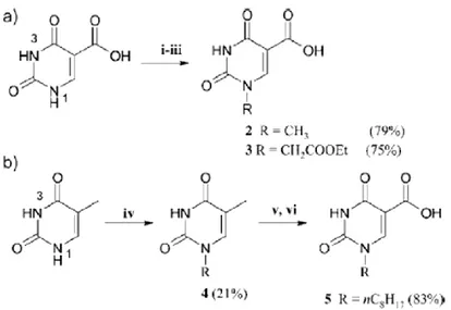
1.1.3 Synthesis of 5-carboxamido uracil dimers
N1-Alkylated uracil derivatives were synthesized from isoorotic acid (uracil-5- carboxylic acid) or thymine. The carboxylic acid derivatives were condensed with diamines in order to produce dimeric compounds or with monoamines in order to obtain reference monomeric compounds. Some of the derivatives, in particular the uracil dimers, were the first to be tested in our laboratories for the erythroid differentiation activity. In the retrosynthetic design, Prof. Corradini and his
collaborators have used as starting material either isoorotic acid (5 uracilcarboxylic acid) or thymine. As a reference compound, isoorotic acid methyl ester was synthesized by reaction with SOCl2 in methanol. Several derivatives were obtained
with the purpose of varying the group at N1, the type (monomeric or dimeric) of the amine residue and the size and rigidity of the spacer, in order to produce chemical diversity. Direct alkylation of isoorotic acid with reactive substrates, such as methyl or allyl halides lead to dialkylated products at both nitrogen atoms; therefore, regioselective mono alkylation of uracil was performed using temporary protection of the carboxylic and carbonyl oxygens with trimethylsilyl groups, through reaction with hexamethyldisilazane (HMDS) in a 3:1 excess and in the presence of trimethylchlorosilane (TMS Cl), as shown in Figure 3.
Reaction of thymine with more hindered long chain primary haloalkanes lead to selective monoalkylation at N(1) (Figure 4), thus affording the subtrate 4 suitable for the synthesis of uracil derivatives with a C8 alkyl chain at N(1). Oxidation of methyl group of 4 with K2S2O8 in the presence of copper(II) leads to the
uracil 5 carboxaldehyde which was then oxidized by reaction with sodium chlorite to the corresponding carboxylic acid 5 bearing a C8 alkyl chain at N1 position.
Figure 3. Synthesis of N1-alkylated isoorotic acid derivatives (a) via regioselective alkylation of Isoorotic acid ((i) HMDS,TMS-Cl, reflux 4 h; (ii) CH3I or ethyl bromoacetate in excess, reflux,18 h; (iii) H2O/ CH3COOH, room temp, 20 min.) and (b) via oxidation of thymine derivatives ((iv) [Br-n-C8H17, NaH, DMF, 80 °C,4 h]; (v) 2,6-lutidine, K2S2O8, CuSO4, H2O/AcCN, 80 °C, 1,5 h; (vi) NaClO2, NaH2PO4; t-BuOH/THF, r.t., 24 h)
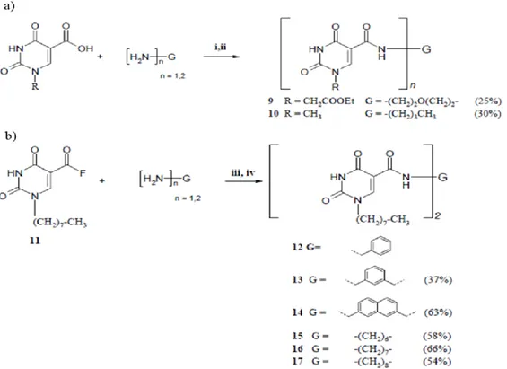
Several other commercially available diamines were used as linkers for the reaction with the carboxylic derivatives. Butylamine was used for generating reference monomeric compound 10 (Figure 4), and benzylamine was used for generating compound 12, both containing a uracil moiety with a carboxamide group at C(5) (Figure 4). The first series of derivatives, containing either a methyl or an ethoxycarbonylmethyl group at N1, was synthesized using reaction with thionyl chloride to generate the corresponding acyl chloride, followed by reaction with the corresponding amine in pyridine (Figure 4a). Since the yields obtained with this method were not optimal (25-30%), mainly because of loss of product during workup, a second series of derivatives was obtained by activation of the carboxylic moiety of 5 (Figure 3) with fluoride using 2,4,6-trifluoro-1,3,5-triazine as fluorinating agent, thus providing the stable intermediate 11 which could be isolated. Subsequent reaction of the acyl fluoride 11 with the corresponding diamine or monoamine in acetonitrile gave the compounds 12-17 (Figure 4b).
Figure 4. Synthesis of monomeric and dimeric uracil derivatives: (a) 12 and 13 via acyl chloride ((i) SOCl2, DMF, 70-80 °C, 2 h; (ii) (H2N)n-G, Py, 2 h); (b) 19-23 via acyl fluoride ((iii) H2N-G-NH2 or H2N-G-NH2. 2HCl, DIEA, AcCN, 80 °C, 7 h; (iv) 1 M HCl, 0.5 h)
We have recently reported a novel class of C(5) linked N(1) alkylated uracil dimers rationally designed to potentially interact with adenine in biological systems: these pyrimidine derivatives have shown antiproliferative and erythroid differentiation activities toward human chronic myelogenous leukemia K562 cells (Accetta et al., 2009). Results are described in the following Section 1.3.
1.2 Terminal differentiation therapy of human cancer
Cancer morbidity and mortality continue to be major problems worldwide, despite significant improvements and innovations in the diagnosis, prevention and therapy of specific cancers, delimiting longevity and the quality of human life. Although the precise reason for the lack of greater efficacy of current therapies is not known, it may relate to the fact that cancer is not caused by a single genetic change, but instead represents the combination of common and distinct multi-step changes in specific neoplasms.
Cancer is a process in which changes in regulating circuits are produced, such as proliferation control, the balance between cellular survival and programmed cellular death (apoptosis), the communication with neighboring cells and extracellular matrix, angiogenesis, and, finally, migration of the tumoral cell, invasion and metastatic dissemination. This process implies the progressive development of a more malign phenotype with an increase of genetic alteration involving genes at several levels of expression during long periods of time. These genetic changes uncouple the normal balance between multiplication and cellular differentiation with an increase in the rate of proliferating cells; it results in the formation of new abnormal cellular products which block final differentiation of cells leading to accumulation of immature cells (Sell et al., 2006). Moreover, the progeny of mutated cell is maintained in a self-renewing tissue stem cell and its immediate progeny or due to cellular components that display stem cell like properties known as cancer
stem cells (CSCs), that grow into cancer. Failure to achieve complete and safe
eradication of cancer is due to the presence of quiescent population of CSCs (Garg, 2009).
Once cancer is diagnoses, a variety of treatment options are considered, mostly depending on tumor type, extent, and location of the cancer lesion. The most
commonly used therapeutic modalities are radiation therapy and chemotherapy. These therapies have proven successful for some tumors; nevertheless, even if classic chemotherapeutical agents have been very important, the mechanism of action of these drugs depends on the cytodestruction of the neoplastic cells, and their beneficial effect are normally accompanied by a notable morbidity, cytotoxicity and drug resistance (both single and multidrug) (Marchel et al., 2006).
The knowledge of the mechanisms involved in differentiation and malignant transformation has allowed the search of alternative routes for anticancer therapy that does not imply cellular death. Example of these alternative routes of cancer therapy are radiation therapy, immunotherapy, differentiation therapy and angiogenesis inhibition therapy (Fisher et al., 1985, 1986; Rosen, 2000).
The objective of these multiple treatment protocols is to eliminate the problems associated with cancer cell resistance to a particular drug or class of drugs and to diminish toxicity associated with high-dose chemotherapy.
1.2.1 Cell differentiation
Cancer begins when a normal cell, which replicates only slightly or not at all, suffers a somatic mutation in a gene and starts to proliferate uncontrollably. Everything indicates that several successive mutations are necessary to change a normal cell into invading carcinoma (Burnet M., 1957) and demonstrates the monoclonal origin of tumors (cellular clone is produced from mutated cell) in which each one of its components reproduces the mutations.
Differentiation program is a complex multistep developmental process of cell specialization that follows the determination (set of progressive restrictions in cell developmental potentials) of a genetic program specific for cell lineage. Development of the differentiation program includes the cellular type and that distinguish the specialized cells, such as muscle cells, nerve and skin. Terminal differentiation is the end stage of this process where the cells irreversibly lose their proliferative capacity and which represents a form of negative control of growing. Regulating molecules interact to produce the correct balance between cellular multiplication and differentiation during embryogenesis and the normal behavior of an adult.
In cancer, neoplastic cells exhibits defects in their ability to differentiate, and the development of these defects in the process of differentiation appears to be an intimate part of the transformation process. Compared with normal cells, tumor cells are less differentiated, which is one of their important biological characteristics. This “undifferentiated state” of tumors is the direct consequence of the “uncontrolled cell proliferation state”: cells are blocked in a stage in which they retain infinite proliferative capably. This hallmark has suggested a novel and potentially less toxic form of cancer therapy involves the use of agents, alone or in combination, that modify the state of differentiation and growth of cancer cells: the differentiation therapy (Fischer et al., 1985).
1.2.2 The differentiation therapy
Because normal terminal differentiation often results in non-proliferating cells that often undergo apoptosis as they complete their normal life span, it was plausible to develop strategies to activate normal pathways of differentiation in premalignant and malignant cells using physiological or pharmacological agents that can bypass the epigenetic and genetic abnormalities that abrogate differentiation. This approach is called differentiation therapy.
Differentiation therapy is based on the concept that cancer cells are normal cells that have been arrested at an immature or less differentiated state, lack the ability to control their own growth, and so multiply at an abnormal fast rate. Differentiation therapy aim to force the cancer cell to resume the process of maturation, so as to prevent, suppress or reverse the malignant phenotype by inducing differentiation with the associated growth arrest, senescence and apoptosis (Figure 5). Although differentiation therapy does not destroy the cancer cell, it restrains their growth and allows the application of more conventional therapy (such as chemotherapy) to eradicate the malignant cells.
Even if only partially successful, differentiation therapy can convert malignant tumors into benign tumors. The mechanism by which differentiation inducing agents cause phenotypic changes in tumor cells is believed to involve the selective activation of defined sets of genes that negatively control cell proliferation and the suppression of genes facilitating expression of the cancer state (Jiang et al., 1994).
By defining the spectrum of genes that are modified as a consequence of induction or irreversible growth arrest, terminal cell differentiation and loss of tumorigenic potential, it should be possible to identify potentially important target genes and molecules for therapeutic intervention in cancer.
The first differentiation agent found to be successful was all-trans-retinoic acid (ATRA) in the treatment of acute promyelocytic leukemia (APL); APL, a distinct subtype of acute myelogenous leukemia (AML), results from the arrest of the maturation of hematopoietic progenitors at the promyelocyte stage. It has been shown that APL is associated with a reciprocal chromosomal translocation, involving chromosomes 15 and 17, which fuses the gene encoding the retinoic acid receptor a (RARa) and the promyelocytic leukemia (PML) gene. The introduction of all-trans retinoic acid (ATRA) in 1987 changed the treatment paradigm of APL (Huang et al., 1988). Huang and collaborators demonstrated for the first time a complete remission of APL following ATRA treatment. Several studies conducted during the early 1990s found that APL patients receiving induction therapy consisting of ATRA followed by chemotherapy fared significantly better than patients treated with chemotherapy alone.
In addition, ATRA therapy has significant and potentially fatal adverse effects, known as retinoic acid syndrome, which consists of elevated white blood cell (WBC) counts, fever, respiratory distress, interstitial pulmonary infiltration, pleural effusion, and weight gain. Among patients in remission following treatment with ATRA alone, the incidence of retinoic acid syndrome is approximately 25%, (Vahdat et al., 1994) whereas that in patients treated with a combination of ATRA plus chemotherapy is reduced to 5%.(Sanz et al., 1999). Based on the results of these studies, the combination of ATRA plus chemotherapy became the standard approach for treating newly diagnosed APL. Most APL patients are actually treated first with ATRA; this compound induces a complete remission in about 70% of cases. ATRA is considered the “prototype” of differentiation therapy agents.
Another example of terminal differentiation induction regarding the therapy of melanoma. Cutaneous malignant melanoma is the fifth most common cancer in the United States and it is also the most frequent cause of death from malignancy in young Caucasian females (Meier et al., 1998).
Figure 5. Targets for differentiation therapy. In a normal differentiation pathway, a hypothetical stem cell designated A is required to undergo a series of progressive changes in gene expression and resultant phenotypic changes represented by the letters B, C and D before reaching the terminally differentiated mature cell stage designated E. Cancer development is associated with aberrant differentiation. This can be represented by a block in differentiation that can occur already in premalignant cells. The scheme shows three possible blocks at late stages of the differentiation pathway (stage D) and at early stages of the pathway (stage B)
Although the focus of intense research, the molecular changes regulating melanoma development and progression remain to be defined, since melanomas are extremely heterogeneous in their genetic background. Numerous studies confirm that melanoma cell exhibits characteristics similar to those observed in de-differentiated cells. Jiang and Fisher (1993) have demonstrated that the treatment of metastatic human melanoma cells with a combination of human fibroblast interferon (IFN-b) and antileukemic compound mezerein (MEZ) results in a loss of tumorigenic potential, irreversible growth arrest, antigenic modulation, enhanced melanin synthesis, profound changes in gene expression and terminal cell differentiation. The mechanism by which IFNs cause growth arrest and differentiation remains to be clearly define; however, many evidence suggested that the signaling pathway for
IFNs (Jag/STAT) may interact with other major pathways in order to elicit effect on tumor growth, such as MAPK, PKA and PKC pathways (Weber et al., 1998).
In the literature is also reported, by more than three decades, the terminal differentiation effect of suberoylanilide hydroxamic acid, also named SAHA (Vorinostat).
SAHA is an HDAC inhibitor and it is currently used for the treatment of cutaneous T cell lymphoma. But, early indications about the differentiating effect of SAHA are older than three decades ago, when Breslow and collaborators (1991), showed that, if used at lower concentrations of toxic, SAHA caused growth arrest and differentiation in murine
erythroleukemia MELC cells (Breslow et al., 1991). This drug is also used in clinical trials against both hematological and solid tumors (Marks PA., 2007).
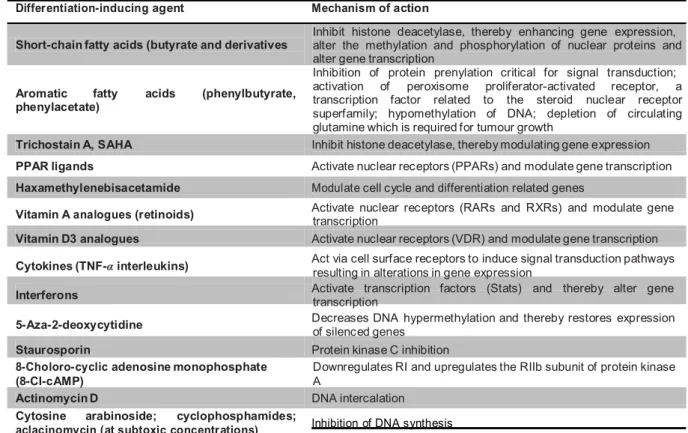
A variety of anticancer agents (Table 1), both natural and synthetic, have been found to induce terminal differentiation and/or apoptosis rather than by cytotoxic action in vitro in various cell lines, including murine erythroleukemia cells, human myeloid leukemia cells and murine and human embryonal carcinoma cells (Leszczyniecka et al., 2001) .
Table 1. Differentiation-inducing agents and their suggested mechanism of action (Lotan R., 2002) Differentiation-inducing agent Mechanism of action
Inhibit histone deacetylase, thereby enhancing gene expression, Short-chain fatty acids (butyrate and derivatives
Aromatic fatty acids (phenylbutyrate, phenylacetate)
alter the methylation and phosphorylation of nuclear proteins and alter gene transcription
Inhibition of protein prenylation critical for signal transduction; activation of peroxisome proliferator-activated receptor, a transcription factor related to the steroid nuclear receptor superfamily; hypomethylation of DNA; depletion of circulating glutamine which is required for tumour growth
Trichostain A, SAHA Inhibit histone deacetylase, thereby modulating gene expression PPAR ligands Activate nuclear receptors (PPARs) and modulate gene transcription Haxamethylenebisacetamide Modulate cell cycle and differentiation related genes
Vitamin A analogues (retinoids) Activate nuclear receptors (RARs and RXRs) and modulate gene transcription Vitamin D3 analogues Activate nuclear receptors (VDR) and modulate gene transcription Cytokines (TNF-α interleukins) Act via cell surface receptors to induce signal transduction pathways resulting in alterations in gene expression Interferons Activate transcription factors (Stats) and thereby alter gene transcription 5-Aza-2-deoxycytidine Decreases DNA hypermethylation and thereby restores expression of silenced genes
Staurosporin Protein kinase C inhibition
8-Choloro-cyclic adenosine monophosphate (8-Cl-cAMP)
Downregulates RI and upregulates the RIIb subunit of protein kinase A
Actinomycin D DNA intercalation
Cytosine arabinoside; cyclophosphamides;
Some of these molecules, such as trichostatin A and cytosine arabinoside, together with other described in the following section, are classified as chemotherapeutic thanks to their high cytotoxic effect; but in addition, when used at sub-toxic concentrations, they are able to induce also the terminal erythroid differentiation.
1.2.3 Antitumor agents acting through induction of erythroid terminal differentiation
In the literature there are numerous publications concerning the study of anticancer agents known as inducers of terminal differentiation. In particular, we want to focus our attention on anticancer possessing also erythro-differentiating ability. It is possible to divide these molecules in different categories, such as (a) cytotoxic agents, (b) HDAC inhibitors, (c) DNA binding drugs (DBD) and (d) from natural world. The same rationale that has led to the successful application of combination of cytotoxic agents with non-overlapping toxicities and distinct mechanisms of action can be applied to differentiation-inducing agents.
1.2.3.1 Cytotoxic agents
Cytarabine, or cytosine arabinoside (AraC), is a chemotherapy agent used mainly in the treatment of cancers of white blood cells such as acute myeloid leukemia (AML) and non-Hodgkin lymphoma. AraC interferes with the synthesis of DNA: inhibiting both DNA and RNA polymerases, leads tumor cells unable to duplicate themselves. Myleran is an alkylating agents used for CML therapy and after bone marrow transplantation; and vinblastine, is currently used for lymphoma, testicular, breast and bladder cancer therapy. These molecules terminate actively cycling progenitors and perturb cellular growth to trigger rapid erythroid-regeneration kinetics and formation of mature red blood cells to contain hemoglobin (Galanello et al., 1988).
1.2.3.2 Hydroxyurea
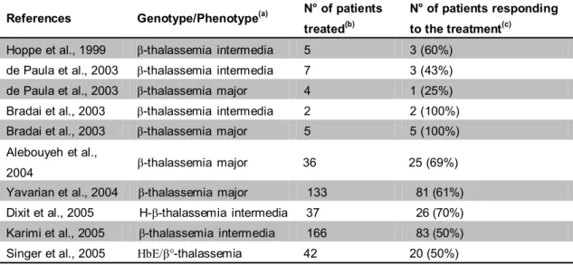
The Multicenter Study of Hydroxyurea was initiated in 1992 to establish the first drug treatment for Sickle Cell Disease (SCD). Hydroxyurea (HU) is a ribonuclease reductase inhibitor that produces blockade of DNA synthesis and cell death. This compound also alters growth rates and gene expression in mammalian cells (Adunyah et al., 1995). One of the first studies showing a clear effect of HU on erythroid differentiation was published in 1993 by Fibach and his collaborators, in which HU was found to have multiple effects on human peripheral blood-derived progenitor cell: an increase in the proportion of fetal hemoglobin (HbF) produced; an inhibition of cell proliferation; and an increase in hemoglobin (Hb) content per cell. Several reports confirmed the HbF augmenting effect of HU both in vitro and in vivo (Wang et al., 2002; Fucharoen et al., 2004; Siriboon et al., 1996; Loukopoulos et al., 2005; Loutradi et al., 2000; Zargari et al., 2004), as summarized in Table 2. For example, Yavarian et al. (Yavarian et al., 2004) reported the treatment with HU of 133 patients with transfusion-dependent β-thalassemia. After one year of treatment these patients were classified into: good responders (61%) who shifted from monthly blood transfusion dependency to a stable transfusion-free condition; moderate responders (23%) who remained transfusion dependent but at longer intervals (6 months or more), and non-responders, who remained at the same level of transfusion dependency.
Table 2. Clinical trials employing Hydroxyurea as In vivo inducer of HbF (from Gambari e
Fibach, medicinal chemistry of fetal hemoglobin inducer for treatment of beta-thalassemia, Curr Med Chem, 2007, 14:199-212)
References Genotype/Phenotype(a) N° of patients treated(b)
N° of patients responding to the treatment(c)
Hoppe et al., 1999 β-thalassemia intermedia 5 3 (60%) de Paula et al., 2003 β-thalassemia intermedia 7 3 (43%) de Paula et al., 2003 β-thalassemia major 4 1 (25%) Bradai et al., 2003 β-thalassemia intermedia 2 2 (100%) Bradai et al., 2003 β-thalassemia major 5 5 (100%) Alebouyeh et al.,
2004 β-thalassemia major 36 25 (69%) Yavarian et al., 2004 β-thalassemia major 133 81 (61%) Dixit et al., 2005 H-β-thalassemia intermedia 37 26 (70%) Karimi et al., 2005 β-thalassemia intermedia 166 83 (50%) Singer et al., 2005 HbE/β°-thalassemia 42 20 (50%)
1.2.3.3 Histone deacetylases Inhibitors (HDACi)
Gene expression is controlled by alterations in chromatin structure produced by acetylation or deacetylation of histone tails, resulting in gene activation or repression, respectively. Histone deacetylase (HDAC) enzymes produce deacetylation of histone tails, causing chromatin condensation and transcriptional silencing.
Several findings suggest that inhibition of the activity of histone deacetylases (HDACs) is associated with an increased expression of the γ-globin genes. Several inhibitors, such as sodium butyrate, trichostatin A, adicipin, and scriptaid, used at low doses, are employed in colorectal cancer therapy. Moreover, they have been shown to induce HbF synthesis in vitro; butyrate also induced HbF in humans (Perrine et al., 1993; McCaffrey et al., 1997; Pace et al., 2003; Cao et al., 2004; Johnson et al., 2001; Pace et al., 2005). Mechanistically, the HDAC inhibitors bind to a central zinc atom in HDACs to block enzymatic deacetylation of histone H3 and H4, leading to an hyperacetylation of ε-amino groups of lysine residues in histones. This in turn causes a decreased association of basic core histone proteins with the DNA, rendering certain genes more accessible to the transcriptional machinery (Cao et al., 2004).
1.2.3.4 DNA-binding drugs
The DNA-binding drugs (DBDs) are molecules able to interact with the major groove of DNA are expected to inhibit complex formation between transcription factors and target DNA elements (Gambari et al., 2003). Several DBDs are or have been used in therapy. Chromomycin and mithramycin (MTH) were used in different kinds of cancer, such as ovarian, stomac, bladder, prostate cancer and hepatoma and they are also employed in the treatment of hypercalcemia; MTH is also used in CML and AML therapy. Tallimustine and tallimustine analogues are anticancer and antiviral agents. Many reports demonstrated that some DBDs display DNA sequence selectivity, and that even similar DBDs differ with respect to stability of their complexes with DNA. Our group has demonstrated that tallimustine (Baraldi et al., 2000; Bianchi et al., 2001; Chiarabelli et al., 2003) and some cisplatin analogues (well-known alkylating agent used for testicular, ovarian, lung, breast, stomach, prostate cancers and also for neuroblastoma, melanoma and sarcoma) (Bianchi et
al., 2000) as well as the GC-rich binders chromomycin and MTH (Bianchi et al., 1999) are powerful inducers of differentiation of K562 cells, suggesting that the expression of crucial genes involved in erythroid differentiation of these cells are influenced by DBDs. Several DBDs, such as tallimustine, MTH and cisplatin, increase of fetal hemoglobin (HbF) production in erythroid precursor cells from normal human subjects. The extent of induction was found to be higher than that of hydroxyurea (HU). Since, among the DBDs studied, MTH displayed the lowest cytotoxicity, we compared it to HU on fetal hemoglobin production by thalassemic erythroid precursors (Fibach et al., 2003). The results demonstrated that in cultures derived from 12 patients, mithramycin increased HbF production in all cases, while HU was not effective in two cases and was toxic in one. In the majority of cases the activity of mithramycin was higher than hydroxyurea. In all cases, HU strongly inhibited cell proliferation, while, at concentrations able to induce HbF production, mithramycin had minimal effect on cell growth.
1.2.3.5 Erythro-differentiating agents from natural world
Several reviews and papers have been published on the possible use of extracts from medicinal plants for biomedical purposes including therapeutic strategies for the treatment of a number of diseases such as dyslipidemia (Alder et al., 2003) and aterosclerosis (Wang et al., 1999), hepatitis (Luper et al., 1998), inflammatory diseases (Nakhai et al., 2007), osteoporosis (Xie et al., 2005), bacterial and virus infections (Khan et al., 2005). In the case of cancer, only few examples are available, as rapamycin and its analogues everolimus and resveratrol, that are briefly described.
Rapamycin (Figure 6a) is a lipophilic macrolide, isolated from a strain of
Streptomyces hygroscopicus found in a soil from Easter Island, that possesses
immunosuppressive, antifungal and anti-tumor properties. This molecule is also approved by the U.S. Food and Drug Administration as an immunosuppressive agent for preventing rejection in patients receiving organ transplantation. The rapamycin, employed in combined therapy with doxorubicin, drive AKT-positive lymphomas into remission in mice. Our group has demonstrated that rapamycin, tested on the human leukemia K562 cell line and the two-phase liquid culture of human erythroid
progenitors isolated from normal donors and patients with beta-thalassemia, has the ability to induce terminal erythroid differentiation and induction of HbF levels. The
interest in rapamycin as an HbF-inducer is related to the fact that its effect is not associated with cytotoxicity and cell growth inhibition, in contrast to other inducers.
Figure 6. Chemical structure of Rapamycin (A) and Everolimus (B)
Several rapamycin-like molecules have been described, exhibiting better characteristics than rapamycin. For instance, everolimus (Figure 6b) is an immunosuppressive macrolide bearing a stable 2-hydroxyethyl chain substitution at position 40 on the rapamycin structure (Dumont et al., 2004; Augustine et al., 2004). Clinical experience, largely limited to its use in kidney transplant patients, indicates that the administration of everolimus is associated with low rates of acute rejection and a tolerable safety profile. Everolimus is a mTOR inhibitor and it acts by the block of proliferation. Recent observations in heart transplant patients suggest that the anti- proliferative effects of this compound may prevent allograft vasculopathy. Zuccato et al., (Zuccato et al., 2007) have determined the effects of everolimus on the erythroid differentiation of K562 cells and on the γ-globin mRNA accumulation in cultures of erythroid precursors isolated from β-thalassaemia patients.
Resveratrol, 3,5,4’-trihydroxystilbene (Figure 7), is a natural phytoalexin present in large quantity in red wine, preferentially in the skin of grapes (Jeandet et al., 1991).
Figure 7. Chemical structure of Resveratrol
Actually, resveratrol is used in support of conventional anticancer chemotherapy in skin cancer, breast and prostate cancer. Rodrigue et al. (Rodrigue et al., 2001) found that resveratrol possesses similar properties to HU toward erythroid differentiation. They firmly demonstrated that resveratrol induces differentiation of K562 cells and augmentation of HbF in erythroid precursor cells isolated from eight sickle cell patients.
In our laboratories, when erythroid precursor cells from normal subjects were treated with increasing concentrations of resveratrol and analysis of accumulation of globin mRNA sequences was performed by quantitative RT-PCR, a clear increase in accumulation of γ-globin mRNA content was found. Increase in accumulation of α- globin and β-globin mRNA was much lower. Taken together these data strongly indicate resveratrol as a strong inducer of HbF and a selective stimulator of the expression in γ-globin genes (Bianchi et al., 2009).
The potential of agents that stimulate cell differentiation to serve for cancer therapy has been studied extensively in vitro and in animal models. Such agents can suppress growth and enhance differentiation, which may also lead to apoptosis. Only a few of the numerous differentiation inducing agents shown in Table 1 have been examined in clinical trials and even for those that have been investigated, most trials were phase I or II. The only definitive demonstration of the efficacy of differentiation therapy is the treatment of acute promyelocytic leukemia patients with ATRA.
1.2.4 K562 cell line as useful experimental model for erythroid differentiation Erythroleukemic K562 cell line was isolated and characterized by Lozzio and Lozzio (Lozzio et al., 1975) from a patient with chronic myelogenous leukemia in blast crisis. K562 cell line has been extensively employed as a very useful in vitro model to study the molecular mechanism(s) regulating the expression of embryonic and fetal human globin genes (Rutherford et al., 1981). K562 cells exhibit a low proportion of hemoglobin-synthesizing cells under standard cell growth conditions, but are able to undergo terminal erythroid differentiation when treated with a variety of compounds, including short fatty acids, 5-azacytidine (Gambari et al., 2007), mithramycin and chromomycin (Bianchi et al., 1999; Fibach et al., 2003), cisplatin and cisplatin analogs (Bianchi et al., 2000), tallimustine (Bianchi et al., 2001; Gambari and Fibach, 2007), rapamycin (Fibach et al., 2006), everolimus (Zuccato et al., 2007), psoralens (Lampronti et al., 2003) and resveratrol (Bianchi et al., 2009). Following erythroid induction, increase of expression of ε and γ globin genes is observed, leading to accumulation of Hb Portland (ζ2γ2) and Hb Gower 1 (ζ2ε2)
(Gambari et al., 2007).
Several antitumor drugs were demonstrated to induce erythroid differentiation of K562 cells. Some of us have recently demonstrated that DNA binding drugs (DBDs) exhibiting antitumor activity are powerful inducers of differentiation of K562 cells, suggesting that the expression of crucial genes involved in terminal erythroid differentiation of these cells is influenced by DBDs.
Thus, this experimental cell system appears to be suitable for the screening of molecules able to inhibit cell growth by acting on the activation of terminal differentiation pathways.
K562 cells grow in culture as single, undifferentiated, cells in suspension, with low production of hemoglobins. When stimulated by various agents, they respond within few days with a significant increase in the production of hemoglobins and γ- globin mRNA (Bianchi et al., 1999).
1.3 “New uracil dimers showing erythroid differentiation inducing activities” As described in section 1.1.3 and reported by Accetta et al. (2009), a series of uracil dimers has been synthesized starting from uracil, thymine or 5-carboxyuracil (isoorotic acid), and tested for antiproliferative and erythroid differentiation activity.
1.3.1 Screening of anti-proliferative and differentiation inducing properties
We first determined for all the synthesized molecules the effects on cell proliferation (Table 3). To this aim, K562 cells were cultured in the presence of increasing concentrations of compounds and cell number per milliliter was determined after 3, 4, and 5 days. These time points were selected because between days 3 and 5 untreated control K562 cells are on the log phase of cell growth.
In order to determine the effects on the erythroid differentiation, cells were seeded at an initial concentration of 3×10⁴ cells/ml and the proportion of benzidine- positive cells was determined after 4-7 days of cell culture using a solution containing 0.2% benzidine in 0.5M glacial acetic acid (10% H2O2) (Bianchi et al., 2001).
Benzidine positivity indicates the presence of intracellular hemoglobin. All methods will be better described in “Materials and Methods” chapter. Table 3 indicates the antiproliferative effects (IC50 values) and the erythroid induction ability (% of
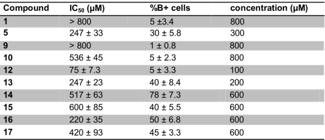
benzidine positive cells) of all the tested compounds. The best erythroid induction ability was displayed by compound 14. The data shown in Table 3 were obtained using concentrations of compounds approaching those giving 50% of inhibition of cell growth (these concentrations were chosen to better compare the potential erythroid inducting activity in experimental conditions leading, for most of the compounds tested, to similar effects on cell proliferation rate). In addition, it should be noted that compound 14 was able to induce erythroid differentiation of K562 cells after 6 days cell culture even if added at concentrations lower than that shown in Table 3 (an average of 52 ± 4.5 % of benzidine positive cells was obtained in four independent experiments with 400 μM compound 14).
Table 3. Antiproliferative Effects (IC50) and Percentage of Benzidine-Positive Cells after Treatment with uracil compounds and concentration used
Compound IC50 (µM) %B+ cells concentration (µM)
1 > 800 5 ±3.4 800 5 247 ± 33 30 ± 5.8 300 9 > 800 1 ± 0.8 800 10 536 ± 45 5 ± 2.3 800 12 75 ± 7.3 5 ± 3.3 100 13 247 ± 23 40 ± 8.4 200 14 517 ± 63 78 ± 7.3 600 15 600 ± 85 40 ± 5.5 600 16 220 ± 35 50 ± 6.8 600 17 420 ± 93 45 ± 3.3 600
Results are presented as average ± SD of three independent experiments performed). The IC50 was
calculated as the concentration of compounds necessary to decrease cell number (after 4 days culture period) at 50% of the values obtained in control untreated K562 cell cultures. The % of benzidine positive (hemoglobin containing) cells was determined after 6 days induction period at concentrations of the tested compounds indicated in the right column.
1.3.2 Conclusions
In conclusion, in this paper, we have demonstrated for the first time that this kind of uracil derivatives can be considered to be a new class of erythroid differentiation inducers, and that dimeric derivatives with suitable spacers have the best performing characteristics: low cytoxicity and higher differentiating ability. Furthermore, the best results were obtained with the compound bearing a naphthalene linker (compound 14), which avoids collapse of the uracil moieties, indicating that a possible recognition of complementary functionalities (such as adenine derivatives) could be implicated in the induction of biological properties.
These findings can be the starting point for the quest for more effective and specific drugs for the induction of terminal erythroid differentiation, ultimately leading to new insights in the treatment of neoplastic diseases with molecules acting by inducing differentiation rather than by exerting cytotoxic effects. In addition, these molecules might be of interest for the experimental treatment of β-thalassemic erythroid cells for which the induction of γ-globin mRNA could be very beneficial.
(Fibach et al., 2003). In this respect it has been demonstrated that inducers of K562
erythroid differentiation are often able to induce fetal hemoglobin production in erythroid cells isolated from β-thalassemia patients (Gambari et al., 2007).
1.4 “C(5) modified uracil derivatives showing antiproliferative and erythroid differentiation inducing activities on human chronic myelogenous leukemia K562 cells”
As just described, we have recently reported a novel class of C(5) linked N(1) alkylated uracil dimers rationally designed to potentially interact with adenine in biological systems: these pyrimidine derivatives have shown antiproliferative and erythroid differentiation activities toward human chronic myelogenous leukemia K562 cells (Accetta et al., 2009). Following these results, in this thesis we would present some biological data obtained from a series of C(5) uracil monomers. These monomers were used, as previously described, as intermediates for the synthesis reactions.
1.4.1 Chemical synthesis of uracil monomers
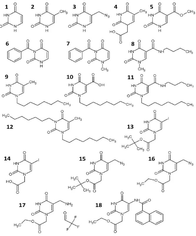
Compounds 1, 2 and 4 were purchased from Sigma-Aldrich and tested without further purification. Synthesis of compounds 5, 8, 9, 10 and 11 is described in section 1.1.3 of this thesis and reported in Accetta et al. (2009). All syntheses started from uracil, thymine or 5-carboxyuracil (isoorotic acid). Also full synthetic procedures and characterization of compounds 3, 12, 13, 14, 15, 16, 17 and 18 are reported section 1.1.3 (Accetta et al., 2010).
The C(5) position of pyrimidine nucleosides can be easily modified with a variety of different residues. Substitution at this position is often used for introducing substituents on DNA oligomers with reporter groups. In a general project, aimed at molecular engineering of peptide nucleic acid (PNA) derivatives (A. Accetta, PhD thesis, 2010), a set of N(1) and/or N(3) alkyl-C(5) modified uracil derivatives were prepared. Since these compounds are structurally analogs of modified nucleosides, they could be considered as potential drugs per se. This set of compounds was chosen for screening since: i) it contains compounds alkylated at N(1) and N(3) positions or both; ii) it contains a series of C(5) derivatives with different functional groups; iii) it has lipophilic groups in both the N(1), N(3), or C(5) region of uracil; iv) it contains derivatives with intact uracil (and thymine) hydrogen bond pattern or lacking of the N(3) hydrogen.
Briefly, modification of uracil at both N(1) and C(5) positions was obtained in several ways. For the more reactive uracil and isoorotic acid, and in the presence of small electrophiles such as methyl iodide, the regioselective alkylation at N(1) can be obtained by temporary protection of the N(3) with trimethylsilyl (Accetta et al., 2009) or benzoyl groups.
The latter method was used in the synthesis of 7 from 6. Azidomethyl derivative 3 was obtained by reaction of uracil with formaldehyde, thus introducing a 5-hydroxymethyl derivative which could be further elaborated to azide by nucleophilic substitution (via a chloromethyl intermediate). Alkylation of 3 with α-bromoacetic acid derivatives led to compounds 15 and 16. The latter was converted to 17, bearing an amino group through Staudinger reduction. The amide derivative 18 was then obtained by reaction of the amino compounds with 2-naphthalenecarboxylic acid after activation of the latter with HBTU. Regioselective alkylation at N(1) was obtained also by exploiting the higher reactivity of the N(1) position in thymine and iodouracil toward sterically hindered electrophiles, which allowed direct synthesis of compounds 9 , according to Coutouli-Argyropoulou and Zachariadou, (Coutouli- Argyropoulou et al., 2005) and 13. The N(1), N(3)- doubly alkylated compound 12 was obtained as a side product of thymine alkylation. Oxidation of thymine methyl group led to the N(1)- alkylated-5-carboxylic derivative 10 (Accetta et al., 2009), which was then converted into the carboxyamide derivative 11 via HBTU activation and reaction with benzylamine. Hydrolysis of 13 with TFA provided derivative 14, bearing a polar substituent in the N(1) position.
Figure 8. Chemical structures of uracil monomers tested. Alternative name: 1, uracil; 2, thymine (5- methyluracil); 3, 5-azidomethyluracil; 4, (thymin-1-yl)acetic acid (1-(carboxymethyl)thymine); 5, methyl 5-uracilcarboxylate (methyl isoorotate); 6, 3-benzoyluracil; 7, 3-benzoyl-1-methyluracil; 8 N- butyl-1-methyl-5-uracilcarboxamide; 9, 1-octylthymine; 10, 1-octyl-5-uracilcarboxylic acid (1- octylisoorotic acid); 11, N-benzyl-1-octyl-5-uracilcarboxamide; 12, 1,3-dioctylthymine; 13, t-butyl (5- iodouracil-1-yl)acetate; 14, (5-iodouracil-1-yl)acetic acid; 15, t-butyl (5-azidomethyluracil- 1- yl)acetate; 16, ethyl (5-azidomehtyluracil-1-yl)acetate; 17, ethyl (5-aminomethyluracil-1-yl)acetate; 18, ethyl [5-(N-(2-naphthylcarboxyl)aminomethyl)uracil-1-yl]acetate
1.5 Aim and thesis outlook
Recently we have reporter a novel class of C(5) linked N(1)-alkylated uracil dimers rationally designed to potentially interact with adenine in biological system. These pyrimidine derivatives have shown antiproliferative and erythroid differentiation activities toward human chronic myelogenous leukemia K562 cells (Accetta et al., 2009). In this thesis we report the screening study on a set of different modified C(5) uracil derivatives; many of them are reaction intermediates for the synthesis of modified uracil dimers. We have evaluated their antiproliferative effect in connection with erythroid differentiation pathways; we would like to propose this molecules as a new class of drugs candidates for the treatment of chronic myelogenous leukemia.
As pointed out from the title of this thesis, we analyzed the biological activity, in particular on erythroid differentiation pathway, of uracil modified monomer used as intermediates of reaction during the synthesis of uracil dimers. These uracil dimers have previously showed interesting biological properties probably in connection with their ability to cooperatively interact with adenine. Using the same geometry, Corradini and his collaborators have synthetized the PNA monomer containing these novel modified dimeric uracils. This new nucleobases performed better than thymine for the adenine recognition on complementary DNA, conferring better selectivity and affinity. These “engineering” uracil were used to the specific design of anti-miR-221 PNAs. The biological activities of these anti-miR-221 PNA were evaluated in a breast cancer cell model, and the results are reported in the Part 2 of this PhD thesis.
-
MATERIALS AND METHODS
-2.1 Cell culture: human erythroleukemic K562 cell line
The human K562 cell line, isolated from a patient with chronic myelogenous leukemia in blast crisis and characterized by Lozzio and Lozzio (Lozzio et al., 1975), were obtained from the American Type Culture Collection (Rockville, Md., USA).
K562 were maintained in a humidified atmosphere of 5% CO2/air at 37 °C in
suspension culture using Roswell Park Memorial Institute 1640 medium (RPMI 1640) (Sigma, St. Louis, MO, USA) supplemented with 10% fetal bovine serum (FBS; Analitical de Mori, Milan, Italy), 50 units/ml penicillin and 50 mg/ml streptomycin (Bianchi et al., 2001).
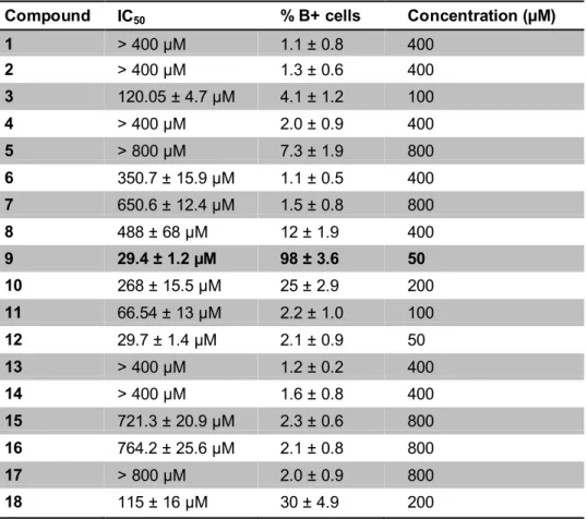
2.2 C(5) modified uracil monomers
For the experiments regarding erythroid differentiation in K562 cell line, we used a serie of uracil monomers and dimers synthesized by Prof. Roberto Corradini and his collaborators, in particular Alessandro Accetta, at University of Parma, Dept. of Organic and Industrial Chemistry. The methods of design and synthesis are well described in Accetta et al., 2009. Here, we report the chemical structure of compounds tested (Table 4).
2.3 Antiproliferative activity
We first determined for all the uracil-derived compounds the effects on cell proliferation. To this aim, K562 cells were seeded at an initial concentration of 3 x 10⁴ cells/ml and cultured in the presence of increasing concentrations of compounds. Non-treated cells were considered as control. Cell number/ ml was determined after 3, 4, and 5 days, using a ZF Coulter Counter (Coulter Electronics, Hialeah, FL, USA). These time points were selected because between days 3 and 5 untreated control K562 cells are on the log phase of cell growth (Bianchi et al., 2001). The IC50 value
(concentration of compound required to inhibit proliferation by 50%) was calculated on three independent experiments.
2.4 Benzidine assay
In order to determine the effects on the erythroid differentiation of uracil- derivatives monomers, K562 cells were seeded at an initial concentration of 3 x 10⁴ cells/ml and the proportion of benzidine- positive cells was determined after 4- 7 days of cell treatment with the compounds, using a solution containing 0.2% benzidine in 0.5 M glacial acetic acid (10% H2O2) as previously described (Bianchi et al, 2001).
The percentage of benzidine-positive cells (blue-cells) was calculated on total cells and indicates the presence of intracellular hemoglobin.
2.5 Transfection of K562 cells with fluorescence protein genes under the γ- globin and the β-globin gene promoters
K562 cells were stably transfected with the pCCL.Promβ.HcRed1. Promγ.EGFP, containing the green and red fluorescence protein (FP) genes under the control of the γ-globin and β-globin gene promoters, respectively (Guerrini et al., 2009; Lampronti et al., 2009). In this system, increases in the green and red signals are consistent with γ-globin and β-globin gene promoter driven activity, respectively. To determine the activity of chemical compounds in inducing the expression of γ- globin and β-globin genes, cells were seeded at 8x10³ cells/ml and treated with the appropriate concentration of the chemical inducer. After 5 days of culture, cells were
assayed for fluorescent proteins expression. First of all they were analyzed under a fluorescence inverted microscope, using filters suitable for both green and red FPs. The fluorescence intensity was then determined by fluorescence- activated cell sorting (FACS) analysis.
2.6 RNA isolation
The total cellular RNA was extracted by TRIZOL® Reagent (Sigma-Aldrich, St.Louis, Missouri, USA). All reagents and materials used were RNase-free. After 6 days of treatment with the compounds, the cells were centrifuged at 1,200 rpm for 10 minutes at 4°C, washed in 1X PBS, re-centrifuged and then lysed with 1 ml TRIZOL®
Reagent. The homogenate was incubated 5 minutes at room temperature and 200 μl
of chloroform were added; the samples were shaken vigorously for 15 second, incubated for 5 minutes at room temperature and finally centrifuged at 12,000 rpm for 15 minutes at 4°C. The aqueous phase was transferred into a clean tube and 500 µl of isopropanol was added. The RNAs were and incubated for 10 minute at room temperature (25°C). After that, the samples were centrifuged at 12,000 rpm for 10 minutes at 4°C; the isolated RNA was precipitated in 2 volumes of absolute ethanol and stored at -80°C, washed once with cold 75% ethanol, dried and dissolved in 10 µl of diethylpyrocarbonate (DEPC)-treated water before use (Sambrook et al., 1989) and conserved at -80°C.
2.6.1 RNA quantification
The concentration of RNA should be determined by measuring the absorbance at 260 nm (A260) in a spectrophotometer. The concentration is obtained by the equation:
g/ml = ODx40xDIL
where OD is the value read from the instrument, 40 is the correction coefficient for reading the RNA at the spectrophotometer (according to the Lambert-Beer law) and DIL is the dilution factor. An absorbance of 1 unit at 260 nm corresponds to 40 µg of