R E S E A R C H A R T I C L E
Open Access
Inhibition of matrix metalloproteinases attenuates
brain damage in experimental meningococcal
meningitis
Susanna Ricci
1,6*, Denis Grandgirard
2,6, Michael Wenzel
3,7, Tiziana Braccini
1, Paola Salvatore
4, Marco R Oggioni
5,6,
Stephen L Leib
2,6†and Uwe Koedel
3,6†Abstract
Background: Approximately 7% of survivors from meningococcal meningitis (MM) suffer from neurological sequelae due to brain damage in the course of meningitis. The present study focuses on the role of matrix metalloproteinases (MMPs) in a novel mouse model of MM-induced brain damage.
Methods: The model is based on intracisternal infection of BALB/c mice with a serogroup C Neisseria meningitidis strain. Mice were infected with meningococci and randomised for treatment with the MMP inhibitor batimastat (BB-94) or vehicle. Animal survival, brain injury and host-response biomarkers were assessed 48 h after meningococcal challenge.
Results: Mice that received BB-94 presented significantly diminished MMP-9 levels (p < 0.01), intracerebral bleeding (p < 0.01), and blood-brain barrier (BBB) breakdown (p < 0.05) in comparison with untreated animals. In mice suffering from MM, the amount of MMP-9 measured by zymography significantly correlated with both intracerebral haemorrhage (p < 0.01) and BBB disruption (p < 0.05).
Conclusions: MMPs significantly contribute to brain damage associated with experimental MM. Inhibition of MMPs reduces intracranial complications in mice suffering from MM, representing a potential adjuvant strategy in MM post-infection sequelae.
Keywords: Neisseria meningitidis, Meningococcal meningitis, Mouse model, Brain damage, Matrix metalloproteinases, Metalloproteinase inhibitors
Background
Neisseria meningitidisis an obligate human pathogen that colonises the nasopharyngeal mucosa of 10-30% individ-uals, with carriage rates increasing with age from small children to young adults. In a minor proportion of carriers, asymptomatic colonisation may progress into invasive meningococcal disease. The clinical manifestations of meningococcal disease (MD) vary considerably, ranging from transient bacteremia to meningitis and/or fulminant
sepsis [1,2]. The most common clinical presentation of MD is meningococcal meningitis (MM), which can affect 30-60% of infected patients, with adolescents and young adults being the age group at higher risk [3,4]. MM is asso-ciated with 5-16% mortality [3]. Approximately 7% of MM survivors suffer from long-term neurological sequelae (i.e., hearing loss, cognitive impairment, motor deficits, sei-zures) due to brain damage in the course of meningitis [5]. Both the host immune response to the pathogen and the direct cytotoxicity of bacterial components are re-sponsible for brain tissue damage [6,7]. Polymorpho-nuclear cells (PMNs), macrophages and microglia that are recruited to the damaged tissue release reactive oxy-gen species, matrix metalloproteinases (MMPs), cyto-kines and excitatory amino acids, finally leading to energy failure and cell death [6,7].
* Correspondence:[email protected] †Equal contributors
1
Department of Medical Biotechnologies, Laboratory of Molecular Microbiology and Biotechnology (LA.M.M.B.), Ospedale Santa Maria alle Scotte (V lotto, piano 1), University of Siena, Viale Bracci, 53100 Siena, Italy 6The ESCMID Study Group for Infectious Diseases of the Brain (ESGIB), Basel, Switzerland
Full list of author information is available at the end of the article
© 2014 Ricci et al.; licensee BioMed Central. This is an Open Access article distributed under the terms of the Creative Commons Attribution License (http://creativecommons.org/licenses/by/4.0), which permits unrestricted use, distribution, and reproduction in any medium, provided the original work is properly credited. The Creative Commons Public Domain Dedication waiver (http://creativecommons.org/publicdomain/zero/1.0/) applies to the data made available in this article, unless otherwise stated.
MMPs are Zn2+-dependent peptidases playing a major role in inflammation, innate immunity and tissue turnover. Not only do they target extracellular matrix proteins, but act as sheddases on non-matrix proteins, such as cyto- and chemokines (and their receptors) and antimicrobial pep-tides [8]. A pathogenic role for MMPs has been suggested for various diseases, including arthritis, cancer, multiple sclerosis, stroke and bacterial meningitis (BM) [9,10]. MMP levels are increased in the cerebrospinal fluid (CSF) of both adults and children with BM [11-13], and high concentra-tions of the gelatinase MMP-9 are considered a risk factor for the development of neurological sequelae [12]. Several experimental models of pneumococcal meningitis (PM) have demonstrated that MMPs contribute to brain damage by participating in blood-brain barrier (BBB) breakdown, PMN recruitment, shedding of cytokines as well as their re-ceptors, and cortical damage [14-16]. Therapy with MMP inhibitors in experimental PM significantly improved ani-mal mortality, neuronal injury, hearing loss, seizures and learning abilities [17-21]. In contrast to plentiful data on MMPs in PM models, the importance of MMPs in experi-mental MM has hardly been reported [11,22], due to the recognised difficulties of mimicking MD/MM in animal models.
Humans are the only natural hosts for N. meningitidis due to the species-specificity of iron meningococcal up-take systems and surface structures. Current animal models of MD consist in mice or rats infected either via the intraperitoneal or intranasal route. In order to compel meningococcal replication in the rodent host, several strategies were attempted, including the use of high bac-terial inocula, neonatal or immunocompromised animals, animal passage prior to infection, or administration of an exogenous source of iron [23-27]. Transgenic mice for human CD46 [28] or transferrin [29] have also been employed to overcome shortcomings of traditional sys-tems. Recently, an MM mouse model based on intracister-nal infection of adult mice with group C N. meningitidis has been developed [30]. Meningococci efficiently multi-plied in the brain and induced meningitis with features mimicking those present in humans [30].
In this study, we have developed a novel mouse model of MM-induced brain damage and demonstrated the therapeutic efficacy of batimastat, a broad MMP inhibitor, in experimental MM. Evaluation of cerebral haemorrhage, BBB breakdown and apoptosis in the hippocampus showed reduced intracranial complications in mice treated with batimastat, suggesting a key role for MMPs in brain damage associated with MM.
Methods
N. meningitidis
The serogroup C 93/4286 isolate, belonging to the ET-37 hypervirulent lineage, was cultured on chocolate GC
agar media (Oxoid, Milano, Italy) or CG broth (Oxoid) supplemented with 1% (v/v) Vitox (Oxoid) at 37°C in 5% CO2. Inocula for mouse challenge were prepared by cul-tivating bacteria in GC broth until mid-logarithmic phase. Viable counts were performed, and bacteria were frozen at -80°C with 10% glycerol until use.
Mice
Six-weeks-old female BALB/c mice were obtained from Charles River (Calco, Italy). Animal experiments were authorised by the local ethics committee (Comitato Etico Locale, Azienda Ospedaliera Universitaria Senese, 21.05.2012) and the Italian Ministry of Health (docu-ment no. 131/2013, 30.05.2013), and were carried out according to institutional guidelines.
Model of MM-induced brain damage
The model of brain damage was developed based on a previously reported MM model in the mouse [30]. Bac-teria were thawed, centrifuged for 5 min at 1800 x g, and suspended in GC broth with iron dextran (5 mg/kg; Sigma-Aldrich, Milano, Italy) at a final concentration of 108 cfu/ml. Approximately 2 h prior to infection, ani-mals were injected intraperitoneally (i.p.) with iron dex-tran (250 mg/kg). Mice were lightly anaesthetised by i.p. injection with Zoletil 20 [(tiletamine and zolazepam hydrochloride), 15 mg/kg; Virbac Srl, Milano, Italy] and Xilor [(xylazine 2%), 4 mg/kg; Bio 98 Srl, Milano, Italy] and infected by the intracisternal (i.c.) route with 10 μl of the bacterial inoculum (106cfu/mouse corresponding to a lethal dose killing 20% of animals, LD20). Mice were monitored for clinical signs according to a previously de-scribed coma scale [21] and euthanised if/when they reached a score of 2. Briefly, coma scale was as follows: 1 = coma, 2 = does not stand upright, 3 = stands upright within 30 sec, 4 = stands upright within 5 sec, minimal ambulatory activities, 5 = normal.
Treatment with BB-94
Mice were treated i.p. with batimastat (BB-94; 50 mg/kg) 1 h before and 24 h post-infection. BB-94 (Merck Che-micals Ltd., Nottingham, United Kingdom) was sus-pended at 50 mg/ml in dimethylsulfoxide (DMSO) and stored frozen at -20°C. Prior to use, it was diluted 20-fold in phosphate buffered saline (PBS), and 500μl were injected into animals. Control mice were injected with 500 μl of 5% DMSO in PBS. Animals were sacrificed 48 h after i.c. challenge.
Experimental design and sample collection
Three independent experiments were performed with a total number of 41 mice (control, n = 20; treated, n = 21). Upon sacrifice, mice were deeply anaesthetised by i.p. in-jection with Zoletil 20 (30 mg/kg; Virbac Srl) and Xilor
(8 mg/kg; Bio 98 Srl) and perfused transcardially with ice-cold PBS. Brains were removed and dissected into the two hemispheres and cerebellum. One hemisphere was fixed in 4% paraformaldehyde (PFA) in PBS (w/v) for histo-logical analysis, while the other one was frozen in dry ice for assessment of intracerebral bleeding and BBB break-down. Cerebella were frozen in dry ice for MMP and cyto-kine analysis. Samples were not collected from animals unsuccessfully perfused, found dead or humanely sacri-ficed before 48 h. Thus, analyses were conducted on a total of 13 control and 20 BB-94-treated mice.
Brain histomorphometry
Cortical necrosis and apoptosis in the dentate gyrus of hippocampus were assessed as previously described [21]. Hemispheres were prepared for cryopreservation by in-cubation in 18% sucrose in PBS (w/v) at 4°C overnight. Forty-five μm-thick coronal sections obtained by ran-dom uniform sampling were stained with cresyl violet for Nissl substance. Histological features of apoptosis (condensed and/or fragmented dark nuclei, apoptotic bodies) were counted in 4 different slices spanning the hippocampus. Three visual fields in the two blades of the dentate gyrus were inspected. The number of apop-totic cells/visual field (apopapop-totic score) was determined, and a mean apoptotic score was calculated for each ani-mal. Cortical damage was defined as areas of decreased neuronal density or frank cortical necrosis and evaluated on digitised brain section images using the ImageJ ana-lysis software (U.S. National Institutes of Health, Be-thesda, Maryland, USA) [31]. The volume of cortical damage was assessed in at least 16 brain sections/mouse and expressed as a percentage of the total cortical vol-ume according to the Cavalieri principle as already de-scribed [32].
Quantification of cyto-/chemokines and MMP-9
Cyto-/chemokines and MMP-9 in cerebella homoge-nates were quantified by using microsphere-based multi-plex assays (Millimulti-plex MAP mouse cytokine/chemokine multiplex assay and Milliplex MAP mouse CVD panel 1 multiplex assay, Merck Millipore, Billerica, MA, USA). Cerebella were homogenised with a glass Dounce hom-ogeniser in 1:7 (w/v) extraction buffer containing 0.1% Triton X-100 and a protease inhibitor cocktail (Roche Diagnostics, Rotkreuz, Switzerland) in PBS. One hun-dredμg (for cyto-/chemokines) or 1 μg (for MMP-9) of homogenates were tested in duplicate. A minimum of 50 beads per analyte was measured using a Bio-Plex 200 station (Bio-Rad Laboratories, Hercules, CA, USA). Cali-bration curves from recombinant standards were calcu-lated with the Bio-Plex Manager software version 4.1.1 using a five-parameter logistic curve fitting.
Gelatin-sepharose affinity binding
Homogenised cerebella were centrifuged at 16000 x g for 10 min at 4°C, and the protein content was deter-mined by the BCA protein assay (Pierce, Thermo Fischer Scientific, Reinach, Switzerland). Gelatinases were enriched by incubating 100 μg of homogenates with 20 μl of Gelatin Sepharose 4B (GE Healthcare GmbH, Glattbrugg, Switzerland) on an orbital shaker overnight at 4°C. Sepharose beads were washed (50 mM Tris-HCl pH 7.6, 150 mM NaCl, 5 mM CaCl2, 0.02% NaN3, 0.05% Brij 25, 1% Triton X-100) and then incubated with 2X zymography sample buffer (30 mM Tris HCl pH 6.8, 20% glycerol, 1% sodium dodecyl sulphate (SDS), 0.02% bromophenol blue) for 5 min at room temperature to elute bound proteins. After centrifugation, supernatants were collected and tested by gelatin gel zymography.
Gelatin zymography
Gelatin zymography was performed as described [21]. Samples were subjected to electrophoresis under non-reducing conditions in polyacrylamide gels containing 1% (v/v) type A gelatin from porcine skin (Sigma-Al-drich). After electrophoresis, SDS was removed from the gels, and the MMP catalytic sites were activated by over-night incubation at 37°C in zymography buffer (10 mM CaCl2, 50 mM Tris, 50 mM NaCl, pH 7.65). Gels were stained with 0.1% Coomassie Brilliant Blue R250 (Sigma-Aldrich), 30% methanol and 10% acetic acid. Gelatinoly-tic activity was determined by densitometric quantifica-tion of the substrate lysis zones around 92 (pro-MMP-9) and 72 (pro-MMP-2) kDa using the ImageJ analysis soft-ware [31]. Purified human neutrophil MMP-9 and MMP-2 (Calbiochem, Darmstadt, Germany) were used as standards for normalisation and quantification of MMP-9 and MMP-2 as a function of the lysis zone. A strong correlation was calculated between MMP-9 quan-tification obtained by gel zymography and by the Milli-plex assay (Pearson’s test, ρ = 0.83, p < 0.0001).
Analysis of cerebral bleeding
Cerebral haemorrhages were assessed as previously de-scribed [33]. Briefly, brain hemispheres were cut in a frontal plane into 30μm-thick sections, and serial sections were photographed with a digital camera at 0.3 mm-inter-vals. For each animal, 5 comparable brain sections were analysed. The bleeding spots were counted, and the rela-tive areas of bleeding were measured by using the UTHSCSA Image Tool (Texas, USA). Cumulative bleed-ing areas were divided by the whole slice area and com-puted into a total bleeding area/whole slice area x 1000.
Assessment of BBB integrity
The integrity of BBB was estimated by quantifying mouse serum albumin in brain homogenates by
enzyme-linked immunosorbent assay (ELISA) as previously re-ported [34]. In brief, brain slices were dissolved in lysis buffer containing 0.1% NaN3 and homogenised in an ultrasound bath for 20 sec. After centrifugation, superna-tants were collected and subjected to the Roti®-Nanoquant assay (Carl Roth GmbH + Co.KG, Karlsruhe, Germany) to quantify the total protein content. For each sample, 0.5μg of total protein were subjected to albumin ELISA (Bethyl/ Biomol GmbH, Hamburg, Germany) according to the manufacturer’s instructions.
Statistical analyses
Results are presented as mean values ± standard deviation. Mouse survival was estimated by the Kaplan-Meier sur-vival analysis, and differences were compared using the log-rank test (p < 0.05). Differences in MMP-9 gelatinoly-tic activity, apoptosis, BBB integrity and cerebral bleeding between mice treated with BB-94 and controls were evalu-ated using the Mann-Whitney U test (p < 0.05). Correl-ation was measured by the Pearson’s analysis (p < 0.05).
Results
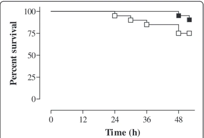
The impact of treatment with BB-94 on MM outcome in infected mice was evaluated by assessing animal survival, brain damage, and levels of MMPs and cyto-/chemo-kines 48 h after meningococcal challenge. Upon sacri-fice, the clinical scores of surviving mice were 3 (2-4) [median (range)] irrespective of the group (p > 0.05). Kaplan-Meier analysis of survival (at 48 h) showed that animals treated with BB-94 had increased survival (95.2%) in comparison with controls (75%), and differ-ences were almost statistically significant (p = 0.064), (Figure 1). The concentrations of interleukin (IL)-1β, IL-10, monocyte chemoattractant protein 1 (MCP-1) and macrophage inflammatory protein 1α (MIP-1α) were slightly reduced in cerebella of treated animals, without reaching statistical significance (data not shown).
BB-94 reduces MMP-9 levels in the brain of mice with MM
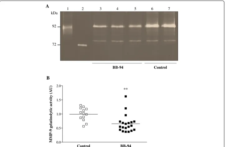
To evaluate whether BB-94 diminished the levels of MMPs in the brain, gelatin zymography was performed on cerebella of treated and control animals after MM in-duction. Densitometric analysis showed no relevant changes in MMP-2 levels between samples from the two mouse groups. In contrast, the amount of MMP-9 was diminished in cerebella of mice that had received BB-94 compared to those injected with vehicle (Figure 2A). Quantitative assessment of MMP-9 based on den-sitometric analysis of MMP-9 substrate lysis zones evidenced a significant reduction (p = 0.0011) between treated (0.6523 ± 0.3079) and control (0.9893 ± 0.2310) mice (Figure 2B).
Treatment with BB-94 ameliorates intracranial complications in infected mice
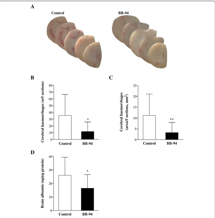
Preliminary observations upon MM model development indicated that mice infected with N. meningitidis pre-sented multifocal cortical haemorrhages visible to the naked eye compared to naïve animals (Ricci, unpublished data). Therefore, the number and size of cerebral bleed-ings in BB-94-treated mice were compared to those from control subjects. Macroscopical examination of brain sam-ples showed reduced bleeding in animals treated with BB-94 in comparison with controls (Figure 3A). Quantitative analysis revealed a 3-fold reduction both in the number of haemorrhagic spots (Figure 3B; p = 0.0122) and the area of bleeding (Figure 3C; p = 0.0061) in treated mice. However, cortical haemorrhages did not translate into large regions of reduced neuronal density as detectable by Nissl staining (data not shown).
BB-94 was also beneficial for the integrity of the BBB, as proven by reduced diffusion of serum albumin in the brain of treated mice in respect to those injected with vehicle. Differences between the concentration of albu-min in BB-94-treated (16,47 ± 10,11 ng/μg total protein) and control (26,00 ± 13,37 ng/μg) animals were statisti-cally significant (p = 0.037; Figure 3D).
Evaluation of apoptosis in the dentate gyrus of hippo-campus showed a trend in the reduction of the apoptosis score in the treatment group (0,1438 ± 0,1396) compared to the control one (0,1898 ± 0,1217; p > 0.05).
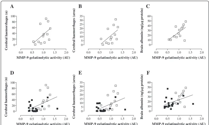
MMP-9 contributes to brain damage associated with MM
To evaluate whether levels of MMP-9 are associated with intracranial complications resulting from MM, Pearson correlation analysis was performed on data from both control and BB-94-treated mice (Figure 4
0 12 24 36 48 0 25 50 75 100
Time (h)
P
ercen
t s
u
rvi
val
Figure 1 Effect of treatment with BB-94 on survival of mice with MM. BALB/c mice were infected i.c. with 106cfu/mouse of strain 93/4286 (group C) of N. meningitidis, injected i.p. with either BB-94 (n = 21) or vehicle (n = 20), and sacrificed 48 h later. Treatment with BB-94 (50 mg/kg) was carried out 1 h before and 24 h post-infection. Data are represented as a Kaplan-Meier survival curve.
and Additional file 1: Table S1). In infected control ani-mals, the amounts of MMP-9 significantly correlated with the extent of BBB disruption (ρ = 0.59, p = 0.031; Figure 4C). Correlation between MMP-9 levels and the number of haemorrhagic spots (ρ = 0.51, p = 0.079) or the size of bleeding (ρ = 0.54, p = 0.054) also showed a tendency towards statistical significance (Figure 4A-B). In contrast, no significant correlation was observed be-tween amounts of MMP-9 and intracranial complications in treated mice (Additional file 1: Table S1). Finally, when data from both control and BB-94-treated animals were pooled, MMP-9 levels presented a robust correlation with the number (ρ = 0.46, p = 0.0067; Figure 4D) and area (ρ = 0.47, p = 0.0049; Figure 4E) of cerebral bleeding, and with the breakdown of BBB (ρ = 0.44, p = 0.0104; Figure 4F).
Discussion
Successful treatment of BM not only requires antibiotics to eradicate the pathogen but also adjuvant therapeutic molecules targeting the host immune and inflammatory responses. Potential targets of adjuvant therapy are
cyto-and chemokines, leukocytes, coagulation cascade mole-cules, oxidants, caspases, and degrading enzymes [35,36]. To this purpose, several therapeutic approaches have been attempted in animal models of meningitis caused by Streptococcus pneumoniae and Streptococcus agalactiae, including the use of nonbacteriolytic antibiotics, antioxi-dants, antagonists of excitatory amino acids, and also in-hibitors of transcription factors, MMPs, caspases, or complement factors [17-21,33,37-41]. In contrast, data on adjuvant therapy in MM animal models are scant partially due to limitations of experimental systems mimicking the human disease.
In this work, we have fine-tuned a recently described MM model [30] to study brain damage associated with MM in the mouse. The model is based on i.c. infection of adult mice with a sub-lethal dose (~LD20) of live men-ingococci in order to limit a rapid fatal outcome and per-mit the development of brain damage. Model readouts included animal survival and clinical signs, cyto-/chemo-kines profiling, MMP levels, apoptosis, BBB breakdown, and intracerebral haemorrhages. Compared to some 1 2 3 4 5 6 7 A B ** kDa 92 72 Control BB-94 0.0 0.5 1.0 1.5 2.0 M M P -9 g ela ti n o ly tic a ctv ity (A U ) Control BB-94
Figure 2 Zymography on cerebella of control and BB-94-treated mice infected with meningococci. Mice were infected and treated as described in Figure 1. Animals were perfused with cold PBS, and cerebella were harvested and homogenised. As samples were not collected from dead or unsuccessfully perfused animals, a total of 20 treated mice and 13 control were analysed. Gelatinases in cerebella were enriched by incubation with gelatin-sepharose and subjected to gelatin zymography. A. Densitometry of gelatinolytic activity around 92 kDa and 72 kDa. Lane 1: human MMP-9; lane 2: human MMP-2; lanes 3-5: BB-94-treated mice; lanes 6-7: controls. B. Quantification of MMP-9 gelatinolytic activity. Results are combined from 3 independent experiments. AU, arbitrary units. **p < 0.01.
experimental model of PM [14,15], animals were not treated with antibiotics, which may influence the course and profile of the inflammatory response. The availability of a simple and reliable MM mouse model allowed, for the first time, to provide evidence that MMP-9 is involved in the pathogenesis of MM-induced
brain damage and MMP-9 inhibition significantly im-proves intracranial complications.
Common histopathologic features of PM include brain oedema, cortical haemorrhages, focal necrotic lesions in cortical/subcortical structures and neuronal apoptosis in the hippocampus [6,7]. PM animal models have largely
A
B
D
Control BB-94 * **C
Control BB-94 0 10 20 30 40 50 60 70 80Cerebral haemorrhages (n/5 sections)
* Control BB-94 0 5 10 15 20 25
Cerebral haemorrhages (area/5 sections, mm
2) Control BB-94 0 10 20 30 40 Brain albumin (ng/ g protein)
Figure 3 Brain damage in control and BB-94-treated mice after meningococcal challenge. Mice were infected and treated as described in Figure 1. Brains from perfused animals were collected. A-C. Brain hemispheres were cut in 30μm cryosections and photographed to determine the number of haemorrhagic spots and the areas of bleeding. Macroscopical assessment of cerebral haemorrhages in animals injected with BB-94 or vehicle (A). Enumeration of bleeding spots (B) and measurement of haemorrhagic areas (C) were carried out on 5 comparable brain sections/ mouse. D. Following bleeding assessment, brain hemispheres were homogenised, and supernatants were subjected to mouse albumin ELISA to quantify serum albumin in the brain as a marker of BBB breakdown. Data are displayed as ng of albumin perμg of total protein content of the brain. Data are from 3 independent experiments. *p < 0.05; **p < 0.01.
proven the role of MMPs in several mechanisms of brain damage, including PMN extravasation, neuronal damage and BBB opening [14-16]. Concerning MM models, challenge of rats with heat-killed meningococci increased the levels of MMP-9 mRNA expression and proteolytic ac-tivity [22] and contributed to BBB disruption [11]. A re-cent study demonstrated that infection of human brain microvascular endothelial cells with N. meningitidis in-duced both the detachment of brain endothelial cells from the matrix and the cleavage of the tight junction protein occludin; interestingly, both processes were mediated by MMP-8, thereby proposing a molecular mechanism be-hind BBB breakdown during MM [42]. However, these studies were carried out either in vivo with inactivated bacteria [11,22] or in vitro [42]. Our work, instead, employed live meningococci in mice and provided in vivoevidence that MMP-9 levels significantly corre-lated with BBB breakdown and cerebral bleeding.
The blood vessel is a major target of both pathogen-and host-mediated injury in MD. During haematogenous spread, N. meningitidis interacts with the endothelial cells of blood vessels throughout the body, including brain (in MM) and skin (in sepsis). Recently, the skin le-sions typical of purpura fulminans were reproduced in
an experimental model of meningococcemia [43], dem-onstrating how ‘vascular colonisation’ by meningococci initiates the thrombotic process [44] that ultimately leads to vasculitis and vascular rupture. A similar mech-anism may occur in the blood vessels of the brain during experimental MM, resulting in cerebral bleeding. On the host side, MMPs also contribute to the dysfunction of brain vasculature by disrupting endothelial cell junctions [42] and promoting BBB opening [11]. MMPs participate in cerebral haemorrhage of diverse origins [45], and blocking MMPs is an effective strategy to control brain (vasogenic) oedema and bleeding size in animal models of intracerebral haemorrhage [46-49]. In our case, the correlation between MMP-9 levels and bleeding is con-sistent with a key role of MMPs in brain haemorrhage, suggesting that both meningococci and MMPs may have contributed to intracerebral bleeding in the model.
Inhibitors of MMPs have shown beneficial effects in experimental studies of neuroinflammatory and neuro-degenerative conditions, including multiple sclerosis, is-chaemic and haemorragic stroke, vascular dementia and meningitis [9,10]. In an infant rat PM model, several molecules with different inhibitory profiles (MMPs or TNF-α converting enzyme (TACE)) were able to reduce
0.0 0.5 1.0 1.5 2.0 0 20 40 60 80 100
MMP-9 gelatinolytic activity (AU)
C er ebral haem or rh a g es (n ) 0.0 0.5 1.0 1.5 2.0 0 5 10 15 20 25 30 35
MMP-9 gelatinolytic activity (AU)
C er ebr al hae m or rh a g es (a re a) 0.0 0.5 1.0 1.5 2.0 0 10 20 30 40 50 60
MMP-9 gelatinolytic activity (AU)
B ra in a lb u m in (n g / g p ro te in )
A
E
C
B
D
F
0.0 0.5 1.0 1.5 2.0 0 20 40 60 80 100MMP-9 gelatinolytic activity (AU)
C er eb ral h a em or rh a g es (n ) 0.0 0.5 1.0 1.5 2.0 0 5 10 15 20 25 30 35
MMP-9 gelatinolytic activity (AU)
Cere b ra l h a em o rr h a g es (a re a ) 0.0 0.5 1.0 1.5 2.0 0 10 20 30 40 50 60
MMP-9 gelatinolytic activity (AU)
B ra in a lb u m in (n g / g p ro te in )
Figure 4 Correlation between MMP-9 levels and intracranial complications in mice with MM. Brain samples from control and treated animals were subjected to different assays as described in Figures 2 and 3. A-C. Control mice (open squares). D-F. Pooled data of control (open squares) and BB-94-treated animals (closed squares). The amount of MMP-9 assessed by zymography was associated with the number of bleeding spots (A, D), the size of haemorrhages (B, E), and the disruption of BBB (C, F). Correlation analysis was performed by the Pearson’s test (p < 0.05).
animal mortality [19,21], cortical necrosis [15,17-19,21], hippocampal apoptosis [17,21], BBB disruption [19], cyto-/chemokine release [21], and also post-infection se-quelae [17-19]. Batimastat (BB-94) is a hydroxamate peptidomimetic broad-spectrum MMP inhibitor, but in-active against TACE. Screening of a wide range of differ-ent MMP inhibitors in a lipopolysaccharide (LPS)-based rodent model showed that BB-94 was the most effective compound at reducing BBB breakdown in both rats and mice [50]. BB-94 was also useful to diminish BBB dis-ruption and intracranial pressure in rats infected with heat-killed N. meningitidis [11]. Moreover, BB-94 signifi-cantly reduced the incidence of haemorrhage associated with administration of thrombolytic drugs in rabbits suf-fering from stroke, demonstrating the added value of BB-94 in controlling experimental brain bleeding [46].
In this study, treatment with BB-94 increased mouse survival and alleviated the severity of intracranial compli-cations. Kaplan-Meier survival curves showed a tendency towards a significant difference between treated and con-trol animals. The lack of statistical significance may be due to the relatively small sample size of animal groups af-fecting statistical power. In contrast, brain damage was significantly reduced in BB-94-treated subjects compared to controls. To emphasise this result, it is worth noticing that mice (of which 87.5% were controls) that died before the experimental endpoint at 48 h were not assessed for brain injury, thereby potentially excluding the most acute cases from the analyses. BB-94 may have operated via dif-ferent mechanisms, including: (i) a direct effect on the brain microvasculature by limiting vascular rupture and BBB breakdown, or (ii) an indirect action on the systemic inflammatory response by reducing MMP-dependent acti-vation of pro-inflammatory mediators. However, the fact that levels of brain cyto- and chemokines were not signifi-cantly reduced in treated mice argues for a direct vasculo-protective effect by BB-94. In BB-94 treated mice, MMP-9 levels did not significantly correlate with intracerebral complications, suggesting that other factors than MMP-9 may be responsible for the residual brain damage detect-able in treated animals.
Drugs that block MMPs are considered a double-edged sword in clinical practice. On one hand, they represent a therapeutic asset in neuroinflammation, while on the other, they may wedge crucial recovery functions, such as neurovascular remodelling and neurogenesis [10]. Never-theless, MM is an acute condition for which a short-term adjuvant therapy with MMP inhibitors may be envisioned to protect the neurovascular unit.
Conclusions
This study describes the development of a novel murine model of MM-induced brain damage that allows the assessment of different clinical, histopathological and
immunological parameters. The model may be valuable in the MM/MD field where experimental systems mim-icking the human disease are lacking. The use of such in vivo model enabled us to demonstrate that: (i) MMP-9 contributes to neuronal injury in MM, and (ii) MMP-MMP-9 inhibition improves disease outcome, indicating that MMP-9 may represent an important target for adjuvant therapy in MM. The present data are consistent with the literature highlighting the role of MMPs in PM, thus suggesting MMPs as universal mediators of brain dam-age in acute BM irrespective of the causative dam-agent. Fu-ture studies will investigate the efficacy of MMP inhibitors as adjuvant therapy in combination with anti-biotics in experimental MM.
Additional file
Additional file 1: Table S1. Describing the statistical analysis related to correlation between zymography and brain damage in mice with MM.
Abbreviations
BM:Bacterial meningitis; MD: Meningococcal disease; MM: Meningococcal meningitis; PM: Pneumococcal meningitis; MMP: Matrix metalloproteinase; TNF-α: Tumor necrosis factor α; IL: Interleukin; MCP-1: Monocyte chemoattractant protein 1; MIP-1α: Macrophage inflammatory protein 1α; TACE: TNF-α converting enzyme; PMN: Polymorphonuclear cell; BBB: Blood-brain barrier; CSF: Cerebrospinal fluid; LPS: Lipopolysaccharide; i.c.: Intracisternally; i.p.: Intraperitoneally; LD20: Lethal dose killing 20% of animals; DMSO: Dimethylsulfoxide; PBS: Phosphate buffered saline; PFA: Paraformaldehyde; SDS: Sodium dodecyl sulphate; ELISA: Enzyme-linked immunosorbent assay.
Competing interests
The authors declare that they have no competing interests. Authors’ contributions
SR, co-ordination and design of the study, data analysis and interpretation, supervision of animal work, manuscript writing. DG, assessment of MMP levels and brain damage. MW, analysis of brain damage. TB, animal experiments. PS, critical reading of manuscript. MRO, experimental design and draft of the manuscript. SLL and UK, study conception and design, critical reading of the manuscript. All authors have read and approved the manuscript.
Acknowledgements
The study was funded by ESCMID Study Group Research Grant (to SR). Author details
1Department of Medical Biotechnologies, Laboratory of Molecular Microbiology and Biotechnology (LA.M.M.B.), Ospedale Santa Maria alle Scotte (V lotto, piano 1), University of Siena, Viale Bracci, 53100 Siena, Italy. 2
Institute for Infectious Diseases, University of Bern, 3010 Bern, Switzerland. 3Department of Neurology, Klinikum Großhadern, Ludwig-Maximilians University, 81377 Munich, Germany.4Department of Molecular Medicine and Medical Biotechnology, Federico II University Medical School, Naples, Italy. 5
Department of Genetics, University of Leicester, Leicester LE1 7RH, UK.6The ESCMID Study Group for Infectious Diseases of the Brain (ESGIB), Basel, Switzerland.7Present address: Columbia University, Biological Sciences, 901 NWC Building, 550 West 120th Street, New York, NY 10027, USA. Received: 7 October 2014 Accepted: 18 December 2014
References
1. Stephens DS, Greenwood J, Brandtzaeg P: Epidemic meningitis, meningococcaemia, and Neisseria meningitidis. Lancet 2007, 369:2196–2210.
2. Pace D, Pollard AJ: Meningococcal disease: clinical presentation and sequelae. Vaccine 2012, 30S:B3–B9.
3. Centers for Disease Control and Prevention: Active Bacterial Core Surveillance Report. Emerging Infectious Program Network, Neisseria meningitidis 2012. http://www.cdc.gov/abcs/reports-findings/survreports/mening12.pdf. 4. European Centre for Disease Control and Prevention: Annual Epidemiological
Report 2013. Reporting on 2011 surveillance data and 2012 epidemic intelligence data. Stockholm: ECDC. http://www.ecdc.europa.eu/en/ publications/Publications/annual-epidemiological-report-2013.pdf. 5. Edmond K, Clark A, Korczak V, Sanderson C, Griffiths UK, Rudan I: Global
and regional risk of disabling sequelae from bacterial meningitis: a systematic review and meta-analysis. Lancet 2010, 10:317–328. 6. Koedel U, Klein M, Pfister H-W: New understandings on the pathophysiology
of bacterial meningitis. Curr Opin Infect Dis 2010, 23:217–223.
7. Gerber J, Nau R: Mechanisms of injury in bacterial meningitis. Curr Opin Neurol 2010, 23:312–318.
8. Parks WC, Wilson CL, Lopez-Boado Y: Matrix metalloproteinases as modulators of inflammation and innate immunity. Nat Rev Immunol 2004, 4:617–629.
9. Leppert D, Lindberg RL, Kappos L, Leib SL: Matrix metalloproteinases: multifunctional effectors of inflammation in multiple sclerosis and bacterial meningitis. Brain Res Rev 2001, 36:249–257.
10. Rosenberg GA: Matrix metalloproteinases and their multiple roles in neurodegenerative diseases. Lancet Neurol 2009, 8:205–216.
11. Paul R, Lorenzl S, Koedel U, Sporer B, Vogel U, Frosch M, Pfister H-W: Matrix metalloproteinases contribute to the blood-brain barrier disruption during bacterial meningitis. Ann Neurol 1998, 44:592–600.
12. Leppert D, Leib SL, Grygar C, Miller KM, Schaad UB, Hollander GA: Matrix metalloproteinase (MMP)-8 and MMP-9 in cerebrospinal fluid during bacterial meningitis: association with blood-brain barrier damage and neurological sequelae. Clin Infect Dis 2000, 31:80–84.
13. Lindberg RL, Sorsa T, Tervahartiala T, Hoffmann F, Mellanen L, Kappos L, Schaad UB, Leib SL, Leppert D: Gelatinase B (matrix metalloproteinase (MMP)-9) and collagenases (MMP-8/-13) are upregulated in cerebrospinal fluid during aseptic
and bacterial meningitis in children. Neuropathol Appl Neurobiol 2006, 32:304–317.
14. Azeh I, Mader M, Smirnov A, Beuche W, Nau R, Weber F: Experimental pneumococcal meningitis in rabbits: the increase of matrix metalloproteinase-9 in cerebrospinal fluid correlates with leucocyte invasion. Neurosci Lett 1998, 256:127–130.
15. Leib SL, Leppert D, Clements J, Täuber MG: Matrix metalloproteinases contribute to brain damage in experimental pneumococcal meningitis. Infect Immun 2000, 68:615–620.
16. Sellner J, Leib SL: In bacterial meningitis cortical brain damage is associated with changes in parenchymal MMP-9/TIMP-1 ratio and increased collagen type IV degradation. Neurobiol Dis 2006, 21:647–656. 17. Leib SL, Clements JM, Lindberg RL, Heimgartner C, Loeffler JM, Pfister L-A,
Täuber MG, Leppert D: Inhibition of matrix metalloproteinases and tumour necrosis factorα converting enzyme as adjuvant therapy in pneumococcal meningitis. Brain 2001, 124:1734–1742.
18. Meli DN, Loeffler JM, Baumann P, Neumann U, Buhl T, Leppert D, Leib SL: In pneumococcal meningitis a novel water-soluble inhibitor of matrix metalloproteinases and TNF-α converting enzyme attenuates seizures and injury of the cerebral cortex. J Neuroimmunol 2004, 151:6–11. 19. Meli DN, Coimbra RS, Erhart DG, Loquet G, Bellac C, Täuber MG, Neumann
U, Leib SL: Doxycycline reduces mortality and injury to the brain and cochlea in experimental pneumococcal meningitis. Infect Immun 2006, 74:3890–3896.
20. Echchannaoui H, Leib SL, Neumann U, Landmann RMA: Adjuvant TACE inhibitor treatment improves the outcome of TLR2-/-mice with experimental pneumococcal meningitis. BMC Infect Dis 2007, 7:25. 21. Liechti FD, Grandgirard D, Leppert D, Leib SL: Matrix metalloproteinase
inhibition lowers mortality and brain injury in experimental pneumococcal meningitis. Infect Immun 2014, 82:1710–1718.
22. Kieseier BC, Paul R, Koedel U, Seifert T, Clements JM, Gearing AJ, Pfister H-W, Hartung HP: Differential expression of matrix metalloproteinases in bacterial meningitis. Brain 1999, 122:1579–1587.
23. Holbein BE, Jericho KWF, Likes GC: Neisseria meningitidis infection in mice: influence of iron, variations in virulence among strains, and pathology. Infect Immun 1979, 24:545–551.
24. Salit IE, Tomalty L: A neonatal mouse model of meningococcal disease. Clin Invest Med 1984, 9:119–123.
25. Saukkonen K: Experimental meningococcal meningitis in the infant rat. Microb Pathog 1988, 4:203–211.
26. Mackinnon FG, Gorringe AR, Funnell SGP, Robinson A: Intranasal infection of infant mice with Neisseria meningitidis. Microb Pathog 1992, 12:415–420. 27. Gorringe AR, Reddin KM, Funnell SG, Johansson L, Rytkönen A, Jonsson AB:
Experimental disease models for the assessment of meningococcal vaccines. Vaccine 2005, 23:2214–2217.
28. Johansson L, Rytkönen A, Bergman P, Albiger B, Källström H, Hökfelt T, Agerberth B, Cattaneo R, Jonsson AB: CD46 in meningococcal disease. Science 2003, 301:373–375.
29. Zarantonelli ML, Szatanik M, Giorgini D, Hong E, Huerre M, Guillou F, Alonso JM, Taha MK: Transgenic mice expressing human transferrin as a model for meningococcal infection. Infect Immun 2007, 75:5609–5614. 30. Colicchio R, Ricci S, Lamberti F, Pagliarulo C, Pagliuca C, Braione V, Braccini
T, Talà A, Montanaro D, Tripodi S, Cintorino M, Troncone G, Bucci C, Pozzi G, Bruni CB, Alifano P, Salvatore P: The meningococcal ABC-type L-glutamate transporter GltT is necessary for the development of experimental men-ingitis in mice. Infect Immun 2009, 77:3578–3587.
31. Schneider CA, Rasband WS, Eliceiri KW: NIH image to ImageJ: 25 years of image analysis. Nat Methods 2012, 9:671–675.
32. Gehre F, Leib SL, Grandgirard D, Kummer J, Bühlmann A, Simon F, Gäumann R, Kharat AS, Täuber MG, Tomasz A: Essential role of choline for pneumococcal virulence in an experimental model of meningitis. J Intern Med 2008, 264:143–154.
33. Koedel U, Frankenberg T, Kirschnek S, Obermaier B, Häcker H, Paul R, Häcker G: Apoptosis is essential for neutrophil functional shutdown and determines tissue damage in experimental pneumococcal meningitis. PLoS Pathog 2009, 5:e1000461.
34. Koedel U, Rupprecht T, Angele B, Heesemann J, Wagner H, Pfister H-W, Kirschning CJ: MyD88 is required for mounting a robust host immune response to Streptococcus pneumoniae in the CNS. Brain 2004, 127:1437–1445.
35. van der Flier M, Geelen SP, Kimpen JL, Hoepelman IM, Tuomanen EI: Reprogramming the host response in bacterial meningitis: how best to improve outcome? Clin Microbiol Rev 2003, 16:415–429.
36. Koedel U, Klein M, Pfister H-W: Modulation of brain injury as a target of adjunctive therapy in bacterial meningitis. Curr Infect Dis Rep 2010, 12:266–273. 37. Koedel U, Winkler F, Angele B, Fontana A, Flavell RA, Pfister H-W: Role of
caspase-1 in experimental pneumococcal meningitis: evidence from pharmacologic caspase inhibition and caspase-1-deficient mice. Ann Neurol 2002, 51:319–329.
38. Woehrl B, Brower MC, Murr C, Heckenberg SG, Baas F, Pfister H-W, Zwinderman AH, Morgan BP, Barnum SR, van der Ende A, Koedel U, van de Beek D: Complement component 5 contributes to poor disease outcome in humans and mice with pneumococcal meningitis. J Clin Invest 2011, 121:3943–3953. 39. Leib SL, Kim YS, Ferriero DM, Täuber MG: Neuroprotective effect of
excitatory amino acid antagonist kynurenic acid in experimental bacterial meningitis. J Infect Dis 1996, 173:166–171.
40. Mook-Kanamori BB, Rouse MS, Kang CI, van de Beek D, Steckelberg JM, Patel R: Daptomycin in experimental murine pneumococcal meningitis. BMC Infect Dis 2009, 9:50.
41. Gerber J, Lotz M, Ebert S, Kiel S, Huether G, Kuhnt U, Nau R: Melatonin is neuroprotective in experimental Streptococcus pneumoniae meningitis. J Infect Dis 2005, 191:783–790.
42. Schubert-Unkmeir A, Konrad C, Slanina H, Czapek F, Hebling S, Frosch M: Neisseria meningitidis induces brain microvascular endothelial cell detachment from the matrix and cleavage of occludin: a role for MMP-8. PLoS Pathog 2010, 6:e1000874.
43. Join-Lambert O, Lécuyer H, Miller F, Lelievre L, Jamet A, Furio L, Schmitt A, Pelissier P, Fraitag S, Coureuil M, Nassif X: Meningococcal interaction to microvasculature triggers the tissular lesions of purpura fulminans. J Infect Dis 2013, 208:1590–1597.
44. Melican K, Duménil G: Vascular colonization by Neisseria meningitidis. Curr Opin Microbiol 2012, 15:50–56.
45. Xue M, Yong VW: Matrix metalloproteinases in intracerebral hemorrhage. Neurol Res 2008, 30:775–782.
46. Lapchak PA, Chapman DF, Zivin JA: Metalloproteinase inhibition reduces thrombolytic (tissue plasminogen activator)-induced hemorrhage after thromboembolic stroke. Stroke 2000, 31:3034–3040.
47. Power C, Henry S, Del Bigio MR, Larsen PH, Corbett D, Imai Y, Yong VW, Peeling J: Intracerebral hemorrhage induces macrophage activation and matrix metalloproteinases. Ann Neurol 2003, 53:731–742.
48. Wang J, Tsirka SE: Neuroprotection by inhibition of matrix
metalloproteinases in a mouse model of intracerebral haemorrhage. Brain 2005, 128:1622–1633.
49. Lee CZ, Xue Z, Zhu Y, Yang GY, Young WL: Matrix metalloproteinase-9 inhibition attenuates vascular endothelial growth factor-induced intracerebral hemorrhage. Stroke 2007, 38:2563–2568.
50. Rosenberg GA, Estrada EY, Mobashery S: Effect of synthetic matrix metalloproteinase inhibitor on lipopolysaccharide-induced blood-brain barrier opening in rodents: differences in response based on strains and solvents. Brain Res 2007, 1133:186–192.
Submit your next manuscript to BioMed Central and take full advantage of:
• Convenient online submission
• Thorough peer review
• No space constraints or color figure charges
• Immediate publication on acceptance
• Inclusion in PubMed, CAS, Scopus and Google Scholar
• Research which is freely available for redistribution
Submit your manuscript at www.biomedcentral.com/submit