Università degli Studi di Ferrara
DOTTORATO DI RICERCA IN
Farmacologia ed Oncologia Molecolare
CICLO XXIVCOORDINATORE Prof. Antonio Cuneo
Leucine-rich repeat kinase 2, synaptic morphology
and motor behavior
Settore Scientifico Disciplinare BIO/14
Dottorando Tutore
Dott. Volta Mattia Prof. Morari Michele
_______________________________ _____________________________
(firma) (firma)
2
Mutations in the leucine-rich repeat kinase 2 (LRRK2) gene are known to cause late-onset, familial forms of Parkinson‟s Disease, with prevalence up to 40% in specific populations. These mutations are also linked to sporadic PD, highlighting the importance of the related protein in the development of the disease.
LRRK2 is a large, complex, multi-domain protein, with two main enzymatic activities: a GTPase domain (bearing three pathogenic mutations) and a kinase domain, in which the most common mutation (G2019S) is found.
Even though the precise cellular functions of LRRK2 are not known, LRRK2 has been proposed to take part in key signaling pathways, ultimately governing cellular functions such as synaptic transmission, synaptic vesicle dynamics, autophagy and membrane-to-Golgi trafficking.
Major efforts have been dedicated to development and characterization of animal models of LRRK2-induced parkinsonism. However, transgenic models reported so far failed in showing parkinsonian phenotype and neuropathology.
In this work, we used three different mouse lines (non-transgenic, BAC hLRRK2-G2019S and LRRK2 knockout) to explore spontaneous and physiologically-stimulated motor behavior as well motor responses to dopaminergic compounds. We found that the G2019S mutation leads to motor impairment which is responsive to dopamine agonists, while ablation of the protein causes hyperactivity and reduced anxious-like behavior. Moreover, mice expressing the G2019S mutation and knockout mice showed opposite responses to dopamine receptor stimulation.
In order to investigate the role of endogenous LRRK2 on synaptic morphology and connectivity, we co-cultured striatal medium spiny neurons (MSN) and cortical neurons and applied the LRRK2 kinase inhibitor LRRK2-IN1. We observed a tendency for an increased presynaptic drive towards MSNs and reduction of their postsynaptic receptiveness.
These results show that LRRK2 is profoundly involved in regulation of motor and non-motor behavior, with pathogenic mutation leading to the development of a parkinsonian phenotype. This
4
role of LRRK2 is intimately associated with dopamine machinery. We also propose LRRK2 to participate in the maintenance and regulation of cortico-striatal synapses in vitro.
ABSTRACT 3
INTRODUCTION 8
Parkinson‟s Disease: clinical aspects 9
Parkinson‟s Disease: therapeutic interventions 11
Evolution of the classical view of PD 14
Genetics of PD 15
Dominantly inherited mutations 16
α-synuclein 16
Leucine-rich repeat kinase 2 18
Vacuolar protein sorting 35 19
Eukaryotic translation initiation factor 4-gamma
20
Recessively inherited mutations 22
Parkin 22
PTEN-induced kinase 1 23
DJ-1 24
LRRK2: structure, properties and neurobiology 25
Biochemistry and cellular biology of LRRK2 25
LRRK2 functions in animal models 29
AIMS OF STUDIES 33
MATERIALS AND METHODS 35
IN VIVO EXPERIMENTS 36
Subjects 36
6
Physiologically-stimulated motor activity 36
Spontaneous motor activity 38
Injected drugs 39
IN VITRO EXPERIMENTS 40
Primary neuronal culture and transfection 40
Treatment protocol 41
Immunocytochemistry 41
Microscopy and image analysis 42
DATA PRESENTATION AND STATISTICAL ANALYSIS
43
RESULTS 44
IN VIVO EXPERIMENTS 45
Characterization of motor phenotype in wild-type, BAC hLRRK2-G2019S and LRRK2-KO mice
45
Effect of aging 45
Physiologically-stimulated motor activity: motor characterization
46
Spontaneous motor activity 48
Pharmacological testing in 6 month-old mice 49
L-DOPA 50 Pramipexole 50 Haloperidol 51 SCH23390 52 SKF38393 53 IN VITRO EXPERIMENTS 55
Co-cultured striatal and cortical neurons 55
Effect of LRRK2-IN1 in CTX neurons 61
DISCUSSION 65
LRRK2 plays an age-dependent role on motor abilities
66 LRRK2 and the DA system interact in the
modulation of motor behavior
69 LRRK2 kinase activity partially modulates
synaptic morphology
71
Concluding remarks 73
ORIGINAL PAPERS 74
8
Parkinson’s Disease: clinical aspects
Parkinson‟s disease (PD) is a complex, multifactorial neurologic disorder. It is the second most common neurodegenerative disease after Alzheimer‟s disease and the most common motor disease. The disease was first described in 1817 by James Parkinson in the publication “An Essay on the Shaking Palsy”, where he defined the disorder as “Shaking palsy (Paralysis Agitans): Involuntary tremulous motion, with lessened muscular power, in parts not in action and even when supported; with a propensity to bend the trunk forwards, and to pass from a walking to a running pace: the senses and intellects being uninjured”.
This definition largely influenced our current vision of PD, which is still identified by a triad of motor manifestations including rigidity, bradykinesia and resting tremor. This triad accurately follows the original definition by James Parkinson. However, PD is now defined as a chronic and progressive neurodegenerative disorder characterized by motor and non-motor symptoms (Jankovic, 2008). In fact, “senses and intellects” are heavily affected during PD.
The appearance of motor symptoms is related to the degeneration of dopamine (DA) neurons in the substantia nigra pars compacta (SNc), resulting in depletion of DA in the striatum and other projection areas. Some of the surviving neurons present eosinophilic intracytoplasmic inclusions termed Lewy bodies (LB), which are proteinaceous in nature (Weintraub et al., 2008) and appear to represent the aftermath of the underlying pathology. However, recent evidence suggests these LBs are not deleterious to cells but may be even cytoprotective (McNaught and Olanow, 2006).
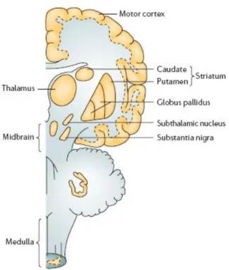
10
Fig. 1. Main brain areas affected in PD.
The standard method to assess motor symptoms and disability in PD is the Unified Parkinson‟s Disease Rating Scale (UPDRS; Ramaker et al., 2002), which can be applied both for patient management and clinical research. The cardinal motor symptoms of PD are rigidity, bradykinesia and resting tremor, complicated in later phases by postural instability, due to impairment of postural reflexes (Pallone 2007). Motor symptoms usually begin asymmetrically but gradually extend to the other side of the body (contralateral side). Asymmetric resting tremor is the common initial symptom (70%-90% of patients; Pallone 2007) and even though it is the most evident, it is rarely a major cause of disability.
On the other hand, the most disabling motor symptom is bradykinesia (slowness of movement), with akinesia (inability to initiate a movement) as its extreme. Bradykinesia also presents as very slow movement, hypophonia (weak voice or whispering as a result of uncoordination of muscles of vocalization), reduced dexterity, a masked face and drooling, decreased blink rate and a slow, shuffling gait.
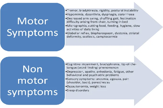
The clinical manifestation of PD is not limited to motor deficit but also includes a variety of non-motor symptoms that significantly affect patients‟ quality of life (Chaudhuri et al., 2006). In this view, the UPDRS is undergoing revisions with the aim to improve sensitivity and integrate non-motor features of the disease (Goetz et al., 2007). Indeed, these signs and symptoms could even precede motor deficits by years and be underestimated in the clinic. It is now accepted that motor features, defined as parkinsonism, represent only one trait of a complex and multifaceted disorder as PD, comprising REM sleep disorders, anosmia, depression, dementia and autonomic failure (Langston 2006; Chaudhuri and Schapira, 2009). These features contribute to motor disability along the course of the disease, dominating the clinical picture as PD progresses, and eventually may contribute to shortened life expectancy (Chaudhuri et al., 2006).
Fig. 2. Table summarizing PD symptomatology.
Parkinson’s Disease: therapeutic interventions
Since its introduction, L-DOPA remains the most effective symptomatic treatment of motor deficits, although it has less impact on non-motor symptoms (Chaudhuri and Schapira, 2009). Currently, no
12
treatment has proved effective in halting or slowing the progression of the neurodegenerative process (Meissner et al., 2011), leaving neuroprotection as an unmet clinical need.
Thus, the most important goals of clinical management are to preserve functional independence and health-related quality of life (Weintraub et al., 2008). In this view, treatment is directed towards symptomatic relief for both motor and non-motor features (Rezak 2007).
As mentioned above, L-DOPA is the cornerstone of the therapy of PD. As a prodrug, levodopa crosses the blood-brain barrier (BBB) and is decarboxylated to DA in the residual nigrostriatal neurons (Pallone 2007) and, as DA loss progresses, also in striatal serotonin (5-HT) terminals. The drug is always given in combination with a peripheral dopa-decarboxylase inhibitor, in order to improve central bioavailability and minimize adverse effects, such as nausea and hypotension, caused by peripheral action of DA (Rao et al 2006). L-DOPA is highly effective in the first phases of therapy, usually up to 5 years („honeymoon period‟). As treatment continues and PD progresses, motor complications, namely wearing-off (shortening of effect duration), dyskinesia (abnormal, involuntary movement) and motor fluctuations appear in virtually all patients.
Since the aim of L-DOPA therapy is to rescue the impairment in DA transmission, other therapeutic strategies involve the use of direct DA receptor agonists. These are used as monotherapy in early PD or in combination with L-DOPA in more advanced phases of the disease. There is an increasing use of these agents as initial therapy to help delay the need for DOPA and reduce overall L-DOPA dosage. However, effects are less robust and patients will eventually require L-L-DOPA. Other pharmacological agents are currently used in the treatment of PD, and most of them maximize the beneficial effects of L-DOPA by augmenting its efficacy and reducing its adverse effects, or cover symptoms not adequately managed by L-DOPA alone. These agents include: MAO-B (monoamino oxidase-B) inhibitors, that prevent DA metabolism in the brain; COMT (Catechol O-Methyltransferase) inhibitors, that inhibit peripheral metabolism of L-DOPA and/or central metabolism of DA; anticholinergic agents that antagonize muscarinic receptors and mainly relieve resting tremor.
Apart from pharmacological treatments, surgery options are also considered in PD therapy. Deep brain stimulation (DBS) of the subthalamic nucleus (STN) is an effective surgical procedure in selected candidates for treating medically resistant motor symptoms of PD (Okun et al., 2007). Gene therapy is a novel therapeutic strategy applied to PD treatment, but with little results obtained so far (Meissner et al., 2011). For example, different clinical studies investigated the effects of the injections of viral vectors that drive the expression of neurturin/glial-derived neurotrophic factor (GDNF) in neurons (Gill et al., 2003; Lang et al., 2006; Marks et al., 2010; Bartus et al., 2011), failing to lead to significant improvements when a large placebo-controlled design was employed (Lang et al., 2006).
Recently, there has been great interest in human stem cells, which have been shown to survive, innervate to some extent and reverse motor dysfunction in rodent and nonhuman primate models of PD (Bjorklund et al., 2002; Cai et al., 2009; Redmond et al., 2007). Human embryonic stem cells are easy to manipulate, but they can form teratomas (Bjorklund et al., 2002). Immunological reactions could be avoided using induced pluripotent stem cells (iPS cells; Kiskinis et al., 2010; Soldner et al., 2009). Unfortunately, they did not show particular efficacy. More experimental data are needed to validate this strategy (see Obeso et al., 2010).
14
Evolution of the classical view of PD
The neuropathological origin of motor symptoms in PD is surprisingly still questionable. Neuronal cell loss has been reported in pre-supplementary motor cortical area in PD patients, and it has been suggested that this damage could occur early in the disease (MacDonald and Halliday 2002). In fact, changes in cortical activation patterns have been reported in concurrence with onset of motor symptoms (Fukuda et al., 2001; Sabatini et al., 2000; Thobois et al., 2000; Cunnington et al., 2001), questioning about the primary cause of motor dysfunction.
The neuronal damage associated with PD causes alterations in neurotransmitter release, not limited to a decrease of DA release in striatum and prefrontal cortex, as could be deducted by the classical histopathological view. The reported functional antagonism of DA and acetylcholine (Ach) transmission at the striatal level leads to increased Ach release (Pisani et al., 2007) although the opposite is true at the cortical level (Francis and Perry, 2007). Indeed, as mentioned earlier, anticholinergic drugs were among the first treatments clinically used to control PD motor symptoms. The 5-HT system is also affected: neuronal loss in the 5-HT-producing raphe nuclei has been reported in PD (Paulus and Jellinger, 1991; Gai et al., 1995). Consistently, reduced 5-HT levels appeared lower in the caudate nucleus and putamen (see Huot et al., 2011) and neocortex (Francis and Perry, 2007). However, animal models of parkinsonism gave conflicting results regarding the 5-HT system. In the MPTP-lesioned mouse a reduction of striatal 5-HT levels accompanied by 5-HT hyperinnervation was reported (Rozas et al., 1998). In other PD animal models the situation appears more variable (for a review, see Huot et al., 2011).
A major advancement in the field has been provided by Braak and colleagues (2003) who analysed and staged the LBs accumulation and pattern in PD patients of different ages, leading to the conclusion that PD spreads from lower brainstem to higher cortical areas. Central to the hypothesis of PD as a prionopathy, was also the evidence that α-synuclein acts and spreads like the prion protein, capable of neuron-to-neuron transmission (Olanow and Prusiner 2009; Desplats et al.,
2009), and that Lewy body-like pathology spreads from host tissue to embryonic nigral transplants (Kordower et al., 2008).
Among non-motor symptoms, dementia is one of the most common (Hely et al., 2008) and difficult to treat (Meissner et al., 2011). PD dementia (PDD) is close in phenotype to Dementia with Lewy Bodies (DLB), which is characterized by α-synuclein-positive inclusions and parkinsonism (Langston 2006), suggesting PD, PDD and DLB might be the same disease but with different points of origin.
It appears clear that the current view of PD is mostly an end-stage one, with attention focused on motor symptoms. The burst on the scene of SNCA (α-synuclein) has paved the way to disclose earlier events in PD pathogenesis.
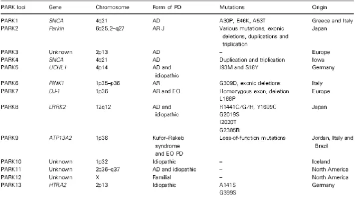
Genetics of PD
Hereditability in PD has long been debated, until Polymeropoulos and colleagues (1997) firstly described pathogenic missense mutations in the SNCA gene (coding for α-synuclein), followed by the discovery of additional genes.
At the moment, gene mutations shown to be causative of PD are grouped in 2 categories: dominantly inherited and recessively inherited mutations.
16
Fig. 4. The 13 PARK loci described to date. VPS35 and eIF4G1 are excluded from this table since they do not map on
the original loci. AD, autosomal dominant; AR, autosomal recessive; J, juvenile; EO, early onset.
Dominantly inherited mutations
α-Synuclein
As said, genetics studies in PD were boosted by the discovery of pathogenic, mutant forms of the SNCA gene. The related protein, α-synuclein, is expressed throughout the brain and has potential roles in learning, synaptic plasticity, vesicle dynamics and DA synthesis (Lotharius and Brundin 2002; Sidhu et al., 2004).
Missense mutations and gene multiplication increase the propensity of α-synuclein to aggregate, and indeed aggregates of beta-sheet-rich fibrillar forms of α-synuclein are the main constituents of LBs in PD and other LB diseases (Spillantini et al., 1998). The function of α-synuclein is still unclear but localization within nerve terminal and interaction with membranes suggest a role in regulation of vesicle dynamics, trafficking at the presynaptic terminal, brain lipid metabolism (Willingham et al., 2003; Darios et al., 2010) and neurotransmitters release at the presynaptic level
through promotion of assembly of the SNARE complex (Burre et al., 2010). Thus, it appears that membrane and lipid regulation pathways could be crucial in developing α-synuclein-related PD. In addition to single-point mutations, duplication and triplication of the SNCA locus elevate α-synuclein expression and cause familial PD (Singleton et al 2003; Farrer et al 2004). However, it remains unclear whether α-synuclein expression is elevated in the brains of sporadic PD patients. The implication of α-synuclein in PD pathology appears to be related to its structural properties. Given its presence in LBs as fibrillar forms, it has been thought that insoluble aggregates are cytotoxic. However, α-synuclein monomers interact to form pre-fibrillar aggregates or protofibrils, which in turn can form insoluble fibrils (Gosavi et al., 2002; Conway et al., 2000; Conway et al., 2001). A recent hypothesis proposes that protofibrils are the cytotoxic species, whereas the fibrillar aggregates of the protein could represent a cytoprotective mechanism in PD (Dev et al., 2003; Caughey and Lansbury, 2003). Supporting this hypothesis, α-synuclein protofibrils are increased in the brains of PD and DLB patients (Sharon et al., 2003), and have been associated with neurotoxicity in α-synuclein-overexpressing cells and mouse models (Masliah et al., 2000; Gosavi et al., 2002).
A number of animal models have been developed to study α-synuclein role in PD pathology. Overexpression of the protein in transgenic animals has been the most common method. In this regard, the promoter used to drive the overexpression appears to play an important role: the tyrosine hydroxylase (TH) promoter was used in several mouse lines, leading to interesting results on the induction of neurodegeneration (Thiruchelvam et al., 2004). However, this does not represent an ideal strategy because expression is restricted to catecholaminergic neurons, and models should mimic the broad but regionally selective α-synuclein pathology observed in patients (Halliday et al., 2006). In fact, other promoters like PDGFβ and Thy1 have been used. Both confer broad expression of α-synuclein in neurons but with different patterns (Rockenstein et al., 2002) and functional outcomes. In particular, one line with the PDGFβ promoter develops DAergic deficits in the striatum (Masliah et al., 2000, 2005). The Thy1 lines have a wide range of phenotypes, depending
18
on mutations, background strain, level of expression and/or the insertion site. Mice overexpressing wild-type α-synuclein under the control of Thy1 promoter have been extensively characterized. In these mice the protein is widespread at high levels in cortical and subcortical regions (including SNc; Rockenstein et al., 2002) with proteinase K-resistant inclusions of α-synuclein in key regions such as olfactory bulb, SN and locus coeruleus (Fernagut et al., 2007). This pattern represents a hallmark of α-synuclein pathology in PD brains (Neumann et al., 2004; Halliday et al., 2006). In addition to transgenic methods, α-synuclein has been overexpressed using viral vectors. This approach leads to a rapid degeneration of nigrostriatal neurons, which is not yet reproduced by genetic mutations in mice and rats (Kirik and Bjorklund, 2003), even with the wild-type protein (Kirik et al., 2002).
Even though genetic models often lack nigrostriatal DA cell loss, considered a “hallmark of PD”, they strikingly reproduce early characteristics of the disease (Halliday and McCann, 2008; Braak et al., 2004) making them particularly useful for assessing pathogenic mechanisms and early therapeutic strategies.
Leucine-rich repeat kinase 2
Mutations in leucine-rich repeat kinase 2 (LRRK2) gene have recently been demonstrated to cause autosomal-dominant, late-onset PD (Zimprich et al., 2004; Paisan-Ruiz et al., 2004). The gene is located in the locus PARK8, which was originally mapped as an autosomal dominant trait in a Japanese family with asymmetrical, L-DOPA-responsive, late-onset PD (Funayama et al., 2002). Glycine to serine substitution in position 2019 is the most common LRRK2 mutation, and in specific populations it might account for 18-30% of PD cases (Ozelius et al., 2006; Lesage et al., 2006). Of note, disease penetrance in G2019S carriers is age dependent, representing a critical consideration in genetic counseling (Kachergus et al., 2005). Interestingly, the G2019S mutation is associated with a common haplotype throughout the world. This haplotype is smallest in Arabic
patients and indicates that this mutation might have originated in the Middle East (Kachergus et al., 2005; Ozelius et al., 2006; Lesage et al., 2006).
Since the LRRK2 protein is the main subject of this thesis, its structure, properties and biological significance will be introduced in a dedicated chapter.
Vacuolar protein sorting 35
Very recently, pathogenic mutations, and in particular the D620N substitution, were identified within vacuolar protein sorting 35 (VPS35) as a novel genetic determinant of autosomal dominant late-onset parkinsonism (Vilarino-Guell et al., 2011; Zimprich et al., 2011).
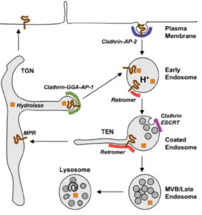
VPS35 is a component of the retromer cargo-recognition complex, critical for membrane-protein recycling and endosome-trans-Golgi network (TGN) trafficking. The TGN is the last sorting station of the secretory pathway from which soluble or membrane proteins and lipids are sorted for subsequent transport to different destinations: the cell surface, the endosomal system and synaptic domains in neurons (Anitei et al., 2010). Within the TGN, transmembrane proteins are sorted for delivery to the plasma membrane and the endosomal/lysosome systems. This includes transmembrane proteins cycling between the TGN and endosomes, such as mannose-6-phosphate receptors (MPR), which deliver bound lysosomal enzymes to endosomes in a mannose-6-phosphate (M6P) dependent manner (Munier-Lehmann et al., 1996; Ghosh et al., 2003). After unloading their bound ligands in endosomes, they return either to cell surface or TGN; membrane coats containing clathrin, GGA and AP-1 mediate the exit of hydrolase-MPR from TGN, whereas the retromer complex mediates the retrieval of unoccupied MPR from endosomes (Anitei et al., 2010; Rojas et al., 2008; Bonifacino and Hurley, 2008).
In addition to MPR recycling, retromer function is necessary for normal endocytosis, as in the case of progranulin, linked to fronto-temporal dementia (Anitei et al., 2010; Hu et al., 2010), and for processing of the amyloid precursor protein (APP) linked to Alzheimer‟s Disease (Nielsen et al., 2007; Vieira et al., 2010). Moreover, the retromer complex has important roles in actin cytoskeletal
20
organization, synapse formation (Korolchuk et al., 2007), Wnt signaling (Franch-Marro et al., 2008), mitochondrial peroxisome trafficking (Braschi et al., 2010) and nuclear export (Mingot et al., 2004). The role of retromer might be particularly relevant in neurons, given they are excitable cells that support ~10.000 times more plasma membrane surface than other cell types. Indeed, protein trafficking, recycling and degradation are essential for the creation and maintenance of neuronal architecture, synaptic plasticity and connectivity (Lasiecka and Winckler, 2011; Sann et al., 2009). VPS35 has not been extensively studied in the context of neurological disorders, but these recent genetic findings will prompt the investigation of its role in neurobiology.
Fig. 5. An example of retromer function: sorting of acid hydrolases by transmembrana MPR cycling between the TGN
and endosomes. From Bonifacino and Hurley (2008).
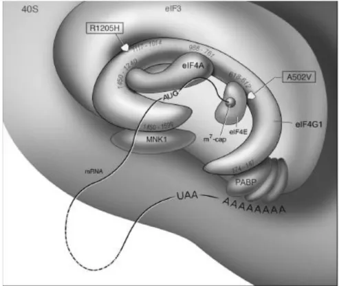
Eukaryotic translation initiation factor 4-gamma
Very recently, genome-wide analysis of families with autosomal-dominant parkinsonism revealed a missense mutation (R1205H) in eukaryotic translation initiation factor 4-gamma (eIF4G1; Chartier-Harlin et al., 2011). The observed clinical phenotype is consistent with late-onset PD. Symptoms
start insidiously with asymmetric resting tremor or akinetic rigidity, and become progressively mixed. In the same study, eIF4G1-R1205H was found to segregate with idiopathic PD. Moreover, the shared haplotype observed suggests that the mutation originates from an ancestral founder and segregates with disease in seemingly unrelated families.
eIF4G1 is the core scaffold of a multisubunit complex that regulates translation initiation of mRNAs encoding mitochondrial, cell survival and growth genes in response to different stresses (Ramirez-Valle et al., 2008; Silvera et al., 2009). This complex is composed, other than eIF4G1, by cap-binding protein eIF4E and ATP-dependent RNA helicase eIF4A. Notably, the R1205H mutation in eIF4G1 perturbs binding with eIF4E while the wild-type protein does not; moreover, oxidative stress results in loss of mitochondrial membrane potential selectively in cells overexpressing the mutant protein (Chartier-Harlin et al., 2011), an effect also observed after eIF4G1 silencing (Ramirez-Valle et al., 2008). This data indicate that the mutation causes a loss of function of the protein.
The mutation in eIF4G1 affects mRNA translation initiation in parkinsonism and might point to a convergent pathway for different forms of disease (Hawkes et al., 2009; Zhou et al., 2008). In this regard, it is important to note that availability of eIF4E is generally the rate-limiting step of translation initiation and is regulated by phosphorylation of eIF4E-binding proteins (4E-BP) through the mammalian target of rapamycin (mTOR) pathway (Ramirez-Valle et al., 2008; Ma and Blenis, 2009). The 4E-BP protein has been shown to be a substrate of human LRRK2, with pathogenic mutants of this protein causing hyperphosphorylation of 4E-BP and reduced resistance to oxidative stress (Gehrke et al., 2010). Finally, activation of mTOR signaling and 4E-BP phosphorylation are associated with L-DOPA-induced dyskinesia (Santini et al., 2009), further stressing the involvement of translation initiation in the context of parkinsonism.
22
Fig. 6. Scheme of eIF4G1 protein interactions. Approximate positions of A502V and R1205H mutations are
highlighted.
Recessively inherited mutations
Recessively inherited deletions and missense mutations have been identified in family-based linkage studies, but they are rare overall, causing <1% of early-onset parkinsonism (Lockhart et al., 2004).
Parkin
Mutations in the parkin gene were originally identified in Japanese families with autosomal recessive, juvenile parkinsonism (Kitada et al., 1998). The protein contains an N-terminal ubiquitin-like domain and two RING-finger domains. It is thought to function as an E3-ligase, conjugating ubiquitin to proteins targeted to proteasome degradation (Shimura et al., 2000). Point mutations are the most common genetic lesions in parkin, although exonic rearrangements, deletions and duplications are also common (Mata et al., 2004). Other than that, patients with homozygous exonic deletions leading to complete loss of parkin expression show selective loss of DA neurons in SN
and locus coeruleus without LB or neurofibrillary tangle pathology, in contrast to that described in patients with heterozygous mutations (Pramstaller et al., 2005). These different outcomes might be mutation-specific (difference in alteration of parkin E3-ligase activity and substrate specificity; Sriram et al., 2005), but, as shown for LRRK2 mutations, different end-stage pathologies might share the same primary cause (Farrer 2006).
Animal models modeling parkin loss-of-function have been described; in parkin-null mice mitochondrial dysfunction and oxidative stress have been implicated in disease pathogenesis. However, none of the parkin knockout mice have any substantial DAergic or behavioral abnormalities (Goldberg et al., 2005; Itier et al., 2003; Perez and Palmiter, 2005; Von Coelln et al., 2004). Interestingly, parkin overexpression appears to be neuroprotective, indicating its upregulation as one route to therapy (Lo Bianco et al., 2004; Greene et al., 2005).
PTEN-induced kinase 1
Homozygous mutations in PTEN-induced kinase 1 (PINK1) were originally found to co-segregate with early-onset parkinsonism in a family-based linkage study (Valente et al., 2001). Mutations were then identified in 1-2% of cases of early-onset disease (Hatano et al., 2004). PINK1 protein comprises a mitochondrial targeting motif and a highly conserved kinase domain, which is shared with the Ca2+/calmodulin family of serine-threonine kinases (Valente et al., 2001).
Protein mutations has different impacts on protein stability, localization and kinase activity (Petit et al., 2005; Beilina et al., 2005). The wild-type protein seems to protect from stress-induced mitochondrial dysfunction and apoptosis (Deng et al., 2005). Also in this case, transgenic mouse models have been developed. Similar to parkin knockout mice, PINK1 knockout mice did not show any major abnormalities (Gautier et al., 2008; Gispert et al., 2009), particularly in DA neurons (Kitada et al., 2007). Again, similar to parkin knockout mice, ablation of PINK1 resulted in mitochondrial defects (Gautier et al., 2008; Palacino et al., 2004). Of note, Drosophila flies lacking PINK1 exhibited behavioral deficits along with mitochondrial pathology. Transgenic expression of
24
parkin in these flies ameliorated the PINK1 loss-of-function phenotype, whereas transgenic expression of PINK1 had no effect on parkin loss-of-function phenotypes (Clark et al., 2006; Park et al., 2006). This led to the accepted hypothesis that parkin and PINK1 are engaged in a common signaling pathway, with PINK1 working upstream of parkin.
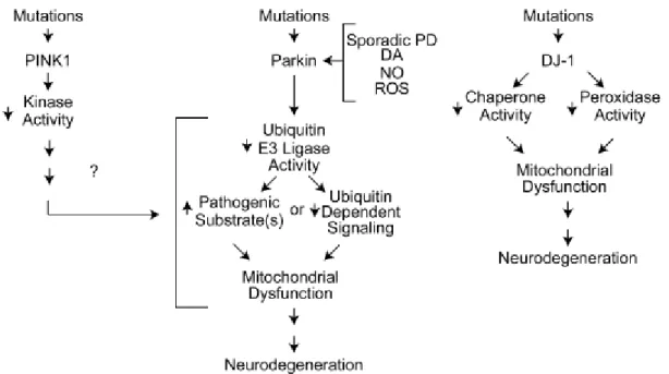
DJ-1
The DJ-1 protein is a member of the ThiJ/PfpI family of molecular chaperones (Gasser, 2009; Moore et al., 2006). These chaperones are induced during oxidative stress and the protein primarily exists as a dimer localized to mitochondria (Tao et al., 2003; Zhang et al., 2005). PD-associated mutations produce DJ-1 loss of function by causing defective dimer formation or lack of expression (Macedo et al., 2003; Moore et al., 2003). The DJ-1 protein is a redox-sensitive molecular chaperone regulating, among other, redox-dependent kinase signaling pathways, and acting as a regulator of antioxidant gene expression (Kahle et al., 2009). Indeed, primary cultures form DJ-1-null mice showed increased sensitivity to oxidative stress, whereas overexpression provided a protective effect (Goldberg et al., 2005; Kim et al., 2005).
DJ-1 knockout mice have been developed as well, without any major detectable abnormalities. However, in some DJ-1 knockout animals changes in the nigrostriatal transmission and mitochondrial dysfunction were reported (Andres-Mateos et al., 2007; Goldberg et al., 2005; Kim et al., 2005).
Fig. 7. Possible molecular mechanisms in autosomal recessive PD (Dawson et al., 2010).
LRRK2: structure, properties and neurobiology
Biochemistry and cellular biology of LRRK2
The LRRK2 protein, also named dardarin, derives its name for the presence of leucine-rich repeats (LRR) and a kinase domain in its structure. Between these two regions, a GTPase sequence called ROC (Ras of complex proteins; Bosgraaf and Van Haastert, 2003) and an adjacent COR (C-Terminal of ROC) domain can be found. These domains are characteristic of the ROCO superfamily of proteins (Marin et al., 2008), which all contain ROC-COR tandem domains, but not all have kinase domains. LRRK2 kinase domain has been hypothesized to derive evolutionarily from different sources and to be quite divergent in sequence from the origin (Marin 2006, 2008; Marin et al., 2008). It was initially proposed that the kinase domain was related to the MLKs (mixed lineage kinases), but analysis of all kinase domains throughout the human genome suggests that LRRK2 is part of a small offshoot group of the RIPK (receptor-interacting protein kinase) family of kinases, which are partially similar to the IRAK (interleukin 1 receptor-activated kinase) family and more distant from the MLKs (Manning et al., 2002).
26
The N-terminal region of LRRK2 is composed of different repeat sequences (LRR, heat, Ankyrin). These are very likely to be protein-protein interaction motifs, indicating that LRRK2 can act as a scaffold for several other proteins. This view is confirmed by the presence of a WD40 domain at the C-end of the protein, which can also interact with lipids (McArdle and Hofmann, 2008). Indeed LRRK2 has been reported to interact with lipid rafts in synaptic terminals and to associate with membrane structures (Hatano et al., 2007). The complexity of LRRK2 function is increased by its ability to self-interact (Gloeckner et al., 2006) and to form a dimer (Greggio et al., 2008).
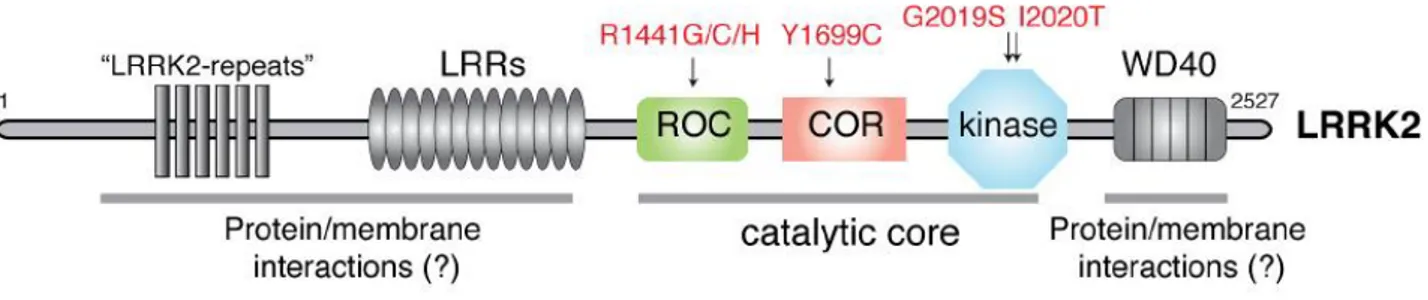
Fig. 8. Schematic representation of LRRK2 protein structure (in red, PD-causing mutations).
Mutations in LRRK2 proven to be pathogenic affect different domains of the protein: R1441C/G/H substitutions are located in the enzymatic ROC domain, Y1699C is found in the COR domain, G2019S and I2020T mutations are in the kinase domain (Paisàn-Ruìz et al., 2008). The most common mutation, G2019S, is proposed to exert its pathogenicity through an increase of kinase activity (Greggio et al., 2006; Jaleel et al., 2007; Nichols et al., 2010; West et al., 2005). On the other hand, point mutations located in the ROC and COR domains have been shown to decrease GTPase activity (Guo et al., 2007; Lewis et al., 2007). Finally, the I2020T substitution has been reported to stimulate phosphorylation of 4E-BP [eIF4E (eukaryotic initiation factor 4E)-binding protein] in some models (Imai et al., 2008) but has no effect on kinase activity in other assays (Nichols et al., 2010). Thus, there might be mechanistic differences between different mutations located in the same domain, with the caveat that these observations could also be due to methodological differences in the various assays (Greggio and Cookson, 2009).
The role of LRRK2 in the cellular context has not yet been defined, mostly due to the uncertainty about molecular substrates and/or partners of LRRK2. The first indications came from studies showing cytotoxic effects of mutant LRRK2. Iaccarino and colleagues (2007) reported R1441C-LRRK2 to cause apoptosis in cell lines, through release of cytochrome c and caspase-3 activation. Moreover, they showed that LRRK2-mediated cell death is dependent upon mitochondrial dysfunction and apoptosome formation. Interestingly, LRRK2 constructs devoid of LRR or WD40 domains did not induce apoptosis, highlighting the importance of non-enzymatic sequences in the protein function. Later, LRRK2 has been reported to interact with the adaptor FADD (Fas-associated protein with death domain), with PD-mutants inducing apoptosis through caspase-8 activation (Ho et al., 2009). This study proposed the apoptotic extrinsic pathway (mediated by cell surface death receptors) as opposed to the intrinsic (mitochondria-mediated) pathway, as a mechanism for LRRK2 cytotoxicity.
However, signaling pathways and mechanisms underlying LRRK2-mediated effects are still under debate. Great efforts have been spent on the identification of molecules interacting with LRRK2 in cells. As previously mentioned, 4E-BP has been shown to undergo phosphorylation in cell cultures overexpressing LRRK2. This leads to inactivation of 4E-BP and, possibly, dysregulation of protein translation (Imai et al., 2008). The Ezrin-Radixin-Moesin (ERM) family of proteins may also be a LRRK2 target (Parisiadou et al., 2009). These proteins are involved in cell maintenance, regulating cellular shape, growth and motility (Mangeat et al., 1999; Bretscher et al., 2002). Far from being the only interactions reported, LRRK2 has been shown to activate the ERK (extracellular signal regulated kinase) pathway in a kinase-dependent manner: PD mutations delay the phosphorylation of MEK2, the upstream activator of ERK, resulting in delayed activation of ERK itself (Carballo-Carbajal et al., 2010). Consistently, oxidative stress stimulated phosphoERK to a lower extent and induced greater cell death in Y1699C-LRRK2 overexpressing cells compared to cells expressing the wild-type protein (Liou et al., 2008). In addition, inactivation of LRRK2 led to decreased amount of phosphorylated 4E-BP, thus probably acting along the Akt/TOR signaling pathway (in
28
which ERK is also involved; Tain et al., 2009). The involvement of LRRK2 in MAPK (mitogen-activated protein kinases) pathway is further suggested by its ability to phosphorylate MAPKK members MKK3/6 and MKK4/7. Moreover, PD mutations located in the LRRK2 kinase domain showed increased phosphorylation of MKK6 (Gloeckner et al., 2009). Along their signaling pathways, MKK3/6 and MKK4/7 activate, respectively, the generically pro-apoptotic p38 and JNK (c-Jun N-terminal kinase; Berwick and Harvey, 2011). Disappointingly, different studies reported no effect of LRRK2 on p38 phosphorylation and JNK activity (West et al., 2007; Carballo-Carbajal et al., 2010), when overexpressed in HEK cell lines. Only West and colleagues (2007) reported activated JNK signaling in SH-SY5Y cell lines overexpressing LRRK2.
Finally, LRRK2 has also been linked to the Wnt signaling pathway through the interaction with central components such as disheveled (DVL) proteins 1-3 (Sancho et al., 2009) and glycogen synthase kinase-3β (GSK-3β; Lin et al., 2010). In the same studies, pathogenic LRRK2 mutations modified these interactions.
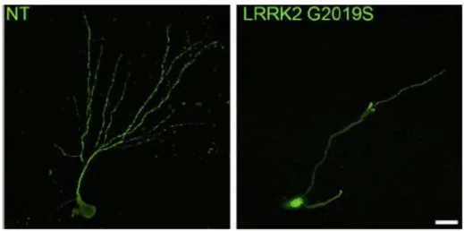
Along with cell lines, LRRK2 functions have been extensively studied in primary neuronal cultures, in order to disclose the role of the protein on neuronal cell physiology. Transgenic expression of human G2019S-LRRK2 in neurons derived from Drosophila induced dendrite arborization defects in DA but not 5-HT cells. In this context, mutant LRRK2 toxicity is mediated by phosphorylation of tau through GSK3β (Lin et al., 2010). Consistently, evidence of reduced neurite outgrowth and branching (Dachsel et al., 2010), reduced dendritic arborization and length together with reduced neurogenesis in dentate gyrus (DG) and subventricular zone (SVZ; Winner et al., 2011) have been obtained in primary cultures from LRRK2 transgenic mice. Interestingly, neuronal cultures from LRRK2 knockout mice showed increased process length and branching (Dachsel et al., 2010). Moreover, LRRK2-G2019S reduced the density of dendritic spines, with a particular reduction in mushroom spines (Winner et al., 2011), which are usually more abundant in mature neurons (Nimchinsky et al., 2002; Sala, 2002). Thus, it has been hypothesized that mutant LRRK2 might
cause neurotoxic effects through inhibition of neurite outgrowth and interference with maturation of filopodia into mature spines.
Fig. 9. Defects in neurite outgrowth in neurons from LRRK2-G2019S mice (Winner et al., 2011).
LRRK2 functions in animal models
The LRRK2 protein has been extensively studied in a variety of animal models, including
Drosophila, C. Elegans and rodents. A thorough analysis of non-mammalian models is beyond the
scope of this thesis, thus only rodents studies will be reviewed.
In the mouse brain, LRRK2 expression was detected at high levels in the hippocampus, striatum, cortex and cerebellum, with no prominent glial staining. Notably, intense labeling was found in striatal medium spiny neurons. At the midbrain level, LRRK2 staining was higher in SNc than SNr (Melrose et al., 2007). Consistently, the majority of DA neurons in rat SN express LRRK2. Of note, LRRK2 is preferentially expressed in SN compared to the adjacent VTA (ventral tegmental area; Han et al., 2008).
Mice overexpressing human LRRK2 through bacterial artificial chromosome (BAC) transgenesis were firstly reported. This model provided a genetic tool for studying gene function in vivo. BAC carries an intact genomic regulatory sequence and confers precise temporal and spatial expression pattern of the transgene under the control of the endogenous promoter (Heintz, 2001).
30
In one of the initial studies, R1441G-LRRK2 BAC mice showed an age-dependent, L-DOPA responsive motor deficit. Moreover, these mice showed slightly decreased striatal DA release and axonal pathology with hyperphosphorylated tau. However, no nigral neuronal degeneration was reported (Li et al., 2009). At variance, BAC G2019S-LRRK2 mice failed to reproduce a parkinsonian behavioral phenotype (Li et al., 2010; Melrose et al., 2010), although a reduction of striatal DA content as well as spontaneous and stimulus-evoked extracellular DA release was found (Li et al., 2010; Melrose et al., 2010). Histopathological signs, e.g. accumulation of hyperphosphorylated tau, were reported in older mice (Melrose et al., 2010), although these were not accompanied by DA cell death in SN.
At the same time, different groups induced a targeted insertion of a PD pathogenic mutation in the LRRK2 gene (knock-in models, KI). This technique generates a transgenic mouse with physiological levels of expression of the gene of interest.
KI mice bearing the R1441C mutation (Tong et al., 2009) are behaviorally normal even at old ages. However, pharmacological manipulation of the DA system mice showed reduced locomotor response to amphetamine and quinpirole, with respect to controls. While the number of DA neurons in SNc and striatal DA content were unchanged, spontaneous firing activity in mesencephalic brain slices was less responsive to DA drugs, consistent with behavioral data. Thus, dysregulation of the nigrostriatal DA system is suggested by the different models so far presented (Tong et al., 2009). Lately, KI G2019S mice have also been generated (Herzig et al., 2011). However, these mice did not show abnormalities in DA transmission and responses to DA agonists. Recently, mice expressing full-length human LRRK2 under the control of a cytomegalovirus-enhanced (CMVE) human platelet-derived growth factor β-chain (CMVE-PDGFβ) promoter (Ramonet et al., 2011) have been generated. In these mice, a different histopathological situation has been reported at
19-21 months of age: G2019S-LRRK2 mice displayed reduction in the number of TH+ SNc neurons
but not striatal TH+ fiber density. This might be due to compensatory re-sprouting of surviving
previous studies, primary mesencephalic cell cultures obtained from these animals showed reduced neurite length and branching.
Overall, these results suggest that LRRK2 transgenic animals hardly show neurodegeneration in SNc (considered a hallmark of PD), but do have alterations in the activity and morphology of DA neurons. Moreover, mutant LRRK2 impairs maturation of dendrites and spines, thus negatively affecting synaptic communication.
Fig. 10. Summary of LRRK2 transgenic models reported to date (Yue and Lachenmayer, 2011).
Different studies independently reported generation of animal lines with targeted deletion of the endogenous LRRK2 gene (knock-out, KO). Data presented so far indicate that the LRRK2 gene is not essential for mouse survival and does not play a role in early neural development, differentiation or viability. In addition, all KO mice are viable and fertile and exhibit no detectable motor function
32
abnormality (Tong et al., 2010; Lin et al., 2009; Andres-Mateos et al., 2009). Interestingly, deletion of the LRRK2 gene induced renal abnormality at old age, with cell type-dependent impairment of protein degradation pathways, accumulation of α-synuclein and enhanced apoptotic cell death (Tong et al., 2010). However, these observations have not been mechanistically linked to pathological changes in the brain, which actually appears unaffected in these animals.
Along with genetically modified models, viral delivery of LRRK2 was also reported (Lee et al., 2010; Dusonchet et al., 2011). In striking contrast with the previously reported models, intrastriatal delivery of LRRK2 mutants through HSV (Lee et al., 2010) or AAV (Dusonchet et al., 2011) carriers induced neuronal cell death in SNc. This difference may be due to activation of glial inflammatory pathways elicited by the viral construct and the specific spatial and temporal expression of the protein (Yue and Lachenmayer, 2011).
34
The main aim of this study was to investigate the role played by LRRK2 in cell and animal neurophysiology, with particular attention to motor behavior and DA transmission in vivo, and synaptic morphology in vitro. The study has been divided in two independent parts, corresponding to the work performed at the University of Ferrara (part #1) and the University of British Columbia (part #2).
Part 1. In vivo phenotypic characterization of LRRK2 effects on motor activity and motor responses to DA receptors ligands. The first part of the study involved the use of three different mouse lines: wild-type mice as controls; transgenic BAC mice carrying the G2019S mutation in the human LRRK2 gene; knock-out (LRRK2-KO) mice bearing deletion of the endogenous LRRK2 gene. First, we performed a motor characterization of these mice through tests specific for spontaneous and exercise-induced motor activity. Mice were divided in age-matched groups (3, 6 and 12 month-old), in order to investigate whether LRRK2 affects the worsening of motor function associated with aging. Then, the motor responses of these mice to DA receptor agonists and antagonists were assessed.
Part 2. In vitro investigation of LRRK2 effects on synaptic morphology in cortistriatal co-cultures. This study was designed to fulfill two aims at the same time. First, we planned to test the novel LRRK2 selective kinase inhibitor LRRK2-IN1 (Deng et al., 2011) in rat primary neuronal cultures. Second, we investigated the involvement of LRRK2 in the development and maintenance of dendritic spines and synaptic contacts. In this view, we employed a novel approach using co-cultures of two different neuronal populations, namely cortical cells and striatal medium spiny neurons (MSNs).
MATERIALS AND
METHODS
36
IN VIVO EXPERIMENTS Subjects
Non-transgenic wild-type, BAC hLRRK2-G2019S and LRRK2-KO mice, backcrossed on a C57BL/6J strain, were obtained from Mayo Clinic (Jacksonville, FL, USA) through a collaboration with Prof. Matthew Farrer and Dr. Heather Melrose. Colonies were grown in the vivarium of the Section of Pharmacology, at the Department of Experimental and Clinical Medicine (University of Ferrara). Animals were kept under regular lighting conditions (12 h light/dark cycle) and given food and water ad libitum. The experimental protocols performed in the present study were approved by the Italian Ministry of Health and the Ethical Committee of the University of Ferrara, and adequate measures were taken to minimize animal pain and discomfort.
Behavioral experiments
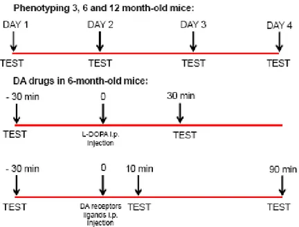
Physiologically-stimulated motor activity
Three behavioral tests were used to evaluate different motor functions as previously described: the bar, drag and rotarod tests (Marti et al., 2005; Viaro et al., 2008). The bar test (Kuschinski and Hornykiewicz, 1972; for a review see Sanberg et al., 1988) measures the animal ability to respond to an externally imposed static posture, and can be taken as a measure of akinesia (time to initiate a movement). The drag test (modification of the “wheelbarrow test”; Schallert et al., 1979) measures animal ability to balance body posture using the forelimbs in response to an externally applied dynamic stimulus (i.e. backward dragging), and can be taken as an index of akinesia and bradykinesia (slowness of movements). The fixed-speed rotarod test (Rozas et al., 1997) measures overall motor performance as an integration of coordination, gait, balance, muscle tone and motivation to run. The three tests were performed in a fixed sequence (bar, drag and rotarod). During behavioral phenotyping the sequence was performed daily during the light cycle (09.00 – 11.00), for four consecutive days (D1 to D4). For pharmacological testing, the sequence was
repeated within the same experimental session, before (control) and after (at 10 and 90 min) drug injection. When L-DOPA was tested, post injection time analysis was set at 30 min.
- Bar test: mice were placed on a table and each forepaw was placed alternatively on blocks of increasing heights (1.5, 3 and 6 cm). Total time (in s) spent by each paw on the blocks was recorded (cut-off time 20 s). Performance was expressed as total time spent on bar. Since performance at the left and right forepaw did not differ, data were pooled together.
Fig. 12 . Mouse undergoing the bar test.
- Drag test: mice were lifted from the tail (allowing forepaws to rest on the table) and dragged backwards at a constant speed (~20 cm/s) for a fixed distance (100 cm). The number of steps made by each forepaw was counted by two separate observers. Performance was expressed as total number of steps, pooling together the left and right forepaw.
38
- Rotarod test: the fixed-speed rotarod test was employed using an established protocol (Marti et al., 2005; Viaro et al., 2008). Briefly, mice were tested on a rotating rod (diameter of the cylinder 8 cm), whose speed was stepwise increased (every 180 s) from 5 to 55 rpm. The time spent on the rod was recorded.
Fig. 14. Mouse performing the rotarod test.
For behavioral phenotyping, untrained mice were tested at three different ages: 3, 6 and 12 months old. Pharmacological testing was then performed on 6 month-old mice.
Spontaneous motor activity
Experiments were performed during the light cycle (between 09.00 and 13.00) according to Guerrini et al. (2009). Six-months old wild-type, BAC hLRRK2-G2019S and LRRK2-KO mice were monitored for 30 min. For these experiments the ANY-maze video tracking system was used (Ugo Basile, application version 4.52c Beta). Mice were positioned in a square plastic cage (40 cm × 40 cm), one mouse per cage. Four mice were monitored in parallel. The central zone of the open field was defined as the central 20 cm × 20 cm square. Mouse horizontal activity was monitored by a camera while vertical activity was measured by an infrared beam array. The parameters measured were: cumulative distance traveled (total distance in meter that the animal traveled during the test), immobility time (the animal is considered immobile when 90% of it remains in the same place for a
minimum of 2.5 s), number of rearings (the number of beam breaks due to vertical movements), number of entries in the central zone, and time spent by the animal in the central area of the field. An entry in the central zone was marked when the entire area of the animal was in the central square, and the time in the central zone is defined as the amount of time in seconds that the animal spent in the central square.
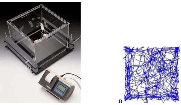
A B
Fig. 15. A. Activity cage for open field test, equipped with infrared laser beams for assessment of vertical activity. B.
Typical tracking of mouse horizontal locomotion in the cage.
Injected drugs
L-DOPA and benserazide were purchased from Sigma Chemical Company (St. Louis, MO, USA). Haloperidol, SCH23390 and SKF38393 were purchased from Tocris Bioscience (Bristol, UK). Pramipexole was purchased from McTony Bio and Chem (Vancouver, BC, Canada). All drugs were freshly dissolved in saline just prior to use and the volume injected was 10 μl/g body weight.
40
Fig 16. Experimental design adopted in physiologically-stimulated motor behavior and pharmacological experiments.
IN VITRO EXPERIMENTS
Primary neuronal culture and transfection
Cultures were prepared as in Fan et al (2010). Briefly, pregnant Wistar rats were anesthetized with halothane and decapitated at embryonic day 17-18. Embryos were extracted and the brains removed and placed on ice in Hank‟s Balanced Salt Solution (HBSS, GIBCO). For transfections, 3-5 millions cells were suspended in 100 μl of electroporation buffer (Mirus Bio) with 1-5 μg of endonuclease-free DNA, placed in a cuvette (0.2 μm, Biorad) and electroporated (AMAXA nucleofector I: program 03 for cortex and 05 striatum). Solution was removed from the cuvette and resuspended in D minimum essential medium (GIBCO) plus 10% fetal bovine serum (FBS; DMEM+), and plated as described above. After 2-4h DMEM+ was replaced with 500 μl plating medium (PM, 2% B27, Invitrogen; penicillin/streptomycin; 2 mM α-glutamine; neurobasal medium, GIBCO). At day in vitro (DIV) 4, 500 μl was added, with subsequent half media changes every 3-5 days. DNA plasmid was a YFP on a CAG promoter (a gift from S. Kaech and G. Banker, Oregon Health Sciences University, Portland, with permission of J. Miyazaki for the CAG promoter; Kaech and Banker, 2006; Niwa et al., 1991). In cortico-striatal co-cultures, either cortical
(CTX) or striatal MSNs were YFP-nucleofected to differentiate between cell types and then plated with non-transfected cells, either MSNs or CTX, at a ratio of 1:1.
Treatment protocol
Cell cultures were divided in three treatment groups, namely DMSO (vehicle), LRRK2-IN1 1 μM and LRRK2-IN1 10 μM. Each treatment was applied for 3, 24 or 72 h before fixation. However, all cells were fixed at 21 DIV, to obtain comparable growth trends. LRRK2-IN1 was obtained by the laboratory of Prof. Matthew Farrer (Centre for Applied Neurogenetics, University of British Columbia, Vancouver, BC, Canada) from the Michael J. Fox Foundation LRRK2 Consortium, through Drs. Dario Alessi and Nathaniel Gray (University of Dundee, UK).
Immunocytochemistry
Coverslips were fixed in 4% paraformaldehyde (PFA) + 4% sucrose for 10 min and rinsed 3 times with phosphate buffered saline (PBS). Next, coverslips were washed (5 min, PBS plus 0.03% triton X-100; Sigma, PBST) and blocked (30 min, 10% normal goat serum, NGS, in PBS). Primary antibodies were incubated overnight with shaking at 4°C in PBST plus 2% NGS, subsequently incubated at RT for 1h, washed 3 times with PBST, and incubated for 1.5h at RT with secondary antibodies (Alexa-488 and Alexa-568 conjugated chicken and mouse, Molecular probes; α-guinea pig AMCA, Jackson Laboratories). For live staining, cells were incubated for 10 min at 37°C with α-GFP (cross reactive with YFP) in conditioned media, rinsed 2 times with media, fixed in PFA-sucrose for 10 min, rinsed 3 times with PBS, and incubated with secondary antibody for 1.5h. For labeling with VGlut1 and PSD95, cells were subsequently permeabilized with methanol for 5 min at -20°C, rinsed 3 times with PBS, washed for 5 min with PBST, then treated with primary antibodies overnight, washed 3 times with PBST, and incubated with secondary antibodies as above. Coverslips were slide mounted with fluoromount (Southern Biotech).
42
Antibodies included: α-green fluorescent protein (GFP, chicken, AbCam ab13970, 1:1000), α-post synaptic density protein 95 (PSD-95, mouse, Thermo Scientific, MA 1-045, 1:1000), α-vesicular glutamate transporter 1 (VGluT1, guinea pig, Chemicon, AB5905, 1:4000).
Microscopy and image analysis
Images were acquired using an Olympus Fluoview-1000 confocal laser scanning microscope; acquisition was performed using a 63x objective (1.4 Oil Plan-Apochromat; 4-8 stacks of 0.5 μm) and extended focus projections were created from 4-5 images containing all of the visible dendritic surface staining and the glass-attached somatic membrane. For cluster detection and localization images were manually thresholded by a blinded experimenter, and analysis of co-localization was conducted in small regions of interest around three secondary dendrites. Cluster analysis was performed in ImageJ; co-localization was calculated using an ImageJ colocalization plugin (http://rsb.info.nih.gov/ij/plugins/colocalization.html) as in Tapia et al. (2011). Points of colocalisation were defined as regions > 1 pixel in size where the intensity ratio of the two channels was > 50.
DATA PRESENTATION AND STATISTICAL ANALYSIS
For in vivo experiments, data are presented as absolute values with mean ± SEM. Statistical analysis was performed by one-way repeated measure (RM) ANOVA, implemented on a excel spreadsheet, followed by Bonferroni post-hoc test. In both cases, limit of significance was set at p<0.05. For in vitro experiments, data are presented as mean ± SEM obtained from 3 different cultures. Statistical analysis was performed with one-way analysis of variance (ANOVA) followed by Turkey‟s Multiple Comparison post-hoc test, using GraphPad Prism software (San Diego, CA, USA).
44
IN VIVO EXPERIMENTS
Characterization of motor phenotype in wild-type, BAC hLRRK2-G2019S and LRRK2-KO mice
Effect of aging
The fixed sequence of bar, drag and rotarod tests was performed on untrained mice of 3, 6 and 12 months. The comparison of the different ages within each genotype yielded to a differential aging profile. In the bar test, (Fig. 17A) wild-type mice showed an age-dependent increase of the time spent on the blocks, with 12 month-old mice being more akinetic than younger ones (~411%). In the same conditions, BAC hLRRK2-G2019S were significantly akinetic yet at 6 months (~141%), reaching the highest level of akinesia at 12 months (~365%). The responses of LRRK2-KO mice did not worsen over time.
In the drag test (Fig. 17B), wild-type and BAC hLRRK2-G2019S mice showed reduction of the number of steps, which was evident at 6 months (~23% and ~24%, respectively) and maintained at 12 months (~22% and ~28%, respectively). Conversely, LRRK2-KO mice did not show worsening of stepping activity over time and displayed the same stepping activity at 3, 6 and 12 months of age. Finally, in the rotarod test (Fig. 17C), no differences were observed in any of the three genotypes at all ages under investigation.
Fig. 17. Motor profile of mice during aging (3, 6 and 12 months of age) as observed in the bar (A), drag (B) and rotarod
(C) tests. Data are expressed as time on bar (A), number of steps (B) and time on the rod (C) and are means ± SEM of 14-16 determinations per group. *p<0.05, **p<0.01, significantly different from 3 months of age; #p<0.05, ##p<0.01,
46
Physiologically-stimulated motor activity: motor characterization
Previous studies in the same types of mice (Melrose et al., 2010) did not report major differences in motor behavior, even at old ages. Thus, we designed a behavioral protocol in which mice were tested for 4 consecutive days, in order to disclose motor deficits as a consequence of repetitive behavioral stimulation and motor learning.
- 3 month-old mice. In the bar test (Fig. 18A), no major differences in motor profiles among genotypes were observed, with BAC hLRRK2-G2019S mice showing occasional (D1 and D3) mild increases in immobility time. Likewise, the only difference detected in the drag test (Fig. 18B) was a greater activity of LRRK2-KO mice at D2. Finally, in the rotarod test (Fig. 18C) no difference was observed among genotypes over the whole testing period.
Fig. 18. Effect of a 4-day training protocol in the bar (A), drag (B) and rotarod (C) tests in 3-month old mice. Data are
expressed as time on bar (A), number of steps (B) and time on the rod (C) and are means ± SEM of 14-16 determinations per group. **p<0.01, significantly different from wild-type; #<0.05, significantly different from BAC
hLRRK2-G2019S.
- 6 month-old mice. In the bar test (Fig. 19A), BAC hLRRK2-G2019S mice showed a trend towards higher immobility time, reaching significance at D4 (~112%). Oppositely, LRRK2-KO mice showed significantly less akinesia than wild-type mice at D3 (~56%) and D4 (~52%). Likewise, BAC hLRRK2-G2019S mice developed a deficit in stepping activity at
D3 (~15%) and D4 (~19%) in the drag test (Fig. 19B) whereas LRRK2-KO mice showed greater stepping activity at D1 through D4 (~20%). No significant difference among genotypes was found in the rotarod test (Fig. 19C).
Fig. 19. Effect of a 4-day training protocol in the bar (A), drag (B) and rotarod (C) tests in 6-month old mice. Data are
expressed as time on bar (A), number of steps (B) and time on the rod (C) and are means ± SEM of 14-16 determinations per group. *p<0.05, **p<0.01, significantly different from wild-type; ##p<0.01, significantly different
from BAC hLRRK2-G2019S.
- 12 month-old mice. In the bar test (Fig. 20A), higher levels of akinesia in BAC hLRRK2-G2019S were observed at D3 (~74%) and D4 (~84%) whereas LRRK2-KO animals showed a reduction of immobility time throughout the 4 testing sessions, although this did not reach the level of significance. In the drag test (Fig. 20B), behavioral outcomes were similar to 6 month-old mice, with BAC hLRRK2-G2019S mice showing motor impairment at an earlier time point (i.e. D2; ~18%). LRRK2-KO animals were consistently hyperactive, from D1 (~22%) through D4 (~21%). In the rotarod test (Fig. 20C), the motor performance of wild-type and BAC hLRRK2-G2019S mice was not different whereas that of LRRK2-KO animals was significantly higher at D1 (~25% increase), D3 (~21%) and D4 (~28%).
48 Bar 6 months day 1 day 2 day 3 day4 0 2 4 6 8 10 12 14 16 18 20 22
Day 2 Day 3 Day 4
BAR TEST Day 1 A WT LRRK2-G2019S LRRK2 KO ## * ## ** ## ## T im e o n b a r (s e c ) Drag 6 months day 1 day 2 day 3 day4 8 10 12 14 16 18 20 22 24 26
Day 2 Day 3 Day 4
DRAG TEST Day 1 B WT LRRK2-G2019S LRRK2 KO ## ** ## ** * ## ** ** ## ** ** N um be r o f s te ps Rotarod 6 months
day 1 day 2 day 3 day4
0 100 200 300 400 500 600 700 800 900 1000 1100 1200
Day 2 Day 3 Day 4
ROTAROD TEST Day 1 WT LRRK2-G2019S LRRK2 KO C ** # # # T im e o n r o d ( s e c )
Fig. 20. Effect of a 4-day training protocol in the bar (A), drag (B) and rotarod (C) tests in 12-month old mice. Data are
expressed as time on bar (A), number of steps (B) and time on the rod (C) and are means ± SEM of 14-16 determinations per group. *p<0.05, **p<0.01, significantly different from wild-type; #p<0.05, ##p<0.01, significantly
different from BAC hLRRK2-G2019S.
Spontaneous motor activity
In order to assess the spontaneous locomotion activity and exploratory behavior, we challenged the three mouse lines in the open field test (Fig. 21). The total distance traveled and the immobility time during the 30-min session did not differ among wild-type, BAC hLRRK2-G2019S and LRRK2-KO mice (Fig. 21A-B). Conversely, vertical motor activity was higher in LRRK2-KO mice (~63%) with a greater number of rearings recorded by the system (Fig. 21C). Exploratory behavior showed similar trends, with a non significant increase in the number of entries in the central zone of the arena (Fig. 21D), but a significantly greater amount of time spent in the same zone for LRRK2-KO mice (~78%), with respect to both wild-type and BAC hLRRK2-G2019S mice (Fig. 21E).
Fig. 21. Spontaneous locomotor activity and anxious-like behavior in 6-month old mice. Data are expressed as distance
traveled (A), immobility time (B), number of rearings (C), number of entries in the central zone (D) and time spent in the central zone (E) and are means ± SEM of 18-20 determinations per group. **p<0.01, significantly different from
wild-type; #p<0.05, ##p<0.01, significantly different from BAC hLRRK2-G2019S.
Pharmacological testing in 6 month-old mice
The identification of an age-dependent motor deficit in BAC hLRRK2-G2019S mice prompted us to investigate the nature of this impairment. In particular, we set to test whether this motor deficit could be reversed by DA agonists, and thus be reminiscent of a parkinsonian phenotype. Thus, we performed the fixed sequence of bar, drag and rotarod tests in 6 month-old mice before and after systemic administration of L-DOPA, DA receptors agonists and DA receptors antagonists.
50
L-DOPA
The antiparkinsonian drug of reference, L-DOPA, was systemically injected (i.p.) at a dose (10 mg/Kg) previously found effective in reversing the impairment of stepping activity in the MPTP-treated mouse, a well-established model of parkinsonism (Viaro et al., 2010). L-DOPA was given in combination with peripheral dopa-decarboxylase inhibitor benserazide (25 mg/Kg, i.p.).
L-DOPA failed to reduce the immobility time of wild-type, BAC hG2019S and LRRK2-KO mice (Fig. 22A). Conversely, in the drag test (Fig. 22B) L-DOPA rescued stepping activity in BAC hLRRK2-G2019S mice (~40%), being ineffective in the other genotypes. Finally, L-DOPA did not affect the rotarod performance of the 3 genotypes (Fig. 22C). Overall, these data suggest that the motor impairment induced by G2019S mutation is DA-dependent.
Fig. 22. Effect of systemic injection of L-DOPA (10 mg/Kg plus benserazide 25 mg/Kg, i.p.) in the bar (A), drag (B)
and rotarod (C) tests in 6-month old mice. Data are expressed as time on bar (A), number of steps (B) and time on the rod (C) and are means ± SEM of 8-10 determinations per group. **p<0.01, significantly different from T0.
Pramipexole
To further confirm the parkinsonian nature of the phenotype observed in BAC hLRRK2-G2019S mice, we tested the D2/D3 receptor agonist pramipexole (PPX), which is widely employed in PD therapy. We selected a dose of 0.001 mg/Kg (i.p.) which has been demonstrated to positively modulate motor behavior in MPTP-treated mice (Viaro et al., 2010). In the bar test (Fig. 23A), no motor response was detected in wild-type and BAC hLRRK2-G2019S mice whereas an increase of immobility time was recorded in LRRK2-KO mice 90 min after PPX injection (~259%). In the drag