Rusnati and Domenico Ribatti
Stabile, Maura Camozzi, German Andrés Hernandez, Stefania Mitola, Patrizia Dell'Era, Marco
Chiara Urbinati, Antonella Bugatti, Roberto Ronca, Stefania Nicoli, Emanuela Moroni, Helena
Marco Presta, Pasqua Oreste, Giorgio Zoppetti, Mirella Belleri, Elena Tanghetti, Daria Leali,
Fibroblast Growth Factor Antagonists
Low-Molecular-Weight-Sulfated Escherichia coli K5 Polysaccharide Derivatives as
Antiangiogenic Activity of Semisynthetic Biotechnological Heparins:
Print ISSN: 1079-5642. Online ISSN: 1524-4636
Copyright © 2004 American Heart Association, Inc. All rights reserved. Greenville Avenue, Dallas, TX 75231
is published by the American Heart Association, 7272
Arteriosclerosis, Thrombosis, and Vascular Biology
doi: 10.1161/01.ATV.0000148863.24445.b4
2005;25:71-76; originally published online October 28, 2004;
Arterioscler Thromb Vasc Biol.
http://atvb.ahajournals.org/content/25/1/71
World Wide Web at:
The online version of this article, along with updated information and services, is located on the
http://atvb.ahajournals.org//subscriptions/
at:
is online
Arteriosclerosis, Thrombosis, and Vascular Biology
Information about subscribing to
Subscriptions:
http://www.lww.com/reprints
Information about reprints can be found online at:
Reprints:
document.
Question and Answer
Permissions and Rights
page under Services. Further information about this process is available in the
which permission is being requested is located, click Request Permissions in the middle column of the Web Copyright Clearance Center, not the Editorial Office. Once the online version of the published article for
can be obtained via RightsLink, a service of the
Arteriosclerosis, Thrombosis, and Vascular Biology
in
Requests for permissions to reproduce figures, tables, or portions of articles originally published
Permissions:
by guest on October 27, 2013
http://atvb.ahajournals.org/
Downloaded from http://atvb.ahajournals.org/ by guest on October 27, 2013 Downloaded from http://atvb.ahajournals.org/ by guest on October 27, 2013 Downloaded from http://atvb.ahajournals.org/ by guest on October 27, 2013 Downloaded from http://atvb.ahajournals.org/ by guest on October 27, 2013 Downloaded from http://atvb.ahajournals.org/ by guest on October 27, 2013 Downloaded from http://atvb.ahajournals.org/ by guest on October 27, 2013 Downloaded from http://atvb.ahajournals.org/ by guest on October 27, 2013 Downloaded from http://atvb.ahajournals.org/ by guest on October 27, 2013 Downloaded from http://atvb.ahajournals.org/ by guest on October 27, 2013 Downloaded from http://atvb.ahajournals.org/ by guest on October 27, 2013 Downloaded from
http://atvb.ahajournals.org//subscriptions/
at:
is online
Arteriosclerosis, Thrombosis, and Vascular Biology
Information about subscribing to
Subscriptions:
http://www.lww.com/reprints
Information about reprints can be found online at:
Reprints:
document.
Question and Answer
Permissions and Rights
page under Services. Further information about this process is available in the
which permission is being requested is located, click Request Permissions in the middle column of the Web Copyright Clearance Center, not the Editorial Office. Once the online version of the published article for
can be obtained via RightsLink, a service of the
Arteriosclerosis, Thrombosis, and Vascular Biology
in
Requests for permissions to reproduce figures, tables, or portions of articles originally published
Permissions:
by guest on October 27, 2013
http://atvb.ahajournals.org/
Antiangiogenic Activity of Semisynthetic
Biotechnological Heparins
Low-Molecular-Weight–Sulfated Escherichia coli K5 Polysaccharide
Derivatives as Fibroblast Growth Factor Antagonists
Marco Presta, Pasqua Oreste, Giorgio Zoppetti, Mirella Belleri, Elena Tanghetti, Daria Leali,
Chiara Urbinati, Antonella Bugatti, Roberto Ronca, Stefania Nicoli, Emanuela Moroni, Helena Stabile,
Maura Camozzi, German Andre´s Hernandez, Stefania Mitola, Patrizia Dell’Era,
Marco Rusnati, Domenico Ribatti
Objective—Low-molecular-weight heparin (LMWH) exerts antitumor activity in clinical trials. The K5 polysaccharide from Escherichia coli has the same structure as the heparin precursor. Chemical and enzymatic modifications of K5 polysaccharide lead to the production of biotechnological heparin-like compounds. We investigated the fibroblast growth factor-2 (FGF2) antagonist and antiangiogenic activity of a series of LMW N,O-sulfated K5 derivatives. Methods and Results—Surface plasmon resonance analysis showed that LMW-K5 derivatives bind FGF2, thus inhibiting
its interaction with heparin immobilized to a BIAcore sensor chip. Interaction of FGF2 with tyrosine-kinase receptors (FGFRs), heparan sulfate proteoglycans (HSPGs), and␣v3integrin is required for biological response in endothelial
cells. Similar to LMWH, LMW-K5 derivatives abrogate the formation of HSPG/FGF2/FGFR ternary complexes by preventing mediated attachment of FGFR1-overexpressing cells to HSPG-bearing cells and inhibit FGF2-mediated endothelial cell proliferation. However, LMW-K5 derivatives, but not LMWH, also inhibit FGF2/␣v3integrin
interaction and consequent FGF2-mediated endothelial cell sprouting in vitro and angiogenesis in vivo in the chick embryo chorioallantoic membrane.
Conclusions—LMW N,O-sulfated K5 derivatives affect both HSPG/FGF2/FGFR and FGF2/␣v3 interactions and are
endowed with FGF2 antagonist and antiangiogenic activity. These compounds may provide the basis for the design of novel LMW heparin-like angiostatic compounds. (Arterioscler Thromb Vasc Biol. 2005;25:71-76.)
Key Words: angiogenesis 䡲 endothelium 䡲 FGF 䡲 heparin 䡲 integrin
H
eparin is a natural sulfated glycosaminoglycan (GAG) used as anticoagulant and antithrombotic drug.1Heparinis heterogeneous in size: unfractionated heparin (UFH) shows an average molecular weight (MW) of 13 000 to 15 000 with chains ranging from 5000 to 30 000. Heparin structure is largely accounted for by regular trisulfated disaccharide sequences made of alternating␣-1,4-linked residues of 2-O-sulfated L-iduronic acid (IdoA) and N,6-di2-O-sulfated D-glucosamine (GlcN). These sequences are occasionally interrupted by nonsulfated uronic acids and undersulfated hexosamines,1 including 3-O–sulfated GlcNs present in the
antithrombin III– binding pentasaccharidic sequence relevant for anticoagulant activity.2
Heparin binds to a variety of biologically active polypep-tides.3 This capacity may be exploited to design
heparin-derived drugs for pharmacological interventions in a variety
of pathologic conditions besides coagulation and thrombosis, including neoplasia. Actually, heparins show antitumor ac-tivity in clinical trials4 – 6 without affecting the incidence of
thrombotic and bleeding complications, thus suggesting a direct effect of heparins on cancer progression. Indeed, heparins may inhibit malignant growth by different mecha-nisms,6,7including the suppression of tumor
neovasculariza-tion after interacneovasculariza-tion with angiogenic growth factors (re-viewed in Presta et al8).
Fibroblast growth factor-2 (FGF2) is a major heparin-binding angiogenic growth factor and a possible target for antiangiogenic therapies.9FGF2 exerts its activity on
endo-thelial cells by interacting with tyrosine-kinase receptors (FGFRs)10 and heparan sulfate proteoglycans (HSPGs),11
thus forming HSPG/FGF2/FGFR ternary complexes.12
Hep-arin competes with HSPGs and FGFRs for the binding to
Original received March 9, 2004; final version accepted October 7, 2004.
From the Unit of General Pathology and Immunology, Department of Biomedical Sciences and Biotechnology (M.P., M.B., E.T., D.L., C.U., A.B., R.R., S.N., E.M., H.S., M.C., G.A.H, S.M., P.D., M.R.), School of Medicine, University of Brescia; Glycores 2000 Srl (P.O., G.Z.), Milano; and the Department of Human Anatomy and Histology (D.R.), University of Bari, Italy.
Correspondence to Marco Presta, General Pathology and Immunology, Department of Biomedical Sciences and Biotechnology, viale Europa 11, 25123 Brescia, Italy. E-mail [email protected]
© 2005 American Heart Association, Inc.
Arterioscler Thromb Vasc Biol. is available at http://www.atvbaha.org DOI: 10.1161/01.ATV.0000148863.24445.b4
FGF2.13Therefore, synthetic molecules and chemically
mod-ified heparins able to interfere with HSPG/FGF2/FGFR interaction may act as angiogenesis inhibitors (reviewed in Presta et al8). Also, FGF2 binds to endothelial␣
v3integrin,
and this interaction is required for biological response.14,15
Accordingly, FGF2/␣v3integrin interaction antagonists are
endowed with antiangiogenic activity in vitro and in vivo.16
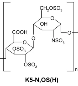
The capsular K5 polysaccharide from Escherichia coli has the same structure [3 4)--D-GlcA-(1 3 4)-␣-D
-GlcNAc-1(1 3]n as the heparin precursor N-acetyl heparosan17 in
which GlcA is glucuronic acid and GlcNAc is N-acetyl-glucosamine. Chemical and enzymatic modifications of the K5 polysaccharide lead to the synthesis of heparin-like compounds18 endowed with different biological properties,
including anticoagulant/antithrombotic,19,20 antineoplastic,21
and anti-AIDS22activities. Recently, we demonstrated that a
highly N,O–sulfated K5 derivative [K5-N,OS(H)] binds FGF2 with high affinity and exerts a potent antiangiogenic activity.23Thus, K5-N,OS(H) may provide the basis for the
design of novel angiostatic compounds with therapeutic implications in different angiogenesis-dependent diseases, including cancer.
Low-molecular-weight heparins (LMWHs) are obtained by chemical or enzymatic depolymerization of UFH. LMWH has favorable pharmacokinetics compared with conventional heparin.24,25Relevant to the use of heparins in cancer therapy,
LMWH prolongs disease-free survival time and reduces death rate in cancer patients.6In this study, we synthesized a
series of LMW derivatives of K5-N,OS(H) that were assessed for their FGF2 antagonist activity in vitro and angiostatic capacity in vivo.
Methods
Materials
Recombinant FGF2 was from Pharmacia-Upjohn (Milan, Italy). UFH (average MW⫽13 700; sulfate/carboxyl ratio [SO3⫺/
COO⫺]⫽2.14) was obtained from unfractionated beef mucosa so-dium heparin (Laboratori Derivati Organici, Milan, Italy). LMWH (average MW⫽5000; SO3⫺/COO⫺⫽2.13) was provided by C. Pisano
(Sigma-Tau, Pomezia, Italy). K5 polysaccharide (average MW⫽30 000) was prepared as described.23
Cyclo(-Arg-Gly-Asp-D-Phe-Val) peptide [c(RGDfV)] and cyclo(-Arg-Ala-Asp-D-Cyclo(-Arg-Gly-Asp-D-Phe-Val) peptide [c(RADfV)] were from Bachem AG.
LMW-K5 Derivatives
Derivatives were generated by nitrous acid depolymerization and subsequent reduction from a single batch of K5-N,OS(H)23 (for
details, please see the online Methods, available at http://atvb. ahajournals.org.).13C-NMR spectrum analysis, SO
3⫺/COO⫺
analy-sis, and MW determinations of the different samples were performed as described.23
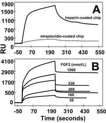
BIAcore Binding Assay
Biotinilated UFH was immobilized (6.0 fmol/mm2) to a
streptavidin-activated sensor chip (BIAcore Inc, Piscataway, NJ). Increasing concentrations of FGF2 in 10 mmol/L HEPES, 150 mmol/L NaCl, 3.4 mmol/L EDTA, 0.005% surfactant P20 (pH 7.4), were then injected over the heparin-coated surface for 4 minutes and washed until dissociation was observed. The signal was expressed as resonance units. Also, FGF2 (160 nmol/L) was injected over the heparin-coated sensor chip in the presence of increasing GAG concentrations. A streptavidin-activated sensor chip was used for blank subtraction.
FGF2-Mediated Cell–Cell Adhesion Assay
Chinese hamster ovary (CHO)-K1 cells were seeded at 52 000 cells per cm2
in 24-well plates. After 24 hours, cell monolayers were fixed with 3% glutaraldehyde in PBS. Then, A745 CHO flg-1A cells, generated by transfection of GAG-deficient A745 CHO cells26with
the IIIc variant of murine FGFR1 cDNA,27were added at 52 000
cells per cm2
to CHO-K1 monolayers in serum-free medium plus 10 mmol/L EDTA with no addition or with 30 ng/mL FGF2 in the presence of increasing concentrations of the different GAGs. After 2 hours at 37°C, cells bound to the monolayer were counted under an inverted microscope.23
Cell Proliferation Assay
Bovine endothelial GM7373 cells (Human Genetic Mutual Cell Repository, Camden, NJ) were seeded at 70 000 cells per cm2
in 24-well plates. After overnight incubation in Eagle’s minimal essen-tial medium containing 10% FCS, cells were incubated in medium containing 0.4% FCS and 10 ng/mL FGF2. After 8 hours, increasing concentrations of GAGs were added to cell cultures without chang-ing the medium. Sixteen hours thereafter, cells were trypsinized and counted.23
Fibrin Gel Sprouting Assays
FGF2-transfected murine endothelial FGF2-T-MAE cell aggre-gates28were prepared on agarose-coated plates and seeded within
fibrin gel.23Then, culture medium with or without K5 derivatives
(100 g/mL) or cyclic pentapeptides (60 mol/L) was added. Formation of radially growing cell sprouts was quantified after 24 hours by image analysis of the digitized images using the Image-Pro Plus software (Media Cybernetics). Freshly prepared rat aorta rings29
were cultured in fibrin gel for 7 days in the presence of the different antagonists. Rings were examined daily and neovessels counted under an inverted microscope.
Cell Adhesion Assay
Polystyrene nontissue culture 96-well plates were coated with 20 g/mL FGF2, vitronectin, or fibronectin as described.14 Next,
50 000 GM7373 cells were seeded in the absence or presence of the antagonist. After 2 hours at 37°C, adherent cells were quantified by methylene blue/Azur II staining.14
Chick Embryo Chorioallantoic Membrane Assay
At day 8 of incubation, sterilized gelatin sponges (1 mm3; Gelfoam,
Upjohn Co) adsorbed with the K5-derivative dissolved in 3L of PBS (50g/embryo) were implanted on the top of growing cho-rioallantoic membranes (CAMs).23Sponges containing vehicle alone
were used as negative controls. CAMs were examined under a stereomicroscope until day 12, and blood vessels around the sponges were counted (10 eggs per group).
Results
Production of LMW-K5 Derivatives
K5 derivatives with reduced size were prepared by controlled chemical depolymerization of K5-N,OS(H) (SO3⫺/
COO⫺⫽3.87; average MW of 20 700). Three LMW com-pounds [LMW-K5(A), LMW-K5(B), and LMW-K5(C) with MW equal to 6000, 5000, and 4200, respectively] and 1 derivative with an intermediate MW of 11 000 (IMW-K5) were obtained. As for K5-N,OS(H),23the compounds carry 1
N-sulfated group and 1 6-O–sulfated group in all GlcN
residues, with 70% of their sequence being represented by GlcA2,3SO3⫺-GlcNSO3⫺,6SO3⫺disaccharide units (Figure I,
Binding of LMW-K5 Derivatives to FGF2
LMW-K5 derivatives were evaluated for the capacity to interact with FGF2 by preventing its binding to biotinilated heparin immobilized onto a streptavidin-activated BIAcore sensor chip. FGF2 binds to the heparin-coated sensor chip, but not to the streptavidin-activated sensor chip, in a dose-dependent manner (Figure II, available online at http://atvb.ahajournals.org). An association rate constant equal to 9.0⫻103 M⫺1 s⫺1 and a
dissociation rate constant equal to 3.8⫻10⫺4s⫺1characterize the interaction that occurs with high affinity (Kd⫽42.5 nmol/L),
consistent with previous determinations.30,31
On this basis, increasing concentrations of LMW-K5 derivatives or UFH were preincubated with FGF2 and then injected onto the heparin-coated sensor chip. All the com-pounds caused a dose-dependent inhibition of FGF2/heparin interaction (Figure 1). LMW-K5(A), LMW-K5(B), and LMW-K5(C) showed a similar potency (ID50⫽100 to 120
nmol/L), 4 to 5⫻ lower than that observed for K5-N,OS(H) and IMW-K5. These differences were, however, abolished when the concentration of the different compounds was expressed on a weight basis (ID50 values ranging from 0.3
g/mL to 0.6 g/mL). When compared with K5 derivatives, UFH (MW of 13 700) appeared to be a more active compet-itor (ID50⫽6.0 nmol/L, corresponding to 0.08 g/mL).
Fi-nally, a LMWH preparation (MW of 5000) inhibited the binding of FGF2 to immobilized heparin with a similar potency (ID50⫽20 nmol/L, corresponding to 0.1 g/mL;
Figure 1). No effect was instead exerted by unmodified K5 (not shown).
Effect of LMW-K5 Derivatives on HSPG/FGF2/FGFR Ternary Complex
FGF2 mediates cell– cell attachment by linking FGFRs and HSPGs on neighboring cells through the formation of HSPG/ FGF2/FGFR ternary complexes.32 Indeed, HSPG-deficient
FGFR1-transfected A745-CHO flg-1A cells adhere to a monolayer of HSPG-bearing CHO-K1 cells when incubated in the presence of 30 ng/mL FGF2 but not in the absence of the growth factor.27 K5-N,OS(H) inhibits FGF2-mediated
cell– cell attachment in this model.23
When tested under the same experimental conditions, all the LMW-K5 compounds and IMW-K5 hamper the forma-tion of the HSPG/FGF2/FGFR ternary complex with an activity equal to that shown by the parent compound K5-N,OS(H) and LMWH (ID50⫽0.3 g/mL) and only slightly
less potent than that exerted by UFH (ID50⫽0.1 g/mL).
Unmodified K5 was ineffective (Figure 2A).
Effect of LMW-K5 Derivatives on Endothelial Cell Proliferation and Sprouting
To evaluate a possible angiosuppressive activity of LMW-K5 derivatives, we evaluated their capacity to affect
FGF2-Figure 1. Surface plasmon resonance analysis of LMW-K5 derivative/FGF2 interaction. FGF2 (160 nmol/L) was injected over a heparin-coated BIAcore sensor chip in the presence of increasing concentrations of the different GAGs. The response was recorded at the end of injection and plotted as a function of the GAG concentration.
Figure 2. Effect of LMW-K5 derivatives on FGF2-mediated cell– cell adhesion and endothelial cell proliferation. A, HSPG-deficient FGFR1 transfectants were added to CHO monolayers in the presence of 30 ng/mL FGF2 and increasing concentra-tions of the different GAGs. After 2 hours at 37°C, adherent cells were counted. B, The different GAGs were tested for the capac-ity to inhibit FGF2-mediated proliferation in GM7373 cells. Data are expressed as percentage of the proliferation measured in the absence of any competitor. In both A and B, experiments were performed in triplicate and repeated 3⫻ with similar results.
mediated cell proliferation in bovine endothelial GM7373 cells. Similar to heparin and LMWH, LMW-K5 derivatives and IMW-K5 inhibit endothelial cell proliferation (ID50⫽1 to
3g/mL). No inhibition was instead exerted by unmodified K5 (Figure 2B).
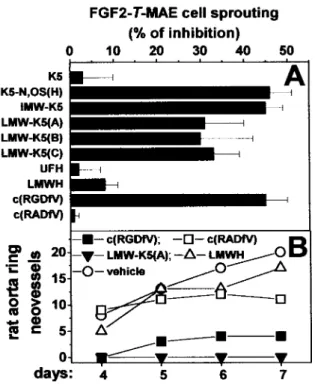
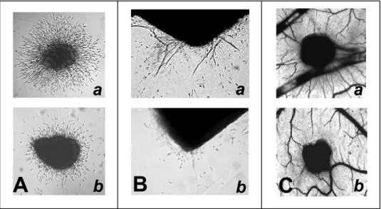
The capacity of K5 derivatives to affect angiogenesis was investigated further by an in vitro sprout formation assay. In this assay, FGF2-transfected endothelial FGF2-T-MAE cell aggregates are embedded into a fibrin gel in which they form solid cell sprouts after 1 to 2 days in culture.33As shown in
Figure 3A (see also Figure III, available online at http:// atvb.ahajournals.org), K5 derivatives exert a significant in-hibitory effect on FGF2-T-MAE cell sprouting, whereas unmodified K5, UFH, and LMWH are ineffective. Also, endothelial cell sprouting was suppressed by the␣v3integrin
antagonist cyclic pentapeptide c(RGDfV)34 but not by the
control peptide c(RADfV; Figure 3A).
Accordingly, LMW-K5(A) and c(RGDfV), but not LMWH and c(RADfV), inhibit endothelial cell sprouting in an ex vivo assay in which new capillary-like structures originate spontaneously from the endothelium of fibrin-embedded rat aorta rings29(Figure 3B and Figure III).
Effect of LMW-K5 Derivatives on FGF2/␣v3
Integrin Interaction
Integrins, including␣v3, play an important role in
angiogen-esis.35Also, FGF2 binds␣
v3, and this interaction is required
for endothelial cell response to the growth factor.14 –16 The
capacity of K5 derivatives to inhibit endothelial cell sprouting similar to the integrin antagonist c(RGDfV) prompted us to assess the possibility that K5 derivatives may affect FGF2/ ␣v3 interaction. Endothelial cells adhere and spread on
FGF2-coated plastic through FGF2/␣v3interaction.14,15Both
K5-N,OS(H) and LMW-K5(C) prevent GM7373 cell adhe-sion to immobilized FGF2, whereas UFH and LMWH are ineffective (Figure 4). Similar results were obtained with LMW-K5(A) and the integrin antagonist c(RGDfV), whereas control c(RADfV) was ineffective (data not shown). The effect of K5 derivatives on FGF2/␣v3-mediated endothelial
cell adhesion was specific, because K5-N,OS(H) and LMW-K5(C) did not inhibit cell adhesion to the prototypic integrin ligands vitronectin and fibronectin.
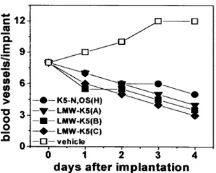
Effect of LMW-K5 Derivatives on Chick Embryo CAM Angiogenesis
Various FGF2 antagonists, including heparin-like compounds and␣v3 antagonists, exert a significant antiangiogenic
ac-tivity in the chick embryo CAM.16,23,36,37When delivered on
the top of the CAM through a gelatin sponge implant, all the K5 derivatives inhibited blood vessel growth. This was observed in 70% of the embryos treated with K5-N,OS(H), in 80% of those treated with LMW-K5(A) or LMW-K5(B), and in 90% of the embryos treated with LMW-K5(C). Accord-ingly, the number of blood vessels surrounding the implants decreased rapidly during incubation with K5-N,OS(H) or with the different LMW-K5 compounds compared with the physiological increase in vascularization observed in vehicle-treated embryos (Figure 5 and Figure III). In agreement with previous observations, unmodified K5 and heparin did not affect CAM neovascularization.23,36,37
Discussion
Here, a series of LMW-K5 compounds (MW ranging from 6000 to 4200) obtained by controlled chemical
depolymer-Figure 3. Effect of LMW-K5 derivatives on endothelial cell sprouting. A, FGF2-T-MAE cell aggregates were seeded in fibrin gel in the presence of the different GAGs (100g/mL) or cyclic peptides (60mol/L). After 24 hours, sprouting was evaluated by computerized image analysis. Data are the mean⫾SEM of 3 experiments in triplicate. B, Rat aorta rings were embedded in fibrin gel in the presence of vehicle or of LMW-K5(A), LMWH (both at 100g/mL), or cyclic peptides (60 mol/L). Neovessels that sprout out of the rings were counted during the next 7 days. Data represent 4 rings per experimental condition. Similar results were obtained in a second independent experiment.
Figure 4. Effect of LMW-K5 derivatives on FGF2/␣v3
interac-tion. GM7373 cells were seeded on plastic coated with FGF2, fibronectin (FN), or vitronectin (VN) in the absence or presence of UFH, LMWH, K5-N,OS(H), or LMW-K5(C). Adherent cells were counted after 2 hours.
ization of a high MW N,O-sulfated K5 derivative23exert a
potent FGF2 antagonist activity in vitro and show antiangio-genic capacity in vivo. Thus, as few as 8 monosaccharide units [as in LMW-K5(C)] are sufficient to confer to K5-N,OS(H) derivatives a full angiostatic capacity.
The minimal FGF2-binding sequence in heparan sulfate is a pentasaccharide that contains the disaccharide units IdoA2SO3⫺-GlcNSO3⫺ or IdoA2SO3⫺-GlcNSO3⫺,6SO3⫺.38
K5 derivatives can be chemically and enzymatically modified to reproducible structures in which sulfated groups and GlcA/IdoA residues are distributed along the GAG chain in a statistically homogenous manner.18,39 – 41 Surface plasmon
resonance analysis demonstrates that free K5-N,OS(H), IMW-K5, and all LMW-K5 derivatives (⬇70% of their sequence represented by GlcA2,3SO3⫺-GlcNSO3⫺,6SO3⫺
dis-accharide units) compete with immobilized heparin for the binding to FGF2 with similar potency and only slightly less efficiently than free UFH or LMWH when GAG chain concentration was expressed on a weight basis. This is in keeping with the hypothesis that more FGF2 molecules bind to a single heparin chain and that the stoichiometry of FGF2/heparin complexes depends on the size of the GAG. A single UFH or LMWH chain binds 6 or 2 FGF2 molecules, respectively, both heparin types giving a heparin mass of 2300 per FGF2-binding site.42 Similarly, it is possible to
hypothesize that LMW-K5 chains can bind 2 to 3 FGF2 molecules per complex and that longer IMW-K5 and K5-N,OS(H) chains will bind a proportionally higher number of growth factor molecules.
FGF2 interaction with nonsignaling HSPGs is required for the binding to FGFRs. FGFR occupancy will then lead to multiple biological responses in cultured endothelial cells and eventually to in vivo neovascularization.11 Similar to
K5-N,OS(H) and IMW-K5, LMW-K5 derivatives abrogate FGF2-mediated attachment of FGFR1-expressing cells to neighboring HSPG-bearing cells, thus indicating their ability to prevent the formation of the HSPG/FGF2/FGFR ternary complex.23 Synthetic molecules and chemically modified
heparins that interfere with HSPG/FGF2/FGFR interaction
act as angiogenesis inhibitors (reviewed in Presta et al8).
Accordingly, K5-N,OS(H) derivatives suppress neovascular-ization in the chick embryo CAM.
Similar to K5 derivatives, heparin binds FGF2 and pre-vents HSPG/FGF2/FGFR interaction and FGF2-driven endo-thelial cell proliferation in vitro. However, at variance with K5 derivatives, heparin does not affect endothelial cell sprouting in vitro and angiogenesis in vivo. This apparent discrepancy may be explained by the observation that K5 derivatives, but not heparin, also prevent the interaction of FGF2 with ␣v3 integrin. ␣v3 plays an important role in
angiogenesis,35 and FGF2/␣
v3 interaction is required for
endothelial cell response to the growth factor.14 –16 Also,
numerous observations point to an intimate cross-talk be-tween integrin-mediated intracellular signaling and angio-genic growth factor tyrosine kinase receptor activation.43
Accordingly, ␣v3 represents a target for angiostatic
com-pounds.44 Thus, the capacity of K5 derivatives to interfere
with both HSPG/FGF2/FGFR and FGF2/␣v3 interactions
results in a potent angiostatic activity.
K5 derivatives do not inhibit integrin-mediated endothelial cell adhesion to vitronectin (a prototypic ␣v3 ligand) or
fibronectin (a prototypic ␣51 ligand), indicating that the
observed FGF2/␣v3 antagonist activity is because of the
capacity of these compounds to bind the growth factor rather than the integrin molecule. The characterization of the struc-tural requirements for the FGF2/␣v3 antagonist activity of
K5 derivatives deserves further investigation.
Both thrombotic and hemorrhagic complications have been reported in cancer patients undergoing antiangiogenic therapy (discussed in Daly et al45). The occurrence of these
life-threatening complications suggests that the use of antiangio-genic compounds endowed with anticoagulant activity or in association with anticoagulants should be approached with caution. Relevant to this point, K5-N,OS(H) and its LMW derivatives are endowed with negligible anticoagulant activ-ity (P.O. and G. Z., unpublished data, 2003). LMW-K5 compounds may therefore provide the basis for the design of novel LMW angiostatic compounds with therapeutic impli-cations in angiogenesis-dependent diseases, including cancer.
Acknowledgments
This work was supported by grants from Ministero dell’ Istruzione, Universita` e Recerca (MIUR) (Centro di Eccellenza, Innovazione Diagnostica e Terapeutica [IDET], Firb 2001, Cofin 2002), Associa-zione Italiana per la Ricerca sul Cancro (AIRC), and Istituto Superiore di Sanita´ (ISS; Oncotechnological Program) to M.P., and from MIUR (Cofin 2003) to M.R. and (Firb 2001) to D.R.
References
1. Roden L, Ananth S, Campbell P, Curenton T, Ekborg G, Manzella S, Pillion D, Meezan E. Heparin: an introduction. Adv Exp Med Biol. 1992; 313:1–20.
2. Casu B, Lindahl U. Structure and biological interactions of heparin and heparan sulfate. Adv Carbohydr Chem Biochem. 2001;57:159 –206. 3. Lindahl U, Lidholt K, Spillmann D, Kjellen L. More to “heparin” than
anticoagulation. Thromb Res. 1994;75:1–32.
4. Lebeau B, Chastang C, Brechot JM, Capron F, Dautzenberg B, Delaisements C, Mornet M, Brun J, Hurdebourcq JP, Lemarie E. Subcu-taneous heparin treatment increases survival in small cell lung cancer. “Petites Cellules” Group. Cancer. 1994;74:38 – 45.
Figure 5. Effect of LMW-K5 derivatives on chick embryo CAM vascularization. Gelatin sponges adsorbed with vehicle or K5 derivatives (all at 50g per embryo) were implanted on the top of CAMs at day 8. CAMs were examined daily, and blood ves-sels around the sponges were counted (n⫽10).
5. von Tempelhoff GF, Heilmann L. Antithrombotic therapy in gynecologic surgery and gynecologic oncology. Hematol Oncol Clin North Am. 2000; 14:1151–1169, ix.
6. Zacharski LR, Ornstein DL, Mamourian AC. Low-molecular-weight heparin and cancer. Semin Thromb Hemost. 2000;26(suppl 1):69 –77. 7. Engelberg H. Actions of heparin that may affect the malignant process.
Cancer. 1999;85:257–272.
8. Presta M, Leali D, Stabile H, Ronca R, Camozzi M, Coco L, Moroni E, Liekens S, Rusnati M. Heparin derivatives as angiogenesis inhibitors.
Curr Pharm Des. 2003;9:553–566.
9. Gerwins P, Skoldenberg E, Claesson-Welsh L. Function of fibroblast growth factors and vascular endothelial growth factors and their receptors in angiogenesis. Crit Rev Oncol Hematol. 2000;34:185–194.
10. Klint P, Claesson-Welsh L. Signal transduction by fibroblast growth factor receptors. Front in Biosci. 1999;4:D165–D177. Review. 11. Schlessinger J, Lax I, Lemmon M. Regulation of growth factor activation
by proteoglycans: what is the role of the low affinity receptors? Cell. 1995;83:357–360. Review.
12. Schlessinger J, Plotnikov AN, Ibrahimi OA, Eliseenkova AV, Yeh BK, Yayon A, Linhardt RJ, Mohammadi M. Crystal structure of a ternary FGF-FGFR-heparin complex reveals a dual role for heparin in FGFR binding and dimerization. Mol Cell. 2000;6:743–750.
13. Coltrini D, Rusnati M, Zoppetti G, Oreste P, Grazioli G, Naggi A, Presta M. Different effects of mucosal, bovine lung and chemically modified heparin on selected biological properties of basic fibroblast growth factor.
Biochem J. 1994;303(pt 2):583–590.
14. Tanghetti E, Ria R, Dell’Era P, Urbinati C, Rusnati M, Ennas MG, Presta M. Biological activity of substrate-bound basic fibroblast growth factor (FGF2): recruitment of FGF receptor-1 in endothelial cell adhesion contacts. Oncogene. 2002;21:3889 –3897.
15. Rusnati M, Tanghetti E, Dell’Era P, Gualandris A, Presta M. ␣v3 integrin mediates the cell-adhesive capacity and biological activity of basic fibroblast growth factor (FGF-2) in cultured endothelial cells. Mol
Biol Cell. 1997;8:2449 –2461.
16. Kumar CC, Malkowski M, Yin Z, Tanghetti E, Yaremko B, Nechuta T, Varner J, Liu M, Smith EM, Neustadt B, Presta M, Armstrong L. Inhi-bition of angiogenesis and tumor growth by SCH221153, a dual␣(v)3 and ␣(v)5 integrin receptor antagonist. Cancer Res. 2001;61: 2232–2238.
17. Vann WF, Schmidt MA, Jann B, Jann K. The structure of the capsular polysaccharide (K5 antigen) of urinary-tract-infective Escherichia coli 010:K5:H4. A polymer similar to desulfo-heparin. Eur J Biochem. 1981; 116:359 –364.
18. Naggi A, Torri G, Casu B, Oreste P, Zoppetti G, Li JP, Lindahl U. Toward a biotechnological heparin through combined chemical and enzymatic modification of the Escherichia coli K5 polysaccharide. Semin
Thromb Hemost. 2001;27:437– 443.
19. Razi N, Feyzi E, Bjork I, Naggi A, Casu B, Lindahl U. Structural and functional properties of heparin analogues obtained by chemical sul-phation of Escherichia coli K5 capsular polysaccharide. Biochem J. 1995; 309(pt 2):465– 472.
20. Oreste P, Zoppetti G. Glycosaminoglycans derived from K5 polysaccha-ride having high anticoagulant and antithrombotic activities and process for their preparation. US patent application No. US 2002 062019; 2002. 21. Borgenstrom M, Jalkanen M, Salmivirta M. Sulfated derivatives of Esch-erichia coli K5 polysaccharides as modulators of fibroblast growth factor signaling. J Biol Chem. 2003;278:49882– 49889.
22. Vicenzi E, Gatti A, Ghezzi S, Oreste P, Zoppetti G, Poli G. Broad spectrum inhibition of HIV-1 infection by sulfated K5 Escherichia coli polysaccharide derivatives. AIDS. 2003;17:177–181.
23. Leali D, Belleri M, Urbinati C, Coltrini D, Oreste P, Zoppetti G, Ribatti D, Rusnati M, Presta M. Fibroblast growth factor-2 antagonist activity and angiostatic capacity of sulfated Escherichia coli K5 polysaccharide derivatives. J Biol Chem. 2001;276:37900 –37908.
24. Hirsh J, Levine MN. Low molecular weight heparin. Blood. 1992; 79:1–17.
25. Weitz JI. Low-molecular-weight heparins. N Engl J Med. 1997;337: 688 – 698.
26. Esko JD. Genetic analysis of proteoglycan structure, function and me-tabolism. Curr Opin Cell Biol. 1991;3:805– 816.
27. Liekens S, Leali D, Neyts J, Esnouf R, Rusnati M, Dell’Era P, Maudgal PC, De Clercq E, Presta M. Modulation of fibroblast growth factor-2 receptor binding, signaling, and mitogenic activity by heparin-mimicking polysulfonated compounds. Mol Pharmacol. 1999;56:204 –213. 28. Sola F, Gualandris A, Belleri M, Giuliani R, Coltrini D, Bastaki M,
Tosatti MP, Bonardi F, Vecchi A, Fioretti F, Ciomei M, Grandi M, Mantovani A, Presta M. Endothelial cells overexpressing basic fibroblast growth factor (FGF-2) induce vascular tumors in immunodeficient mice.
Angiogenesis. 1997;1:102–116.
29. Nicosia RF, Ottinetti A. Growth of microvessels in serum-free matrix culture of rat aorta. A quantitative assay of angiogenesis in vitro. Lab
Invest. 1990;63:115–122.
30. Thompson LD, Pantoliano MW, Springer BA. Energetic characterization of the basic fibroblast growth factor-heparin interaction: identification of the heparin binding domain. Biochemistry. 1994;33:3831–3840. 31. Li LY, Seddon AP. Fluorospectrometric analysis of heparin interaction
with fibroblast growth factors. Growth Factors. 1994;11:1–7. 32. Richard C, Liuzzo JP, Moscatelli D. Fibroblast growth factor-2 can
mediate cell attachment by linking receptors and heparan sulfate proteo-glycans on neighboring cells. J Biol Chem. 1995;270:24188 –24196. 33. Gualandris A, Rusnati M, Belleri M, Nelli EE, Bastaki M,
Molinari-Tosatti MP, Bonardi F, Parolini S, Albini A, Morbidelli L, Ziche M, Corallini A, Possati L, Vacca A, Ribatti D, Presta M. Basic fibroblast growth factor overexpression in endothelial cells: an autocrine mechanism for angiogenesis and angioproliferative diseases. Cell Growth
Differ. 1996;7:147–160.
34. Ruoslahti E. RGD and other recognition sequences for integrins. Annu
Rev Cell Dev Biol. 1996;12:697–715.
35. Ruegg C, Mariotti A. Vascular integrins: pleiotropic adhesion and sig-naling molecules in vascular homeostasis and angiogenesis. Cell Mol Life
Sci. 2003;60:1135–1157.
36. Casu B, Guerrini M, Naggi A, Perez M, Torri G, Ribatti D, Carminati P, Giannini G, Penco S, Pisano C, Belleri M, Rusnati M, Presta M. Short heparin sequences spaced by glycol-split uronate residues are antagonists of fibroblast growth factor 2 and angiogenesis inhibitors. Biochemistry. 2002;41:10519 –10528.
37. Casu B, Guerrini M, Guglieri S, Naggi A, Perez M, Torri G, Cassinelli G, Ribatti D, Carminati P, Giannini G, Penco S, Pisano C, Belleri M, Rusnati M, Presta M. Undersulfated and glycol-split heparins endowed with antiangiogenic activity. J Med Chem. 2004;47:838 – 848.
38. Maccarana M, Casu B, Lindahl U. Minimal sequence in heparin/heparan sulfate required for binding of basic fibroblast growth factor. J Biol
Chem. 1994;269:3903. Erratum.
39. Casu B, Grazioli G, Hannesson H, Jan B, Jann K, Lindahl U, Naggi A, Oreste P, Razi N, Torri G, Tursi F, Zoppetti G. Biological active, heparan sulfate-like species by combined chemical and enzymic modification of the Escherichia coli polysaccharide K5. Carbohydr Lett. 1994;1:107–114. 40. Kusche M, Hannesson HH, Lindahl U. Biosynthesis of heparin. Use of Escherichia coli K5 capsular polysaccharide as a model substrate in enzymic polymer-modification reactions. Biochem J. 1991;275(pt 1):151–158.
41. Casu B, Grazioli G, Razi N, Guerrini M, Naggi A, Torri G, Oreste P, Tursi F, Zoppetti G, Lindahl U. Heparin-like compounds prepared by chemical modification of capsular polysaccharide from E. coli K5.
Car-bohydr Res. 1994;263:271–284.
42. Arakawa T, Wen J, Philo JS. Stoichiometry of heparin binding to basic fibroblast growth factor. Arch Biochem Biophys. 1994;308:267–273. 43. Eliceiri BP. Integrin and growth factor receptor crosstalk. Circ Res.
2001;89:1104 –1110.
44. Kumar CC. Integrin␣ v  3 as a therapeutic target for blocking tumor-induced angiogenesis. Curr Drug Targets. 2003;4:123–131.
45. Daly ME, Makris A, Reed M, Lewis CE. Hemostatic regulators of tumor angiogenesis: a source of antiangiogenic agents for cancer treatment?