DOCTORAL SCHOOL IN BIOLOGY
Section: Biology applied to human health
CYCLE XXV
Identification and functional analysis of virulence determinants of
Verocytotoxin-producing Escherichia coli (VTEC)
Valeria Michelacci
MSc. (Hons), Genomic Biotechnology
Tutor
Dr. Stefano Morabito
Coordinator
Prof. Paolo Visca
External Referees
Prof. Herbert Schmidt (Ext. Examiner)
Prof. Maria Teresa Muniesa Perez
Dr. Patrick Fach
Thesis submitted for the Degree of Doctor of Philosophy
December 2012
i
Abstract
Verocytotoxin(VT)-producing Escherichia coli (VTEC) are important zoonotic pathogens whose natural reservoir is the gastrointestinal tract of ruminants. The transmission of the infections mainly occurs via the ingestion of contaminated food of animal origin. VTEC pathogenicity relies on the production of the VTs and on the action of accessory virulence factors constituting the virulome, which has not been completely identified yet.
The main objective of this piece of research was the identification of the genomic structures forming the VTEC virulome. An additional goal was the analysis of their distribution in different VTEC sub-populations. Finally, a function for some of the factors identified and involved in the pathogenetic mechanism has been proposed.
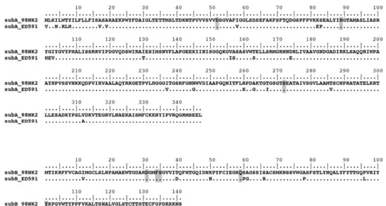
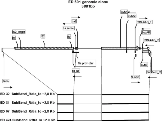
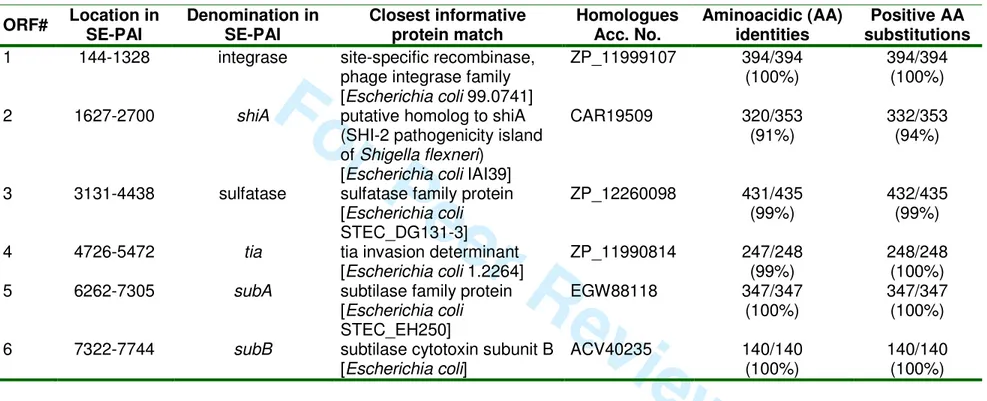
The work presented here has been largely based on the genomic comparison of VTEC strains isolated from human and animal sources and held in the collections of the EU RL VTEC and of the collaborating institutions Statens Serum Institut (Copenhagen, DK) and University of Extremadura (Caceres, ES). This approach led to the identification and characterisation of two pathogenicity islands (PAIs) proposed to be part of the virulome of VTEC strains commonly isolated from cases of human disease. The two PAIs harbour genes encoding factors involved in the colonization (adfO, OI-57, Chapter 4 and tia, SE-PAI, Chapter 3) or specifying an allelic variant of the Subtilase cytotoxin (subAB2, SE-PAI, Chapter 3). The identification and characterization of the open reading frames carried by the two PAIs allowed making inference on the function of the encoded proteins and on their role in the pathogenetic process. As a matter of fact, the tia and shiA genes (SE-PAI, Chapter 3) have been proposed to be part of the colonization machinery of VTEC strains lacking the ability to cause attaching-and-effacing (A/E) lesions on the intestinal mucosa, a typical feature of the VTEC associated with the most severe forms of the infection. The products of genes homologues to tia and shiA have been in fact described respectively to have a role in the process of cell invasion used by some enterotoxigenic E. coli strains or in the attenuation of host inflammatory response upon infection of another invasive enteric pathogen: Shigella
flexneri.
The genomic approach has also been used to investigate on the VT-producing Enteroaggregative E. coli O104:H4 that caused an outbreak in Germany in 2011. This peculiar VTEC is characterised by a rare combination of virulence traits from Enteroaggregative E. coli (EAEC) and VTEC, two different E. coli pathotypes responsible for enteric infections in low-income areas and common in industrialized countries respectively. Before the German outbreak, VTEC O104:H4 had been isolated in a few sporadic cases of infection, sometimes with an epidemiological connection with travel in North Africa. Similarly, the strain that caused the German outbreak was introduced into the EU with fenugreek seeds produced in Egypt. These observations, together with the genomic stability observed for some of the VTEC O104 strain (Chapter 5), led to the hypothesis that such chimeric strains may have emerged in developing countries. The high loads of enteric pathogens causing diarrhoea and lack of wastewater treatment in these geographic regions, cause the diffuse faecal contamination of the environment, particularly of the aquatic ecosystem often shared with ruminants, facilitating the exchange of mobile genetic elements between human and animal E. coli pathotypes and leading to the formation of new combinations of virulence features such as those characterising the VT-producing EAEC O104:H4.
iii
Acknowledgements
This PhD thesis is the outcome of the last three years spent at the Department of Food Safety and Veterinary Public Health of the Istituto Superiore di Sanità (ISS) in Rome: a period of highly valuable professional and human experience in a widely international atmosphere and during which times of frustration have always been swept away by exciting and rewarding moments.
I am indebted to many people for making the time working on my PhD an unforgettable experience My first debt of gratitude goes to my advisor, Dr Stefano Morabito, who patiently provided the vision, encouragement and advise necessary for me to proceed through the doctorial programme and complete my dissertation. My best wish for my future would be to be able to command an audience someday as well as he can.
I am also very grateful to Dr Alfredo Caprioli for his constant and prompt availability in discussing scientific questions and giving interesting suggestions especially in revising papers or oral presentations, no matter how busy he could be.
I owe a lot to my dear friend and supervisor Rosy, who has worked together with me during all the period and has become busier and busier while proceeding with her scientific career, but who has never regret to help me in any way she could, either discussing about my scientific doubts or helping with any kind of benchwork or finally supporting me in my personal affairs.
I had the wonderful opportunity to spend several periods as a host in outstanding foreign laboratories, where I was always so lucky to meet other great scientists taking care about me with the same kindness I was used to at home. In this respect, it has been a real honor and pleasure to me to collaborate with Prof. Pina Fratamico at the U.S. Department of Agriculture in Pennsylvania, who has acted as a chief and a mother at the same time, making me feel completely at home even at the other edge of the world; Dr Flemming Scheutz at the Statens Serum Institut in Copenhagen, who I have had the opportunity to meet, work with and discuss with in a number of occasions during these years and always demonstrated kindness and availability to me, as only great people like him can do; and Prof Patrick Fach at ANSES in Maison-Alfort, who gave me the opportunity to “play” with some of the most futuristic RealTime-PCR-machines I had ever heard about. Many thanks also go to all the other people who assisted me kindly and patiently during my stages: David Needleman at USDA, who patiently helped me during my stage at USDA; Susanne and Pia, who provided me with any kind of help during the long and cold periods spent in Copenhagen; and Sabine Dellanoy at ANSES, who dealt with the analysis of a huge amount of data together with me, helping me not to get lost in the resulting excel files.
Many thanks also to Prof. Paolo Visca for acting as a supervisor during my PhD and to Prof Schmidt, Dr Muniesa and Prof Fach for accepting to review my thesis.
Special thanks also go to my friends and colleagues at ISS: Maria Luisa, Clarissa, Fabio, Paola and especially Laura and Antonella, who have been helping me in the lab work and have supported me especially in the last year of my PhD programme, when I traveled so much that I almost forgot where I was coming from. They, and Rosy as well, were always ready to remind me that I had nice people in the lab in Rome willing to help me in any way, even when they were facing hard periods on their own. Many other people passed by the lab for shorter periods, like Lejla and Remi, who worked a lot together with me and separately helped for the publication of two of the papers part of this thesis, besides becoming
iv
nice friends to go out with and spend a nice time, no matter where we meet: it has already happened in Rome, Amsterdam and Copenhagen!
Thanks to my dearest friends Ilaria, Elena and Maria Letizia, who have always supported me and found a way to make me laugh even if I have often been too busy to meet them frequently.
Special thanks go to my family for their support and understanding, even in my hardest periods.
Finally, Luca wouldn’t need me to write here how thankful I am to him: he has taken an active part in the most difficult as well in the nicest moments of the last years and has always been the closest person to me, no matter how far we were. He would deserve a honoris causa degree for the knowledge he has patiently acquired on E. coli while listening to me.
I strongly believe that these three years have opened my mind in many ways; now I can’t wait to see what other interesting opportunities are coming up for me in the next future.
v
Table of contents
Chapter 1: Introduction 1
1.1 Escherichia coli 3 ff667
1.2 Genomic plasticity of E. coli 3 7
1.3 Mobile genetic elements 4
1.3.1 Bacteriophages 5
1.3.2 Plasmids 5
1.3.3 Genomic Islands 5
1.4 Pathogroups and pathotypes of E. coli: emergence and characteristics 6
1.4.1 Extraintestinal pathogenic E. coli (ExPEC) 6
1.4.2 Diarrhoeagenic E. coli 7
Enteropathogenic E. coli (EPEC) 8
Enterotoxigenic E. coli (ETEC) 8
Enteroinvasive E. coli (EIEC) 9
Enteroaggregative E. coli (EAEC) 9
Diffusely-adherent E. coli (DAEC) 10
Verocytotoxin-producing E. coli (VTEC) 10
1.5 Did VTEC emerge as a toxigenic clone of “attaching-and-effacing
Escherichia coli”? 12
1.6 Emergence of new pathogroups: the O104 example 13
1.7 References 16
Chapter 2: Aims of the work 23
Chapter 3: Characterization of the genetic determinant of a novel allelic variant of
the Subtilase cytotoxin 27
3.1 Publication: Production of the subtilase AB5 cytotoxin by Shiga toxin-negative
Escherichia coli 31
3.2 Publication: A new pathogenicity island carrying an allelic variant of the Subtilase cytotoxin is common among Shiga toxin producing Escherichia coli
of human and ovine origin 37
Chapter 4: Identification of a virulence-associated MGE of VTEC serogroups
associated with severe human disease (OI-57) 63
4.1 Publication: OI-57, a genomic island of Escherichia coli O157, is present in other seropathotypes of Shiga toxin-producing E. coli associated with severe
vi
Chapter 5: Analysis of VTEC strains possessing rare combinations of MGEs
vehiculating virulence factors 75
5.1 Publication: Similarity of Shiga toxin-producing Escherichia coli O104:H4
strains from Italy and Germany 79
5.2 Publication: Detection and identification of Verocytotoxin-producing
Escherichia coli (VTEC) O104:H4 in food by Real Time PCR 81
Chapter 6: Discussion 95
6.1 Identification and characterization of VTEC virulome: Characterization of a
novel pathogenicity island encoding Subtilase cytotoxin 98 ff667
6.2 Identification and characterization of VTEC virulome: The Genomic Island
(GI) OI-57 101 7
6.3 VTEC strains possessing rare combinations of MGEs vehiculating virulence
factors: the O104 example 104
6.4 Concluding remarks 105
6.5 References 108
Chapter 7: Appendices 111
7.1 Appendix I: List of Abbreviations 113
7.2 Appendix II: List of tables and figures 114
1
1.Introduction
Chapter 1
3
1.INTRODUCTION
1.1 Escherichia coli
Escherichia coli is a Gram-negative bacterium belonging to the family Enterobacteriaceae and represents
an important component of the human and animal intestinal microflora.
This bacterial species colonizes the gastrointestinal tract during the first phases of the life establishing mutual beneficial relationships with the host and playing an important role in maintaining the equilibrium between the numerous bacterial species colonising the gut, in regulating the turnover of the intestinal epithelium and in promoting the development of local immunity system against pathogens. At the same time, E. coli is one of the most diffuse bacterial species in the environment, being present in almost all the niches including water and soil.
Some strains evolved the capability to harm and cause disease. The pathogenic variants of E. coli can cause a wide spectrum of diseases and are generally divided into two main large categories, or pathogroups, based on the district of the host they colonize: Extraintestinal Pathogenic E. coli (ExPEC) and Diarrhoeagenic E. coli (DEC).
ExPEC produce virulence features conferring the ability to colonize host districts other than the gastrointestinal tract. The pathogroup is divided into two main distinct populations: Uropathogenic E. coli (UPEC) and neonatal meningitis E. coli (NMEC) (Smith JL et al., 2007) causing a variety of human disease including urinary tract infections, neonatal meningitis, sepsis, pneumonia and surgical site infections.
Diarrhoeagenic E. coli (DEC) comprise different groups, or pathotypes, of E. coli, which infect the gut of the host inducing a wide spectrum of symptoms spanning from uncomplicated diarrhoea to haemorrhagic colitis and systemic complications like haemolytic uremic syndrome. DEC infections represent a major problem in low-income countries, where mortality from diarrhoea is one of the main causes of death every year (Kosek M. et al., 2003; Bern C. et al., 2004).
1.2 Genomic plasticity of E. coli
The exceptional capability of E. coli strains to colonize a broad range of hosts and environments is linked to their ability of establishing successful relationships with the other microorganisms and it is largely due to their extraordinary genomic plasticity. This is the capability to exchange genetic material with other bacteria also belonging to other species, through events of gain and loss of DNA traits.
The E. coli genome is shaped by a multitude of evolutionary forces derived from its habitat, in which either biotic (e. g competitors, host defence mechanisms) or abiotic (e. g. pH, temperature, UV, mineral depletion) factors determine the selection of the individuals most adapted to survive in any given niche (Van Elsas J.D. et al., 2011). As a result of this selective pressure, the E. coli genomes vary in size from 4.6 to 5.6 Mb (Bergthorsson U. & Ochman H., 1995). These differences are due to the presence of different amounts of strain-specific genetic information, which may represent up to 30% of the complete genome content (Rasko D.A. et al., 2008; Touchon M. et al. 2009, Dobrindt U. et al., 2010).
Chapter 1
4
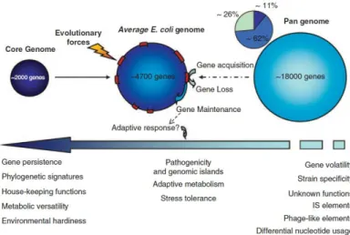
Comparative genome analysis revealed that E. coli genomes have a mosaic-like structure consisting of a conserved part, the ‘‘core genome’’, and interspersed regions of variable DNA. The core genome is common to all the E. coli strains and comprises about 2,000 genes, governing the basic metabolic functions. It represents about 11% of the totality of the so-called E. coli “pangenome”, which includes all the E. coli genetic features discovered so far (Fig. 1). A large portion (62%) of the pangenome is composed of so-called ‘persistent' genes that, in different combination with the core genome, identify the different E. coli pathogroups. Finally, the remaining 26% of the pangenome is composed by ‘volatile' genes conferring pathotype/strain specificity (Touchon M. et al., 2009).
Remodelling of the pangenome resulted in the evolution of specific E. coli types (Van Elsas J.D. et al., 2011) including both the commensal and pathogenic variants (pathotypes) (Ochman H. et al. 2000). The process of exchange of DNA traits between bacteria is defined as horizontal gene transfer (HGT) and is characterized by the integration and excision of mobile genetic elements (MGE). These elements consist in DNA regions encoding all the components required for their mobilisation and able to allocate part of the genetic material deriving from the host genome, variable in size from a few to several thousand base pairs.
Figure 1. The E. coli pangenome. (Van Elsas J.D. et al., 2011)
Through HGT, bacteria can easily acquire MGEs, vehiculating genes encoding advantageous functions or loose genetic determinants encoding unwanted or obsolete characters.
The horizontal transfer of MGE represents the key for the success of E. coli as ubiquitous bacterial species. HGT increases the genetic variability favouring the selection of the individuals most adapted to win the “struggle for life”.
1.3 Mobile genetic elements
Mobile genetic elements include bacteriophages, plasmids, genomic islands, integrons, transposons and insertion sequence elements (Dobrindt U. et al., 2005). The first three categories play the most important role in the evolution of pathogenic E. coli (Kelly B.G. et al., 2009) and will be discussed in detail.
Chapter 1
5
1.3.1 BacteriophagesBacteriophages had a profound impact on the evolution of bacterial pathogens (Campbell A., 1996). As a matter of fact, the spread of virulence-associated genes by lysogenic phages is a common phenomenon in many bacterial species (Boyd E.F. & Brussow H., 2002). Moreover, temperate and cryptic phages are considered possible ‘‘ancestors’’ of pathogenicity islands (Dobrindt U. et al., 2004).
The role of phages in modifying bacterial virulence was first demonstrated by Freeman in 1951, when he observed that the infection of non-toxigenic strains of Corynebacterium diphtheria with a bacteriophage resulted in the production of the diphtheria toxin (Freeman V.J., 1951; Tinsley C.R. et al., 2006). Since then, many toxins and other virulence-associated factors have been demonstrated to be encoded by genes carried by cryptic or intact phages in Gram-positive and negative bacteria (Casjens S., 2003).
The analyses of the E. coli genomes published so far, indicated that the sequenced strains differ considerably in the number of prophage-related sequences (Blattner F.R. et al., 1997; Hayashi T. et al., 2001; Perna N.T. et al., 2001; Welch R.A. et al., 2002) and that genome size differences correlate with the number of these MGEs, thus confirming the importance of the role of phages in structuring E. coli genome.
1.3.2 Plasmids
Plasmids are circular, self-replicating DNA molecules existing in bacterial cells as extra chromosomal replicons and are very common in all bacterial species. They represent a distinct genetic resource as they may confer many advantageous features such as resistance to antibiotics, expression of bacteriocins and siderophores, (Gomez-Lus R., 1998). Plasmids vehiculate genes playing important roles in the virulence mechanisms of pathogenic bacteria including DEC and ExPEC.
Plasmids can be easily exchanged and quickly disseminated in the bacterial populations representing one of the most characterised vehicles for the horizontal gene transfer between bacteria.
1.3.3 Genomic Islands
The concept of genomic islands (GI) was conceived following the discovery of particular regions in the bacterial chromosomes that contributed to pathogenicity and were termed pathogenicity islands (PAI) (Hacker J. & Kaper J.B., 2000). GI are characterized by a significant difference in G + C content, compared to the average base composition of the bacterial chromosome, an alternative codon usage, presence of mobility genes, and, in PAIs, of virulence genes. The majority of the GIs carry functions that are useful for the survival of the organism, by providing a selective advantage over the populations that do not harbour the islands (Hacker J. & Carniel E., 2001). Many types of GIs have been described, including those carrying genes governing metabolic properties enhancing the individual fitness, conferring resistance to antimicrobials or involved in the pathogenetic mechanisms (Dobrindt U. et al., 2004). The type of island that an organism carries depends on the genetic background of the host and on its ecological niche.
Pathogenicity islands carry one or more virulence genes, are present in the genomes of pathogenic bacteria but absent from the non-pathogenic variant of the same species and often exist in the size range of hundreds bp to more than 200 kb (Schmidt H. & Hensel M., 2004). Not much is known about the
Chapter 1
6
origin of these GIs, but it is thought that they originated from co-integrated plasmids and/or phages that have lost the ability to self-transfer.
GIs are typical features of E. coli and played an important role for evolution of the different variants or pathotypes. Many virulence-associated genes of pathogenic E. coli are located on PAIs, including those encoding adhesins, toxins, iron uptake systems, secretion systems and molecules involved in inhibiting the host response.
In pathogenic E. coli strains up to 13 PAI-like genetic entities have been identified (Hayashi T. et al., 2001; Perna N.T. et al., 2001; Welch R.A. et al., 2002).
Many PAIs have a mosaic-like modular structure. They can have different structural organizations or chromosomal localizations in different strains, still vehiculating the same virulence determinants (Guyer D.M. et al., 1998; Johnson J.R. et al., 2001; Redford P. & Welch R.A., 2002). This is probably due to the presence of multiple copies of accessory DNA elements facilitating the homologous recombination within the same or between different islands leading to rearrangements, deletions and acquisition of ‘‘foreign’’ DNA.
1.4 Pathogroups and pathotypes of E. coli: Emergence and characteristics
Horizontal gene transfer has driven E. coli evolution resulting in the definition of specific E. coli lineages, comprising commensal and pathogenic variants (Ochman H. et al. 2000).
All E. coli strains were initially thought to be non-pathogenic commensal organisms. In the 1940s E. coli strains were first identified in association with severe outbreaks of infantile diarrhoea (Bray J., 1945). Since then, E. coli has been associated with a range of clinical conditions and several pathogenic groups have been defined (Kaper J.B. et al., 2004; Nataro J.P. & Kaper J.B., 1998).
1.4.1 Extraintestinal pathogenic E. coli (ExPEC)
E. coli strains isolated from infections outside of the intestinal tract, e.g., uropathogenic E. coli (UPEC),
neonatal meningitis-associated E. coli (NMEC) and sepsis-causing E. coli (SEPEC) have been grouped as Extraintestinal pathogenic E. coli (ExPEC) (Smith J.L. et al., 2007). ExPEC are part of the intestinal microflora in a fraction of the healthy population and normally asymptomatically colonize the gut. Once they get access to niches outside of the gut, they are able to efficiently colonize these niches and cause disease, i.e. urinary tract infection (UTI), septicemia or meningitis in newborns in humans and in many animal species (Köhler C.D. & Dobrindt U., 2011). Several important virulence factors of ExPEC and their role during pathogenesis have been described (Kaper J.B. et al., 2004; Smith J.L. et al., 2007), however the complete gene asset conferring virulence has not been identified yet. As a matter of fact, ExPEC strains can use multiple virulence determinants in a mix-and-match fashion.
These virulence associated factors can be grouped by functional category, as they comprise, for example, adhesins, siderophore systems, toxins, surface polysaccharides, features involved in the invasion process and serum resistance-associated traits (Johnson J.R. & Russo T.A., 2005).
The comparison between the genomes of CFT073 UPEC strain and non-pathogenic E. coli strain Nissle 1917 revealed that, although the overall genetic structure of many genomic islands is very similar, important differences exist which are responsible for the non-pathogenic nature of strain Nissle 1917
Chapter 1
7
(Grozdanov L. et al., 2004). The pheV-associated PAI of strain CFT073 carries the complete hly and pap gene clusters respectively encoding the alpha-hemolysin and P fimbriae virulence factors. Interestingly, only a fragmented pap operon is present in a similar DNA region on the chromosome of Nissle 1917 associated with a transposon-like element, suggesting that the original content of the PAI may have been lost due to the insertion of mobile genetic elements and recombination events.Five cryptic prophages have been detected in the genome of CFT073 strain (Welch R.A. et al., 2002), which exhibit some structural features similar to some of the phage-like regions in the E. coli O157:H7 genome, besides containing different putative virulence-associated genes (Dobrindt U., 2005)
Other virulence-associated features, like the cytotoxic necrotizing factor 2 (Caprioli A. et al., 1987) and the characteristic F17 fimbria, are encoded by genes harboured on a virulence plasmid (Mainil J.G. et al., 2000).
1.4.2 Diarrhoeagenic E. coli (DEC)
Diarrhoeagenic E. coli (DEC) is the E. coli pathogroup with the highest variability, involving both the virulence mechanisms and the complexity of the genomes. As a matter of fact, DEC pathogroup is subdivided in six different pathotypes: Enteropathogenic E. coli (EPEC), Verocytotoxin-producing E.
coli (VTEC), Enterotoxigenic E. coli, Enteroaggregative E. coli (EAEC), Enteroinvasive E. coli (EIEC)
and Diffusely-Adherent E. coli (DAEC).
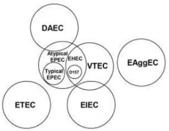
Figure 2. Schematic representation of phylogenetic relations among the different pathotypes of Diarroheagenic E. coli (Adapted from: Donnenberg M. (Ed.) Academic Press 2002.)
The phylogenetic relations existing between the different DEC pathotypes are particularly complex. Strains belonging to VTEC, EPEC, EIEC and DAEC pathotypes share several genetic determinants belonging to the “persistent” set of genes in the pangenome (Figure 1). Some groups, such as EPEC and VTEC share even a greater number of mobile genetic elements vehiculating virulence factors including some of the “volatile” genes indicating a closer correlation between the pathotypes (Figure 2).
Phylogenetic evidences indicate that these two pathotypes could derive from a common ancestor or from convergent evolutionary events, representing a good model for understanding evolutionary relations which have driven the emergence of the existing pathotypes of E. coli.
Chapter 1
8
A brief overview of DEC pathotypes is presented here, with particular attention for Verocytotoxin-producing E. coli, which will be the focus of the present piece of research.
Enteropathogenic E. coli (EPEC)
After the first association of E. coli with outbreaks of infantile diarrhoea in the United Kingdom in the 1940s (Bray J., 1945), similar outbreaks were observed in several Western countries and found to be associated with particular serotypes of E. coli, collectively referred to as enteropathogenic E. coli (EPEC).
EPEC was the first pathotype of E. coli to be described. Although large outbreaks of infant diarrhoea due to EPEC have now largely disappeared from industrialized countries, EPEC remain an important cause of potentially fatal infant diarrhoea in low-income countries. For decades, the mechanisms by which EPEC caused diarrhoea remained unknown. However, since 1979, numerous advances in the understanding of the pathogenesis of EPEC diarrhoea have been made, such that EPEC is now among the best characterised pathogenic E. coli. (Kaper J.B. et al. 2004).
The main virulence factors governing the adhesion mechanism of EPEC strains to the intestinal mucosa are vehiculated by a PAI termed locus of enterocyte effacement (LEE) (McDaniel T.K. et al., 1995), which governs the production of typical attaching-and-effacing (A/E) lesions in the ileum: the bacteria stimulate large-scale cytoskeletal changes in the epithelial cells, resulting in the effacement of the microvilli brush border and the formation of a pedestal which accommodates the bacterial cell (Nataro J.P. & Kaper J.B., 1998), causing its intimate attachment to the eukaryotic cell. Following its first identification in EPEC strains (McDaniel T.K. et al., 1995), the LEE was later on shown to be present in some subpopulations of VTEC strains (Karmali M.A. et al., 2003).
Typical EPEC strains contain a large plasmid termed EAF (EPEC Factor for Adherence) which carries the bfp locus, coding for bundle forming pilus, and the per gene, whose product regulates the expression of the LEE genes involved in the A/E adhesion (Donnenberg M.S. et al., 1992). The presence of this plasmid is associated to EPEC strains responsible for the majority of gastroenteritis cases in low-income countries. Some EPEC strains, termed atypical EPEC (aEPEC), are EAF-negative and possess a virulence plasmid resembling the one typically present in the most pathogenic VTEC serogroups (see below).
Enterotoxigenic E. coli (ETEC)
Enterotoxigenic E. coli (ETEC) are a major cause of infantile diarrhoea in the developing world and are one of the leading causes of diarrhoea in travellers visiting these areas of the globe. The first description of ETEC in human infections dates back to the early 60s, when it was reported that certain E. coli isolates from the stools of children with diarrhoea elicited fluid secretion in ligated rabbit intestinal loops (Taylor J. et al., 1961). Ten years later, DuPont et al. showed that ETEC strains were able to cause diarrhoea in adult volunteers (DuPont H.L. et al., 1971).
ETEC strains were first recognized as causes of diarrheal disease in piglets. Studies on ETEC in this species first elucidated the mechanisms of disease and led to the characterization of two plasmid-encoded enterotoxins: the heat-Stable and the heat-Lable Toxins (ST and LT respectively) (Levine M.M., 1987; Mainil J.G. et al., 1998; Gomez-Duarte O.G. et al., 1999). LTs are similar to the enterotoxins produced
Chapter 1
9
by Vibrio cholerae (Sixma T.K. et al., 1993), bind to membrane gangliosides and, upon internalization, produce an osmotic stress causing the release of great amounts of water in the faeces. STs exert a different molecular mechanism but, similarly to the LTs, cause a massive release of fluids in the gut.Enteroinvasive E. coli (EIEC)
Enteroinvasive E. coli (EIEC) strains were first shown to be capable of causing diarrhoea during the studies on volunteers conducted by DuPont et al. in 1971 (DuPont H.L. et al., 1971). EIEC can cause an invasive inflammatory colitis and occasionally dysentery, but in most cases elicit watery diarrhoea indistinguishable from that caused by other pathogenic E. coli (Nataro J.P. & Kaper J.B., 1998). Numerous studies have shown that EIEC strains are biochemically, genetically and pathogenetically closely related to Shigella spp. (Nataro J.P. & Kaper J.B., 1998) and taxonomically indistinguishable at the species level (Pupo G.M. et al., 2000). Nevertheless, a nomenclature distinction is still maintained owing to the clinical significance of Shigella.
In particular, Enteroinvasive E. coli strains share with Shigella flexneri the large (140 MDa) virulence plasmid pInv. This plasmid vehiculates the invasion-related genes, which encode a type III secretion system, secreted proteins involved in the invasion phenotype (Ipa) and the Shigella enterotoxin 2 (Nataro J.P. et al., 1995).
Enteroaggregative E. coli (EAEC)
In the early 1990s, two other diarrhoeagenic E. coli pathogroups were defined as distinct from EPEC for the mechanism of adhesion to the surface of Hep-2 cultured cells: Enteroaggregative E. coli (EAEC), showing a characteristic ‘‘stacked-brick’’ pattern of aggregation (Nataro J.P. et al., 1987), and Diffusely adherent E. coli (DAEC).
Pathogenesis of EAEC infections is believed to be initiated with adherence to the terminal ileum and colon in an aggregative, stacked-brick-type pattern by means of one of several different hydrophobic aggregative adherence fimbriae (AAFs), whose genetic determinants are vehiculated by a 55–65 MDa plasmids, collectively called pAA (Vial P.A. et al., 1988). The same plasmid also encodes most of the other known virulence factors of EAEC: the gene encoding a small protein termed antiaggregation protein (Aap), or dispersin, which neutralizes the strong negative charge of the LPS helping the displaying of the positively-charged AAFs out from the surface (Harrington S.M. et al., 2006); the aat operon encoding the ABC transporter responsible for the secretion of dispersin (Nishi J. et al., 2003); the 108 kDa plasmid-encoded toxin (Pet) (Eslava C. et al., 1998); the EAST1 cytotoxin, homologous to the ST of ETEC; the AggR global transcriptional activator, which regulates the expression of all these virulence factors.
In addition to these plasmid-encoded genes, other virulence genes are vehiculated by chromosomally located PAIs: the setBA genes, encoding ShET1 cytotoxin, are contained in a the 117 kb PAI inserted at the pheU-tRNA locus (Harrington S.M. et al., 2006); another PAI, integrated in glyU locus, encodes a type III secretion; a third PAI vehiculates genes for a transcriptional regulator and type III effector proteins and is inserted in selC (Harrington S.M. et al., 2006).
Chapter 1
10
Diffusely Adherent E. coli (DAEC)
Diffusely Adherent E. coli (DAEC) are defined by a diffuse pattern of adherence on Hep-2 cultured cells with little aggregation. DAEC strains are associated with watery diarrhoea that can become persistent in young children (Le Bouguenec C., 1999).
The term “diffusely adherent E. coli” was initially used to refer to any HEp-2-adherent E. coli strain that did not form EPEC-like microcolonies. After the discovery of EAEC, DAEC have started to be recognized as an independent category of potentially diarrhoeagenic E. coli. Bilge et al. have described a surface fimbria produced by these strains and designated F1845, responsible for the DA phenotype (Bilge S.S. et al., 1989), which is encoded by genes that can be located either on the chromosome or vehiculated by a plasmid (Nataro J.P. & Kaper J.B., 1998). Beside the fimbriae, other adhesins, known as the afimbrial or nonfimbrial adhesins, have been associated with an amorphous, outer membrane-associated structure on the surface of DAEC strains as well as in some ExPEC strains (Soto G.E. & Hultgren S.J., 1999). The first determinant to be identified encoding an adhesin not associated with visible fimbriae on the bacterial surface was the one specified by the afa operon, encoding factors that can be involved either in the adhesion or in the internalization in the host cells, as well as in the cell signalling on the surface of the intestinal epithelium promoting tight attachment of bacteria.
Verocytotoxin-producing E. coli (VTEC)
Verocytotoxin-producing E. coli (VTEC) is one of the most recently defined E. coli pathotypes.
The first evidence of VTEC infection in humans dates back to 1983 (Riley L.W. et al., 1983) when two outbreaks of a distinctive gastrointestinal illness characterized by severe bloody diarrhoea occurred in the US. In both the episodes stool cultures from patients yielded a previously rarely isolated E. coli serotype, O157:H7. The outbreaks were associated with the ingestion of undercooked hamburgers at a fast-food restaurant chain and the disease was designated haemorrhagic colitis (HC). Similarly, the strains belonging to this pathotype were initially termed Enterohaemorragic E. coli (EHEC).
In the same year, Karmali et al. (Karmali M.A. et al., 1983) reported the association of sporadic cases of haemolytic uremic syndrome (HUS) with a faecal cytotoxin, termed Verocytotoxin (VT), and with Verocytotoxin-producing E. coli in the stools of patients. HUS is characterised by the triad of symptoms including acute renal failure, thrombocytopenia and microangiopathic hemolytic anemia and it is usually preceded by a bloody diarrhoeal illness indistinguishable from HC. The production of the Verocytotoxin was then demonstrated in the E. coli O157:H7 from the human cases and a novel and increasingly important class of enteric pathogens was recognised: Verocytotoxin-producing E. coli (VTEC), or Shiga-toxin producing E. coli (STEC).
Many efforts have been made in the last three decades to unveil the virulence factors implied in the pathogenesis of the disease caused by VTEC strains. However the complete set of virulence genes, generally referred to as the “virulome”, still needs to be completely identified.
Verocytotoxins are the main virulence feature of VTEC. The holotoxins are about 70 kDa and consist of a single catalytic 32 kDa subunit and a pentamer of small 7.7 kDa subunits involved in the binding to specific receptors on the surface of target cells (O’Brien A.D. & Holmes R.K., 1987). Following the binding, the VTs are internalized by a receptor-mediated endocytosis mechanism and reach the
Chapter 1
11
endoplasmic reticulum where they are activated through a proteolytic cleavage of the catalytic subunit (Sandvig K. et al., 1992). The activated toxin exerts an N-glycosidicasic activity on the 28S rRNA, causing the protein synthesis inhibition (Paton J.C. & Paton A.W., 1998).VTs include two main antigenically distinct types, VT1 and VT2, and numerous subtypes identified by differences in the DNA sequence of the coding genes. Three subtypes of VT1 and seven subtypes of VT2 have been described so far, which can be found in any combination of type and subtype into a single VTEC (Persson S. et al., 2007). The VT-coding genes (vtx) are carried by bacteriophages belonging to the lambda family (Reid S.D. et al., 2000), which are usually maintained in a lysogenic state in the bacterial chromosome but retain the capability to enter the lytic cycle and move from a host to the other spreading the vtx-genes (Plunkett G. et al., 1999).
The presence of the vtx-converting bacteriophages is necessary condition for E. coli strains to cause the disease, though it seems not to be sufficient. As a matter of fact, VTEC associated with HC or HUS produce additional virulence factors boosting their pathogenicity. Some of these VTEC strains possess the Locus of Enterocyte Effacement (LEE) PAI encoding factors responsible for the “attaching and effacing” (A/E) hystopathological lesion (McDaniel T.K. et al, 1995). As in EPEC strains, the LEE encodes a type III secretion system (T3SS), consisting of a needle used to directly inject bacterial effectors into the host cell, causing the rearrangement of the cytoskeleton of the enterocyte and the typical brush border effacement. In addition to the T3SS, the LEE encodes an outer membrane protein called intimin (eaeA gene), which mediates the direct binding of the bacterium to the host cell surface, and its translocated receptor, Tir, which is vehiculated into the host cell plasma membrane by the T3SS (Yoon J.W. & Hovde C.J., 2008).
The presence of the LEE locus marks a subpopulation of VTEC which has been frequently associated to the most severe forms of human infections: HC and HUS.
Several other virulence genes have been described in VTEC strains isolated from human cases of severe disease, all carried by mobile genetic elements.
The PAI known as OI-122 hosts a large gene, termed efa1/lifA, encoding an immunomodulator, which inhibits the host lymphocyte activation. This PAI is consistently present in LEE-positive VTEC strains but is also present in the EPEC strains, suggesting the interaction of factors encoded by the two PAIs in governing the induction of the A/E lesion upon infection. Interestingly, OI-122 and the LEE have been found to be physically associated in a mosaic PAI in some VTEC and EPEC strains, suggesting the possibility that the hybrid PAI may have been first acquired as a co-integrated structure that may have undergone recombination events leading to the separation on two PAIs in several VTEC clones (Morabito S. et al., 2003).
VTEC strains isolated from HC and HUS in humans also possess a large virulence plasmid, called pO157 in VTEC of serogroup O157, vehiculating a number of additional virulence factors including the enterohaemolysin (ehxA gene), a catalase peroxidase (katP gene) and a serine-protease (espP gene) (Brunder W. et al., 1996). The pO157 also carries toxB, whose product seems to have a function similar to that of Efa1/lifA (Makino K. et al., 1998; Deng W. et al., 2012).
Chapter 1
12
Some VTEC do not have the LEE locus and the pO157 virulence plasmid but are isolated from human cases of infection. These strains generally do not cause severe disease although they have been rarely isolated from bloody diarrhoea and HUS.
Such LEE-negative VTEC usually have other virulence-associated genes complementing the action of the LEE locus in the colonization of the host gut. The LEE-negative VTEC strains belonging to O113 serogroup, as an example, carry a virulence plasmid enconding a number of unique virulence-associated determinants including the autotransporter protein EpeA (Leyton D.L. et al., 2003), the autoagglutinating adhesin Saa (Paton A.W. et al., 2001) and a subtilase-like serine protease cytotoxin, termed Subtilase cytotoxin (SubAB) (Paton A.W. et al., 2004).
1.5 Did VTEC emerge as a toxigenic clone of “attaching and effacing Escherichia coli”?
VTEC are a heterogeneous population including strains with different assets of virulome. An important group of VTEC causes severe disease in humans such as the Haemorrhagic Colitis and the Haemolytic Uremic Syndrome. This VTEC group shares with EPEC genomic and pathogenetic analogies. They both have the LEE locus and cause the attaching and effacing (A/E) lesion to the enterocyte as the main mechanism for the host colonization (Nataro J.P. & Kaper J.B., 1998). Due to this feature, the two groups have been frequently included in a super-group termed “Attaching-and-Effacing Escherichia Coli” (AEEC). The genomic similarity within the AEEC pathotype is not restricted to the LEE locus. As a matter of fact, other PAIs, probably involved in the A/E phenotype, are present in all the AEEC strains. At a certain moment in the evolution of AEEC, some strains evolved the capability to elaborate the verocytotoxins dividing this group into two distinct subpopulations marked by differences in the virulence features and the epidemiology of the infection: EPEC and VTEC.
Typical EPEC strains are responsible for prolonged diarrhoea in developing countries with inter-human transmission of the infection through the classical oral-faecal route. Conversely VTEC are zoonotic pathogens and the diffusion of the infections is mainly mediated by food of animal origin or by environmentally contaminated vehicles. The natural reservoir of VTEC is represented by the gastrointestinal tract of ruminants, particularly cattle (Nataro J.P. & Kaper J.B., 1998).
As with most of the food-borne bacterial pathogens, VTEC infections mainly occur in industrialized countries where they can cause very large outbreaks, being the spreading of cases facilitated by the large-capacity distribution of food items.
Some EPEC strains can be placed at the interface between typical EPEC and VTEC. These strains have been defined as a group during the Second International Conference on EPEC, held in São Paulo, Brazil in 1995, where the strains of E. coli possessing the LEE locus but lacking the VT genes and the EAF plasmid have been termed «atypical EPEC» (aEPEC). Studies on the characterization of aEPEC showed that these strains have a genomic asset resembling that of VTEC (Trabulsi L.R. et al., 2002). As a matter of fact, they usually possess a large virulence plasmid vehiculating the operon encoding the enterohaemolysin and other virulence genes also present in the pO157 plasmid (Cookson A.L. et al., 2007; Hernandes R.T. et al., 2009).
Several theories have been proposed for the emergence of aEPEC strains. They could represent VTEC strains that simply lost the ability to produce VTs following the excision of the prophage vehiculating vtx
Chapter 1
13
genes. However, some evidences suggest that aEPEC emerged from tEPEC following a “plasmid displacement” event (Wick L.M. et al., 2005).One of the typical features of the virulence plasmid pO157, is the presence of the large virulence gene
toxB. This gene, in its entire form or as remnant, is quite diffuse in the virulence plasmids present in
several VTEC serogroups and part of toxB gene is also present in the EAF plasmid of the prototype tEPEC strain E2348/69 (Tobe T. et al., 1999). Similarly to toxB, the per gene, located on the EAF plasmid and encoding a regulator of some LEE genes in tEPEC, has been detected in aEPEC, lacking the genes coding for the Bfp fimbriae, considered a hallmark for the EAF plasmid (Contreras C.A. et al., 2010).
These observations suggest that the two MGE may have come in contact and that a plasmid displacement event may have occurred causing the substitution of the EAF plasmid with a pO157-derived MGE. The appearance of aEPEC may be considered as an intermediate step in the evolution of VTEC according to the following step-wise model: an E. coli strain may have acquired the LEE locus and the MGEs encoding the accessory adhesins (e.g. OI-122) and the non-LEE encoded effectors probably from different bacterial species, thus triggering the selection of an attaching-and-effacing E. coli ancestor. Afterwards, the acquisition of a plasmid carrying the bfp operon could have occurred into the chimeric E.
coli, originating a tEPEC-like ancestor. Finally, the displacement of the bfp-carrying plasmid (EAF) by a
pO157-like structure, followed by the acquisition of a vtx-converting bacteriophage, may have led to the emergence of the VTEC prototype.
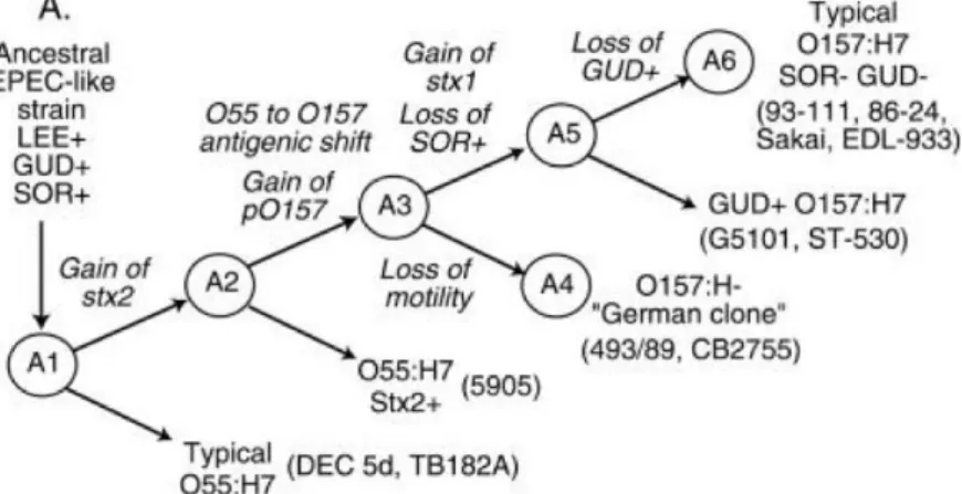
Such a model has been proposed for the evolution of VTEC O157 from a tEPEC of serotype O55:H7 (Figure 3) (Wick L.M. et al., 2005).
Figure 3. Model for the evolution of VTEC O157:H7 from a tEPEC belonging to serotype O55:H7 (Wick LM et al., 2005)
1.6 Emergence of new pathogroups: the O104 example
Horizontal gene transfer can cause pathogenic bacterial populations to emerge from harmless species. Moreover the exchange of genetic material by HGT may favour the formation of new pathotypes by mixing virulence features deriving from different pathogenic types in a single bacterial cell.
The appearance of the VTEC O104:H4 E. coli strain, which caused the large outbreak of infections in Germany in May 2011, is paradigmatic of this scenario. The episode was one of the largest outbreaks
Chapter 1
14
ever occurred, it counted about 4000 ill and more than 850 HUS cases with the heavy toll of 50 deaths (Frank C. et al., 2011; Jansen A. & Kielstein J.T., 2011).
The causative agent did not match the typical definition of pathogenic VTEC. It did not possess the LEE locus or the enterohaemolysin-coding gene and it did not belong to any of the serogroups known to cause HUS. The whole-genome sequencing of the outbreak strain revealed a rare asset of virulence determinants where the bacteriophage carrying the VT-coding genes was inserted in the genomic backbone of a different E. coli pathotype: the Enteroaggregative E. coli (EAEC).
This particular genomic arrangement was not novel. It had been sporadically observed during the last twenty years. In the early 1990s a VT2-producing O111 EAEC strain was isolated during a small outbreak of HUS in France (Morabito S. et al., 1999). An EAEC that produced VT2 has been isolated from cases of HC and HUS in patients with HIV in Bangui, Central African Republic (Mossoro C. et al., 2002) and a few sporadic cases have been reported in the EU (Scheutz F. et al., 2011; Mellmann A. et
al., 2008). In 2009 a single case of infection, described in Italy, was later on identified as caused by a
VT-producing EAEC O104:H4 showing a molecular profile similar to the strains that caused the German outbreak (This study, Scavia G. et al., 2011). Finally, an HUS case ascribed to infection with VT-producing EAEC O104:H4 has been reported in Finland in 2010 (Scheutz F. et al., 2011).
It has to be considered that for such a chimeric pathogen to emerge, an EAEC strain should have come in contact with a VT-converting bacteriophage. In general this scenario should not occur easily. In fact EAEC have a human reservoir, circulate predominantly in the human host and the infections are diffuse mainly in developing countries. Conversely, VTEC have an animal reservoir and VTEC infections are common in industrialized countries and thus the two pathotypes are kept separated epidemiologically and geographically.
Nevertheless, EAEC can easily survive in the environment where they can form strong biofilms. In developing countries, the burden of infection caused by these microorganisms is very high. In these countries, they are massively released with the faeces in the sewage and treatment of wastewaters are generally not in place or are scarcely effective, causing the contamination of the aquatic environment. Water in turn represents the main re-infection vehicle for humans leading to the establishment of an amplification of the pathogen’s populations.
Figure 4. Promiscuous conditions illustrating the overlapping between the human and the animal reservoir of pathogenic E. coli in low-income countries (Pakistan Associated Press/Pervez Masih)
Chapter 1
15
In order to get a figure of this phenomenon, it has been estimated that about 2.6 billion of people in underdeveloped areas of the globe do not have access to water suitable for potable or personal hygiene uses (http://www.unwater.org/statistics_san.html). Moreover, it is not infrequent that, in these parts of the world, the same aquatic environment is shared by human and animals, including ruminants, which are the main reservoir of VTEC, setting the proper condition for the contacts between EAEC and the VT-converting bacteriophages to occur (Figure 4).The coexistence of human and animal E. coli strains in the same ecological niche could thus have triggered the remodelling of the versatile genome characterizing this bacterial species and may have facilitated the exchange of mobile genetic elements through events of horizontal gene transfer.
Chapter 1
16
1.7 References
- Bergthorsson U, Ochman H. 1995. Heterogeneity of genome sizes among natural isolates of
Escherichia coli. J Bacteriol. 177(20):5784-9.
- Bergthorsson U, Ochman H. 1998. Distribution of chromosome length variation in natural isolates of Escherichia coli. Mol Biol Evol 15:6–16.
- Bern C. 2004. Diarrhoeal Diseases In The Global Epidemiology of Infectious Diseases. ed. Murray C.J.L., Lopez A.D. and Mathers C.D. 1-27. Vol. 4 of Global Burden of Disease and Injury Series. Geneva: World Health Organization.
- Bilge SS, Clausen CR, Lau W, Moseley SL. 1989. Molecular characterization of a fimbrial adhesin, F1845, mediating diffuse adherence of diarrhea-associated Escherichia coli to HEp-2 cells. J. Bacteriol. 171: 4281–4289.
- Blattner FR, Plunkett G 3rd, Bloch CA et al. 1997. The complete genome sequence of
Escherichia coli K-12. Science. 277(5331):1453-62.
- Boyd EF, Brussow H. 2002. Common themes among bacteriophage-encoded virulence factors and diversity among the bacteriophages involved. Trends in Microbiology 10, 521– 529.
- Bray J. 1945. Isolation of antigenically homogeneous strains of Bact. coli neopolitanum from summer diarrhoea of infants. J. Pathol. Bacteriol. 57, 239–247.
- Brunder W, Schmidt H, Karch H. 1996. KatP, a novel catalase-peroxidase encoded by the large plasmid of enterohaemorrhagic Escherichia coli O157:H7. Microbiology. 142 ( Pt 11):3305-15.
- Campbell A, 1996. Bacteriophages. In: Neidhardt F, Curtis III J, Ingraham, J. (Eds.)
Escherichia coli and Salmonella:Cellular and Molecular Biology. ASM Press, Washington, DC,
pp. 2325–2338.
- Caprioli A, Falbo V, Ruggeri FM et al. 1987. Cytotoxic necrotizing factor production by hemolytic strains of Escherichia coli causing extraintestinal infections. J Clin Microbiol. 25(1):146-9.
- Casjens S. 2003. Prophages and bacterial genomics: what have we learned so far? Mol. Microbiol. 49: 277-300.
- Contreras CA, Ochoa TJ, Lacher DW et al. 2010. Allelic variability of critical virulence genes (eae, bfpA and perA) in typical and atypical enteropathogenic Escherichia coli in Peruvian children. J Med Microbiol. 59(Pt 1):25-31.
- Cookson AL, Bennett J, Thomson-Carter F, Attwood GT. 2007. Molecular subtyping and genetic analysis of the enterohemolysin gene (ehxA) from Shiga toxin-producing
Escherichia coli and atypical enteropathogenic E. coli. Appl Environ Microbiol.
73(20):6360-9.
- Deng W, Yu HB, de Hoog CL et al. 2012. Quantitative proteomic analysis of type III secretome of enteropathogenic Escherichia coli reveals an expanded effector repertoire for attaching/effacing bacterial pathogens. Mol Cell Proteomics. 11(9):692-709.
Chapter 1
17
- Dobrindt U, Chowdary MG, Krumbholz G, Hacker J. 2010. Genome dynamics and its impacton evolution of Escherichia coli. Med Microbiol Immunol 199:145–154.
- Dobrindt U, Hochhut B, Hentschel U et al. 2004. Genomic islands in pathogenic and environmental microorganisms. Nat Rev Microbiol 2:414–424.
- Dobrindt U. (Patho-)Genomics of Escherichia coli. 2005. International Journal of Medical Microbiology 295. 357–371.
- Donnenberg M. (Ed.) 2002. Escherichia Coli: Virulence Mechanisms of a Versatile Pathogen. Academic Press.
- Donnenberg MS, Girón JA, Nataro JP, Kaper JB. 1992. A plasmid-encoded type IV fimbrial gene of enteropathogenic Escherichia coli associated with localized adherence. Mol Microbiol. 6(22):3427-37.
- DuPont HL, Formal SB, Hornick RB et al. 1971. Pathogenesis of Escherichia coli diarrhea. N. Engl. J. Med. 285:1–9.
- Eslava C, Navarro-García F, Czeczulin JR et al. 1998. Pet, an autotransporter enterotoxin from enteroaggregative Escherichia coli. Infect Immun. 66(7):3155-63.
- Frank C, Werber D, Cramer JP et al. HUS Investigation Team. 2011. Epidemic profile of Shiga-toxin-producing Escherichia coli O104:H4 outbreak in Germany. N Engl J Med. 365(19):1771-80.
- Freeman VJ. 1951. Studies on the virulence of bacteriophage-infected strains of
Corynebacteria diphtheriae. J. Bacteriol. 61: 675-688.
- Gomez-Duarte OG, Ruiz-Tagle A, Gomez DC et al. 1999. Identification of lngA, the structural gene of longus type IV pilus of enterotoxigenic Escherichia coli. Microbiology 145, 1809–1816.
- Gomez-Lus, R, 1998. Evolution of bacterial resistance to antibiotics during the last three decades. Int. Microbiol. 1, 279–284.
- Grozdanov L, Raasch C, Schulze J et al. 2004. Analysis of the genome structure of the nonpathogenic probiotic Escherichia coli strain Nissle 1917. J Bacteriol. 186(16):5432-41. - Guyer, DM, Kao JS, Mobley HL. 1998. Genomic analysis of a pathogenicity island in
uropathogenic Escherichia coli CFT073: distribution of homologous sequences among isolates from patients with pyelonephritis, cystitis, and catheter-associated bacteriuria and from fecal samples. Infect. Immun. 66, 4411–4417.
- Hacker J, Carniel E. 2001. Ecological fitness, genomic islands and bacterial pathogenicity. A Darwinian view of the evolution of microbes. EMBO Rep. 2, 376–381.
- Hacker J, Kaper JB. 2002. Pathogenicity islands and the evolution of pathogenic microbes. Curr. Top. Microbiol. Immunol. 264 (1,2).
- Harrington SM, Dudley EG, Nataro JP. 2006. Pathogenesis of enteroaggregative Escherichia
coli infection. FEMS Microbiol Lett. 254(1):12-8.
- Hayashi T, Makino K, Ohnishi M et al. 2001. Complete genome sequence of enterohemorrhagic Escherichia coli O157:H7 and genomic comparison with a laboratory strain K-12. DNA Res. 8(1):11-22. Erratum in: DNA Res. 8(2):96.
Chapter 1
18
- Hernandes RT, Elias WP, Vieira MA, Gomes TA. 2009. An overview of atypical enteropathogenic Escherichia coli. FEMS Microbiol Lett. 297(2):137-49.
- Jansen A, Kielstein JT. 2011. The new face of enterohaemorrhagic Escherichia coli infections. Euro Surveill. 16(25).
- Johnson JR, O’Bryan TT, Kuskowski M, Maslow JN. 2001. Ongoing horizontal and vertical transmission of virulence genes and papA alleles among Escherichia coli blood isolates from patients with diverse-source bacteremia. Infect. Immun. 69, 5363–5374.
- Johnson JR, Russo TA. 2005. Molecular epidemiology of extraintestinal pathogenic (uropathogenic) Escherichia coli. Int J Med Microbiol. 295(6-7):383-404.
- Kaper JB, Nataro JP, Mobley HL. 2004. Pathogenic Escherichia coli. Nat Rev Microbiol. 2(2):123-40.
- Karmali MA, Mascarenhas M, Shen S et al.. 2003. Association of genomic O island 122 of
Escherichia coli EDL 933 with verocytotoxin-producing Escherichia coli seropathotypes
that are linked to epidemic and/or serious disease. J Clin Microbiol. 41(11):4930-40.
- Karmali MA, Steele BT, Petric M, Lim C. 1983. Sporadic cases of haemolytic uremic syndrome associated with faecal cytotoxin and cytotoxin-producing Escherichia coli in stools. Lancet i:619–620.
- Kelly BG, Vespermann A, Bolton DJ. 2009. The role of horizontal gene transfer in the evolution of selected foodborne bacterial pathogens. Food and Chemical Toxicology 47. 951– 968.
- Köhler CD, Dobrindt U. 2011. What defines extraintestinal pathogenic Escherichia coli? Int J Med Microbiol. 301(8):642-7.
- Kosek M, Bern C, Guerrant RL. 2003. The global burden of diarrhoeal disease, as estimated from studies published between 1992–2000. Bull. World Health Organ. 81: 197–204.
- Le Bouguenec C. 1999. Diarrhea-associated diffusely adherent Escherichia coli. Clin Microbiol Rev. 12: 180–181.
- Levine MM. 1987. Escherichia coli that cause diarrhea: enterotoxigenic, enteropathogenic, enteroinvasive, enterohemorrhagic, and enteroadherent. J Infect Dis. 155(3):377-89. - Leyton DL, Sloan J, Hill RE et al. 2003. Transfer region of pO113 from enterohemorrhagic
Escherichia coli: similarity with R64 and identification of a novel plasmid-encoded
autotransporter, EpeA. Infect Immun. 71(11):6307-19.
- Mainil JG, Daube G, Jacquemin E et al. 1998. Virulence plasmids of enterotoxigenic
Escherichia coli isolates from piglets. Vet. Microbiol. 62, 291–301.
- Mainil JG, Gérardin J, Jacquemin E. 2000. Identification of the F17 fimbrial subunit- and adhesin-encoding (f17A and f17G) gene variants in necrotoxigenic Escherichia coli from cattle, pigs and humans. Vet Microbiol. 73(4):327-35.
- Makino K, Ishii K, Yasunaga T et al. 1998. Complete nucleotide sequences of 93-kb and 3.3-kb plasmids of an enterohemorrhagic Escherichia coli O157:H7 derived from Sakai outbreak. DNA Research 5(1), 1–9.
Chapter 1
19
- McDaniel TK, Jarvis KG, Donnenberg MS, Kaper, JB. 1995. A genetic locus of enterocyte effacement conserved among diverse enterobacterial pathogens. Proc. Natl. Acad. Sci. USA 92, 1664–1668.- Mellmann A, Bielaszewska M, Kock R et al. 2008. Analysis of collection of hemolytic uremic syndrome-associated enterohemorrhagic Escherichia coli. Emerg Infect Dis. 14:1287–1290. - Morabito S, Karch H, Schmidt H et al. 1999. Molecular characterisation of verocytotoxin-producing Escherichia coli of serogroup O111 from different countries. J Med Microbiol. 48(10):891-6.
- Morabito S, Tozzoli R, Oswald E, Caprioli A. 2003. A mosaic pathogenicity island made up of the locus of enterocyte effacement and a pathogenicity island of Escherichia coli O157:H7 is frequently present in attaching and effacing E. coli. Infect Immun. 71(6):3343-8. - Mossoro C, Glaziou P, Yassibanda S et al. 2002. Chronic diarrhea, hemorrhagic colitis, and hemolytic-uremic syndrome associated with HEp-2 adherent Escherichia coli in adults infected with human immunodeficiency virus in Bangui, Central African Republic. J Clin Microbiol. 40(8):3086-8.
- Nataro JP, Kaper JB, Browne RR et al. 1987. Patterns of adherence of diarrheagenic
Escherichia coli to HEp-2 cells. Pediatr. Infect. Dis. J. 6:829–831.
- Nataro JP, Kaper JB. 1998. Diarrheagenic Escherichia coli. Clin. Microbiol. Rev. 11, 142–201. - Nataro JP, Seriwatana J, Fasano A et al. 1995. Identification and cloning of a novel
plasmid-encoded enterotoxin of enteroinvasive Escherichia coli and Shigella strains. Infect Immun. 63(12):4721-8.
- Nishi J, Sheikh J, Mizuguchi K et al. 2003. The export of coat protein from enteroaggregative Escherichia coli by a specific ATP-binding cassette transporter system. J Biol Chem. 278(46):45680-9.
- O’Brien AD, Holmes RK. Shiga and Shiga-like toxins. 1987. Microbiol. Rev., 51:206–220. - Ochman H, Lawrence JG, Groisman EA. 2000. Lateral gene transfer and the nature of
bacterial innovation. Nature 405:299–304.
- Paton AW, Srimanote P, Talbot UM et al. 2004. A new family of potent AB(5) cytotoxins produced by Shiga toxigenic Escherichia coli. J Exp Med. 200:35–46.
- Paton AW, Srimanote P, Woodrow MC, Paton JC. 2001. Characterization of Saa, a novel autoagglutinating adhesin produced by locus of enterocyte effacement-negative Shiga-toxigenic Escherichia coli strains that are virulent for humans. Infect Immun. 69(11):6999-7009.
- Paton JC, Paton AW. 1998. Pathogenesis and diagnosis of Shiga Toxin-producing
Escherichia coli infections. Clin. Microbiol. Rev. 11(3):450–479.
- Perna NT, Plunkett G 3rd, Burland V et al. 2001. Genome sequence of enterohaemorrhagic
Escherichia coli O157:H7. Nature. 409: 529-533.
- Persson S, Olsen KE, Ethelberg S, Scheutz, F. 2007. Subtyping method for Escherichia coli shiga toxin (verocytotoxin) 2 variants and correlation to clinical manifestations. J Clin Microbiol. 45:2020-45.
Chapter 1
20
- Plunkett G, Rose DJ, Durfee TJ, Blattner FR. 1999. Sequence of Shiga toxin 2 phage 933W from Escherichia coli O157:H7: Shiga toxin as a phage late-gene product. J. Bacteriol. 181:1767-1778.
- Pupo GM, Lan R, Reeves PR. 2000. Multiple independent origins of Shigella clones of
Escherichia coli and convergent evolution of many of their characteristics. Proc. Natl Acad.
Sci. USA 97, 10567–10572.
- Rasko DA, Rosovitz MJ, Myers GS et al. 2008. The pangenome structure of Escherichia coli: comparative genomic analysis of E. coli commensal and pathogenic isolates. J Bacteriol 190: 6881–6893.
- Redford P, Welch RA. 2002. Extraintestinal Escherichia coli as a model system for the study of pathogenicity islands. Curr. Top. Microbiol. Immunol. 264, 15–30.
- Reid SD, Herbelin CJ, Bumbaugh AC et al. 2000. Parallel evolution of virulence in pathogenic Escherichia coli. Nature. 406:64-67.
- Riley LW, Remis RS, Helgerson SD et al. 1983. Hemorrhagic colitis associated with a rare
Escherichia coli serotype. N Engl J Med. 308(12):681-5.
- Sandvig K, Garred O, Prydz K et al. 1992. Retrograde transport of endocytosed Shiga toxin to the endoplasmic reticulum. Nature. 358:510–512.
- Scavia G, Morabito S, Tozzoli R et al. 2011. Similarity of Shiga toxin-producing Escherichia
coli O104:H4 strains from Italy and Germany. Emerg Infect Dis. 17(10):1957-8.
- Scheutz F, Nielsen EM, Frimodt-Møller J et al. 2011. Characteristics of the enteroaggregative Shiga toxin/verotoxin-producing Escherichia coli O104:H4 strain causing the outbreak of haemolytic uraemic syndrome in Germany, May to June 2011. Euro Surveill. 16(24). - Schmidt H, Hensel M. 2004. Pathogenicity islands in bacterial pathogenesis. Clin Microbiol
Rev. 17(1):14-56.
- Sixma TK, Kalk KH, van Zanten BA et al. 1993. Refined structure of Escherichia coli heat-labile enterotoxin, a close relative of cholera toxin. J Mol Biol. 230(3):890-918.
- Smith JL, Fratamico PM, Gunther NW. 2007. Extraintestinal pathogenic Escherichia coli. Foodborne Pathog Dis. 4(2):134-63.
- Soto GE, Hultgren SJ. 1999. Bacterial adhesins: common themes and variations in architecture and assembly. J Bacteriol. 181(4):1059-71.
- Taylor J, Wilkins MP, Payne JM. 1961. Relation of rabbit gut reaction to enteropathogenic
Escherichia coli. Br. J. Exp. Pathol. 42:43–52.
- Tinsley CR, Bille E, Nassif X. 2006. Bacteriophages and pathogenicity: more than just providing a toxin? Microbes Infect. 8: 1365-1371.
- Tobe T, Hayashi T, Han CG et al. 1999. Complete DNA sequence and structural analysis of the enteropathogenic Escherichia coli adherence factor plasmid. Infect Immun. 67(10):5455-62.
- Touchon M, Hoede C, Tenaillon O et al. 2009. Organised genome dynamics in the
Chapter 1
21
- Trabulsi LR, Keller R, Tardelli Gomes TA. 2002. Typical and atypical enteropathogenicEscherichia coli. Emerg Infect Dis. 8(5):508-13.
- Van Elsas JD, Semenov AV, Costa R, Trevors JT. 2011. Survival of Escherichia coli in the environment:fundamental and public health aspects. The ISME Journal. 5; 173–183. - Vial PA, Robins-Browne R, Lior H et al. 1988. Characterization of
enteroadherent-aggregative Escherichia coli, a putative agent of diarrheal disease. J. Infect. Dis. 158, 70–79. - Welch RA, Burland, V, Plunkett G 3rd et al. 2002. Extensive mosaic structure revealed by the complete genome sequence of uropathogenic Escherichia coli. Proc. Natl. Acad. Sci. USA 99, 17020–17024.
- Wick LM, Qi W, Lacher DW, Whittam TS. 2005. Evolution of genomic content in the stepwise emergence of Escherichia coli O157:H7. J Bacteriol. 187(5):1783-91.
- Yoon JW, Hovde CJ. 2008. All blood, No stool: enterohemorrhagic Escherichia coli O157:H7 infection. J. Vet. Sci. 9(3):219-231.
Chapter 1