DOCTORAL SCHOOL OF BIOLOGY
Section “Biomolecular and Cellular Sciences (SBC)”
XXV CYCLE
CAV1 Protein in Skin Cancer Pathogenesis
Ruolo della Caveolina-1 nella Patogenesi del
Cancro della Pelle
Dr. Franco Capozza
DOCTORAL SCHOOL OF BIOLOGY
Section “Biomolecular and Cellular Sciences (SBC)”
XXV CYCLE
CAV1 Protein in Skin Cancer Pathogenesis
Ruolo della Caveolina-1 nella Patogenesi del Cancro della
Pelle
__________________________ Dr. Franco Capozza,
PhD Candidate Department of Biology,
Roma Tre University, Rome, Italy
___________________________ Prof. Gino Cingolani,
PhD Tutor (External), Department of Biochemistry & Molecular Biology, Thomas Jefferson University, Philadelphia, PA, USA
________________________ Prof. Sandra Moreno, PhD Tutor
Department of Biology,
Roma Tre University, Rome, Italy
_________________________ Prof. Paolo Mariottini, PhD Program Coordinator,
Biomolecular and Cellular Sciences (SBC) Department of Biology,
Roma Tre University, Rome, Italy
ABSTRACT……… I
RIASSUNTO……… IV
INTRODUCTION AND OBJECTIVES ……… 1 CHAPTER 1. CAV1 inhibits metastatic potential in melanoma cells
by suppressing the Intergrin/Src/FAK signaling pathway……...16 CHAPTER 2. Genetic ablation of Cav1 differentially affects melanoma tumor
growth and metastasis in mice: role of Cav1 in Shh heterotypic signaling and transendothelial migration………17 CHAPTER 3. Cav1 inhibits benign skin tumor development in a two-stage
carcinogenesis model by suppressing epidermal proliferation...18 CHAPTER 4. Cav1 Suppresses Tumor Growth and Metastasis in a Murine
Model of Cutaneous SCC through Modulation of MAPK/AP-1 Activation………19 DISCUSSION AND CONCLUSIONS………20 REFERENCES……… 30
Caveolins are a class of oligomeric plasma membrane proteins that function to compartmentalize signaling molecules that are involved in several signal transduction processes. Several lines of in vitro and in vivo evidence suggest that
Caveolin-1 (Cav1) is implicated in the pathogenesis of oncogenic cell
transformation, tumorigenesis, and metastasis. Although previous studies indicate that Cav1 expression is modulated during both melanoma and non melanoma skin cancers, strong experimental evidence that describes the function of Caveolin proteins in skin carcinogenesis (tumor growth and metastasis) is still lacking. Therefore, the work discussed herein aims to examine this issue by determining Cav1 regulated mechanisms in both melanoma cells and/or the surrounding micro-environment that may affect primary tumor growth and metastatic dissemination. We will also describe mechanisms by which Cav1 expression may affect skin carcinogenesis in a two stage carcinogenesis protocol and in a murine model of cutaneous squamous cell carcinoma (cSCC).
In melanoma, Cav1 is demonstrated to suppress the metastatic ability of melanoma cells by inhibiting signaling along the Integrin/Src/FAK pathway. Accordingly, Cav1 expression is shown to be significantly reduced in human metastatic lesions indicating that it may function in late stage melanomas. To determine possible functions of Cav1 in the melanoma microenvironment, we used Cav1 KO mice to determine whether loss of stromal Cav1 may affect the growth and the metastatic ability of B16F10 melanoma cells. Our findings demonstrating that loss of stromal Cav1 has a tumor promoting effects in primary melanoma while having a suppressive function for lung metastasis, illustrate the ability of this protein to affect different biological processes in a tissue specific manner. Furthermore, in non melanoma skin cancer, Cav1 is demonstrated to suppress benign tumorigenesis and inhibit epidermal proliferation both in primary keratinocytes in
vitro and promoter-treated epidermis in vivo. In addition, Cav1 functions to
suppress proliferation, invasion, and metastasis in a murine model of cSCC, attributed in part to its ability to inhibit signaling along the Ras/Erk/AP-1 pathway. In summary, the work described herein provides evidence that Cav1 may function as a suppressor of tumor progression stages in both melanoma and non melanoma skin cancers and is therefore a possible biomarker for tumor aggression and is a potential target for therapeutic intervention in skin cancer.
responsabili della regolazione di molecole coinvolte in vari processi di trasduzione del segnale. Diversi studi in vitro e in vivo suggeriscono che la Caveolina-1 (Cav1) sia implicata nella trasformazione oncogenica cellulare, nella tumorigenesi e nei processi metastatici. Sebbene precedenti studi indichino che l’espressione della Cav1 sia modulata nel melanoma e in altri tipi di tumori cutanei, ad oggi mancano chiare evidenze sperimentali che descrivano la precisa funzione delle caveoline nel processo di carcinogenesi della pelle (crescita tumorale e metastasi). Perciò il presente lavoro si propone di esaminare questa questione, determinando i meccanismi Cav1-regolati coinvolti sia nella crescita dei tumori primari sia nella diffusione metastatica. Nel corso del progetto di Dottorato tali studi sono stati affrontati esaminando la funzione di Cav1 sia nelle cellule tumorali che nello stroma circostante. In questo lavoro sono anche descritti i meccanismi mediante i quali l'espressione di Cav1 influenza la carcinogenesi in topi sottoposti a trattamento di induzione di cancro della pelle secondo il two-stage carcinogenesis
protocol, che implica l'uso di un iniziatore (DMBA) e di un promotore (TPA). E'
stato inoltre valutato l'effetto dell’espressione di Cav1 in una linea cellulare che forma in vivo tumori a cellule a squamose (cSCC).
Nel melanoma si dimostra che Cav1 sopprime la capacità metastatica delle cellule, inibendo il pathway di segnalazione Integrina/Src/FAK. In accordo con questi risultati, l'espressione di Cav1 è significativamente ridotta in lesioni metastatiche umane, indicando che la proteina possa svolgere un ruolo in stadi avvanzati di melanoma. Per determinare le possibili funzioni di Cav1 nel microambiente del melanoma, abbiamo utilizzato topi Cav1-/-, dimostrando che la perdita di Cav1 nello stroma esercita un effetto promotore sulla crescita di tumori primari della pelle, mentre produce un effetto inibitore sulla capacità metastatica delle cellule di melanoma B16F10. I dati raccolti dimostrano come questa proteina influenzi il fenotipo tumorale in modo tessuto-specifico. Inoltre, in tipi di cancro della pelle diversi dal melanoma, dimostriamo che Cav1 si comporta da soppressore di tumori benigni della pelle, inibendo la proliferazione sia dei cheratinociti primari in vitro che quella dell'epidermide precedentemente trattata con un agente promotore. Infine, lo studio di un modello murino di cSCC ha permesso di attribuire a Cav1 una funzione inibitoria nei riguardi della proliferazione, invasione e metastasi, realizzata attraverso l'inibizione del pathway Ras/Erk/AP-1.
In conclusione, il lavoro qui presentato dimostra che Cav1 può funzionare come un soppressore tumorale in entrambi i tipi di tumore della pelle (melanoma e cSCC) e di conseguenza può essere considerato un marcatore di malignità tumorale e un possibile target di intervento terapeutico contro il cancro della pelle.
electron microscopy in endothelial and epithelial cells. They have been described as 50-100nm flask-shaped invaginations of the plasma membrane morphologically distinct from the more electron dense and larger “clathrin coated pits” (1, 2). However, further investigations have led scientists to the discovery of alternative forms of caveolae that are present intracellularly in the form of grape like clusters, rosettes and/or elongated tubules (3, 4). These structures have been prevalently identified at the plasma membrane of numerous well differentiated tissues and cell types including adipocytes, endothelial cells, Type 1 pneumocytes, smooth and
skeletal muscles. Interestingly, neurons and lymphocytes lack these invaginations (5, 6). According to earlier views, the plasma membrane was considered a “fluid mosaic” where integral membrane proteins were thought to diffuse freely thought the phospholipids of the plasma membrane (7). A more contemporary view, describes a more discrete distribution for these proteins that are clustered within special microdomains of the plasma membrane termed lipid rafts. These lipid rafts are believed to form in the Golgi via the aggregation of cholesterol and sphingolipids (glycosphingolipids and sphingomielin) and then are delivered to the plasma membrane. These lipid rafts are also enriched in several resident proteins including glycophosphatidylinositol (GPI)-linked proteins (8, 9). Their atypical lipid composition confers resistance to solubilization to mild nonionic detergents such Triton X-100 at 4 oC (10). These properties constitute the base for the
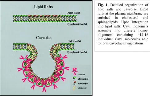
Fig. 1. Detailed organization of
lipid rafts and caveolae. Lipid rafts at the plasma membrane are
enriched in cholesterol and
sphingolipids. Upon integration into lipid rafts, Cav1 monomers assemble into discrete
homo-oligomers containing ~14-16
individual Cav1 molecules able to form caveolae invaginations.
plasma membrane (11).
Caveolin-1: initial discovery. Caveolin-1 (Cav1) was initially identified as one of
the phosphorylated substrate in Rous sarcoma virus transformed fibroblasts (12) (13). Interestingly, a separate group identified the VIP-21 (vesicular integral protein of 21 kDa), an integral protein component of trans-Golgi-derived transport vesicles (14). It turned out, the caveolin sequence was identical to that of VIP-21, thereby showing that the same protein could possibly serve as a structural component of plasma membrane caveolae, as well as have roles in oncogenesis and vesicular trafficking, all at the same time (15). In Fig. 1 a schematic on the current view of lipid rafts and caveolae is shown. Briefly, upon integration of the Caveolin-1 into lipid rafts, adjacent CAV1 homo-oligomers (containing 14-16 individual caveolin molecules are thought to pack side-by-side within caveolae membranes thereby providing the structural backbone for caveolae invagination.
Caveolin genes. Three members of the caveolin (CAV) gene family have been
identified (Fig. 2). Caveolin was the first gene discovered and is composed of three exons that are highly conserved in sequence and structure across species. Caveolin-2 was discovered when micro-sequencing of purified adipocyte caveolae membrane domains revealed a strikingly similarity to caveolin-1, differing in several key conserved residues (16). Caveolin-3 was cloned from a cDNA library using a caveolin-1-related sequence found immediately downstream of the rat oxytocin receptor (17). In addition to these two new members of the caveolin gene family, it was also found that both caveolin-1 and -2 have multiple isoforms. Caveolin-1 has two isoforms, termed α and β, with the α-isoform consisting of residues 1–178 and the β-isoform containing residues 32–178, resulting in a protein 3 kDa smaller in size (18). The β-isoform was originally thought to derive from an alternate translation initiation site occurring at a methionine in position 32. However, later on the existence of two mRNA isoforms was reported using an RNAase protection assay (19). This was later confirmed by another group which detected two mouse caveolin-1 mRNA variants. The full length variant generated mostly caveolin-1α while the 5-variant, lacking the first exon, generated exclusively caveolin-1β (20). The authors confirmed that both α and β caveolin-1 isoforms are produced when a caveolin-1 cDNA construct, having no 5-untranslated region (UTR), is transfected into mammalian cells. They further showed that in vivo expression of the full length and 5’-variant mRNAs correlated with expression of the α and β isoforms, respectively (21). While the exact functional significance of these distinct isoforms remains unclear, studies have suggested that the caveolin-1α isoform is localized predominantly to deeply
been identified, the full-length caveolin-2 (α), and two truncated Caveolin-2 variants, termed -2β and -2γ. However, it remains unknown the functional significance of the Cav-2 isoforms (22). The caveolin proteins share significant homology. Human Caveolin-2 is ~58% similar to human Caveolin-1, while Caveolin-3 is 85% similar to Caveolin-1. A short stretch of eight amino acids has been identified (FEDVIAEP) that constitutes the “caveolin signature sequence,” a motif that is identical between all three caveolin proteins. The pattern of expression of Cav3, instead, is distinct from that of Cav1 and Cav2 that are not co-expressed in differentiated cardiac and muscle cells (23). Interestingly, reconstitution experiments in fibroblasts have demonstrated that Cav1 expression is required for Cav2 to be correctly targeted to the plasma membrane/Caveolae domains of cells (23). The Cav-3 protein is muscle specific and, similarly to Cav1, is known to form high-molecular-mass homo-oligomeric complexes and is sufficient to drive Caveolae formation in cells lacking Caveolins (23).
Membrane topology and post-translational modifications of Cav1. Initial studies
have identified Cav1 as an integral membrane protein, as it was found to be resistant to sodium carbonate and high salt concentration extraction. In addition, further studies have shown evidence that Cav1 has an unusual topology with both the NH2 and COOH termini of the protein facing the cytoplasm with a connecting
hairpin hydrophobic intramembrane domain (residues 103-134) (Fig. 1). This structure is consistent with the posttranslational modifications of Caveolin such as phosphorylation and palmitoylation that can only occur in the cytoplasmic side of the plasma membrane. One of the most important properties of this unusual protein is its ability to homo- or hetero-oligomerize (with Cav2) that may be one of the possible mechanisms driving Caveolae formation. In fact, Cav1 after being synthesized as an integral membrane protein in the endoplasmatic reticulum (ER) goes through a first stage of homo- or hetero-oligomerization in the ER forming oligomers containing 14-16 individual Caveolin monomers. A further stage of oligomerization between Cav1 oligomers is described to happpen at the level of the trans-Golgi, where several Cav1 oligomers self-associate via C-terminal domain (TD domain, Fig. 2) and interact with cholesterol to form an extensive networks of Cav1 proteins that may be able to drive the invagination of Caveolae at the plasma membrane. Interestingly, deletion mutagenesis analysis has demonstrated that the hydrophobic residues constituting the transmembrane domain (TM 102-134) is not necessary for membrane attachment. Two other regions instead termed N-MAD (82-101) and C-MAD (135-150) are required to target Cav1 to Caveolae membrane and cis Golgi complex respectively. In fact, when fused to the C-MAD of CAV1, the GFP protein was resistant to extraction
(KYWFYR) in the N-MAD domain sufficient to confer membrane localization to GFP protein but not to Caveolae (24). As we will discuss below, the N-MAD region, also called the Caveolin Scaffolding Domain (CSD), is the region of the Caveolin protein believed to directly interact and modulate the activity of several signaling molecules within Caveolae microdomains. Interestingly, Cav1 undergo a series of post-translational modifications that are important in defining its function in several biological processes. Caveolin-1 is palmitoylated on three Cysteines (133,143,156) located in the C-terminus of the protein (25) and these post-translational modifications appear not necessary for caveolar targeting of the protein. However, studies using palmitoylation mutants of the Cav1 protein show that palmitoylation at Cys-156 is required to facilitate the coupling of Cav1 and the c-Src tyrosine kinase within Caveolae (26, 27). Other studies have suggested that palmitoylation may be a mechanism to stabilize Cav1 oligomers and to couple Cav1 with others lipid modified signaling proteins such as Abl, Cbl, Src, Gα subunits, Ras related GTPases, eNos and to cholesterol (28) (26). Caveolin has also been described undergoing phosphorylation on Tyr 14 (Y14) (12) and on Ser-80 (SSer-80) (29). The role of Y14 phosphorylation has only partially been clarified. It occurs in response to oxidative (30) and osmotic stress (31) and also in response to growth factor and hormonal stimulation (26). In addition, recent studies demonstrate the importance of Y14 phosphorylation in regulating migration/invasion and Caveolin mediated endocytosis in several cell types (32) (33). Furthermore, Cav1 resulted to be phosporylated on S80 and it seems important in regulating ER localization and in regulating the secretory pathway. Mutation of Serine 80 to Ala impairs the ability of Cav1 to be secreted in certain cell types (29) (34).
Vesicular Trafficking. Based on their structural features as plasma membrane
invaginations, Caveolae were initially proposed to function in potocytosis (35), a kind of receptor mediated endocytosis that involves the selective uptake of small molecules. With the development of new more sophisticated techniques it appears clear that caveolae take part in other vesicular trafficking processes such as transcytosis and endocytosis that constitute an alternative endocytic pathway to clathrin-coated pits for trafficking to Golgi, ER or lysosome. Several lines of experimental evidence have demonstrated the existence of caveolae mediated endocytosis. First, caveolae endocytosis is significantly slower than clatrin coated pits endocytosis. Second, the phosphatase inhibitor okadaic acid stimulates caveola-mediated endocytosis but inhibits the formation of clathrin-coated vesicles. Third, the sterol binding agent filipin has little or no effect on clathrin-mediated endocytosis, yet inhibits the internalization of caveolae. Finally,
endothelial cell layer. Elegant experiments were performed using Caveolar proteins specific antibodies clearly demonstrating the movement of caveolar proteins across the endothelial cells from the luminal side to the interstizial side (37). In endocytosis, Caveolae have been shown to mediate the intracellular trafficking of molecules to the ER and Golgi either directly or through an intracelluar organelle termed Caveosome or VVO. Macromolecules such as Cholera toxin, folic acid and albumin are internalized by a Caveolae mediated endocytosis to intracellular compartments that are negative for endosomal markers (EEA1 and TfR). Although the exact process mediating caveolar endocytosis remains unknown, several recent findings suggest that the molecular machinery involved in the generation of caveolar vescicles is the same used for numerous other vesicular transport processes that requires, dynamin, VAMP, SNAP-25, the SNARE complex, and GTP hydrolysis (36) (8). Interestingly, several pathogens, including viruses, bacteria, fungi, parasites and even prions can use caveolar endocytosis as a mean to bypass the classical clathrin mediated endocytosis and as a consequence avoid the degradative compartment constituted by the endosome-lysosome vescicular system. The intracellular trafficking of simian virus 40 is perhaps the most well studied infectious agent that selectively uses caveolae to enter the cells. After its initial binding, and following its accumulation in the “caveosome,” SV40 is delivered directly to the ER. In doing so, SV40 avoids the inactivation that would have occurred by using the clathrin coated pits pathway (38) (39). Interestingly, other viruses have been shown to utilize the folate receptors as a cofactor to entry into the cells (40). Importantly, caveolae as a common mechanism to gain entry into cells avoiding the intracellular degradative compartments may be used as a strategy to deliver therapeutic agents (41).
Lipid homeostasis. Since their discoveries Caveolae/Caveolins have a strict
interrelationship with cholesterol and with its homeostasis. Initially described as a cholesterol binding proteins, Caveolins and Caveolae are sensitive to agents such as filipin and nystatin that can reduce Caveolar structures, intracellular localization and their stability (42). Additionally, cholesterol transcriptionally regulates Cav1 expression through two steroid regulatory binding elements in the Cav1 promoter. This results in decreased Cav1 mRNA in cells depleted of cholesterol (43). Thus, Cav1 expression and its intracellular distribution are clearly dependent upon cellular cholesterol levels. Other findings suggest that Caveolin can regulate cholesterol homeostasis by modulating its cellular influx or efflux (44). Interestingly, Cav1 and Cav2 have been found associated with lipid droplets, metabolically active organelles serving for lipid storage functions (45).
kinases and heterotrimeric G-proteins. These initial observations led the authors to the “Caveolae signaling hypothesis”that describes Caveolae as hubs that may serve to specifically compartmentalize certain signaling molecules and thereby rapidly
modulating signal transduction events and the cross-talk between different signaling pathways (46) (47). This hypothesis was confirmed by studies demonstrating that the activity of G-protein-alpha subunit could be suppressed by a peptide mimicking the Caveolin scaffolding domain (CSD; 82-101) of Cav1. Similar results were obtained with other signaling proteins such as Src/Fyn, EGF-R, Neu, PKC, and PKA (48). With the proteomic analysis of purified Caveolae it has been possible to identify a wide assortment of proteins that are localized to these structures in several different tissues and cell types. For instance proteomic analysis of purified caveolae from lung tissue identified a prevalence of cytoplasmatic signaling molecules (Src-like kinases and heterotrimeric G protein subunits), including the small GTPases Rap 1, Rap 2 and cytoskeletal elements,
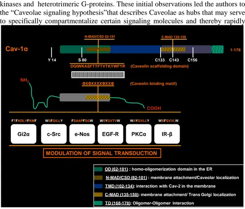
1-178
N-MAD/CSD 82-101 C-MAD 135-150
DGIWKASFTTFTVTKYWFYR (Caveolin scaffolding domain)
ΦXΦXXXXΦXXΦ (Caveolin binding motif) Cav-1α
NH2
COOH
Gi2α c-Src e-Nos EGF-R PKCα IR-β
FTFKDLYFKMF WSFGILLY FSAAPFSGW WSYGVTVW WAYGVLLY WSFGVVLW
MODULATION OF SIGNAL TRANSDUCTION
Y 14 S 80 C133 C143 C156
N-MAD/CSD (82-101) : membrane attachment/Caveolar localization TMD (102-134): interaction with Cav-2 in the membrane
OD (62-101) : homo-oligomerization domain in the ER
C-MAD (135-150): membrane attachment/ Trans Golgi localization TD (168-178): Oligomer-Oligomer interaction
Fig. 2. The sequence of the caveolin-scaffolding domain and the caveolin binding motif sequences
of several proteins are shown. In most cases, this caveolin interaction is inhibitory, leading to inactivation of the signaling pathway and thereby modulation of downstream signal trusduction.
obtained during the proteomic analysis of Caveolae purified from other cell types (49). These and other studies led to the development of the idea that caveolae are actively engaged in the compartmentalization of various signaling molecules and thereby behaving as active signaling organelles in the cell. There is now evidence that the region termed the caveolin scaffolding domain (CSD) seems to be responsible for many of the biological functions attributed to Cav1 by interacting with a sequence called the Caveolin binding domain (CBD) identified in several Cav1 binding proteins including tyrosine/serine threonine kinases and eNos. Site-directed mutagenesis performed to modify the caveolin binding sequance within the Caveolin binding domain of eNos, identifiyed the following motifs that are always found in Caveolin interacting proteins: ΦXΦXXXXΦ, ΦXXXXΦXXΦ,
ΦXΦXXXXΦXXΦ, where Φ is an aromatic residue (Phe, Tyr, or Trp) and X is any
amino acid (36) (50). Except few exceptions, the current view is that upon binding to signaling molecule with the CSD, Cav1 leads to inhibition of downstream signaling and thereby functioning as a negative regulator of signal transduction (Fig.2). Given the ability of many of these molecules to be involved in cellular transformation (i.e. H-Ras, MAPK, PDGFR, EGFR) when hyperactivated, it is reasonable to speculate that Cav1 may behave as tumor suppressor gene in vivo.
Caveolin1: role in cancer pathogenesis. The initial discovery that Cav1 is a major
phosphorylation substrate in Rous sarcoma virus transformed cells indicated it was a potential target in the process of oncogenesis (12). Indeed, subsequent experiments showed that both Cav1 mRNA and protein levels decrease when NIH3T3 cells are transformed by a variety of oncogenes, including H-RasG12V, polyoma virus mTAg, and p210bcr-abl (51) (52). Transformation with H-RasG12V was further shown to alter transcription factor binding at the Cav1 promoter and decreases transcriptional activity (53). Additionally, the Cav1 promoter contains p53-responsive elements and the p53 tumor suppressor protein increases Cav1 mRNA and protein levels (54). Finally, the Cav1 promoter has c-myc-repressive elements and expression of the c-myc oncogene decreases Cav1 mRNA levels (55). Because one of the hallmarks of transformation is the downregulation of tumor suppressor proteins (56), the down-regulation of Caveolin-1 at the mRNA and protein levels in response to a broad range of oncogenic stimuli provided evidence that this protein could have a function in cancer. Evidence for the tumor suppressive function of Cav1 comes from the observation that Cav1 is lost in human cancers through gene deletion, misregulation, or mutation. Fragile sites are chromosomal loci sensitive to replication stress, and they are frequently involved in chromosomal rearrangements in human disease (57). The q31 region of Chromosome 7, encompassing the fragile site FRA7G at 7q31.2, is frequently lost
genes to Chromosome 7q31.1 (62) (63), and this discovery led to the hypothesis that CAV1 and CAV2 are potential tumor suppressor genes that contribute to tumor development when this region is lost in human cancers (53, 62). Methylation of the
CAV1 gene promoter provides a second mechanism by which expression of this
protein is lost in human cancers. In addition to mapping the chromosomal location of CAV1 and CAV2, Engelman and colleagues found methylation of CpG islands in the CAV1 promoter region in human breast cancer cell lines (53). Subsequent research found similar results in breast cancer tissue, small cell lung cancer cell lines, a human ovarian cell line and ovarian adenocarcinoma tissue, and human prostate cell lines and prostate tumor samples (64) (65, 66) (67). This work suggests CAV1 expression can be lost in human cancer not only through gene deletion, but also through alterations to promoter methylation status and transcriptional repression. Finally, a third mechanism of functional Cav1 loss in human cancer is through mutation. Hayashi and colleagues examined the CAV1 gene sequence in primary human breast cancer tissue and discovered a mutation— P132L—in 16% of the tumors. The mutation is found mainly in invasive carcinomas, and expression of this mutant protein is able to transform NIH3T3 cells (68). Interestingly, further work on this mutation showed expression of Cav1-P132L in mammary epithelial cells expressing endogenous WT Cav1 results in the retention of both mutated and WT protein in the Golgi, demonstrating that P132L is a dominant negative mutation (69). In addition to these specific mechanisms of Caveolin-1 down-regulation, Cav1 mRNA or protein expression has been examined in specific cancers with varying results. Multiple studies have consistently shown that Cav1 expression is lost or decreased in: 1) ovarian carcinoma, demonstrated at both the mRNA and protein levels in cell lines and tissue, 2) lung carcinoma, demonstrated in some studies for several types of lung cancer and in others as being more common in small cell lung cancer, and 3) breast cancer, shown to correlate with tumor size and grade. For some cancers, Cav1 expression seems to be related to metastatic, but not primary, tumor growth. For example, in head and neck squamous cell carcinoma, Cav1 expression is lost in metastatic tumors and lymph node metastases. It should be noted that Cav1 is not a universal tumor-suppressor gene. Indeed, its role in oncogenesis has been shown to be context-dependent, and Cav1 is up-regulated in the development and progression of certain cancers. For example, in bladder carcinoma, studies have shown that Cav1 expression in not observed in normal epithelium but is associated with higher-grade tumors. In addition, Cav1 has a growth-promoting role in prostate cancer. In the prostate, phosphorylation of Cav1 at serine-80 causes it to be secreted by cancer cells; in human patients with advanced disease, secreted Cav1 can act in an autocrine/paracrine manner to promote prostate cancer cell
role of Cav1 in cancer development. However, the loss of Cav1 expression in several human cancers through gene deletion, transcriptional repression, or mutation indicates an anti-oncogenic function for this protein in many contexts, which is further supported by examination of the effects of gene deletion in a mouse model.
The Caveolin-1 knock-out mouse and tumorigenesis. The generation of the Cav1
KO mouse model allowed for the examination of the effect of complete Cav1 loss on numerous phenotypes. These mice were developed in parallel by two different groups (73) (74). Given the previously-discussed importance of Cav1 in caveolar biogenesis (75), it is not surprising that both groups report loss of caveolae structures in Cav1-expressing tissues (i.e caveolae are still apparent in tissues which predominantly express Caveolin-3). Interestingly, in Cav1 KO cells Cav2 expression is significantly reduced demonstrating that Cav1 is necessary for the stabilization of his binding partmner (76). Cav1 KO mice show a roughly 90% decrease in Cav2 protein levels in Cav1- expressing tissues, which is rescued by proteasome inhibition (74). Due to this phenomenon, Cav1 KO mice are essentially double knock-outs for both Caveolins-1 and 2. Cav1 KO mice display numerous pathologies, including lung hypercellularity and thickening of the alveolar septa, increased eNOS activation resulting in vascular abnormalities, and metabolic defects that include decreased fat accumulation and insulin resistance (74). Given the proposed tumor suppressive function of Cav1, Cav1 mice null have been used extensively in studies of cancer biology and tumorigenesis. Cav1 KO mice do not develop spontaneous tumors throughout the course of their approximately 500 day lifespan (77). The first evidence that loss of Cav1 contributes to in vivo oncogenesis was provided by Capozza and colleagues (78). When C57BL/6 WT and Cav1 KO mice were subjected to chronic topical treatments of the carcinogen 7,12-dimethylbenz[a]anthracene (DMBA), KO mice displayed an increase in epidermal hyperplasia, tumor incidence, tumor multiplicity, and tumor size along with hyperactivation of the Erk1/2 MAPK pathway (78). In addition, Cav1 KO mice display hyperproliferative defects. Specifically, mouse embryonic fibroblasts (MEFs) isolated from these mice show an increase in proliferation, and their lungs show evidence of hypercellularity (74). In addition, these mice display mammary epithelial hyperplasia by 6 weeks of age and increased lobular development with acini formation by 9 months of age (69). Crossing Cav1 KO mice to a model of spontaneous breast tumor development (MMTVPyMT) revealed that Cav1 KO mice develop mammary lesions approximately 5 weeks earlier and show a roughly 2-fold increase in tumor multiplicity when compared to WT mice. In addition, lesions from Cav1 KO mice
stromal compartment surrounding epithelial tumor cells. Interestingly, loss of Cav1 has been shown to affect benign tissue stroma in aged Cav1 KO mice. Yang and colleagues showed that the stromal compartments of various organs, including the lung and liver, are disorganized in KO mice, and this pathology results in defects in epithelial cell growth and differentiation (80). Examination of the effect of Cav1 loss in cancer-associated stroma revealed that the injection of breast cancer cells into the Cav1 KO mammary fat pad increases tumor growth in comparison to injection in the WT fat pad, indicating the KO fat pad is more permissive to malignant growth (81) (82). Similarly, injection of lung carcinoma cells into Cav1 KO mice increases angiogenesis and tumor growth in comparison to cells injected in WT mice (83). Different groups have reported that loss of stromal Cav1 in human breast cancers correlates with poor prognosis, suggesting that Cav1 loss in the stroma could be a biomarker for an aggressive cancer microenvironment (84). However, these findings are controversial, as other researchers assert that the disorganized stroma displayed by the Cav1 KO mouse is actually an inhibitory microenvironment for invasion and metastasis (85). The potential function of Caveolin-1 in the tumor microenvironment is interesting but contentious, and most work still focuses on the role of this protein in the epithelial component of the tumor. As detailed above, the Caveolin-1 knock-out mouse has been used extensively to study the effect of Cav1 loss on tumor development and progression through the examination of both epithelial cancer cells and their associated stroma. These studies demonstrate that the Cav1 KO mouse does not develop spontaneous tumors. Given this phenomenon, it has been proposed that while Cav1 loss is not sufficient in and of itself to transform normal cells, its loss in conjunction with an oncogenic stimulus exacerbates tumor development, growth, and progression (86). Further support of this hypothesis is the extensive research describing the role of Cav1 in the modification of several cancer-associated phenotypes, i.e. proliferation and survival, anchorage-independent growth, and invasion (87).
epidermal layers (Fig. 3). The underlying dermis, composed mainly of fibroblasts and connective tissue, functions as the support system for the epidermis. The epidermis is a stratified epithelium that is constantly undergoing renewal as
outside cells are sloughed off and replaced by cells from the lower layers. Epidermal homeostasis is maintained by actively-dividing stem cell populations in both the hair follicles and the basal layer (88) (89) (90). Keratinocytes are the major cell type of the epidermis, and they undergo a process of terminal differentiation and cell death that culminates in the creation of an environmental barrier. Stem cell division in the basal layer of the epidermis produces new keratinocytes; these cells lose their ability to adhere to the basement membrane between the dermis and the epidermis, causing the keratinocytes to undergo differentiation. The cells exit the cell cycle and are pushed from the basal layer into the upper layers of the epidermis. In the spinous layers, the cells fortify their keratin filament network. In the granular layers, keratins are bundled into larger units called macrofibrils, lipids are produced in epidermal lamellar bodies, and cornified envelope proteins are deposited beneath the plasma membrane. In the final steps of terminal differentiation, the cell membrane disintegrates, triggering
Fig. 3. Skin and epidermal morphology. The two main layers of the skin, dermis and epidermis
separated by the basement membrane. Keratinocytes in the basal layer divide and undergo
terminal differentiation to form a layer of enucleated, dead cells in a matrix of lipids, called the
of lipids called the cornified layer (Fig. 3). As the body’s major barrier, the skin is subjected to constant damage from environmental stresses, including chemicals, toxins, and ultraviolet radiation, and accordingly is a common site for tumorigenesis (88).
Skin cancer characteristics. Malignancies that develop in the epidermis are
broadly termed skin cancer and are categorized into two groups: malignant melanoma and non-melanoma skin cancer. Malignant melanoma (MM) originates in the melanocytes localized at the dermal-epidermal junction. MM is the least common type of skin cancer, but it is also the deadliest due to its higher rate of metastasis. Transformation of skin melanocytes to cutaneous melanoma is a multistep process also called melanomagenesis (91). The first steps, considered as benign, are associated with the formation of a nevus and the radial growth phase (RGP). In a nevus, melanocytes are clustered and have lost their appropriate contact with keratinocytes. During RGP, melanocytes tend to proliferate superficially to the basement membrane of the epidermis. During the next stage, the vertical growth phase (VGP), the cells bypass senescence to proliferate actively in a vertical manner in the dermis, crossing the basement membrane. At this stage, cells migrate and become clearly invasive. The last stage is the acquisition by the cells of metastatic characteristics: the cells are able to enter the bloodstream or lymphatic vessels from which they colonize tissues and organs. In addition, melanomagenesis is accompanied by dysregulation of various signaling pathways which ultimately affect several cancer associated phenotypes such cell cycle, invasion, migration, tumor growth and metastatic dissemination. Several genetic and epigenetic abnormalities have been detected in melanomas that results in inactivation of tumor suppressor genes and hyperactivation of oncogenes, the inhibition of apoptosis, changes in morphology and migration capacity, and also modification of DNA repair enzyme activities. Two of the most common genetic alterations found in melanoma are the loss/inactivation of the tumor suppressor p16Ink4a and the activating mutations in N-Ras and B-Raf (>50%). Additional alterations in terms of signaling may accumulate at later stages of melanoma progression (91). Non-melanoma-skin-cancer (NMSC) is the most prevalently-diagnosed malignancy in the United States and estimated that roughly 45 percent of Americans that live to age 65 will have NMSC at least once in their lives (92). There are two types of non-melanoma skin cancer: basal cell carcinoma (BCC) and squamous cell carcinoma (SCC). BCC is the most common type of skin cancer and comprises a group of tumors that can vary phenotypically, but primarily contain undifferentiated epidermal basal cells and mutations that constitutively activate the Hh pathway are found in the vast majority of these lesions (93).
development of cSCCs is considered a multi-stage process in which an accumulation of genetic events in necessary (94). As such, precursor lesions have been identified for cSCC. These include actinic keratoses (AK), a pre-cancerous lesion with a 5-10% chance of developing into cSCC, and Bowen’s disease (BD), a non-invasive tumor that precedes invasive cancer and is referred to as carcinoma
in situ (95) (96). Another potential precursor lesion is keratoacanthoma (KA), a
low grade lesion with a rapid early growth phase that is histologically very similar to a well differentiated SCC (97). Squamous cell carcinomas are the result of abnormal proliferation of the more-differentiated squamous cells of the epidermis. However, the actual cell of origin for SCC most likely resides in the basal epidermal layer, and the squamous phenotype is a consequence of what and when specific mutations are acquired along the progression to malignancy (98) (99). The outcome of this multi-step process is a squamous cell carcinoma that, unlike BCC, has the potential for metastatic dissemination. Given the potential to metastasize, extensive research has examined the characteristics of the primary tumor that would indicate a potential for tumor progression and metastatic dissemination. Given their prevalence and potential for metastasis, much research is needed to identifying the factors that contribute to cSCC development.
Cav1 expression and function in normal skin. Upon barrier disruption through
tape stripping, Cav1 KO mice display increased raft formation and accelerated barrier reacquisition due to increased lamellar body (LB) secretion and also experimental epidermal hyperplasia due to reduced terminal differentiation (100). Similarly, wound healing studies demonstrated that cutaneous wounds in Cav1 KO mice heal more quickly than those of wild-type (WT) mice (101). Studies in primary human keratinocytes have demonstrated that Cav1 mRNA and protein levels increase dramatically in the later stages of keratinocyte differentiation, further supporting a role for Cav1 in this process (102). In addition, loss of Cav1 confers a hyperproliferative phenotype to basal keratinocytes indicating an enhanced ability to re-populate a wounded site with keratinocytes (100) (103). In addition to functioning in re-epithelization of the epidermis, Cav1 also plays a role in intercellular junctions in the skin. Caveolin-1 has been implicated in the regulation of three different types of junctions in the epidermis: adherens, desmosomes, and gap junctions (104) (105) (100) (106). These studies indicate that Cav1 has a significant function in maintaining structure and homeostasis in the skin. For instance, Connexin-43 (Cx43) is highly expressed in the epidermis where it co-localizes with Cav1 in keratinocytes. Loss of this association between Cav1 and Cx43 renders keratinocytes more susceptible to transformation (107). Thus, loss Cav1 expression decreases intercellular adhesion through adherens and
suggest that its loss could confer sensitivity to the development of skin disease.
Cav1 and skin cancer. The role of Cav1 in melanoma progression remains not
very well defined. Overexpression of Cav1 in a metastatic melanoma cell line SKMEL28 resulted in inhibition of their proliferation and motility. The reduced motility of these cells was attributed to the reduced levels of phosphorylated levels of p130Cas and paxillin in Cav1 expressing cells. In addition, Cav1 expressing cells displayed a diffused staining for Gangliosides GD3 compared to their wild type counterpart that showed a more localized staining to the leading edge of melanoma cells, suggesting that Cav1 expression may inhibit or at least attenuate the malignant properties of this cell line. Alonso and colleagues used a melanoma tissue microarray of 165 samples in which they used a panel of 39 different antibodies for cell cycle proteins, apoptosis, transcription factors and other proteins. Interestingly, this panel of antibodies included Cav1 whose expression was significantly diminished in advanced stages of melanomas relative to nevus, suggesting for the first time that Cav1 could be used as a biomarker to predict melanoma progression and disease outcome. While these results from earlier investigations suggest a tumor suppressive function for Cav1 in malignant melanoma, more recent in vitro studies proposed a tumor promoting role for Cav1. Sargiacomo and colleagues observed increased proliferation and increased motility in melanoma cells expressing Cav1. These authors attributed this phenotype to the ability of Cav1 to hyperactivate the FGFR/Src/Rho GTPases signaling pathway which is critical in regulating cell cycle progression and several aspects of cell motility (108) (109). In another recent article, Del Pozo and colleagues reported that in a melanoma cell line the presence of Cav1 was required for the internalization of the lipid raft protein Rac1 in suspended cells, resulting in the inhibition of downstream effectors proteins (110). Several lines of experimental evidence suggest that tumor growth results from key bidirectional interactions between cancer cells and their surrounding stroma (111). Although the role of stromal Cav1 has been recently investigated in breast cancer (112) the function of Cav1 in melanoma microenvironment and consequent tumor development remains largely unexplored. As described above, Cav1 loss-of-function in the skin may contribute to hyperproliferative disorders of the epidermis by promoting pro-proliferative signals or by affecting the differentiation stage of epidermal keratinocytes (107) (113) (100). Interestingly, in addition to showing a decreased expression in hyperproliferative diseases of the skin, Langlois and colleagues demonstrated that Cav1 expression is significantly reduced in both basal and squamous cell carcinomas of the skin suggesting that Cav1 may have a function in the pathogenesis of skin cancer. Accordingly, Capozza and colleagues showed that
enhanced growth signaling, including cyclin D1 and activated Erk1/2, in the expanded epidermal proliferative compartment. Similarly, Roelandt and colleagues showed that the epidermis of Cav1 KO mice had increased baseline and experimentally-induced proliferating cell nuclear antigen (PCNA) incorporation (100). Collectively, this work indicates that loss of Cav1 may confer a malignant advantage in the context of skin cancer development.
OBJECTIVES
As the main protein component of the membrane lipid rafts caveolae, Cav1 functions as a modulator of cellular signal transduction through both internalization and interaction with various signaling molecules. By means of its scaffolding domain, Cav1 compartmentalizes signaling proteins, often resulting in the suppression of their signaling function. Given its multi-functional nature, Cav1 has been implicated in the pathogenesis of several human diseases, including cancer. Its role seems to be context-specific and dependent on the type of tissue being examined. In several cancers, Cav1 expression is lost through mutation, gene misregulation, or unknown mechanisms. In addition, Cav1 is a negative regulator of many cancer associated phenotypes, including proliferation, anchorage independent growth, and invasion. Published data from us and from other investigators indicates that Cav1 expression is modulated during both melanoma and non melanoma skin cancers. Several lines of in vitro and in vivo evidence suggest that Cav1 is implicated in the pathogenesis of oncogenic cell transformation, tumorigenesis, and metastasis. However, while Cav1 does not appear to have a direct role in tumor initiation, it does synergize with other oncogenes and tumor suppressors to modulate the transformed/tumorigenic phenotype. Recent published data from us and others indicates that Cav1 expression is modulated during both melanoma and non melanoma skin cancers. However, very little work has examined the contribution of Cav1 to specific stages of skin carcinogenesis, namely initiation, promotion of growth, and progression to malignancy and metastasis. Therefore, the work discussed herein aims to address
this issue by determining mechanisms by which modulation of Cav1 protein expression in both melanoma cells and/or the surrounding micro-environment may affect melanoma tumor growth and metastatic dissemination. We will also describe mechanisms by which Cav1 expression may affect skin carcinogenesis in a to two stage carcinogenesis protocol and in a murine model of cutaneous squamous cell carcinoma (cSCC.) In summary, the work described herein provides
evidence that Cav1 may function as suppressor of tumor progression stages in both melanoma and non melanoma skin cancers and is therefore a biomarker for tumor aggression and is a potential target for therapeutic intervention in skin cancer.
Chapter 1
_______________________________________________________
CAV1 Inhibits Metastatic Potential in Melanomas through
Suppression of the Integrin/Src/FAK Signaling Pathway
CAV1 Inhibits Metastatic Potential in Melanomas through Suppression of the Integrin/Src/FAK Signaling Pathway
Casey Trimmer1,2, Diana Whitaker-Menezes2, Gloria Bonuccelli2, Janet N. Milliman1, Kristin M. Daumer1,
Andrew E. Aplin1, Richard G. Pestell1, Federica Sotgia1, Michael P. Lisanti2, and Franco Capozza1,3
Abstract
Caveolin-1 (CAV1) is the main structural component of caveolae, which are plasma membrane invaginations that participate in vesicular trafficking and signal transduction events. Although evidence describing the func-tion of CAV1 in several cancer types has recently accumulated, its role in melanoma tumor formafunc-tion and progression remains poorly explored. Here, by using B16F10 melanoma cells as an experimental system, we directly explore the function of CAV1 in melanoma tumor growth and metastasis. We first show that CAV1 expression promotes proliferation, whereas it suppresses migration and invasion of B16F10 cells in vitro. When orthotopically implanted in the skin of mice, B16F10 cells expressing CAV1 form tumors that are similar in size to their control counterparts. An experimental metastasis assay shows that CAV1 expression suppresses the ability of B16F10 cells to form lung metastases in C57Bl/6 syngeneic mice. Additionally, CAV1 protein and mRNA levels are found to be significantly reduced in human metastatic melanoma cell lines and human tissue from metastatic lesions. Finally, we show that following integrin activation, B16F10 cells expressing CAV1 dis-play reduced expression levels and activity of FAK and Src proteins. Furthermore, CAV1 expression markedly reduces the expression of integrin β3in B16F10 melanoma cells. In summary, our findings provide experimental
evidence that CAV1 may function as an antimetastatic gene in malignant melanoma.Cancer Res; 70(19); 7489–99. ©2010 AACR.
Introduction
Malignant melanoma remains among the most life threat-ening of all cancers, and its incidence has been increasing dramatically in the last decades. Despite great progress in understanding the genetics and biochemistry of malignant melanoma, patients with metastatic disease have very few treatment options available. The establishment of metastases in distant organs of the body is a stepwise process that begins with the invasion of the dermis surrounding the primary tumor and ends with the colonization of ectopic sites (1). Each of the steps of the metastatic cascade is rate limiting. Thus, identifying novel mechanisms and factors reg-ulating melanoma progression may be critical for the devel-opment of new therapeutics in this type of cancer.
Initially identified by electron microscopy (2), caveolae are 50 to 100 nm large plasma membrane invaginations morpho-logically distinct from the classic clathrin-coated vesicles (3). Three different caveolin genes (CAV1, CAV2, and CAV3) encode for the structural components of these organelles (4, 5). CAV1 is the best studied of the three caveolins, and it is considered a multifunctional scaffold protein able to bind and regulate the activity of numerous signaling molecules within caveolae (6). Due to the multitude of interacting proteins described, CAV1 has been implicated in the modulation of several cancer-associated phenotypes, including cell proliferation, death, and transformation (4). Aside from data derived from cell cul-ture experiments, there are several lines of clinical and genetic evidence implicating CAV1 as a tumor suppressor in vivo. First, CAV1 has been found to be downregulated and/or mutated in a number of human tumors, including mammary adenocarcino-mas and squamous cell carcinoadenocarcino-mas (7, 8). Second, the genera-tion of CAV1 knockout (KO) mice has allowed for the validagenera-tion of the hypothesis that CAV1 may behave as a tumor suppressor. Although CAV1 KO mice do not develop spontaneous tumors, they are more susceptible to carcinogen [7,12-dimethylbenz(a) anthracene]– and oncogene-induced cancer in skin and mam-mary tissues, respectively (9, 10). However, the idea that CAV1 may be a “general” tumor suppressor has been recently chal-lenged by reports showing that CAV1 expression is cancer type and/or stage dependent (11). CAV1 is upregulated in the bladder, esophagus, thyroid (papillary subtype), and prostate carcinomas, and this upregulation seems to be associated with multidrug resistance and/or metastasis (12, 13).
Authors' Affiliations: Departments of1Cancer Biology and2Stem Cell Biology and Regenerative Medicine, Kimmel Cancer Center, Thomas Jefferson University, Philadelphia, Pennsylvania and3Department of Biology, University of Roma Tre, Rome, Italy
Note: Supplementary data for this article are available at Cancer Research Online (http://cancerres.aacrjournals.org/).
Corresponding Authors: Franco Capozza, Thomas Jefferson University, Kimmel Cancer Center, 940 BLSB, 233 South 10th Street, Philadelphia, PA 19107. Phone: 215-9555333; Fax: 215-9231098; E-mail: Franco. [email protected] and Michael P. Lisanti, Thomas Jefferson Univer-sity, Department of Stem Cell Biology and Regenerative Medicine, 933 BLSB, 233 S 10th Street, Philadelphia, PA 19107; E-mail: Michael. [email protected].
doi: 10.1158/0008-5472.CAN-10-0900 ©2010 American Association for Cancer Research.
The role of CAV1 in malignant melanoma, however, re-mains poorly understood. Several groups have reported conflicting results for the role of CAV1 in melanoma trans-formation, migration, and invasion (14, 15). Furthermore, the role of CAV1 in melanoma tumor formation and metastasis remains to be determined. Here, to gain better insight into the function of CAV1 in melanoma progression, we used B16F10 melanoma cells as an experimental system to directly explore the function of CAV1 in melanoma tumor growth and metastasis.
In the current study, we show that CAV1 expression inhi-bits the motility of B16F10 melanoma cells in vitro and their ability to form lung metastases in vivo. These results were consistent with reduced CAV1 expression in a panel of hu-man metastatic melanoma cell lines and metastatic lesions of human patients. Finally, recombinant CAV1 expression in B16F10 cells was sufficient to suppress the expression and activity of Src and FAK proteins following integrin en-gagement. In summary, these data underscore the impor-tance of CAV1 as a new antimetastatic gene in malignant melanoma.
Materials and Methods
Materials
Antibodies and their sources were as follows: p-FAK(Y397) and p-Src(Y418) were from Invitrogen. Cyclin D1, cyclin A, Bcl-2, integrin α5, integrin β1, and CAV1(N-20) were from
Santa Cruz Biotechnology. FAK Flotillin-1 and CAV1 were from BD. Src, integrin α6, and integrin αV(Ab1930) were
from Millipore. Integrin β3, AKT, and p-AKT(S473) were from
Cell Signaling. β-Tubulin was from Sigma; S-100b was from Affinity BioReagents; and glyceraldehyde-3-phosphate dehy-drogenase (GAPDH) was from Fitzgerald.
Mice experiments
Orthotopic injections were performed by intradermally in-jecting 106B16F10 cells, whereas i.v. injections of 105cells
were used to assay for experimental metastasis in 3- to 4-month-old C57Bl6/J female mice (16, 17). All in vivo studies were approved by the Institutional Animal Care and Use Committee of Thomas Jefferson University. Detailed descrip-tions are available in Supplementary Methods.
Cell lines
B16F0, B16F10, A-375, WM-115, SK-MEL-28, SK-MEL-5, WM-266-4, WM-35, and normal human epidermal melano-cytes (NHEM) were cultured according to the manufacturer's instructions [American Type Culture Collection (ATCC), iell, and Science Cell Research Laboratories]. ATCC and Cor-iell routinely perform DNA profiling to authenticate their cell lines. For all the in vitro and in vivo experiments, only early passages of these cells (passages 5–6) were used. Retrovirus infection
pBabe-Puro and pBabe-CAV1-Puro retrovectors were used to stably transduce melanoma cells (18).
Western blots
Melanoma cells were sonicated and lysed in a modified radioimmunoprecipitation assay buffer and processed for Western blot analysis as we previously described (19). Protein fractionation and Triton X-100 solubility assay
Triton X-100 solubility assay was performed as previ-ously described (18). Cytoplasm and membrane proteins were extracted using a commercially available kit (Pierce Biotechnology).
Growth curves, cell cycle analysis, and proliferation assay
Growth curves were generated by seeding 2 × 10³cells/cm2
in triplicate. Cells were dissociated and counted with a he-macytometer at 1, 2, 3, and 4 days after seeding. Cell cycle analysis was conducted by flow cytometry analysis of propi-dium iodide–stained cells (20). DNA synthesis in cells was di-rectly analyzed by [3H]thymidine incorporation assay (21).
Cell proliferation was also estimated by immunostaining cells with the proliferation marker Ki67 (Abcam). Immunofluorescence
Cells were grown on glass coverslips and double immunos-tained for CAV1 and CAV2 as previously described (18). Slides were mounted with the Pro-Long Gold antifade reagent (Molecular Probes) and imaged by confocal micros-copy (LSM 510 META Confocal; Zeiss).
Tissue scan melanoma panel and quantitative reverse transcriptase-PCR
As previously described (22), a commercial panel of human cDNAs, obtained from normal human skin tissue and from human melanoma metastatic lesions (stages III and IV), was purchased from OriGene Technologies (MERT501). Quantitative reverse transcriptase-PCR (qRT-PCR) was per-formed using ready-to-use CAV1 and RPL13a primers/SYBR master mixes (SA-Biosciences). Quantitative expression data were acquired using ABI-Prism 7900HT Sequence Detection System (Applied Biosystems), and results were analyzed by the ΔΔCt method (23).
Immunohistochemistry of tissue sections
A tissue microarray of paraffin-embedded human mela-noma tissue samples were purchased from U.S. Biomax (Mel207; 69 cases/207 cores) and was stained for CAV1(N-20) using standard immunohistochemical techniques (9). An expert dermatopathologist carefully analyzed and blindly scored the tissue cores for semiquantitative analysis of immunoreactivity. Detailed descriptions are available in Supplementary Methods.
Migration and invasion assays
Cells (5 × 10⁴) suspended in 0.5 mL of serum-free medium
(SFM) containing 0.1% bovine serum albumin (BSA; Sigma) were added to the wells of an 8-μm-pore polycarbonate membrane, either coated with (for chemoinvasion assays) or without (for chemotaxis assays) Matrigel (Transwells;
BD Biosciences). Serum-free NIH3T3 conditioned medium (48 hours) was used as a chemoattractant. After 6 hours, the cells that had migrated were stained and counted as previous-ly described by others (17). For studies using Src and FAK inhibitors, SKI-606 (Selleck), PF-573,228 (Tocris Bioscience), or DMSO were placed in both the upper and lower chambers. Adhesion/suspension assays
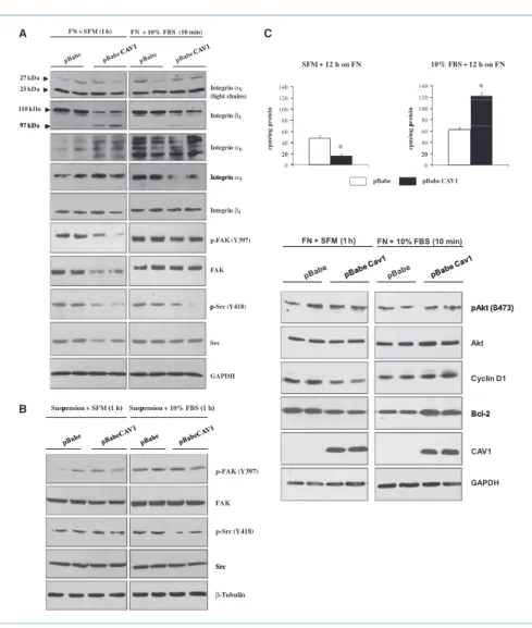
Integrin engagement was performed as described before (24). After being maintained in SFM containing 0.1% BSA for 18 hours, cells were dissociated, suspended in medium contain-ing 0.1% BSA, and replated on fibronectin (FN)-coated plates (BD) for 1 hour at 37°C. Cells were either lysed immediately or following the addition of complete medium [10% fetal bovine serum (FBS)] for 10 minutes. Alternatively, following 18 hours of serum starvation, cells were dissociated and left in suspension for 1 hour, and then processed for Western blot analysis. Statistical analysis
Results are represented as means ± SEM. Statistical anal-yses were performed using the Prism 4.0 Program (GraphPad Software, Inc.).
Results
CAV1 protein is correctly targeted to the plasma membrane of B16F10 melanoma cells
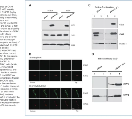
Lack of CAV1 expression has been described in several metastatic melanoma cell lines including B16F10 cells (15, 25, 26). Western blot analysis showed that a high expression level of CAV1 was achieved in B16F10 cells transduced with pBabeCAV1. CAV2 expression was not affected by CAV1 ex-pression in B16F10 melanoma cells. Identical results were obtained with the low metastatic B16F0 melanoma cell line (Fig. 1A). To determine the subcellular localization of CAV1 and CAV2, we next performed confocal microscopy on pBabe and pBabeCAV1 transduced cells. Serial optical images (z sections) of pBabe and pBabeCAV1 B16F10 melanoma cells double immunostained with CAV1 and CAV2 antibodies showed that recombinant CAV1 is correctly targeted to the plasma membrane of B16F10 cells. As expected, CAV2 colo-calized with CAV1 at the plasma membrane, despite the fact that a large portion of CAV2 also colocalized intracellularly (perinuclear) with CAV1 (Fig. 1B). These results were further confirmed by the observation that the CAV1/CAV2 complex
Figure 1.Absence of CAV1 expression in B16F0 (weakly metastatic) and B16F10 (highly metastatic) melanoma cell lines. A, immunoblotting of retrovirally transduced pBabe and pBabeCAV1 B16F10 and B16F0 cells for CAV1 and CAV2. S-100 immunoblot is shown as a loading control. Note the absence of CAV1 expression in both pBabe transduced B16F0 and B16F10 cells. B, confocal microscopy. Serial optical images (z sections) of pBabe and pBabeCAV1 B16F10 melanoma cells double immunostained with CAV1 and CAV2 antibodies show correct targeting of CAV1 to the plasma membrane. CAV2 extensively colocalizes with CAV1 in B16F10pBabeCAV1 cells (scale bar, 20 μm). C, immunoblot analysis of cytoplasmic (C) and membrane (M) fractions reveals that both CAV1 and CAV2 are enriched in the membrane fraction of B16F10pBabeCAV1 cells. Immunoblot for the membrane protein Flotillin-1 is also displayed. D, immunoblot analysis of Triton X-100–soluble (S) and Triton X-100–insoluble (I) fractions reveals that CAV1 is enriched in the Triton X-100–insoluble fraction. Note that CAV1 expression renders CAV2 Triton X-100 insoluble in B16F10 cells.
was enriched in the membrane fraction and in the Triton X-100–insoluble fraction of B16F10 cells expressing CAV1 (Fig. 1C and D). Thus, these results provide evidence that the CAV1/CAV2 complex is correctly targeted to the plasma membrane of B16F10 cells following the reexpression of CAV1 by retroviral strategy.
CAV1 expression promotes proliferation of B16F10 melanoma cells in vitro
Given the role of CAV1 in regulating proliferation and cell cycle progression (27), we next performed a proliferation
assay and cell cycle analysis. Interestingly, growth curves (in 5% and 10% FBS) and [3H]thymidine incorporation assay
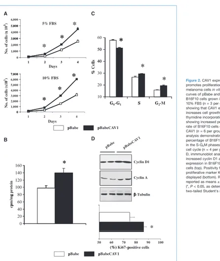
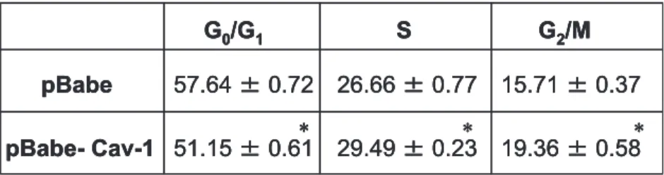
showed enhanced cell growth and increased DNA synthesis in B16F10pBabeCAV1 cells (140 ± 13 versus 98 ± 7 cpm/mg in pBabeB16F10; Fig. 2A and B). Fluorescence-activated cell sorting (FACS) analysis of asynchronously growing cells showed a significantly increased percentage of B16F10pBabe-CAV1 cells in the S and G2M phases of the cell cycle (Fig. 2C;
Supplementary Table S1). CAV1 expression in B16F10 cells was also associated with increased cyclin D1 and cyclin A expression and increased Ki67 positivity as determined by
Figure 2.CAV1 expression promotes proliferation of B16F10 melanoma cells in vitro. A, growth curves of pBabe and pBabeCAV1 B16F10 cells grown in 5% and 10% FBS (n = 3 per group) showing that CAV1 expression increases cell growth. B, a [3H]
thymidine incorporation assay showing increased proliferative rate of B16F10 cells expressing CAV1 (n = 6 per group). C, FACS analysis demonstrating increased percentage of B16F10pBabeCAV1 in the S-G2M phases of the
cell cycle (n = 4 per group). D, immmunoblot analysis showing increased cyclin D1 and cyclin A expression in B16F10pBabeCAV1 cells (top). Positivity for the proliferative marker Ki67 is also displayed (bottom). Results are reported as means ± SEM (*, P < 0.05, as determined by two-tailed Student's t test).
Western blot and immunofluorescence analysis (Fig. 2D). These results show a proproliferative role for CAV1 in the B16F10 melanoma cell line.
CAV1 expression decreases migration and invasion of B16F10 melanoma cells in vitro
Migration and invasion through a basement membrane are hallmarks of malignancy. To determine whether CAV1 expression affects these properties, pBabe and pBabeCAV1 B16F10 cells were subjected to migration (chemotaxis) and chemoinvasion assays. Specifically, we observed a roughly 2-fold reduction in the capacity of pBabeCAV1 B16F10 cells to migrate through the polycarbonate membrane of transwell chambers when NIH3T3 serum-free conditioned medium was used as a chemoattractant. Moreover, when cells were subjected to chemoinvasion assays, we observed a reduced capacity (roughly 2-fold reduction) of pBabeCAV1 B16F10 cells to invade through Matrigel-coated transwell chambers when NIH3T3 conditioned medium was used as a chemoat-tractant (Fig. 3). These results, along with the results from our proliferation assays, suggest that CAV1 inhibits migra-tion and invasion while maintaining a positive effect on cell cycle progression in B16F10 melanoma cells.
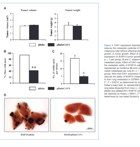
CAV1 expression dramatically reduces the metastatic potential of B16F10 cells in vivo without affecting primary tumor growth
To determine the effect of CAV1 expression on B16F10 tu-mor growth in vivo, 106pBabe and pBabeCAV1 B16F10
mel-anoma cells were orthotopically (intradermally) implanted in the skin of 3- to 4-month-old C57Bl/6 female mice. Eighteen days after injections, the determination of tumor size and
weight revealed that tumor growth was not significantly different between B16F10pBabe and B16F10pBabeCAV1 (Fig. 4A). Additionally, lungs dissected from both groups of mice did not show any spontaneous metastasis formation. To assess whether CAV1 expression was able to affect the me-tastatic potential of B16F10 melanoma cells, 105B16F10pBabe
and B16F10pBabeCAV1 cells were i.v. injected in 3- to 4-month-old C57Bl/6 female mice (experimental lung metas-tasis). After 18 days, examination of lungs revealed that the incidence of metastasis was significantly reduced in the B16F10pBabeCAV1-injected mice (42%) compared with the B16F10pBabe-injected animals (94%; Fig. 4B; Supplemen-tary Table S2). Strikingly, the B16F10pBabeCAV1-injected mice that showed metastasis formation displayed a signifi-cant reduction (roughly 3.5-fold) in the number of visible metastases per lung compared with the B16F10pBabe-injected mice (Fig. 4C and D). Consistent with the ability of CAV1 to reduce the motility of B16F10 cells in vitro, these results show that CAV1 expression suppresses the meta-static potential of B16F10 cells without affecting primary tumor growth in vivo.
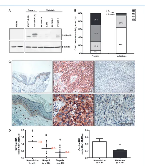
CAV1 expression is reduced in human metastatic melanoma cell lines and human tissue samples derived from metastatic lesions
Because CAV1 expression had no effect on the growth of B16F10-derived tumors, we next wanted to determine CAV1 expression levels in a panel of primary and metastatic mel-anoma-derived cell lines. Immunoblot analysis revealed that CAV1 expression was significantly reduced in metastatic melanoma cell lines (SK-MEL-28, A-375, SK-MEL-5, WM-266-4) compared with primary melanoma–derived cell lines (WM35, WM115). Interestingly, primary human melanocytes displayed a complete absence of CAV1 expression (Fig. 5A). To validate the significance of the expression pattern ob-served in melanoma cell lines, we next determined CAV1 expression by immunohistochemistry in normal skin, primary melanoma samples, and metastatic lesions from 69 melanoma patients (207 tissue cores). CAV1 immunore-activity scores revealed that ∼90% of the metastatic lesions showed absent (scored as 0) or weak (scored as 1) CAV1 staining. In contrast, we observed that only 30% of the primary melanoma samples showed absent or weak CAV1 staining (Fig. 5B). In cores that stained positive, CAV1 was observed to localize in the cytoplasm and at the plasma membrane of melanoma cells (Fig. 5C, center). In the skin, CAV1 immunostaining was observed in the keratinocytes of the basal cell layer as we have previously described (Fig. 5C, left; ref. 9). To further analyze the extent of CAV1 altera-tions in melanoma progression, we determined CAV1 expression by qRT-PCR on cDNA obtained from stage III (n = 20) and stage IV (n = 19) metastatic lesions. Analysis of CAV1 mRNA levels revealed that CAV1 expression was significantly reduced in stage IV metastases compared with stage III metastases (Fig. 5D, left). In addition, when CAV1 mRNA levels for both stages III and IV metastatic lesions were combined, they were significantly reduced (∼2-fold reduction) when compared with CAV1 mRNA levels in
Figure 3.CAV1 expression decreases migration and invasion of B16F10 melanoma cells in vitro. Chemotaxis (A) and chemoinvasion (B) were performed by seeding 5 × 10⁴pBabe and pBabeCAV1 B16F10 cells in
the upper wells of Matrigel-coated (for chemoinvasion) or uncoated (for chemotaxis) transwell chambers in SFM containing 0.1% BSA. Serum-free conditioned medium (48 h) from cultures of NIH3T3 cells was used as chemoattractant in the lower wells. After 6 h, the cells that had migrated to the underside of the membrane were washed with PBS, stained with crystal violet, and counted. Data represent the average of three independent experiments. Five fields per sample were counted. Results are reported as means ± SEM (*, P < 0.05, as determined by two-tailed Student's t test).