Studi sperimentali
Smoking, physical activity and respiratory irregularities
in patients with panic disorder
Fumo, attività fisica e irregolarità respiratorie
in pazienti affetti da disturbo di panico
EMMA FADDA1,2,3, ELISA GALIMBERTI1,2,3, STEFANIA CAMMINO1,2, LAURA BELLODI1,2
E-mail: [email protected]
1Department of Clinical Neuroscience, San Raffaele Scientific Institute of Milan 2School of Psychology, Vita-Salute San Raffaele University of Milan
3Institute of Experimental Neurology (INSPE), Vita-Salute San Raffaele University of Milan
SUMMARY. Background. In the past decades different evidences suggested a relationship between panic disorder (PD) and
respiration, among which the presence of different respiratory irregularities at rest in PD patients. It has been hypothesized that PD could be characterized by a dysfunction of those areas involved in the central control of respiration. The aim of the present study was to elucidate possible differences in breath-by-breath respiratory function at rest between a sample of PD patients with agoraphobia and healthy controls (HC), with particular attention to smoking and physical activity as possible relevant factors in the understanding of respiratory dynamics in PD. Methods. Respiratory physiology was assessed in 32 PD patients and 24 HC. Respiratory rate (RR), tidal volume (VT), minute ventilation (VE), and end-tidal CO2 (pCO2) have been
assessed. Results. A significant diagnosis-by-smoking interaction was found for mean RR and VT. Mean pCO2was
signifi-cantly higher in active than in sedentary patients. Anxiety state did not account for the results. Conclusions. Our findings suggest an abnormal regulation of the respiratory system as a key mechanism in PD. In future studies it should be useful to stratify data taking into account level and intensity of physical activity and smoking behaviour, as well as to consider the car-diac profile and the effect of those variables able to modulate the homeostatic brain functioning.
KEY WORDS: panic disorder, respiration, irregularity, homeostatic brain, smoking, physical activity.
RIASSUNTO. Introduzione. Negli ultimi decenni diverse evidenze suggeriscono una connessione tra disturbo di panico (DP) e
respirazione, tra cui la presenza di molteplici irregolarità respiratorie a riposo in pazienti affetti da DP. Il DP potrebbe essere carat-terizzato da una disfunzione delle aree centrali di controllo del respiro. L’obiettivo di questo studio è stato valutare la funzionalità respiratoria a riposo in un campione di pazienti con DP e di soggetti sani (HC), con particolare attenzione per l’effetto del fumo e dell’attività fisica sul pattern respiratorio. Metodi. È stata misurata la funzionalità respiratoria a riposo in 32 pazienti con DP e 24 HC. Sono stati calcolati i valori di frequenza respiratoria (RR), volume corrente (VT), ventilazione al minuto (VE) e pCO2.
Ri-sultati. È emerso un effetto combinato della diagnosi e del fumo sui valori medi di RR e VT. Nel campione clinico la media di pCO2
è risultata significativamente più elevata nei soggetti che praticavano sport rispetto ai soggetti sedentari. I livelli di ansia non sono sembrati influenzare tali risultati. Conclusioni. I risultati sembrano essere in linea con l’ipotesi di un’alterata regolazione del sis-tema respiratorio nel DP. In studi futuri la stratificazione del campione sulla base dell’ attività fisica e del fumo, così come la valu-tazione del’attività cardiocircolatoria, potrebbe favorire la comprensione del funzionamento del cervello omeostatico nel DP.
PAROLE CHIAVE: disturbo di panico, respirazione, irregolarità, cervello omeostatico, fumo, attività fisica.
INTRODUCTION
Panic disorder (PD) is characterized by the unex-pected and repeated occurrence of panic attacks, in which feelings of extreme fear and dread are
accom-panied by marked neurovegetative symptoms, among which cardiorespiratory symptoms (1).
For a long time the relationship between the respi-ratory system and anxiety disorders, particularly PD, has been investigated (2), starting from evidences that
suggested a prominent role of respiratory control mechanisms in the genesis of abnormal anxiety (3).
Starting from clinical evidences for the common ap-pearance of respiratory symptoms during panic at-tacks, in the last decades some authors postulated the role of respiratory system in the pathophysiology of PD, and different clinical and experimental lines of ev-idence were proposed as distinguishing markers of that relationship (4). These evidences concern the presence of prominent respiratory symptoms during panic at-tacks and the intercritical periods, a childhood history of respiratory diseases in up to 40% of PD patients, the relationship between PD and hyperventilation and hy-peractivity to inhalation of hypercapnic substances. The sensitivity of PD patients to respiratory tests and the presence of prominent respiratory symptoms dur-ing panic attacks seem also to discriminate between a respiratory and non-respiratory subtype (5-8). Respi-ratory subtype patients show lower end-tidal CO2 (pCO2) basal levels (9,10), a more chronic symptoma-tology, more serious and frequent nocturnal panic at-tacks and are more often smokers (11). Moreover, some authors suggested a higher pharmacological re-sponsiveness of respiratory subtype patients compared to the non-respiratory subtype group (12,13).
Another line of evidence that favours a link be-tween PD and respiration refers to the presence of multiple respiratory irregularities at rest in several res-piratory parameters, such as resres-piratory rate (RR), pCO2, tidal volume (VT) and minute ventilation (VE). Investigations on resting pCO2 levels in PD com-pared to healthy controls (HC) have yielded contra-dictory results. Some authors observed similar pCO2 levels in PD patients compared to HC (14-16), even if other studies showed evidences for lower pCO2in PD patients (14,17,18). Several studies found similar RR values in PD patients compared to HC (18-20). Few studies reporting a group difference showed several limitations concerning methodological issues and sam-ple selection criteria. Findings from studies investigat-ing VT and VE at rest seem to be the most inconsis-tent, with increased values in PD patients compared with HC (14,17,21) or no differences between the two groups (18,19,22,23). It has been supposed that incon-sistencies could be due to unbalanced samples with re-gard to gender distribution (22,24), likely to introduce a bias in respiratory outcomes. Great irregularity in breathing pattern of PD patients has been attributed also to frequent sighs (14,25-27).
Finally, some studies have reported greater instabili-ty and higher levels of respiratory irregulariinstabili-ty and com-plexity in PD subjects compared with HC in RR, VT, VE and pCO2, assessed using non-linear measures, such
as approximate entropy (14,19,28,29). Results were in line with the traditional measures of those respiratory parameters (i.e. mean ± standard deviation, SD).
Taken together these findings led to the idea that dysfunctions in respiratory control mechanisms may underlie the occurrence of panic attacks (7,30). It has been proposed that respiratory variability might be a candidate as a biological marker of PD, and abnormal breathing patterns, as those observed in PD compared to controls, might indicate instability of the respiratory homeostasis (29).
However, despite the huge literature available, the nature of respiratory abnormalities remains unclear, and studies have not yielded unequivocal results, mostly those assessing respiratory patterns according-ly to their mean and SD values. Inconsistencies could be probably due to methodological issues, like sample size, patients’ medication status and heterogeneity on experimental procedures. Moreover, most studies used mixed samples with and without agoraphobia, not always controlling for possible effects of this vari-able. Despite the fact that epidemiological and clinical studies focusing on anxiety disorders showed a strong association between smoking and PD (31-36), possible effects of this variable on respiratory function have not been always taken into account. Finally, the rela-tionship between PD and physical activity has not been deepened yet. Early studies focused attention on the relationship between anxiety, agoraphobia and levels of physical activity avoidance. Clark et al. (37) investigated physical activity in patients with PD, and reported that mean hours of daily activity were high-er in patients without phobic avoidance than in con-trols. This result is consistent with recent results from Sakamoto et al. (38). Moreover, some studies showed that physical activity is able to provoke panic attacks in PD patients (39,40), and Broman-Fulks and Storey (41) proposed that anaerobic activity could reduce anxiety sensibility in PD patients. Recently, Pfaltz et al. (42) have hypothesized that respiratory irregulari-ties could be observed in PD patients during higher levels of physical activity. They found stronger in-creases in VT variability during minimal and slow walking in PD patients compared to HC, and atypical respiratory activity was generally not seen in PD dur-ing more active states. Despite the increased interest on this issue, it remains unclear whether and how physical activity could be associated with stable base-line respiratory irregularities in PD patients compared to controls.
The aim of the present study was to elucidate possi-ble differences in breath-by-breath respiratory func-tion at rest between a sample of PD patients with
ago-raphobia and HC, with particular attention to smoking and physical activity as possible relevant factors in the understanding of the respiratory dynamics in PD. Therefore, in this study we investigated 1) the possible effect of smoking on baseline respiratory pattern, and 2) the possible link between physical activity and sta-ble baseline respiratory alterations.
Evaluation of respiratory dynamics included assess-ment of RR, VT, VE and pCO2. We decided to assess respiratory parameters by traditional measures, i.e. mean and SD, because these indexes are the most stud-ied in the literature, and for which there is no general consensus concerning the possible presence of irregu-larities in PD.
METHODS Participants
Thirty-two outpatients with PD with agoraphobia (20 women and 12 men) and 24 HC (12 women and 12 men) were recruited from those consecutively referred to the Anxiety Disorders Clinical and Research Unit of the San Raffaele Turro Hospital (Milan), over a period of 8 months. HC subjects were recruited from the general po-pulation by advertisements placed in the nearby of the Vi-ta-Salute University.
DSM-IV-TR diagnoses were obtained by a senior psy-chiatrist who assessed patients with the Mini-International Neuropsychiatric Interview (M.I.N.I) for DSM-IV Psy-chiatric Disorders (43). HC had to be free of any current or lifetime psychiatric disorders, and never experienced panic attacks, even paucisymptomatic. The presence of concurrent psychiatric disorders was an exclusion criterion for PD patients.
According to a direct physical examination and a care-ful collection of medical history, the exclusion criteria for all subjects were 1) severe organic disease, particularly car-diorespiratory, osteomuscular, vestibular and neurologic diseases; 2) significant hypertension (systolic blood pressu-re >180 mmHg, diastolic ppressu-ressupressu-re >100 mmHg); 3) eviden-ce for mental retardation; 4) pregnancy or epilepsy. Mo-reover, HC had to be free of any psychotropic drugs, whe-reas PD patients had to be free of psychotropic drugs or on stable doses for at least 6 months.
None of the patients had taken fluoxetine in the 6 months before testing. Because many substances can affect respiratory patterns (44), subjects were asked to refrain from alcohol for at least 36 hours, from beverages or food-containing xanthines for at least 8 hours, from nonsteroidal anti-inflammatory drugs for at least 36 hours, and from any eating or smoking for at least 2 hours before assessment of respiratory physiology. All participants gave their written informed consent to the study after a detailed explanation
of the entire procedure. The study procedure was appro-ved by the Ethics Committee of Milan ASL.
Assessment of smoking and physical activity In this study, we included non-smokers (i.e., subjects who declared they had never used cigarettes or other to-bacco products in their lifetime) and smokers (i.e., subjects with an active tobacco use on a daily basis and with a re-gular smoking habit). Rere-gular smokers were defined as subjects who smoked on a daily basis for a period of at le-ast 4 weeks continually (34,36,45) and did not quit smo-king for a period longer than 6 continuous months in their lifetime. Regular smokers were also classified as nonde-pendent smokers if they had never met DSM-IV criteria for nicotine dependence and as dependent smokers if they had. In our sample, all smokers smoked cigarettes.
Physical activity was also assessed. All subjects were ca-tegorized as sedentary if they did not regularly perform sports activity and active if they performed sports, such as volley, running, jogging, fast walking, cycling, swimming, soccer, tennis, and so on, at least once a week during the previous 2 months (46).
Assessment of respiratory physiology (apparatus) We used the Quark b2 stationary testing system (Co-smed, Rome, Italy) to assess respiratory physiology by mo-nitoring respiratory function and pulmonary gas exchange on a breath-by-breath basis, in accordance with the recom-mendations of the American Thoracic Society and the Eu-ropean Respiratory Society (47,48). The apparatus and procedure have been described elsewhere (28). Briefly, the Quark b2 system consists of a mobile unit containing the principal components, which are connected online to a computer to allow continuous breath-by-breath recording of respiratory parameters. An open, light face mask con-nects the subject to the respiratory testing system. Before each test, the turbine and the analyzers were calibrated in order to maintain optimal technical characteristics of the apparatus.
Procedure
A standardized procedure was used during the entire assessment period to minimize any confounding influences (44). The recording was carried out in a quiet room and to-ok 18 minutes. All subjects were tested between 4:00 p.m. and 6:00 p.m. to avoid biases related to circadian rhythms of respiratory control (49,50). Before the recording star-ted, all subjects rested for 20 minutes and were familiari-zed with the study apparatus. All subjects were told that the system would assess baseline respiratory physiology and record the respiratory measures during natural brea-thing at rest. They were instructed to remain seated silen-tly, quiesilen-tly, and with eyes open during the entire session. They were also told they could stop the session whenever they wanted with a hand signal to the examiner. Before the
RESULTS
Epidemiological and clinical variables
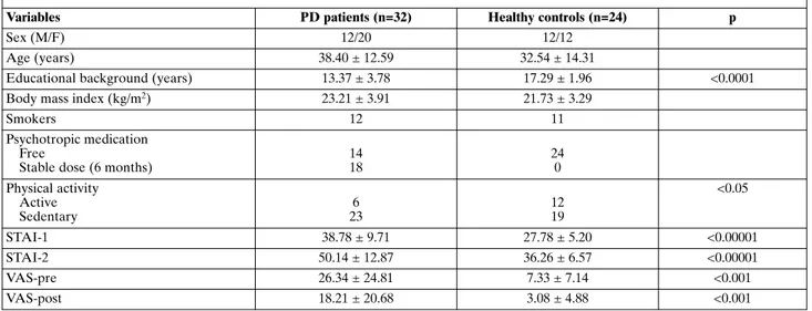
No significant differences in sex distribution (X2=0.875; df=1; p>0.05) and age (t=-1.62; df=54; p>0.05) were observed between HC and PD patients. Educational background was statistically different be-tween the two groups (t=4.61; df=54; p<0.0001), with higher values in HC. Furthermore, the two groups did not differ on body mass index (t=0.44; df=54; p>0.05) (Table 1).
The number of subjects who regularly practiced sports was significantly higher in HC than in PD pa-tients (X2=5.03; df=1; p<0.05), whereas no significant differences were found between groups in smoker dis-tribution (X2=0.282; df=1; p>0.05) (Table 1).
Self-reported anxiety
PD patients had significantly higher baseline anxi-ety levels, as measured by the STAI-1, before assess-ment of respiratory physiology than HC subjects (38.78 ± 9.71 vs 27.78 ± 5.20) (t=-5.529; df=53; p<0.00001). Values of STAI-2 were significantly higher in PD patients than in HC (50.14 ± 12.87 vs 36.26 ± 6.57) (t=-5.648; df=54; p<0.00001). Values on VAS-pre in PD patients were significantly higher than in HC (26.34 ± 24.81 vs 7.33 ± 7.14) (t=-3.633; df=54; p<0.001). Moreover, values on VAS-post in PD pa-tients were significantly higher than in HC (18.21 ± 20.68 vs 3.08 ± 4.88) (t=-3.504; df=54; p<0.001) (Table
1). ANOVA for repeated measures showed significant
effects of diagnosis (F=16.59, df=1, p<0.0001) and time (F=7.03, df=1, p<0.01) in the VAS scores, while no sig-nificant time-by-diagnosis interaction was found (p>0.05).
Respiratory variables
A MANCOVA with the STAI-1 score as covariate showed no significant differences in the mean and SD values of any of the respiratory parameters between PD patients and HC. RR means and SDs were 14.57 ± 3.15 and 16.11 ± 3.84 breaths/minute, respectively; VT means and SDs were 0.54 ± 0.24 and 0.55 ± 0.27 liter; VE means and SDs were 7.43 ± 2.50 and 8.55 ± 5.00 l/min; pCO2 means and SDs were 33.93 ± 2.68 and 35.60 ± 6.54 mmHg (≤).
An ANCOVA with the STAI-1 score as covariate and medication as grouping factor was performed in the PD group. No differences were found between medicated and unmedicated patients in RR (mean: F=0.90; df=1; p=0.34 and SD: F=1.30; df=1; p=0.26), VT (mean: F=0.17; df=1; p=0.67 and SD: F=0.17; df=1; p=0.67), VE (mean: F=0.63; df=1; p=0.43 and SD: F=0.28; df=1; p=0.59) and pCO2(mean: F=0.77; df=1; p=0.38 and SD: F=0.13; df=1; p=0.71).
Covariate was significant for VT and VE means and for VT, VE and pCO2SDs (p<0.05) (Table 2).
Smoking and physical activity
A MANCOVA with the STAI-1 as covariate and di-agnosis and smoking as grouping factors showed no significant diagnosis and smoking effects on all respi-ratory parameters. A significant diagnosis-by-smoking interaction was found for the mean values of RR (F=4.28; df=1; p<0.05) and for the mean values of VT (F=5.31; df=1; p<0.05). Covariate was significant for VT and VE means and for VT, VE and pCO2 SDs (p<0.05) (Table 3).
recording start, baseline anxiety was assessed with the Sta-te (STAI-1) and Trait (STAI-2) Anxiety Inventory (51). A visual analogue scale (VAS) for anxiety, which assesses the degree of global subjective anxiety on a continuum from 0 (no anxiety) to 100 (the worst anxiety imaginable), was ad-ministered immediately before pre) and after (VAS-post) the recording session. During the whole procedure, the examiner monitored on a computer screen the conti-nuous recording of the respiratory parameters breath-by-breath and interacted with the subjects only before and at the end of the evaluation, to administer the psychometric scales. Any disturbances that could modify the respiratory pattern, such as coughs, sneezes, or laughs, were noted by the examiner directly in the data file during the continuous recording, without interrupting the test.
Respiratory parameters
Respiratory physiology was assessed by the following parameters: RR, VT, VE and pCO2. For each respiratory
parameter we calculated the mean and the within-subject SD, which quantify the overall variability of each measu-red parameter. Data from the first 3 minutes of recording were discarded in order to minimize the influence that fa-miliarization with the face mask and study apparatus could have on the respiratory pattern. Likewise, distortions du-ring the breath-by-breath recording due to artifacts, such as coughs, sneezes, or laughs, were discarded.
Statistical analysis
Parametric statistical analyses were employed. Conti-nuous data were analyzed by Student’s t-test, analysis of va-riance (ANOVA), analysis of covava-riance (ANCOVA), and multivariate analysis of covariance (MANCOVA). Nominal data were compared by chi-square (X2) analysis. Statistical
Since the X2analysis showed a statistical difference between groups concerning the distribution of seden-tary and active subjects, physical activity effect on res-piration was analyzed separately in the two groups. An ANCOVA with the STAI-1 score as covariate and sport as grouping factor was performed. In HC, no sig-nificant differences in mean and SD values for all res-piratory parameters were observed between seden-tary and active subjects (all >0.05). Covariate was not significant for all respiratory parameters (all >0.05).
In PD patients, no significant differences in mean and SD values for all respiratory parameters were
ob-served between sedentary and active subjects (all >0.05), except for the pCO2mean value that was sig-nificantly higher in active patients than in sedentary subjects (F=1.81; df=1; p<0.05). Covariate was signifi-cant for VT, VE and pCO2SDs (p<0.05).
DISCUSSION
The aim of the present study was to elucidate possi-ble differences in breath-by-breath respiratory func-tion at rest between a sample of PD patients with ago-raphobia and HC, with particular attention to smoking and physical activity as possible relevant factors in the understanding of respiratory dynamics in PD.
Baseline anxiety assessed by the STAI and VAS scales was significantly higher in PD patients than in HC, and this finding is in line with data from the liter-ature (52,53) that show higher trait-state anxiety levels in PD patients than in HC. Moreover, VAS scores for anxiety during the procedure decreased similarly in the two groups, indicating that the procedure was not more anxiogenic for patients than for comparison sub-jects.
We did not find significant differences between groups in mean and SD for all physiological indexes. Anxiety levels did not account for this result, since we used STAI-1 values as covariate in our analysis. Our findings are in line with part of the available literature. Among those studies that showed similar pCO2levels between groups, there are some showing how antipan-ic medantipan-ications can modulate respiratory physiology,
Table 1. Demographic, epidemiological and clinical characteristics of patients with panic disorder (PD) and healthy controls
Variables PD patients (n=32) Healthy controls (n=24) p
Sex (M/F) 12/20 12/12
Age (years) 38.40 ± 12.59 32.54 ± 14.31
Educational background (years) 13.37 ± 3.78 17.29 ± 1.96 <0.0001 Body mass index (kg/m2) 23.21 ± 3.91 21.73 ± 3.29
Smokers 12 11
Psychotropic medication Free
Stable dose (6 months)
14 18 24 0 Physical activity Active Sedentary 6 23 12 19 <0.05 STAI-1 38.78 ± 9.71 27.78 ± 5.20 <0.00001 STAI-2 50.14 ± 12.87 36.26 ± 6.57 <0.00001 VAS-pre 26.34 ± 24.81 7.33 ± 7.14 <0.001 VAS-post 18.21 ± 20.68 3.08 ± 4.88 <0.001
STAI-1 and -2: State and Trait Anxiety Inventory; VAS: visual analogue scale.
Table 2. Respiratory parameters in patients with panic disor-der (PD) and in healthy controls
Respiratory parameter PD patients
(n=32) Healthy controls (n=24) Respiratory rate 16.11 ± 3.84 14.57 ± 3.15 Tidal volume 0.55 ± 0.27 0.54 ± 0.24 Minute ventilation 8.55 ± 5.00 7.43 ± 2.50 End-tidal CO2partial pressure 35.60 ± 6.54 33.93 ± 2.68
Table 3. Respiratory rate (RR) and tidal volume (VT) in smoker and non-smoker patients with panic disorder (PD) and in healthy controls No. RR VT PD patients Smokers Non-smokers 13 19 14.98 ± 4.09 16.81 ± 3.71 0.62 ± 0.37 0.50 ± 0.18 Healthy controls Smokers Non-smokers 12 12 15.54 ± 2.79 13.28 ± 2.97 0.45 ± 0.11 0.64 ± 0.30
normalizing pCO2by increasing it (54-56). Moreover, studies reporting a group difference in baseline RR values showed as a limitation the PD patients’ medical status. In our sample, medicated and unmedicated pa-tients did not show differences in respiratory physiolo-gy, suggesting that medication did not have an effect on respiration at rest. Our results are in line with those by Siepmann et al. (57) that showed selective serotonin reuptake inhibitors to have no or only minor impact on respiratory pattern.
Results concerning VT and VE are in line with sev-eral studies that reported no differences in these val-ues in PD patients compared to HC (19,22,24,58). In a recent review, Niccolai et al. (29) reported that unbal-anced samples with regard to gender are likely to in-duce a bias, because progesterone has been found to be a respiratory stimulant (59,60) able to increase VE values during the luteal phase of the menstrual cycle (61). As a limitation of our study, we did not control for this variable as a possible factor influencing respira-tion.
No significant differences were found between groups in smoker distribution, in contrast with epi-demiological studies (47) that showed smokers to be more prevalent among patients with PD than in healthy subjects.
Interestingly, we found a significant diagnosis-by-smoking interaction effect for the mean values of RR and VT. When assessing the RR and VT mean and standard deviation values of smokers and non-smok-ers of both groups some observations could be made. In fact, non-smoker patients showed a significantly higher RR compared to non-smoker HC. This evi-dence could suggest the existence of a different respi-ratory function between the two groups, in line with the hypothesis of a greater overall variability in base-line respiratory patterns in PD, indicating greater ir-regularity in their respiratory function. Smoking seems to flatten out this difference, having a dissimilar effect in the two groups. In fact, while in HC the profile of smoker subjects seems to be characterized by higher values of RR and lower values of VT compared with non-smokers, on the contrary in PD patients the pro-file of smokers seems to be characterized by lower val-ues of RR and higher valval-ues of VT compared to non-smokers. Taken together these observations suggest that the effect of nicotine seems to be different in PD patients compared to HC, suggesting the existence of a peculiar equilibrium condition in PD patients. In the literature, daily smoking has been longer associated with an increased risk for later onset of panic attacks or PD (32,34,36). However, the temporal pattern un-derlying such co-occurrence and the biological
mecha-nisms underlying this association are unknown. Ac-cording to the false suffocation alarm theory (7), smoking may increase the risk of panic by impairing respiratory system functioning. Our results seem to be consistent with this hypothesis.
In our study the number of subjects who regularly practiced sports was significantly higher in HC than in PD patients, in contrast with previous studies (28). However, this evidence is in line with the fact that physical activity avoidance and agoraphobia could re-duce time dedicated to exercise (62) and, accordingly to cognitive theories of PD (63), patients tend to avoid those situations in which bodily symptoms increase and are interpreted as a danger signal. Moreover, an-ticipatory anxiety and panic attacks mostly occur while patients are physically active compared to a more sedentary status, thus favouring the latest one.
In HC we did not find a significant sport effect on respiratory physiology confirming our previous results (28) indicating that sports activity, at least as assessed in our sample, does not have an effect on respiratory func-tion. This observation could be done only for the HC group, because, in PD patients we found higher mean values of pCO2in those who regularly practice sports compared to sedentary patients. It is well known that physical exercise has an effect on respiratory function by stimulating receptor activity of carotid bodies, thus producing ventilatory variation and adaptation during exercise. Moreover, several studies showed higher res-piratory variability in PD patients during minimal exer-cise. Our results suggest that in PD patients physical ac-tivity is able to produce a baseline alteration of pCO2 mean values, indicating a possible dysfunction of those areas involved in the control of ventilatory and chemi-cal variations occurring during physichemi-cal exercise. How-ever, we did not take into account for levels of physical activity and hours per week that could help to better understand our results. In fact, the anaerobic threshold level is higher in exercised than in sedentary subjects, and this threshold allows the maintenance of a linear correlation between ventilation, CO2 production and O2consumption. It is likely that in PD patients a mal-functioning of those areas involved in the central venti-latory control may determine a difficulty for the respi-ratory systems to respond adequately to ventilatory variations that are associated with specific levels of physical activity. Respiratory irregularities could be the immediate result of this phenomenon.
Study limitations
This study has several limitation. Sample sizes were rather small, and future investigations with larger
sam-ples would be desirable, particularly for the evaluation of smoking and physical activity effects on respiration. We did not assess levels of physical activity and hours per week. Thus, sample differences in these variables may have concealed group differences in respiratory patterns or may have been responsible for the sample differences in pCO2levels observed. Moreover, all pa-tients were agoraphobic, and this might explain why we found a number of subjects who regularly practiced sports significantly higher in HC compared to PD. Ad-ditionally, we could not be able to search for possible differences between agoraphobic and non-agorapho-bic patients on respiratory function.
Future studies should also assess the possible effect of menstrual cycle on respiration. Moreover, we did not take into account for age of smoking onset and for the number of cigarettes smoked daily, data that could be relevant for better understanding our results. Fur-ther studies with larger samples and controlling for these variables will be necessary.
CONCLUSIONS
The present study provides interesting suggestions to the question of whether PD patients may show res-piratory irregularities at rest, and most importantly how smoking and physical activity may affect respira-tory physiology. In the PD group, higher mean values of pCO2 were found in those who regularly practice sports compared to sedentary patients and a significant diagnosis-by-smoking interaction effect for the mean values of RR and VT was observed. These findings seem to be in line with the idea of an abnormal regu-lation of the respiratory system as a key mechanism in PD, and panic and cigarette smoking appear to serve as a causal/predisposing factor in the development of the other. For this reason, the nature of respiratory abnor-malities in PD and HC smokers/non-smokers should be compared to better understand the role of smoking in inducing clinical or subclinical abnormalities that may favour panic occurrence (64).
Further studies are warranted to confirm and clari-fy our results. However, given the complexity of PD in terms of multiple factors that influence not only the onset and maintenance of the disorder but mostly the homeostatic brain functioning, it is firmly necessary that future studies investigate respiratory dynamics in PD assessing the complex interaction between differ-ent factors and their possible combined effect on mod-ulating homeostatic brain. This could determine the evaluation on one hand of respiratory and cardiac function in PD, on the other of the combined effects of
that variables able to modulate the homeostatic brain functioning. Finally symptom heterogeneity, i.e. symp-tom subtypes, should be considered, in line to building complex PD neurobiological profiles.
Conflict of interest statement:
Emma Fadda conducted this study as partial fulfilment of her PhD in Molecular Medicine, Program in Experimental Neu-rology, San Raffaele University, Milan, Italy.
REFERENCES
American Psychiatric Association. Diagnostic and Statistical 1.
Manual for Mental Disorders. 4th ed. Washington: American Psychiatric Association 1994.
Gorman JM, Kent JM, Sullivan GM, Coplan JD. Neuroanatom-2.
ical hypothesis of panic disorder, revised. Am J Psychiatry 2000; 157: 493-505.
Nardi AE, Freire RC, Zin WA. Panic disorder and control of 3.
breathing. Respir Physiol Neurobiol 2009; 167: 133-43.
Perna G, Caldirola D, Bellodi L. Panic disorder: from respira-4.
tion to the homeostatic brain. Acta Neuropsychiatrica 2004; 16: 57-67.
Briggs AC, Stretch DD, Brandon S. Subtyping of panic disorder 5.
by symptom profile. Br J Psychiatry 1993; 163: 201-9.
Ley R. The many faces of Pan: psychological and physiological 6.
differences among three types of panic attacks. Behav Res Ther 1992; 30: 347-57.
Klein DF. False suffocation alarms, spontaneous panics, and re-7.
lated conditions: an integrative hypothesis. Arch Gen Psychiatry 1993; 50: 306-17.
Kircanski K, Craske MG, Epstein AM, Wittchen HU. Subtypes 8.
of panic attacks: a critical review of the empirical literature. De-press Anxiety 2009; 26: 878-87.
Hegel MT, Ferguson RJ. Psychophysiological assessment of res-9.
piratory function in panic disorder: evidence for hyperventila-tion subtype. Psychosom Med 1997; 59: 224-230.
Moynihan JE, Gevirtz RN. Respiratory and cognitive subtypes 10.
of panic. Preliminary validation of Ley’s model. Behav Modif 2001; 25: 555-83.
Biber B, Alkin T. Panic disorder subtypes: differential responses 11.
to CO2challenges. Am J Psychiatry 1999; 156: 739-44.
Nardi AE, Nascimento I, Valenca AM, et al. Respiratory panic 12.
disorder subtype: acute and long-term response to nortriptyline, a noradrenergic tricyclic antidepressant. Psychiatry Res 2003; 120: 283-93.
Nardi AE, Valenca AM, Nascimento I, et al. A three-year follow-13.
up study of patients with the respiratory subtype of panic disor-der after treatment with clonazepam. Psychiatry Res 2005; 137: 61-70.
Wilhelm FH, Trabert W, Roth WT. Physiologic instability in pan-14.
ic disorder and generalized anxiety disorder. Biol Psychiatry 2001; 49: 596-605.
Gorman JM, Kent J, Martinez J, Browne S, Coplan J, Papp LA. 15.
Physiological changes during carbon dioxide inhalation in pa-tients with panic disorder, major depression, and premenstrual dysphoric disorder: evidence for a central fear mechanism. Arch Gen Psychiatry 2001; 58: 125-31.
Ponto LL, Kathol RG, Kettelkamp R, et al. Global cerebral 16.
blood flow after CO2inhalation in normal subjects and patients
with panic disorder determined with [15O]water and PET. J Anxiety Disord 2002; 16: 247-58.
Abelson JL, Nesse RM, Weg JG, Curtis GC. Respiratory psy-17.
chophysiology and anxiety: cognitive intervention in the doxapram model of panic. Psychosom Med 1996; 58: 302-13. Papp LA, Martinez J, Klein DF, et al. Respiratory psychophysi-18.
ology of panic disorder. Three respiratory challenges in 98 sub-jects. Am J Psychiatry 1997; 154: 1557-65.
Yeragani VK, Radhakrishna RKA, Tancer M, Uhde T. Nonlinear 19.
measures of respiration: respiratory irregularity and increased chaos of respiration in patients with panic disorder. Neuropsy-chobiology 2002; 46: 111-20.
Hoehn- Saric R, McLeod DR, Funderburk F, Kowalski P. So-20.
matic symptoms and physiologic responses in generalized anxi-ety disorder and panic disorder: an ambulatory monitor study. Arch Gen Psychiatry 2004; 61: 913-21.
Gorman JM, Papp LA, Martinez J, et al. High-dose carbon diox-21.
ide challenge test in anxiety disorder patients. Biol Psychiatry 1990; 28: 743-57.
Papp LA, Klein DF, Martinez J, et al. Diagnostic and substance 22.
specificity of carbon-dioxide-induced panic. Am J Psychiatry 1993; 150: 250-7.
Martinez JM, Papp LA, Coplan JD, et al. Ambulatory monitor-23.
ing of respiration in anxiety. Anxiety 1996; 2: 296-302.
Gorman JM, Fyer MR, Goetz R, Askanazi J. Ventilatory physi-24.
ology of patients with panic disorder. Arch Gen Psychiatry 1988; 45: 31-9.
Abelson JL, Weg JG, Curtis GC. Respiratory irregularity in pan-25.
ic patients may reflect excessive sighing. Biol Psychiatry 2000; 47 (suppl 1): 157-8.
Abelson JL, Weg JG, Nesse RM, Curtis GC. Persistent respira-26.
tory irregularity in patients with panic disorder. Biol Psychiatry 2001; 49: 588-95.
Wilhelm FH, Trabert W, Roth WT. Characteristics of sighing in 27.
panic disorder. Biol Psychiatry 2001; 49: 606-14.
Caldirola D, Bellodi L, Caumo A, Migliarese G, Perna G. Ap-28.
proximate entropy of respiratory patterns in panic disorder. Am J Psychiatry 2004; 161: 79-87.
Niccolai V, van Duinen MA, Griez EJ. Respiratory patterns in 29.
panic disorder reviewed: a focus on biological challenge tests. Acta Psychiatr Scand 2009; 120: 167-77.
Bellodi L, Perna G. The panic respiration connection. Milan: 30.
MDM Medical Media, 1998.
Jacobs S, Hansen F, Kasl S, Ostfeld A, Berkman L, Kim K. Anx-31.
iety disorders during acute bereavement: risk and risk factors. J Clin Psychiatry 1990; 51: 269-74.
Pohl R, Yeragani V, Balon R, Lycaki H, McBride R. Smoking in 32.
patients with panic disorder. Psychiatry Res 1992; 43: 253-62. Amering M, Bankier B, Berger P, Griengl H, Windhaber J, 33.
Katschnig H. Panic disorder and cigarette smoking behavior. Compr Psychiatry 1999; 40: 35-8.
Breslau N, Klein DF. Smoking and panic attacks: an epidemio-34.
logic investigation. Arch Gen Psychiatry 1999; 56: 1141-7. Johnson J, Cohen P, Pine D, Klein D, Kasen S, Brook J. Asso-35.
ciation between cigarette smoking and anxiety disorders dur-ing adolescence and early adulthood. JAMA 2000; 284: 2348-51.
Isensee B, Wittchen HU, Stein MB, Hofler M, Lieb R. Smoking 36.
increases the risk of panic: findings from a prospective commu-nity study. Arch Gen Psychiatry 2003; 60: 692-700.
Clark DB, Taylor CB, Hayward C, et al. Motor activity and ton-37.
ic heart rate in panic disorder. Psychiatry Res 1990; 32: 45-53. Sakamoto N, Yoshiuchi K, Kikuchi H, et al. Panic disorder and 38.
locomotor activity. Biopsychosoc Med 2008; 2: 23.
Cameron OG, Hudson CJ. Influence of exercise on anxiety level 39.
in patients with anxiety disorders. Psychosomatics 1986; 27: 720-3. Rief W, Hermanutz M. Responses to activation and rest in pa-40.
tients with panic disorder and major depression. Br J Clin Psy-chol 1996; 35 (Pt 4): 605-16.
Broman-Fulks JJ, Storey KM. Evaluation of a brief aerobic ex-41.
ercise intervention for high anxiety sensitivity. Anxiety Stress Coping 2008; 21: 117-28.
Pfaltz MC, Grossman P, Michael T, Margraf J, Wilhelm FH. Phys-42.
ical activity and respiratory behaviour in daily life of patients with panic disorder and healthy controls. Int J Psychophysiol 2010; 78: 42-9.
Sheehan DV, Lecrubier Y, Sheehan KH, et al. The Mini-Interna-43.
tional Neuropsychiatric Interview (M.I.N.I.): the development and validation of a structured diagnostic psychiatric interview for DSM-IV and ICD-10. J Clin Psychiatry 1988; 59 (suppl 20): 22-33. Akiyama Y, Kawakami Y. Clinical assessment of the respiratory 44.
control system. In: Akiyama Y, Kawakami Y, Altose MD, Kawakami Y (eds). Control of breathing in health and disease. New York: Marcel Dekker, 1999: pp. 251-80.
Sonntag H, Wittchen HU, Hofler M, Kessler RC, Stein MB. Are 45.
social fears and DSM-IV social anxiety disorder associated with smoking and nicotine dependence in adolescents and young adults? Eur Psychiatry 2000; 15: 67-74.
Palatini P, Visentin P, Dorigatti F, et al. Regular physical activity 46.
prevents development of left ventricular hypertrophy in hyper-tension. Eur Heart J 2009; 30: 225-32.
Palange P, Forte S, Onorati P, Manfredi F, Serra P, Carlone S. 47.
Ventilatory and metabolic adaptations to walking and cycling in patients with COPD. J Appl Physiol 2000; 88: 1715-20.
Schena F, Padoin E. Influence of fitness level on metabolic and 48.
cardiac on-response in the elderly [abstr]. J Aging Phys Activity 1999; 7: 251.
Spengler CM, Czeisler CA, Shea SA. An endogenous circadian 49.
rhythm of respiratory control in humans. J Physiol 2000; 526 (Pt 3): 683-94.
Stephenson R, Mohan RM, Duffin J, Jarsky TM. Circadian 50.
rhythms in the chemoreflex control of breathing. Am J Physiol Regul Integr Comp Physiol 2000; 278: R282-6.
Spielberger CD, Gorsuch RL, Lushene RD. STAI Manual. Palo 51.
Alto: Consulting Psychologists Press, 1970.
Holt PE, Andrews G. Hyperventilation and anxiety in panic dis-52.
order, social phobia, GAD and normal controls. Behav Res Ther 1989; 27: 453-60.
Masaoka Y, Homma I. Anxiety and respiratory patterns: their 53.
relationship during mental stress and physical load. Int J Psy-chophysiol 1997; 27: 153-9.
Gorman JM, Fyer AJ, Ross DC, et al. Normalization of venous 54.
pH, pCO2, and bicarbonate levels after blockade of panic at-tacks. Psychiatry Res 1985; 14: 57-65.
Papp LA, Klein DF, Gorman JM. Carbon dioxide hypersensitiv-55.
ity, hyperventilation, and panic disorder. Am J Psychiatry 1993; 150: 1149-57.
Gorman JM, Browne ST, Papp LA, et al. Effect of antipanic 56.
treatment on response to carbon dioxide. Biol Psychiatry 1997; 42: 982-91.
Siepmann M, Grossmann J, Mück-Weymann M, Kirch W. Effects 57.
of sertaline on autonomic and cognitive functions in healthy vol-unteers. Psychopharmacology 2003; 168: 293-8.
Gorman JM, Goetz RR, Uy J et al. Hyperventilation occurs dur-58.
ing lactate-induced panic. J Anxiety Disord 1988; 2: 193-202. Zwillich CW, Natalino MR, Sutton FD, Weil JV. Effects of prog-59.
esterone on chemosensitivity in normal men. J Lab Clin Med 1978; 92: 262-9.
White DP, Douglas NJ, Picket CK, Weil JV, Zwillich CW. Sexual 60.
influence on the control of breathing. J Appl Physiol 1983; 54: 874-9.
Takano N. Resting pulmonary ventilation and dead space venti-61.
lation during the menstrual cycle. Jpn J Physiol 1982; 32: 469-73. Broocks A, Meyer TF, Bandelow B, et al. Exercise avoidance and 62.
impaired endurance capacity in patients with panic disorder. Neuropsychobiology 1997; 36: 182-7.
Clark DM. A cognitive approach to panic. Behav Res Ther 1986; 63.
4: 461-70.
Cosci F, Knuts IJ, Abrams K, Griez EJ, Schruers KR. Cigarette 64.
smoking and panic: a critical review of the literature. J Clin Psy-chiatry 2009; 71: 606-15.