O R I G I N A L A R T I C L E
Substernal hand-assisted videothoracoscopic lung
metastasectomy: Long term results in a selected
patient cohort
Federico Tacconi, Vincenzo Ambrogi, Eugenio Pompeo, Francesco Sellitri & Tommaso Claudio Mineo
Department of Thoracic Surgery, Tor Vergata University Foundation, Rome, Italy
Keywords
Lung resection; metastasectomy; video-assisted thoracoscopy (VATS).
Correspondence
Tommaso Claudio Mineo, Department of Thoracic Surgery, Fondazione Policlinico Tor Vergata, Università Tor Vergata, Viale Oxford 81- 00133, Rome, Italy. Tel:+39 06 20902884 Fax:+39 06 20902881 Email: [email protected] Received: 14 October 2010; accepted 4 December 2010. doi: 10.1111/j.1759-7714.2010.00038.x
Abstract
Background: Substernal hand-assisted thoracoscopy (HATS) has been proposed as a reliable surgical method with which to perform a bilateral lung metastasectomy. Herein, we review our 15-year experience with this approach for the purpose of understanding the long-term results in a large study cohort.
Methods: The study cohort was a series of 87 patients that underwent a HATS lung metastasectomy between 1995 and 2010. We focused on the main surgical findings including the ability of this approach to facilitate the detection of unexpected pul-monary lesions. Overall and disease-free survival rates were analyzed in a long-term follow up using the Kaplan-Meier method.
Results: A total of 219 lesions were removed. Of these, 191 proved to be malignant. This figure accounted for 31 (19.3%) unexpected lung metastases not previously identified at imaging work-up. Eighteen nodules previously suggested as metastatic lesions proved to be benign. On the basis of these findings sensitivity, specificity, positive and negative predictive values for imaging work-up in detecting lung metastases were 79.6%, 41.3%, 86.4%, and 30.5%, respectively. Lesions sized<7 mm showed the highest false negative rate. Postoperatively no major complications occurred. Overall survival rates at 3 and 5 years were 57.9% and 38.4%, respectively. Disease-free interval after primary cancer removal, but not all metastases nor bilat-eral spread, was related to survival (P = 0.015).
Conclusions: In our experience, HATS resulted in a considerable percentage of resected lung metastases not previously detected at imaging work-up. We recom-mend this approach whenever feasible as it can conciliate low invasiveness and completeness of surgical resection. Substernal HATS makes it possible to detect a considerable number of unexpected lesions in patients undergoing lung metasta-sectomy with low surgical trauma. This study reinforces the role of this approach in the setting of lung metastasectomy, due to the large patient series and long-term follow-up.tca_3845..53
Introduction
Surgical resection has been established as a reliable option in selected patients with pulmonary metastases spreading from different primary cancers.1–4 Since survival in this
setting is strictly related to the completeness of resection, aggressive and iterative procedures are deemed justified in most institutions.
In the last few decades, video-assisted thoracic surgery (VATS) has emerged as the standard of care in a broad spectrum of thoracic conditions. Nonetheless, since manual palpation is not allowed, the question has arisen as to whether VATS is adequate in the treatment of pulmonary metastases.5,6In order to overcome this limitation, we
devel-oped a transxiphoid, hand-assisted thoracoscopic (HATS) approach with which one can manually explore both the
lungs in a single procedure,7–9thus avoiding more invasive
surgical access. Since in some favorable instances the xiphoid can be left in place, we decided to generally describe these two approaches with the name substernal HATS.
Herein, we review our 15-year experience with this tech-nique. Our first aim was to assess the ability of the HATS approach to ensure careful exploration of the pleural cavities and to detect occult pulmonary lesions. As a second target, we updated long-term results in terms of overall and disease-free survival rates.
Methods
Our program using transxiphoid HATS metastasectomy started in December 1995, and was approved by the institu-tional Ethics Committee of the Tor Vergata University. Written informed consent was obtained from all patients, who were given basic information about the possible risks of the procedure.
The indications for substernal HATS metastasectomy have not substantially changed over the years and the surgical treatment decision is taken by a multidisciplinary staff including oncologists, thoracic surgeons and radiotherapists. Complete control of the primary tumor and absence of extra-pulmonary metastases represent the main prerequisites. Patients with lesions sited at more than 2 cm from the visceral pleura or greater than 3 cm in maximum diameter are excluded. Patients with cardiomegaly or arrhythmia are also excluded due to technical difficulties and an increased risk of intraoperative rhythm disturbances. On the other hand, pre-vious thoracic or abdominal surgeries are not deemed as absolute contraindications, unless the presence of thick tho-racic adhesions is suspected.
Preoperative work-up
After being scheduled for lung metastasectomy, patients underwent a total-body computed tomography (CT) scan. If appropriate, liver ultrasonography and whole-body bone scan scintigraphy were obtained to exclude any local or distant relapses, other than pulmonary involvement. Fiber-optic bronchoscopy was routinely performed to rule out the presence of intrabronchial lesions.
Malignancy was presumed according to standardized imaging-based criteria (Table 1). The CT scanning protocol included a tube voltage of 120 kilovolts (peak), a tube current of 250 mA, a slice thickness of 5 mm, and a table increment of 5 mm per rotation. Reconstructions were performed at 5 mm intervals and the images were printed at a window center of -530 Hounsfield (H), with a window width of 1500 H.
In 200518-fluorodeoxyglucose positron emission
tomogra-phy (PET) became available in our institution. Since then integrated CT/PET imaging has been included in the
standard work-up protocol. A standard uptake value (SUV) greater than 2.5 was arbitrarily considered as the cut-off value to suggest the presence of tumor tissue.
Videomediastinoscopy was performed as a part of the pre-operative work-up if there was recent enlargement in medias-tinal lymph nodes (short-axis diameter>15 mm) and/or if metabolic activity was detected at PET assay.
Surgical technique
The basic surgical steps of the procedure have been widely described elsewhere. Briefly, the patient is placed in a 60° off-center position, and a 15 mm flexible trocar is inserted in the fourth intercostal space between the midclavicular and ante-rior axillary lines. Another two thoracoscopic ports are placed in the fifth and seventh intercostal spaces along the posterior and midaxillary lines, respectively. A midtransverse arcuate skin incision is performed just along the inferior margin of the thoracic cage. The rectus abdominis muscle is divided and the xiphoid appendix used to be resected, though under favorable circumstances the appendix may now sometimes be left in place and just freed and raised with the help of a retrac-tor. Next, the parietal pleura is divided under thoracoscopic assistance and the pleural cavity is entered to allow lung palpation between the thumb and forefinger. All palpated nodules are excised with minimal resection by an endo-stapler or Nd:YAG laser beam, as described in our previous reports.7–9During these maneuvers, it is helpful to have one
hand inside the hemithorax to hold the lesions to be resected and to protect the healthy lung parenchyma. At the end of the procedure one or two chest tubes are inserted.
Size, location and the main imaging-based findings of the resected lesions are immediately recorded in a prospective database form. The same surgical maneuvers are then repeated on the opposite side using the substernal incision. Refraining from bilateral exploration was usually due to the presence of pleural adhesions in the side determined to be radiologically free from metastases.
Postoperative work-up
Postoperative care was conducted according to a fast-track protocol, including early mobilization and physiotherapy as needed. Pain control was obtained with intravenous
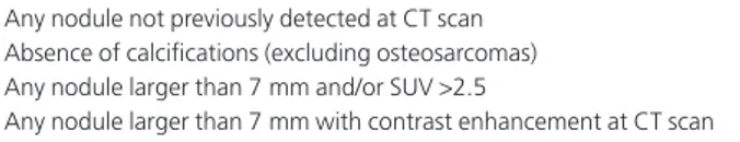
Table 1 Established criteria for defining high likelihood of metastatic lesion at imaging work-up
Any nodule not previously detected at CT scan Absence of calcifications (excluding osteosarcomas) Any nodule larger than 7 mm and/or SUV>2.5
Any nodule larger than 7 mm with contrast enhancement at CT scan CT, computed tomography; SUV, standard uptake value.
tramadol, 200 to 300 mg a day, and reinforced on demand with boluses of 30 mg ketorolac. Postdischarge follow up was arranged at 1, 3, and 6 months after surgery in a multidisci-plinary fashion, where subsequent control check-ups were performed in the Thoracic Oncology Unit of our institution. Patients underwent total-body CT scans twice a year for the first 2 years, and annually thereafter. In addition, ultrasonog-raphy, whole-body bone scan, and a laboratory examination were performed where indicated. All patients with metastatic osteosarcoma had adjuvant chemotherapy consisting of 100 mg/m2 of cisplatinum, 60 mg/m2of doxorubicin, and
high-dose methotrexate. One patient with laryngeal carci-noma also had postoperative chemotherapy, which consisted of 1 mg/m2of vincristine, 150 mg/m2of bleomycin sulfate,
and 20 mg/m2of methotrexate.
Statistical analysis
A retrospective analysis of a prospective 15-year database which included patients’ baseline, operative and postsurgical findings was made. The database was updated over the years in cooperation with the Thoracic Oncology Unit of our insti-tution, and the status of the patients was recorded in an alphanumeric codex according to their current conditions. Patients not updated within the last 12 months were con-tacted by personal phone call and invited to a follow-up visit. Whenever patient results were unavailable their status was checked through either the institutional or regional demo-graphic registers.
Any pulmonary lesion radiologically suspected for metastasis and confirmed as tumor tissue of any origin was considered a true positive. Any pulmonary lesion suspected for metastasis, and histologically proven as benign tissue or not found intraoperatively was a false positive; any pulmo-nary lesion predicted as indolent in nature at CT/PET scan and confirmed as histologically benign tissue was a true nega-tive. Finally, any unexpected metastatic lesion found intraop-eratively and any pulmonary lesion predicted as indolent and proven to be malignant was a false negative. Lesions not found at surgical exploration were assumed to be benign, unless proven to be malignant at follow up.
Time-dependent analysis was performed using the Kaplan-Meier method and the log–rank test where appropri-ate. Frequencies were analyzed by means of the two-tailed Fisher’s exact test. All data were processed through the Statis-tica software, (StatSoft, Tulsa, OK, USA, release 7.0).
Results
Baseline findings
Throughout a 15-year period, a total of 239 patients with radiological findings suggestive for lung metastases arising
from different primary tumors were referred to our institu-tion. Of these, 59 patients were judged unfit for curative intent surgery, even though 41 of these patients underwent VATS resection of a single lung lesion for restaging or diag-nostic purpose. Sixty-five patients were scheduled for cura-tive metastasectomy via a lateral thoracotomy (n = 34, 14%) or VATS (n = 31, 13%). In particular, thoracotomy was pre-ferred for patients with lesions of more than 3 cm in diam-eter (n = 14, 6%) and/or lesions located more than 2 cm from the visceral pleura (n = 27, 11.3%), as well as in cases of mediastinal nodal spread unmanageable thoracoscopically (n = 12, 5%).
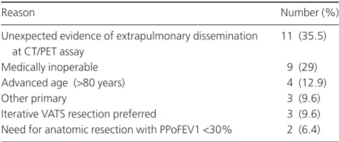
The remaining 115 patients were initially scheduled for a HATS metastasectomy. Thirty-one of these (26.7%) were subsequently judged unfit for surgery after the completion of a preoperative work-up and were referred to the Thoracic Oncology Unit of our institution for re-evaluation. Reasons for exclusion are reported in Table 2. In three instances, VATS metastasectomy was preferred, in agreement with the patients, due to extremely favorable localization of their lesions. Another seven (6%) patients denied consent to surgi-cal treatment in favor of alternative non-surgisurgi-cal treatments which included radiofrequency ablation and radio-stereotaxis. Out of a total of 14 patients undergoing staging videomediastinoscopy, 12 were found to be free from nodal involvement, while two patients had single-station tumor spread. These latter patients underwent HATS metastasec-tomy and limited nodal dissection.
Thus, a total of 87 patients were included in this database, which accounts for 40.4% of all patients with lung metastases referred for surgical evaluation at our institution. There were 47 men and 40 women ranging in age from 16 to 82 years (mean: 60.5⫾ 11). Primary tumors originated from carci-noma of the colon–rectum (n = 45), kidney (n = 13), uterus (n = 7), larynx (n = 5), breast (n = 3), prostate gland (n = 2), ovary (n = 2), limb osteosarcoma (n = 6), chest wall fibrosarcoma (n = 1) and melanoma (n = 3). Most of the patients were asymp-tomatic and the presence of the metastases was discovered as
Table 2 Reasons for exclusion from hand-assisted thoracoscopy follow-ing preoperative work-up
Reason Number (%)
Unexpected evidence of extrapulmonary dissemination at CT/PET assay
11 (35.5)
Medically inoperable 9 (29)
Advanced age (>80 years) 4 (12.9)
Other primary 3 (9.6)
Iterative VATS resection preferred 3 (9.6) Need for anatomic resection with PPoFEV1<30% 2 (6.4) CT/PET, computed tomography/ positron emission tomography; PPo FEV1: predicted-postoperative forced-expiratory volume at 1 second; VATS, video-assisted thoracoscopy.
an unexpected finding at follow-up assessment. Major symp-toms were respiratory (cough and/or dyspnea, n = 11), fatigue (n = 11), weight loss (n = 3) and anemia (n = 2). In four cases, the metastases were synchronous with the resected primary tumor, while in another six patients pulmonary metastases were discovered at the same time as hepatic ones. In patients with metachronous onset the median disease-free interval (DFI) was 26⫾ 9.4 months. DFI was longer than 36 months in 32 patients and in two of those cases it was more than 100 months. According to the prognostic classification formu-lated by the International Registry of Lung Metastases, 10 patients were classified as group I, 50 as group II, and 27 as group III.
Surgical findings
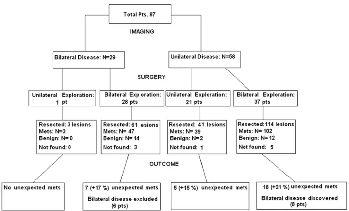
Overall, 186 lung lesions were detected preoperatively. Of these, 160 were presumed to be metastatic foci on the basis of previously established criteria, while 26 were deemed as indo-lent lesions. In 29 patients preoperative imaging work-up scans revealed bilateral disease. HATS metastasectomy was possible in 80 patients (94%), while seven patients required conversions to alternative therapies, including non-rib spreading minithoracotomy to manage deeply located nodules (n = 4) or lateral, muscle-sparing thoracotomy due to unexpected adhesions (n = 2) and difficulty in palpating the
left-lower lobe (n = 1). Of the procedures performed, a total of 65 were bilateral, while in the remaining 22 patients (25%), contralateral exploration was not performed due to the pres-ence of thick adhesions (Fig 1). A brief summary of baseline and operative data is reported in Table 3.
Overall, 219 lung lesions were removed, 191 of which proved metastatic at definitive histology. This figure accounts for a total of 31 metastatic lesions not previously indicated at imaging assessment (+19.3% difference). One hundred and sixty metastatic lesions were correctly predicted on the basis of imaging assay and proved to be metastatic lesions at defini-tive histology. One of these resected lesions proved to be an early non-small cell lung cancer of bronchiole-alveolar histol-ogy and was considered as a true positive result for statistical purposes. A total of 18 lesions were confirmed as benign in nature and included fibrotic tissue (n = 8), rheumatoid nodules (n = 4), anthracotic subpleural lymph nodes (n = 3) and benign chondromas (n = 3). Nine more nodules diag-nosed using CT scan were not detected at palpation and were interpreted as small blood vessels or partial volume effects. Another 10 indolent lesions (nine granulomas/anthracotic nodes and one small chondroma) were found accidentally at manual palpation. On the basis of the reported findings, overall sensitivity, specificity, positive predictive value and negative predicted value for helical CT scans identifying pre-operatively metastatic lesions were 79.6%, 41.3%, 86.4% and
30.5%, respectively. These values significantly changed when categorizing the radiological findings according to different characteristics including diameter, localization and SUV. In particular, lesions sized<7 mm showed the highest rate of false negative findings, as showed by the extremely low nega-tive predicnega-tive value (Table 4). Conversely, no remarkable changes were found when analyzing the diagnostic value of imaging work-up according to methodological changes and increased experience over the years.
Long term outcomes
The dataset was 96% complete. After a mean follow up of 65 ⫾ 12 months (range, 8–72 months), no patient experienced significant procedure related complications, while minimal incisional hernia was found in two instances. Overall 3- and 5-year survival rates were 57.9% and 38.4%, respectively. DFI after primary cancer removal was related to survival, but survival was not significantly affected by the number of metastases, nor by the presence of bilateral spread. When restricting the analysis to just patients with colorectal cancer, 3- and 5-year survival rates were 63.9% and 49.4%, similar to that of patients with other primary cancers(Fig 2). Eighteen patients underwent iterative procedures, which included redo-HATS (n = 4), VATS (n = 6), and thoracotomy (n = 14). No local relapse was found in lung areas previously subjected to resection or failed exploration. Disease-free survival rates
Table 3 Main baseline and operative findings in the patients’ cohort
Baseline findings Mean (⫾SD)† Range
Age (years) 60.5⫾ 11 16–82
Sex (Male/Female) 47/40 NA
Previous KMT 52 pts (59%) NA
Previous VATS metastasectomy 5 pts (5.7%) NA Operative data
Operative time (min) 102⫾ 39 45–170
Bleeding (mL) 250⫾ 80 150–420
Resected lesions 2.5⫾ 1.7 2–10
Bilateral/unilateral explorations 65/22 NA Postoperative data
Hospital stay (days) 6⫾ 2 4–12
Mortality rate 0% NA
Major morbidity rate‡ 2 pts (2.2%) NA
Minor morbidity rate 11 pts (12.6%) NA
Definitive histology Colorectal (n) 102 NA Kidney (n) 31 NA Breast (n) 7 NA Sarcomas (n) 26 NA Others (n) 24 NA
†Values are expressed as Mean⫾ SD unless differently required. ‡Major morbidity entailed postoperative acute lung injury in two patients. KMT, chemotherapy; NA, not applicable; pts, patients; VATS, video-assisted thoracoscopy.
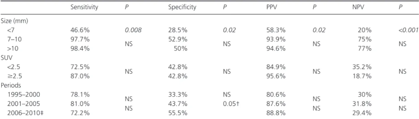
Table 4 Stratified correlation between imaging-based and surgical findings
Sensitivity P Specificity P PPV P NPV P Size (mm) <7 46.6% 0.008 28.5% 0.02 58.3% 0.02 20% <0.001 7–10 97.7% NS 52.9% NS 93.9% NS 75% NS >10 98.4% 50% 94.6% 77% SUV <2.5 72.5% NS 42.8% NS 84.9% NS 35.2% NS ⱖ2.5 87.0% 42.8% 95.6% 18.7% Periods 1995–2000 78.1% NS 33.3% NS 80.6% NS 30% NS 2001–2005 81.0% NS 43.7% 0.05† 87.6% NS 31.8% NS 2006–2010‡ 72.2% 55.5% 88.8% 29.4%
†P-value versus 1995–2000 period. ‡Since 2005, CT/PET scan entered our preoperative work-up study protocol. NPV, negative predictive value; NS, not significant; PPV, positive predictive value; SUV, standard uptake value.
Figure 2 Comparative survival analysis in patients with metastases from colorectal cancer and other primary cancers.
were 53.5% and 40.9%, with a mean post-metastasectomy DFI of 43⫾ 11 months. Categorized overall and disease-free survival curves are depicted in Figures 3 and 4.
Thirty-four patients relapsed at various sites; however, only 18 patients developed pulmonary metastases after a mean interval of 12.8 months. In seven of these 18 patients, tumors metastasized in the unexplored lung after intervals of 6, 9, and 12 months. No recurrence was found in the subster-nal incision, along the thoracoscopic ports, or in proximity to the previous resection margins.
Discussion
Completeness of resection is considered worldwide the ideal target of surgery in patients with lung metastases.1–4This aim
is limited by the inaccuracy of preoperative radiological studies for detecting all pulmonary metastases and by the dif-ficulty of accurately exploring both lungs with a minimally-invasive approach. The use of helical CT-scan and the subsequent introduction of PET imaging in this setting have resulted in a remarkable diagnostic advantage, but small-sized metastases still remain difficult to identify even when using both methods.
In 1995 our group developed the transxiphoid hand-assisted approach with the rationale of allowing careful manual palpation during video-assisted metastasectomy without the need for open access to the pleural cavities. Since then, this surgical method has proved quite feasible and safe. The hypothetical concerns of provoking cardiac tamponade or arrhythmia during lung palpation were acceptable and counterbalanced by the indubitable advantages of a minimally-invasive procedure – namely decreased postop-erative pain, shorter hospitalization, and easier patient accep-tance. On the basis of this, we subsequently enlarged the indication of this approach to other thoracic surgery fields, including Morgagni’s hernia repair.9In addition, as a
conse-quence of increased familiarity with the approach we can now avoid resecting the xiphoid appendix in favorable instances, especially in thin, tall subjects. This has not affected the overall surgical results.
At present, only limited, single institution reports dealing with substernal HATS are available in the literature. Detter-beck and Egan focused on a consecutive series of 24 patients undergoing HATS metastasectomy, with findings similar to ours.10In 2003, Wright et al. proposed a modified technique
using diaphragmatic division to enter the pleural cavity.11
Despite the use of helical CT scan which is presumed the most accurate preoperative assessment in this setting,12,13we
were able to identify 31 unexpected small-sized metastases, accounting for more than 19% of potentially missed lesions without manual palpation. This is consistent with a recent report by Cerfolio et al. of patients undergoing a thorac-otomy.14On the other hand, 10 of the 160 lesions deemed
metastatic at helical CT scan, ultimately proved to be benign on histological examination, thus accounting for a total of six patients in whom bilateral disease was excluded.
Although the prognostic relevance of undetected, small-sized metastases still remains controversial, it is generally believed that complete resection of pulmonary metastases confers both disease-free and overall survival advantage, as proven by the International Registry of Lung Metastases on a population of 5206 patients.2Indeed, the low sensitivity of
imaging methods and subsequent incomplete resection may affect recurrence rates and long-term survival. In our previ-ous paper, we found that the frequency of recurrence was consistently higher among non-palpated or unexplored lungs.8Recurrence time was shorter in the subset of patients
with non-palpated lungs (9 vs. 16 months) and this is likely be due to occult metastases situated on a non-accessible side. In particular, patients with sarcoma proved to have highest probability of overlooked lesions and false unilateral disease. In contrast with other authors’reports,15we found
multiple and unexpected metastases within the carcinoma group in a consistent percentage of cases. All these findings suggest that whenever technically feasible, bilateral palpa-tion represents the best way to perform radical surgery in these patients. This maneuver can be easily and satisfactorily performed using the HATS approach. The limitations of the procedure may be the difficulty of its use on centrally located metastases or lesions requiring lobectomy, cardi-omegaly, diffuse pleural adhesions, or where there has been previous sternotomy. However, the procedure is feasible in the majority of patients selected for lung metastasectomy. In our series, despite the extensive exclusion criteria, as many as 64.4% of the patients who underwent pulmonary metastasectomy were able to undergo HATS without con-version to an open approach.
In our experience, the overall 5-year survival rate was 38.4%, a figure in concordance with previous reports of pul-monary metastasectomy. Interestingly, neither the number of resected lesions nor the presence of bilateral disease signifi-cantly affected survival, whereas DFI did. This finding is somewhat surprising, since considerable evidence exists that the smaller the number of resected metastases, the better the disease-free and overall survival rates.2,16–19This discrepancy
could be partly explained by our own restrictive inclusion cri-teria. Indeed, HATS metastasectomy was not performed in patients with large (>3 cm) and more deeply located lesions. It is possible that this basic criterion resulted in a higher pro-portion of operated patients having a less aggressive biologi-cal pattern of metastatic disease, resulting in relatively better long-term outcome regardless of the presence of multiple and bilaterally sited nodules. In addition, the prevalent peripheral location of the pulmonary lesions could imply a higher radical resection rate, alongside a lesser likelihood of occult nodal spread.
Figure 3 Overall survival rates categorized according to (a) the disease-free interval (DFI) from primary cancer removal, (b) presence of bilateral spread, (c) and median number of metastases (mets).
Figure 4 Disease-free survival rates categorized according to (a) the disease-free interval (DFI) from primary cancer removal, (b) presence of bilateral spread, and (c) median number of metastases (mets).
An interesting field of investigation is whether minimally invasive approaches in thoracic oncology could result in a survival advantage due to less likelihood of systemic tumor spread. The rationale for this theoretical assumption is that the anticancer defense mechanisms, including natural killer cells activity, can be compromised in the perioperative period due to the surgical trauma and postop-erative pain, which can both affect systemic host response and hormonal release in a dose-dependent fashion. In pre-vious reports we were able to demonstrate that biomarkers of systemic inflammation including interleukine-6 and -8 are reduced after substernal HATS metastasectomy when comparing standard open approaches including median sternotomy and thoracotomy.9 These results are in
accor-dance with those found by other authors in other thoracic surgery fields.20,21 Although a prospective study design is
mandatory to achieve definitive conclusions, we believe that these issues should be included in future studies in this setting.
In 2005 PET scanning entered our established follow-up protocol in thoracic oncology. Not surprisingly, this did not translate into a remarkable improvement in preoperative assessment with respect to the number and site of meta-static foci. Indeed, in a total of 154 resected nodules Fortes
et al reported an overall 32.7% false negative rate for
CT/PET.22 Nonetheless, CT/PET is expected to improve
the preoperative detection of tumor spread at the level of mediastinal lymph nodes. The question of what is the best strategy in these patients, and whether metastasectomy is indicated, is one of vivid speculation.23,24 According to a
recent international survey from the European Society of Thoracic Surgeons, mediastinal sampling is currently per-formed in only 55% of instances during metastasectomy, and 35% of the Society’s members still consider mediastinal node involvement as not being a major contraindication to metastasectomy.24With the refining of diagnostic and
thera-peutic protocols, one of the main fields of development of the HATS approach could be the possibility of performing a safer, hand-assisted mediastinal sampling during metasta-sectomy as a one-stage procedure.
Limitations
We acknowledge a number of limitations in our study. First, we included patients with metastases spreading from differ-ent primary cancers. This could have affected the results in a two-tailed fashion. Nonetheless, our main purpose was to update our findings in terms of the adequacy of the technique in obtaining complete resection with minimal surgical trauma. Therefore, we reasoned that, provided that the inclu-sion morphological criteria were strict, data would not be excessively biased by including a heterogeneous cohort of patients.
Conclusions
We conclude that, despite the introduction of helical CT and integrated CT/PET assessment, the importance of occult metastases is still not negligible. This has led us to prefer routine bilateral palpation of the lung, which can be easily accomplished using the substernal route. We recommend this approach whenever feasible, as it seems to conciliate two apparently incompatible issues: low invasiveness and com-pleteness of resection.
Disclosure
No authors report any conflict of interest.
References
1 Rush VW. Pulmonary metastasectomy: current indications.
Chest 1995; 107: 322S–32S.
2 Pastorino U, Buyse M, Friedel G et al. Long term results of lung metastasectomy: prognostic analyses based on 5206 cases. J Thorac Cardiovasc Surg 1997; 113: 37–49.
3 Kern KA, Pass HI, Roth JA. Surgical treatment of pulmonary metastases. In: Rosenberg SA (ed.). Surgical Treatment of
Metastatic Cancer. Lippincot, Philadelphia, PA
1987; 69–100.
4 Putnam JB, Douglas MS, Natarjan G, Roth JA. Extended resection of pulmonary metastases: is the risk justified?
Ann Thorac Surg 1993; 55: 1440–6.
5 McCormack M, Bains MS, Begg CB et al. Role of
video-assisted thoracic surgery in the treatment of pulmonary metastases: results of a prospective trial. Ann Thorac Surg 1996; 62: 213–7.
6 Landreneau RJ, Hazelrigg SR, Ferson PF et al. Thoracoscopic resection of 85 pulmonary lesions. Ann Thorac Surg 1992; 54: 415–9.
7 Mineo TC, Ambrogi V, Paci M, Iavicoli N, Pompeo E, Nofroni I. Transxiphoid bilateral palpation in video-assisted
thoracoscopic lung metastasectomy. Arch Surg 2001; 136: 783–88.
8 Mineo TC, Pompeo E, Ambrogi V, Pistolese C. Transxiphoid video-assisted approach for bilateral pulmonary
metastasectomy. Ann Thorac Surg 1999; 67: 1808–10. 9 Mineo TC, Ambrogi V, Mineo D, Pompeo E. Transxiphoid
hand-assisted videothoracoscopic surgery. Ann Thorac Surg 2007; 83: 1978–85.
10 Detterbeck F, Egan TM. Thoracoscopy using a substernal handport for palpation. Ann Thorac Surg 2004; 78: 1031–6. 11 Wright GM, Clarke CP, Paiva JM. Hand-assisted
thoracoscopic surgery. Ann Thorac Surg 2003; 75: 1665–7. 12 Ambrogi V, Paci M, Pompeo E, Mineo TC. Transxiphoid
video-assisted pulmonary metastasectomy: relevance of helical computed tomography occult lesions. Ann Thorac Surg 2000; 70: 1847–52.
13 Detterbeck FC, Grodzki T, Gleeson F, Robert J. Imaging requirements in the practice of pulmonary metastasectomy.
J Thorac Oncol 2010; 5: S134–S9.
14 Cerfolio RJ, McCarty T, Bryant AS. Non-imaged pulmonary nodules discovered during thoracotomy for metastasectomy by lung palpation. Eur J Cardiothorac Surg 2009; 35: 786–91. 15 Roth JA, Pass HI, Wesley MN, White D, Putnam JB, Seipp C.
Comparison of median sternotomy and thoracotomy for resection of pulmonary metastases in patients with adult soft-tissue sarcomas. Ann Thorac Surg 1986; 42: 134–8. 16 De Giacomo T, Rendina AE, Venuta F, Ciccone AM, Coloni
GF. Thoracoscopic resection of solitary lung metastases from colorectal carcinoma is a viable therapeutic option. Chest 1999; 115: 1441–3.
17 Putnam JB, Roth JA. Prognostic indicators in patients with pulmonary metastases. Semin Surg Oncol 1990; 291–6. 18 Harlpole DH, Johnson CM, Wolfe WG et al. Analysis of 945
cases of pulmonary metastatic melanoma. J Thorac Cardiovasc
Surg 1992; 103: 743–50.
19 Detterbeck F. The number of metastases and its influence on outcome. J Thorac Oncol 2010; 5: S164–S5.
20 Friscia ME, Zhu J, Kolff JW et al. Cytokines response is lower after lung volume reduction surgery through bilateral thoracoscopy versus sternotomy. Ann Thorac Surg 2007; 83: 252–6.
21 Walker WS, Leaver HA. Immunologic and stress responses following video-assisted thoracic surgery and open
pulmonary lobectomy in early stage lung cancer. Thorac Surg
Clin 2007; 17: 241–9.
22 Fortes DL, Allen MS, Lowe VJ et al. The sensitivity of 18F-fluorodeoxyglucose positron emission tomography in the evaluation of metastatic pulmonary nodules. Eur J
Cardiothorac Surg 2008; 34: 1222–7.
23 Szoke T, Kortner A, Neu R et al. Is the mediastinal lymphadenectomy during pulmonary metastasectomy of colorectal cancer necessary? Interact Cardiovasc Thorac Surg 2010; 10: 694–8.
24 Internullo E, Cassivi S, Van Raemdonck D, Friedel G, Tresaure T. Pulmonary metastasectomy: a survey of current practice amongst members of the European Society of Thoracic Surgeons. J Thorac Oncol 2008; 3: 1257–66.