HLA Class II Antigen Expression in
Colorectal Carcinoma Tumors as a
Favorable Prognostic Marker
1,2Giuseppe Sconocchia*,
Serenella Eppenberger-Castori†, Inti Zlobec‡, Eva Karamitopoulou‡,§, Roberto Arriga¶, Andrea Coppola¶, Sara Caratelli*, Giulio Cesare Spagnoli#, Davide Lauro¶, Alessandro Lugli‡, Junyi Han#,**, Giandomenica Iezzi#, Cristina Ferrone††, Amedeo Ferlosio‡‡, Luigi Tornillo†,
Raoul Droeser#, Piero Rossi§§, Antonio Attanasio¶, Soldano Ferrone††and Luigi Terracciano†
*Laboratory of Tumor Immunology and Immunotherapy, Institute of Translational Pharmacology, Department of Medicine, CNR, Rome, Italy;†Institute of Pathology, University of Basel, Basel, Switzerland;‡Institute of Pathology, University of Bern, Bern, Switzerland; §
First Department of Pathology, Medical School, University of Athens, Athens, Greece;¶Department of Systems Medicine, University of Rome Tor Vergata, Rome, Italy; #
Institute for Surgical Research and Hospital Management, University of Basel, Basel, Switzerland; **Department of Gastroenterology, Shangai East Hospital, Tongji University, Shangai, PR China;††Department of Surgery, Massachusetts General Hospital, Harvard Medical School, Boston, MA;‡‡Department of Biopathology, University of Rome Tor Vergata, Rome, Italy;§§Department of Experimental Medicine and Surgery, University of Rome Tor Vergata, Rome, Italy
Abstract
The goal of this study was to determine the frequency of HLA class II antigen expression in colorectal carcinoma (CRC) tumors, its association with the clinical course of the disease, and the underlying mechanism(s). Two tissue microarrays constructed with 220 and 778 CRC tumors were stained with HLA-DR, DQ, and DP antigen–specific monoclonal antibody LGII-612.14, using the immunoperoxidase staining technique. The immunohistochemical staining results were correlated with the clinical course of the disease. The functional role of HLA class II antigens expressed on CRC cells was analyzed by investigating their in vitro interactions with immune cells. HLA class II antigens were expressed in about 25% of the 220 and 21% of the 778 tumors analyzed with an overall frequency of 23%. HLA class II antigens were detected in 19% of colorectal adenomas. Importantly, the percentage of
Address all correspondence to: Dr Giuseppe Sconocchia, Laboratory of Tumor Immunology and Immunotherapy, Institute of Translational Pharmacology, Department of Biomedicine, CNR, Via Fosso del Cavaliere 100, 00133, Rome, Italy. E-mail: [email protected]
1This work was supported by the Italian Association for Cancer Research (AIRC; grant IG10555 to G.S.). J.H. has been supported by the Academic Leaders Training Program
of Pudong Health Bureau (Shanghai, PR China; grant PEWd2010-05). G.I. is partially supported by a Swiss National Fond Professorship grant. R.A.D. is partially supported by a grant from the Dr Hans-Altschüler Stiftung and the Werner und Hedy Berger-Janser Stiftung. L.M.T. and L.T. are partially funded by a Swiss National Fond grant to LTe. The authors have no conflicts of interest to declare.
2This article refers to supplementary materials, which are designated by Tables W1 to W3 and are available online at www.neoplasia.com.
Received 2 September 2013; Revised 16 December 2013; Accepted 19 December 2013 Copyright © 2014 Neoplasia Press, Inc. All rights reserved 1522-8002/14/$25.00 DOI 10.1593/neo.131568
stained cells and the staining intensity were significantly lower than those detected in CRC tumors. However, HLA class II antigen staining was weakly detected only in 5.4% of 37 normal mucosa tissues. HLA class II antigen expression was associated with a favorable clinical course of the disease. In vitro stimulation with interferon gamma (IFNγ) induced HLA class II antigen expression on two of the four CRC cell lines tested. HLA class II antigen expression on CRC cells triggered interleukin-1β (IL-1β) production by resting monocytes. HLA class II antigen expression in CRC tumors is a favorable prognostic marker. This association may reflect stimulation of IL-1β production by monocytes.
Neoplasia (2014) 16, 31–42
Introduction
According to genetic profiles, colorectal carcinoma (CRC) tumors are classified in two groups. About 75% of patients have CRC tumors of sporadic origin, with no clear evidence of having inherited the disease. In contrast, about 25% of patients with CRC tumors are likely to have a hereditary contribution. The cause of an inherited CRC tumor risk involves defects in the DNA mismatched repair (MMR) system leading to an increased possibility of colorectal cells to acquire mutations [1]. MMR deficiency condition seems to be associated with high density of lymphocyte infiltration, reduced invasiveness, and improved survival [2].
Besides being expressed on antigen presenting cells, B lympho-cytes, and activated T lympholympho-cytes, HLA class II antigens are also expressed in a variety of malignant tumors of different embryological origin. The frequency of expression reaches 74.5% in medullary breast carcinoma, 17.7% in ductal breast carcinoma [3], 100% in renal cell carcinoma [4], and 60% in primary melanomas [5]. In some malignancies such as melanomas [6] and osteogenic sarcoma [7], HLA class II antigen expression is associated with poor prognosis, while it is associated with favorable prognosis in cervical carcinoma [8] and in squamous cell carcinoma of the larynx [9]. Limited information is available about HLA class II antigen expression in CRC tumors and its clinical significance. To the best of our knowledge, a total of only two large studies involving more than 300 patients have been ac-complished so far. The first study included 357 patients with micro-satellite stable CRC tumors [10]; the second study involved 1016 CRC patients with rectal carcinoma [11]. The average frequency of HLA class II antigen expression has been found to be 38% with ranges from 21% to 55% [12]. Conflicting information is available about the clini-cal significance of HLA class II antigen expression in CRC tumors. HLA class II antigen expression in CRC tumors has been reported to be associated with favorable prognosis by Lovig et al., Matsushita et al., and Morita et al. [10,13–15] and in a population of patients with rectal carcinoma by de Bruin et al. [11] but also with irrelevant prognosis because HLA class II antigen expression in CRC cells was not asso-ciated with the clinical course of the disease by Moller et al., Mulder et al., Diederichsen et al., and Momburg et al. [16–19]. The reason(s) for these conflicting results is (are) not known.
In human, presence of inflammatory infiltrate, in CRC tumors, has been associated with favorable prognosis [2,20]. HLA class II antigens play a pivotal role in stimulating an inflammatory response against pathogen and tumor antigens. They are not expressed in normal colonic epithelium but could be detected in CRC cells. Furthermore, HLA class II antigen expression in colonic epithelial
and CRC cells is inducible on stimulation with interferon gamma (IFNγ) [21]. These data suggest that HLA class II antigen expression in the CRC tumors may result from the activity of pro-inflammatory cytokines and may be associated with immunostimulation.
Information about HLA class II antigen expression in CRC tumors and its clinical significance may contribute to our understanding of the role of these molecules in the interactions of CRC tumors with the host’s immune system and to the design of strategies to modulate these interactions. Therefore, in the present study, we have determined the frequency of HLA class II antigen expression in about 1000 CRC tumors, using two tissue microarrays (TMAs) independently con-structed at two medical centers. Furthermore, we have analyzed the association of HLA class II antigen expression in CRC tumors with their histopathologic characteristics and the clinical characteristics of the disease. Lastly, we have investigated the potential role of pro-inflammatory cytokines in the functional properties of HLA class II antigens expressed by CRC cells.
Materials and Methods
Colorectal Specimens and TMAs
Retrospective materials obtained from formalin-fixed paraffin-embedded biopsies of surgically removed CRC tumors were separately collected and stored in the Institutes of Pathology at the University of Athens (Athens, Greece) and at the biobank of the Institute of Pathology at the University of Basel (Basel, Switzerland). The use of these tissue specimens and data for analysis was approved by the Regional Ethics Committee.
The Athens study comprised 220 nonconsecutive CRC tissue resections. In addition, we included 42 colorectal adenomas and 37 normal colonic mucosa tissues obtained from surgical resections of tumor-free areas adjacent to CRC tumors. The Basel study com-prised 1420 unselected nonconsecutive CRC tissue resections [20]. The TMAs were constructed as described elsewhere [22]. How-ever, only 778 CRC tumor punches, also obtained from surgical resection with tumor-free margins, contained ≥80% of malignant cells and, therefore, were suitable for evaluation, while the remain-ing spots were unavailable because of missremain-ing representative CRC tissues (Table 1).
Briefly, formalin-fixed paraffin-embedded tissue blocks of CRC resec-tions were obtained. Tissue cylinders (0.6-mm diameter) were punched from morphologically representative tissue areas of each donor tissue block in a paraffin block recipient by a semiautomated tissue arrayer.
Each punch was made from the center of the tumor such that each TMA spot consisted of at least 50% tumor cells.
Antibodies
LGII-612.14 monoclonal antibody (mAb) recognizes monomorphic epitope expression in theβ chain of HLA-DR, DQ, and DP anti-gens. The antibody was prepared and characterized as described [23]. Antibody specificity was validated by immunoprecipitation, ELISA, and binding assay. Peroxidase-labeled secondary antibody was pur-chased from Dako (Glostrup, Denmark). Fluorescein isothiocyanate– conjugated goat anti-mouse Ig and anti–HLA-DR antibodies were purchased from BD Biosciences (San Jose, CA). Phycoerythrin-conjugated anti–HLA-DR, DQ, DP antibodies were purchased from Abcam (Cambridge, United Kingdom).
Cell Lines and Peripheral Blood Mononuclear Cells
COLO205, HCT116, HT29, and SW480 cell lines were cultured in Dulbecco’s modified Eagle’s medium supplemented with heat-inactivated FBS. The identity of the indicated cell lines was monitored by HLA class I and II antigen expression and in vitro growth pattern. Peripheral blood mononuclear cells (PBMCs) were isolated from healthy donors’ buffy coats of the Policlinico “Tor Vergata” blood bank, using the Ficoll-Hypaque density gradient separation method [24]. PBMCs were cultured in RPMI 1640 medium (Life Technologies Europe, Milan, Italy) supplemented with heat-inactivated FBS,
gluta-mine (2 mM), streptomycin (100 U/ml), and penicillin (100 U/ml); this medium is referred to as the complete medium.
Immunohistochemistry
TMAs were stained with a two-step procedure using mAb LGII-612.14 as a primary antibody and a peroxidase-labeled rabbit anti-mouse IgG antibody as a secondary antibody. Following dewaxing and rehydration of the TMA slides, in distilled water, endogenous peroxidase activity was blocked with a 0.5% H2O2solution. Colorectal tissue sections were then incubated with mAb LGII-612.14 for 30 min-utes at room temperature. Following three washes with phosphate-buffered saline, tissue sections were incubated with peroxidase-labeled secondary antibody for 30 minutes at room temperature. For antigen visualization, colorectal tissues were soaked for 30 minutes at room temperature in 3-amino-9-ethylcarbazole (Dako) supplemented with substrate-chromogen and counterstained with Gill’s hematoxylin (Dako). CRC punches were evaluated for HLA class II antigen expression by counting the total number of positive cells detected in each tumor punch. HLA class II antigen–positive CRC and inflam-matory cells were clearly identified by morphologic evaluation. Results were validated by at least three independent investigators achieving an optimal concordance rate of 90%.
Quantitative Reverse Transcription–Polymerase Chain
Reaction of Gene Expression in Colorectal Tissues
Following the Basel Internal Review Board (IRB) approval (63/07), freshly obtained specimens from surgically excised CRC, not included in the tumor associated macrophage (TAM) collection, and autologous normal colorectal mucosa samples at a distance from the tumor were submerged in RNAlater (Qiagen, Venlo, The Netherlands) and stored overnight at 4°C. Samples were then frozen at−20°C for long-term storage. Total cellular RNA was extracted using RNeasy Mini Kit (Qiagen) allowing routine purification of high-quality RNA. RNA was then subjected to reverse transcription (RT) using M-MLV reverse transcriptase (Invitrogen, Carlsbad, CA). Quantitative RT–polymerase chain reaction (qRT-PCR) was performed using the TaqMan uni-versal PCR master mix (Applied Biosystems, Foster City, CA) and the following primers and probes:
for IFNγ [25], forward: AGCTCTGCATCGTTTTGGGTT, reverse: GTTCCATTATCCGCTACATCTGAA and probe: FAM-TCTTGGCTGTTACTGCCAGGACCCA-TAMRA; interleukin-1β (IL-1β): primers and probes were purchased as a kit (ref. Hs99999029_m1) from Applied Biosystems and used according to the manufacturer’s instructions;
for IL-6 [26], forward: CAGCCCTGAGAAAGGAGACATG, reverse: GGTTCAGGTTGTTTTCTGCCA and probe: FAM-AGTAACATGTGTGAAAGCAGCAAA-GAGGCAC-TAMRA; for glyceraldehyde-3-phosphate dehydrogenase (GAPDH) [26], forward: ATGGGGAAGGTGAAGGTCG, reverse: TAAAAG-CAGCCCTGGTGACC and probe: FAM-CGCCCAATACG-ACCAAATCCGTT-GAC-TAMRA.
Gene expression was quantified as already described [27]. Normaliza-tion of gene expression was done on the GAPDH housekeeping gene.
Flow Cytometry
CRC cells were incubated on ice, for 30 minutes, in the presence of 1 μg per million cells of the HLA class II antigen–specific mAb
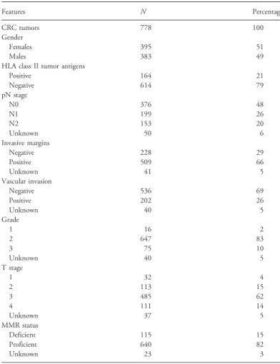
Table 1. Clinicopathologic Features of 778 Patients with Valuable CRC Tumor Punches of the Basel Study. Features N Percentage CRC tumors 778 100 Gender Females 395 51 Males 383 49
HLA class II tumor antigens
Positive 164 21 Negative 614 79 pN stage N0 376 48 N1 199 26 N2 153 20 Unknown 50 6 Invasive margins Negative 228 29 Positive 509 66 Unknown 41 5 Vascular invasion Negative 536 69 Positive 202 26 Unknown 40 5 Grade 1 16 2 2 647 83 3 75 10 Unknown 40 5 T stage 1 32 4 2 113 15 3 485 62 4 111 14 Unknown 37 5 MMR status Deficient 115 15 Proficient 640 82 Unknown 23 3
LGII-612.14. Following two washes, cell surface–bound antibodies were detected using fluorescein-conjugated mouse IgG anti-bodies. Cells then were analyzed using a two-laser BD FACSCalibur equipped with a Cell Quest software package (Beckton-Dickinson, San Jose, CA).
Magnetic Sorting of Peripheral Blood Monocytes
Freshly isolated PBMCs (50-70 × 106) were washed twice in modi-fied MACS buffer (phosphate-buffered saline supplemented with 0.5% bovine albumin). Then, 50 to 70 μl (1 μl per 1 million of PBMCs) of anti-CD14 magnetic beads (Miltenyi Biotec, Bergisch Gladbach, Germany) was added to the PBMC pellet. Following a 30-minute incubation on ice, cells were washed twice in modified MACS buffer. Cells were resuspended in 1 ml of modified MACS buffer and passed through a 0.40-μm cell strainer (BD Falcon, San Jose, CA), in a magnetic column. CD14+cells retained in the column were eluted by a strong mechanical pressure. Following two washes, monocytes were resuspended at the concentration of 1 × 10⁶/ml, in complete medium, for functional experiments.
Cytokine Array
The amount of cytokines in the supernatant harvested from cul-tures of COLO205 and PBMCs was assessed, using a duplicate of 42 human cytokine array system (RayBiotech Inc, Norcross, GA), which detects the antibody-cytokine sandwich by chemiluminescence. For cytokine blocking experiments, before PBMCs or monocytes were mixed with COLO205 cells, FcγRs of PBMCs or monocytes were blocked using mouse IgG (6-10 μg/ml) or FcγR blocking solu-tion (Miltenyi Biotec). HLA class II antigen blockade was accom-plished by incubating IFNγ (10 ng/ml)–treated COLO205 cells with 3μg of LGII-612.14 mAb for 30 minutes on ice in 100 μl of complete medium.
Enzyme-Linked Immunosorbent Assay
IL-1β and IL-6 levels were measured in the supernatants harvested from the cultures using commercially available IL-1β (BD Biosciences) and IL-6 (R&D Systems, Minneapolis, MN) ELISA kits with a sen-sitivity of 0.80 and 0.70 pg/ml, respectively. Supernatants harvested from cultures of COLO205 and PBMCs were added, in triplicates, to wells previously coated with anti–IL-1β or anti–IL-6 capture antibodies. Wells were covered with plate sealers, incubated for 2 hours at room temperature, and washed five times with 300 to 400 μl of wash buffer. Following the last wash, plates were blotted on absorbent paper to remove the remaining buffer. Then, 100μl of biotin-conjugated anti–IL-1β antibody or 200 μl of HRP-conjugated anti–IL-6 antibody was added to each well for 2 hours at room tem-perature. Following extensive washing, substrates were added, and plates were incubated for 30 minutes at room temperature in the dark. Reactions were stopped by adding 50μl of stop solution to each well. The absorbance was read at 450 nm within 30 minutes.
Statistical Analysis
HLA class II antigen–positive CRC cells were analyzed, in each CRC tumor punch, by counting a maximum number of 100 positive cells. The threshold for this marker was calculated by means of receiver operating characteristic curve analysis in the testing collective of Athens. Punches containing ≤15 and >15 HLA class II antigen–
positive CRC cells were scored negative and positive, respectively. The calculated threshold was tested in the collective of Basel. Survival time differences were evaluated using the log-rank test in univariate analysis. Multivariate hazard Cox regression analysis was performed by adjusting for CRC standard prognostic factors including pT, pN, tumor grade, vascular invasion, age, and metastasis. Hazard ratios and 95% confidence intervals were used to calculate the validity of the prognostic effect.
Correlation analysis among biologic markers was assessed using the Spearman rank correlation coefficient.
Statistical analyses were performed using SPlus software (version 6.1; Insightful Corporation, Seattle, WA) and Statistical Analysis System software (SAS Institute, Cary, NC).
Results
Association of HLA Class II Antigen Expression with
Disease Progression in CRC Tumors
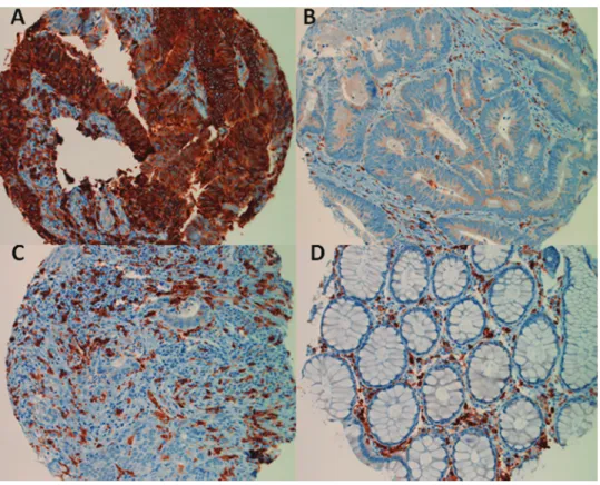
HLA class II antigens were found to be expressed by CRC cells in about 23% of the tumors tested. Representative staining patterns are shown in Figure 1, which indicates that the majority of CRC cells were stained by mAb LGII-612.14 in the tumors expressing HLA class II antigens (Figure 1A). In contrast, HLA class II antigens were detected in about 95% of inflammatory infiltrating cells of the CRC tumors analyzed. Representative examples are shown in Figure 1, B and C. HLA class II antigens were not detected in cells of normal colorectal mucosa but were found to be expressed by immune cells in the interstitial tissues (Figure 1D).
We next investigated whether HLA class II antigen expression was associated with malignant transformation of colorectal cells. To this end, we compared HLA class II antigen expression in 37 normal colorectal mucosa samples, in 42 colorectal adenomas, and 220 CRC tumors. The frequency of HLA class II antigen expression increased with disease progression because it was about 5% in the normal mucosa samples tested, about 19% in the colorectal adenoma samples tested, and about 25% in the CRC tumors tested. Furthermore, in CRC tumors, the percentage of stained malignant cells and their staining intensity were significantly higher than those found in colorectal adenomas (P < .013) and in normal colorectal mucosa (P < .0001; Table 2). The difference, in HLA class II antigen expression, between normal colorectal mucosa and colorectal adenoma was also significant (P = .01) corroborating the conclusion that in colorectal mucosa HLA class II antigen expression is associated with disease progression.
Association of HLA Class II Antigen Expression in
CRC Cells with Patients
’ Prolonged Survival
To assess the clinical significance of HLA class II antigen expression by CRC cells, we correlated it with the histopathologic characteristics of the lesions and with the clinical characteristics of the patients. For this analysis, patients were divided into the following two groups: those who contained >15 malignant cells, per TMA, stained by mAb LGII-612.14 in their tumor punches and those who contained <15.
The presence of ≤15 CRC cells stained by mAb LGII-612.14 in tumor punches was significantly associated with lymph node involvement (P < .0001), presence of metastasis (P < .0021), and lymphatic invasion (P≤ .008). Table W1 shows a detailed des-cription of the association of HLA class II antigen expression with tumor stage in the Athens study. There was a significant association
between low HLA class II antigen–positive CRC cell counts and high tumor stage (P < .0001). Similar results were obtained in the larger collective of the Basel study (data not shown). However, there was no association with the histologic characteristics of the tumors, as well as their grade and location. Furthermore, the survival of patients with≤15 CRC cells stained by mAb LGII-612.14 in their tumor punches was significantly shorter than that of patients with >15 malignant cells stained by mAb LGII-612.14 in their tumor punches. To determine whether HLA class II antigen expression in CRC cells was an independent prognostic marker, we performed a multi-variate survival analysis of HLA class II antigen expression in CRC
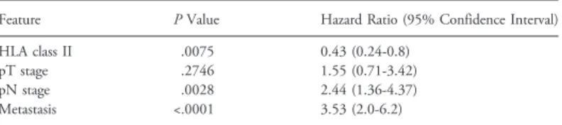
cells and pT, pN, and metastasis. Table W2 shows that HLA class II antigen expression in CRC cells was an independent favorable prognostic factor.
Because the American Society of Clinical Oncology (ASCO) guide-lines for the identification of new biologic tumor markers recommend data validation at least by an independent study, we assessed the im-pact of HLA class II antigen expression on the overall survival (OS) of patients with CRC in an additional group of patients of the Basel study. Of 778 patients with CRC whose tumors were used for immuno-histochemical analysis, 742 patients were also available for an OS estima-tion. Univariate analysis confirmed that patients with (N = 153) CRC tumors having >15 malignant cells, per tumor punch, stained by mAb LGII-612.14 had a significantly (0.007) longer OS time than pa-tients (N = 589) with CRC tumors having≤15 CRC cells, per tumor punch, stained by mAb LGII-612.14.
Furthermore, we investigated in 730 patients with CRC of the Basel study the impact of the HLA class II antigen–positive inflam-matory cells in the CRC tumors on the OS of patients with CRC. Figure 2C shows that patients (N = 688) with CRC tumors having >6 HLA class II antigen–positive inflammatory cells, per tumor punch, had a longer OS time than patients (N = 42) with CRC tumors having≤6 HLA class II antigen–positive inflammatory cells per tumor punch. However, the difference did not reach the level of statistical significance (P = .09).
We then assessed the relationship between HLA class II anti-gen expression and the inflammatory infiltrate of the CRC tumors.
Table 2. Athens Study: HLA Class II Antigens Are Preferentially Expressed in CRC Cells of Colorectal Tissues.
Histopathology Normal Mucosa Adenoma CRC
N 37 42 220
HLA class II+ colorectal tissues 2 8 55
HLA class II− colorectal tissues 35 34 165
Min. number of HLA class II− cells 0 0 0
Max. number of HLA class II+ cells 20 100 100
Mean+ cells 1.4 11.7 21.4
Statistical analysis P < .05 P < .0001
The expression of HLA class II antigens in colorectal mucosae was evaluated by immunohisto-chemistry as described in the Materials and Methods section.
N , sample size; Min. and Max., minimal and maximal absolute cell numbers of HLA class II antigen–positive cells detected in the colorectal tissues.
Figure 1. Expression of HLA class II antigens in the CRC tumors. Post-resection colorectal tissues were stained with the anti–HLA class II antigen LGII-612.14 mAb as indicated in the Materials and Methods section. Positive cells are stained in brown. The upper panel shows representative examples of powerful CRC cell positivity (A) and negativity (B) for HLA class II antigens. B also shows a certain degree of HLA class II antigen–positive inflammatory cells into the interstitial tissues of the CRC tumor. C documents the presence of HLA class II antigen–negative CRC cells and the presence of HLA class II antigen–positive inflammatory cells. D shows a normal colorectal tissue. Normal colorectal glands were clearly HLA class II antigen–negative, while HLA class II antigen–positive cells were restricted to the interstitial tissues.
Table W3 shows that CRC tumors with HLA class II antigen– positive CRC cells correlated with the presence of CD16+myeloid and T cell antigens. Conversely, there was no correlation with natural killer (NK)–NK-T and myeloid markers. Tumors rich in HLA class II antigen–positive inflammatory cells maintained a weak but signif-icant correlation with the majority of immune markers indicated in Table W3. These data suggest that there may be a correlation between HLA class II antigen expression and CD16 myeloid and T cell infiltration [20,28,29].
IFN
γ, IL-1β, and IL-6 Expression in Freshly Removed
CRC Tumors
Because HLA class II antigens are pivotal players of the immune response and their expression can be induced by pro-inflammatory cyto-kines including IFN-γ, we explored the expression of pro-inflammatory
cytokines by qRT-PCR in CRC tumors and normal colorectal mucosae. Among 10 CRC tumors available, 10 were evaluated for IFNγ and IL-1β gene expression and 9 for IL-6 gene expression. CRC tumors contained IFNγ (P = .004), IL-1β (P = .001), and IL-6 (P = .001) gene expression levels significantly higher than those assessed in normal colonic mucosae (Figure 3).
COLO205 Cells and PBMCs Trigger IL-1
β and
IL-6 Inflammatory Cytokine Production
Because inflammatory cytokines are capable of polarizing macro-phage toward M1 phenotype, we investigated whether HLA class II antigen–positive CRC cells in the presence of allogeneic PBMCs could contribute to develop an inflammatory microenvironment in vitro.
Following a 48-hour incubation with IFNγ at 37°C, two of four CRC cell lines including COLO205 and HT29 cells expressed HLA class II antigens (Figure 4). However, IFNγ did not induce CD80, CD86, and CD18 expression (data not shown). These results suggest that HLA class II antigens are heterogeneously expressed among CRC cell lines.
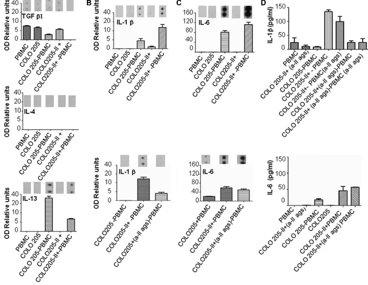
Figure 5A shows that resting PBMCs and COLO205 cells without or with HLA class II antigen expression produced traces of transforming
Figure 2. HLA class II antigen expression is associated with im-proved OS in CRC of the Athens and Basel studies. (A) Survival probability according to the Athens study: The red line shows OS probability of CRC patients with HLA class II antigen–positive CRC cells; the blue line shows OS of CRC patients with HLA class II antigen–negative CRC cells. (B) Survival probability according to the Basel study: The red line shows OS probability of CRC patients with HLA class II antigen–positive CRC cells; the blue line shows OS probability of CRC patients with HLA class II antigen–negative CRC cells. (C) Survival probability according to the level of infiltration of HLA class II antigen–positive or –negative inflammatory cells. The red line shows OS probability of CRC patients with their CRC tumors infiltrated by HLA class II antigen–positive immune cells; the blue line shows OS probability of CRC patients with their CRC tumors noninfiltrated by HLA class II antigen–positive immune cells. Statis-tical differences between groups are indicated. The definition of HLA class II antigen–positive or –negative tumors is described in the Materials and Methods section.
Figure 3. Gene expression of the pro-inflammatory cytokines IL-1β, IFNγ, and IL-6 in freshly resected CRC tumors and normal colorectal mucosa. CRC tumors and normal colorectal mucosae (CM) were ob-tained during surgery and placed in Falcon tubes. Following mecha-nical separation and DNA digestion, total RNA was isolated from CRC and colorectal tissues and subjected to RT. Then, cDNA was analyzed by qRT-PCR for IL-1β, IFNγ, and IL-6 gene expression as de-scribed in the Materials and Methods section. Data are expressed as a ratio toGAPDH housekeeping gene as indicated; solid circles repre-sent normal colorectal mucosa; solid squares reprerepre-sent CRC tumors.
growth factor–β1 (TGF-β1) but did not secrete IL-4 and IL-13. However, supernatants harvested from cultures of COLO205 cells and PBMCs showed changes in M2 cytokine content since there was an increase in the level of IL-13. Furthermore, we noted a reduction of IL-13 production when PBMCs were cultured in the presence of HLA class II antigen–positive COLO205 cells. These results suggest that HLA class II antigen–positive COLO205 cells failed to induce the production of TGF-β1, IL-4, and IL-13 cytokines.
We next investigated whether HLA class II antigen–positive COLO205 cells could affect the production of IL-1β and IL-6.
Following a 48-hour incubation of PBMCs with or without HLA class II antigen–positive or –negative COLO205 cells, supernatants harvested from a culture of PBMCs and HLA class II antigen–positive COLO205 cells showed high contents in IL-1β. In contrast, a lower amount of IL-1β was detected in the supernatants harvested from a culture of PBMCs and HLA class II antigen–negative COLO205 cells (Figure 5B, upper panel). Resting PBMCs and HLA class II antigen– negative or–positive COLO205 cells did not produce IL-6. Con-versely, supernatants obtained from a culture of PBMCs and HLA class II antigen–negative COLO205 cells contained levels of IL-6 lower than those detected in the supernatants harvested from a cul-ture of PBMCs and HLA class II antigen–positive COLO205 cells (Figure 5C, upper panel). These results suggest that the production of IL-1β and IL-6 in the supernatants of PBMCs and COLO205 was associated with HLA class II antigen expression on COLO205 cells.
To assess the impact of HLA class II antigens on IL-1β and IL-6 production, PBMCs were cultured in the presence of HLA class II antigen–positive COLO205 cells coated with or without anti–HLA class II antigen mAb. Figure 5B, lower panel, shows that IL-1β produc-tion in the cell cultures composed of HLA class II antigen–positive COLO205 cells and PBMCs was inhibited by the HLA class II anti-gen–specific mAb LGII-612.14, whereas IL-6 production was not (Figure 5C , lower panel ). These results strongly suggest that HLA class II antigens played a role in the secretion of IL-1β. Similar results were obtained when IL-1β and IL-6 cytokine production was assessed using an ELISA (Figure 5D).
IL-1β Production Involved Monocytes
Because IL-1β is typically produced by monocyte/macrophage, we assessed whether co-cultures of monocytes and HLA class II antigen– positive COLO205 generated IL-1β. Supernatants harvested from a culture of monocytes and HLA class II antigen–positive COLO205 cells obtained from three healthy donors (Figure 6A) showed the high-est content of IL-1β (Figure 6B). Conversely, supernatants harvhigh-ested from a cell culture consisting of monocytes cultured in the absence of COLO205 cells, or PBMCs, depleted of monocytes, with or without HLA class II antigen–positive COLO205 cells did not produce IL-1β, suggesting that IL-1β production requires the presence of monocytes and HLA class II antigen–positive COLO205 cells.
To identify whether monocytes or HLA class II antigen–positive COLO205 cells secreted IL-1β, we incubated monocytes with HLA class II antigen–negative or –positive COLO205 cells. Subsequently, monocytes were magnetically separated from COLO205 cells, and both cells were placed in a single culture condition. Following a 48-hour incubation, the supernatants harvested from resting mono-cytes or COLO205 cells negative and positive for HLA class II antigen expression as well as COLO205, previously stimulated with monocytes, did not show significant production of IL-1β. Conversely, monocytes previously stimulated with HLA class II antigen–positive COLO205 cells produced most of the IL-1β detected in the supernatants of the different culture conditions. As expected, lipopolysaccharides (LPS) and IFNγ stimulation of monocytes, which were used as positive con-trols, induced IL-1β production (Figure 6C). These results suggest that HLA class II antigen–positive COLO205 cells specifically triggered IL-1β by monocytes.
Discussion
In this study, we investigated HLA class II antigen expression in a large collection of about 1000 CRC tumors recruited in two independent European institutions. Immunohistochemical analysis of the CRC tumor showed an overall frequency of HLA class II antigen expres-sion of 23% ranging between 21% of the Basel study and 25% for the Athens study. In contrast, 95% of CRC TMA punches analyzed
Figure 4. IFNγ induced HLA class II antigen expression in COLO205 and HT29 CRC cell lines. CRC cell lines COLO205, HT29, SW480, and HCT116 were cultured in the presence or absence of IFNγ (10 ng/ml). Following a 48-hour stimulation, cells were indirectly stained with the anti–HLA class II antigen mAb LGII-612.14. Cell surface–bound LGII-612.14 was detected using a fluorescein isothiocyanate goat anti-mouse F(ab)2. The blue lines show LGII612.14 staining, while the red lines show the fluorescence intensity given by the secondary antibody used as a negative control.
had HLA class II antigen–positive inflammatory cells. Furthermore, when CRC tumors were classified as positive, in most of the cases, the frequency of the CRC positive cells was high, and the intensity of the staining was particularly strong. Our results are in agreement with the study of de Bruin et al. who find an expression frequency of about 21% but are conflicting with Lovig et al. who found a fre-quency of HLA class II antigen expression of 51% [10]. A possible explanation for this difference may involve the choice of the investiga-tors to consider different cutoff scores to define positive or negative CRC tumors. Our choice to consider the threshold of 15 positive cells
was based on information obtained from receiver operating character-istic curve analysis in the collective of Athens and validated in the independent collective of Basel. Alternatively, it could be also possible to speculate that there were different levels of cytokine production in the CRC tumors used in these studies. Finally, HLA class II antigen expression disparity may reflect different molecular profiles of the CRC tumors included in these studies. However, we tend to exclude this hypothesis because, although the Athens study only included MMR-proficient CRC tumors, while the Basel study included 83% and 17% of MMR-proficient and MMR-deficient CRC tumors,
Figure 5. HLA class II antigen expression on CRC cells regulated IL-1β production in the cell cultures composed of COLO205 cells and PBMCs. COLO205 CRC cells were cultured in the presence or absence of IFNγ (10 ng/ml) for 48 hours in complete medium. HLA class II antigen expression on COLO205 cells was confirmed by flow cytometry. After extensive washes, COLO205 cells and freshly isolated PBMCs were co-cultured, in the absence of IFNγ, for 48 hours at 1:4 ratios, respectively, according to the indicated experimental con-ditions. Following a 48-hour incubation, supernatants were evaluated in duplicates for the presence of TGF-1β, IL-4, IL-13 (A), IL-1β (B, upper panel), and IL-6 (C, upper panel) cytokines using a cytokine array. HLA class II antigen–positive COLO205 cells were cultured in the presence or absence of blocking anti–HLA class II antigen mAbs (6 μg/ml) for 30 minutes at 4°C. Then, cells were washed and cultured in the presence of freshly isolated PBMCs whose FcγRs were previously blocked using mouse IgG. Following a 48-hour incubation, supernatants were harvested and analyzed in duplicates for IL-1β (B, lower panel) and IL-6 (C, lower panel) contents. Tripli-cates of 100μl of supernatants obtained after 30 hours of culture condition indicated in the x-axis were analyzed for IL-1β (D, upper panel) and IL-6 (D, lower panel) protein content by ELISA. In all situations, where blocking conditions were performed, COLO205 cells were pre-coated with the anti–HLA class II antigen mAb LGII-612.14, while PBMC FcγRs were blocked using a commercially available blocking buffer (Miltenyi, Bologna, Italy). Cytokine protein spots were analyzed by photo densitometry and expressed as OD relative units calculated as follows:cytokine spotsOD/standard control spotsOD × 100. Abbreviations: II, HLA class II antigen–positive; a-II ag, anti–HLA class II antigen mAb LGII-612.14; mono, monocytes.
respectively, no significant differences in HLA class II antigen expres-sion were observed between the groups. In agreement with the studies of Lovig et al. and de Bruin et al., we found that HLA class II antigen expression in the CRC tumor cells was a favorable prognostic factor. However, these results are not in agreement with other studies that suggest that HLA class II antigen expression is at least an indifferent prognostic factor without being associated with a favorable clinical course of the disease [16–18]. Explanation(s) for these conflicting re-sults is (are) not known. The existence of such a conflicting situation prompted us the idea that HLA class II antigen expression in the CRC tumor could be significant but not sufficient to generate a tumor microenvironment predisposing to a favorable clinical course of the disease. Then, other factors such as local cytokine production and or type and function of inflammatory immune cells may be involved in such a process.
The expression of HLA class II antigens could be a result of the activation of an inflammatory response mediated by immune cells infiltrating the tumor microenvironment [2,20,30–36]. CRC tumors
for a variety of reasons including their microenvironment, rich in bacterial flora, undergo active infiltration of inflammatory cells com-posed of T lymphocytes and TAMs but limited in NK cells [32]. Thus, the expression of HLA class II antigens is likely to be due to IFNγ producing immune cells during the inflammation process. Indeed, we have found that IFNγ gene expression was upregulated in the CRC tumor microenvironment. Furthermore, in agreement with published results [37], IFNγ triggered HLA class II antigen expression on the cell surface of 50% of CRC cell lines used, includ-ing COLO205 and HT29 cells. Because CRC tumor microenviron-ment is hardly infiltrated by NK cells and rich in T lymphocytes, it is likely to predict that CD4-positive lymphocytes are responsible for the local production of IFNγ.
HLA class II antigen expression in CRC cells, in the presence of IFNγ, is controlled by the methylation of class II transactivator– isoform-PIV (PIV) and is inhibited by somatic mutation of the RFX5 gene [21]. Although, we have not tested our CRC cell lines for FRX5 mutation, we also found that HLA class II antigen expression
Figure 6. HLA class II antigen–dependent IL-1β production was produced by monocytes. PBMCs were isolated from three healthy donors. (A) monocytes were obtained using anti–CD14-conjugated magnetic beads by positive selection. CD14-depleted PBMCs were obtained harvesting unbound cells flowing through the magnetic columns. After cell sorting, monocytes were stained in the presence of an allophycocyanin (APC)-conjugated anti-CD33 mAb. The purity of myeloid cells was higher than 95%. (B) Following monocyte isolation, 1 × 10⁶/ml CD14+or CD14−cells were plated onto 96-well plates in the presence or absence of 0.25 × 10⁶HLA class II antigen–positive COLO205 cells. After a 30-hour incubation, supernatants were harvested and analyzed in triplicates for IL-1β content by ELISA. (C) CD14 magnetic bead–labeled monocytes were cultured in the presence or absence of HLA class II antigen–positive or –negative COLO205 cells for 5 hours. Then, cells were vigorously resuspended and passed through magnetic columns. Unbound COLO205 cells were harvested and seeded in a 96-well plate, while magnetically labeled monocytes retained in the columns were detached by a mechanical pressure. Monocytes were harvested and cultured according to the indicated experimental conditions in 96-well plates. Following a 30-hour incu-bation, supernatants harvested from cultures of COLO205 cells or monocytes were assessed for IL-1β content. Supernatants harvested from cultures of resting HLA class II antigen–positive or –negative cells were also used as negative controls. Abbreviations: II, HLA class II antigen–positive; mono, monocytes.
requires the presence of class II transactivator–PIV (data not shown). These results suggest possible mechanisms controlling HLA class II an-tigen expression in CRC cells on stimulation with IFNγ.
There is an increased consensus supporting the positive effect of a strong inflammatory response, in the CRC tumors, on the OS of patients with CRC [38]. Interestingly, the favorable contribution of TAM infiltration to the clinical course of the patients with CRC makes this disease a compelling exception among solid malignancies investigated so far [20,29,39–42]. Thus, HLA class II antigen expression in the CRC tumors could play a role in counteracting CRC progression. This situation raises questions about mechanisms by which HLA class II antigen expression in CRC cells protects the host against the tumor.
In this context, we suggest that HLA class II antigen expression in CRC may cause an immunologic antitumor response [20,29,31].
HLA class II antigen expression on CRC cells could activate CD4+ T cells by presenting tumor-associated antigens (TAAs). Indeed, a certain number of TAA have been described in CRC [43–48]. How-ever, HLA class II antigen expression is not sufficient to develop a successful CD4+T cell stimulation because it requires the expression of co-stimulatory molecules on tumor cells [49].
Accordingly, in the allogeneic setting, IFNγ induced HLA class II antigen expression but failed to generate B7, B7.1, and CD18 ex-pression on COLO205 cells. As a consequence, we failed to produce a CD4+T cell proliferation (data not shown). However, HLA class II antigen expression on COLO205 cells induced the production of pro-inflammatory cytokines including IL-1β and IL-6 by monocytes but failed to generate anti-inflammatory cytokines including TGF-β, IL-13, and IL-4.
IL-1β is a pro-inflammatory cytokine, and the expression of the IL-1β receptor is required for an efficient activation of dendritic cells (DC). Furthermore, IL-1β induces macrophage recruitment and macrophage auto-stimulation [50] shaping a pro-inflammatory microenvironment with a potential antitumor activity [20,29,33].
IL-1β and IL-6 molecules are pro-inflammatory cytokines mainly produced by granulocyte/monocytes [51,52]. IL-6 molecule promotes the transition from acute to chronic phase of inflammation reducing granulocyte trafficking and increasing monocyte recruitment at the damaged tissues [52]. Thus, a pro-inflammatory infiltrate is likely to have a positive clinicopathologic effect on the clinical course of human CRC tumors. However, these results are not in agreement with mouse models of CRC because inflammation has been found to be involved in the pathogenesis of colitis-associated cancer. According to these studies, in the tumor microenvironments, IL-6 secreted by bone marrow–derived myeloid cells activates signal transducer and activator of transcription 3 (STAT3) signaling pathways leading to the concen-tration ofβ-catenin into the nuclei favoring tumor progression. In addition, IL-1β can also induce IL-6 production promoting colitis-associated cancer progression [53].
Thus, in human and mouse, inflammation is differently linked to CRC tumors. There is evidence that inflammation is connected to cancer intrinsic and extrinsic pathways. The intrinsic pathway is activated by genetic modifications leading to inflammation and can-cer. The extrinsic pathway involves CRC tumors growing in a tumor microenvironment with a preexistence of inflammatory condition such as ulcerative colitis and Chron’s disease [54]. Most of the CRC tumors evaluated rose in mice with inflammatory bowel diseases, while the majority of our patients had no underlying inflammatory conditions. These differences may explain the conflicting results
ob-tained in human and mouse studies. Then, inflammation developing in CRC tumors derived from genetic alteration may have protective activity. In contrast, inflammation of CRC tumors derived from colorectal mucosa chronically affected by inflammatory diseases may generate a qualitative different inflammation that may promote cancer progression. However, ulcerative colitis and Chron’s disease are often associated with immune dysregulation. The idea that inflammation could be good and bad in CRC tumors is also reinforced by Klintrup et al. [38]. These investigators found that inflammatory cell infiltra-tion at the invasive margin, in CRC, was a favorable prognostic factor, while Richards et al. showed that systemic inflammation was a poor prognostic marker in patients with CRC [55].
In human, certain HLA-DR genotype alleles have been associated with ulcerative colitis [56]. It is tempting to speculate that aberrant expression of HLA class II antigens may differentially affect the patho-genesis of malignant and autoimmune diseases having a protective role in CRC but a detrimental one in autoimmune diseases [57]. However, aberrant expression of HLA class II antigens worsened the clinical out-come of thyroiditis, type I diabetes, and biliary cirrhosis [58–60].
According to our study, HLA class II antigen–positive cells induced IL-1β production in peripheral blood monocytes. We and others have shown that CRC tumor is rich in TAMs. Phenotypic analysis of freshly infiltrating cells indicates two types of TAMs: The first is CD16 positive, while the second is CD16 negative. CD16-positive TAMs are CD11c high and are associated with survival. Thus, It is possible to speculate that these cells could be a target of IL-1β in the context of CRC tumors perhaps favoring DC differentiation.
Considering that HLA class II antigen expression may be induced in the presence of IFNγ, it could be possible to use this cytokine as a platform for developing personalized immunotherapeutic studies. To avoid the adverse effect and the nonspecific stimulation of IFNγ on the immune system, we may think to deliver IFNγ into the CRC tumors using anti–TAA-IFNγ fusion proteins. However, Trinh et al. have demonstrated that is possible to deliver IFNβ to human lymphoma using an anti–CD10–IL-1β fusion protein [61]. References
[1] Fearon ER (2011). Molecular genetics of colorectal cancer. Annu Rev Pathol 6, 479–507.
[2] Smyrk TC, Watson P, Kaul K, and Lynch HT (2001). Tumor-infiltrating lym-phocytes are a marker for microsatellite instability in colorectal carcinoma. Cancer 91, 2417–2422.
[3] Lazzaro B, Anderson AE, Kajdacsy-Balla A, and Hessner MJ (2001). Antigenic characterization of medullary carcinoma of the breast: HLA-DR expression in lymph node positive cases. Appl Immunohistochem Mol Morphol 9, 234–241. [4] Dengjel J, Nastke MD, Gouttefangeas C, Gitsioudis G, Schoor O, Altenberend F,
Muller M, Kramer B, Missiou A, Sauter M, et al. (2006). Unexpected abundance of HLA class II presented peptides in primary renal cell carcinomas. Clin Cancer Res 12, 4163–4170.
[5] Taramelli D, Fossati G, Mazzocchi A, Delia D, Ferrone S, and Parmiani G (1986). Classes I and II HLA and melanoma-associated antigen expression and modulation on melanoma cells isolated from primary and metastatic lesions. Cancer Res 46, 433–439.
[6] Moretti S, Pinzi C, Berti E, Spallanzani A, Chiarugi A, Boddi V, Reali UM, and Giannotti B (1997). In situ expression of transforming growth factor beta is associated with melanoma progression and correlates with Ki67, HLA-DR and beta 3 integrin expression. Melanoma Res 7, 313–321.
[7] Trieb K, Lechleitner T, Lang S, Windhager R, Kotz R, and Dirnhofer S (1998). Evaluation of HLA-DR expression and T-lymphocyte infiltration in osteosarcoma. Pathol Res Pract 194, 679–684.
[8] Hilders CG, Houbiers JG, van Ravenswaay Claasen HH, Veldhuizen RW, and Fleuren GJ (1993). Association between HLA-expression and infiltration of immune cells in cervical carcinoma. Lab Invest 69, 651–659.
[9] Esteban F, Ruiz-Cabello F, Concha A, Pèrez-Ayala M, Sànchez-Rozas HA, and Garrido F (1990). HLA-DR expression is associated with excellent prognosis in squamous cell carcinoma of the larynx. Clin Exp Metastasis 8, 319–328. [10] Lovig T, Andersen SN, Thorstensen L, Diep CB, Meling GI, Lothe RA, and
Rognum TO (2002). Strong HLA-DR expression in microsatellite stable car-cinomas of the large bowel is associated with good prognosis. Br J Cancer 87, 756–762.
[11] de Bruin EC, van de Velde CJ, van Krieken JH, Marijnen CA, and Medema JP (2008). Epithelial human leukocyte antigen-DR expression predicts reduced recurrence rates and prolonged survival in rectal cancer patients. Clin Cancer Res 14, 1073–1079.
[12] Gutierrez J, Lòpez-Nevot MA, Cabrera T, Oliva R, Esquivias J, Ruiz-Cabello F, and Garrido F (1987). Class I and II HLA antigen distribution in normal mucosa, adenoma and colon carcinoma: relation with malignancy and invasive-ness. Exp Clin Immunogenet 4, 144–152.
[13] Matsushita K, Takenouchi T, Shimada H, Tomonaga T, Hayashi H, Shioya A, Komatsu A, Matsubara H, and Ochiai T (2006). Strong HLA-DR antigen expression on cancer cells relates to better prognosis of colorectal cancer patients: possible involvement of c-myc suppression by interferon-γ in situ. Cancer Sci 97, 57–63.
[14] Morita M, Tanaka K, Kawanishi H, Tsuji M, Ookusa T, Takada H, Okamura A, and Hioki K (1995). Immunohistochemically demonstrated expression of HLA-DR antigen in colorectal adenocarcinomas and its relation to clinico-pathological features. J Surg Oncol 59, 233–238.
[15] Walsh MD, Dent OF, Young JP, Wright CM, Barker MA, Leggett BA, Bokey L, Chapuis PH, Jass JR, and Macdonald GA (2009). HLA-DR expression is associated with better prognosis in sporadic Australian clinicopathological stage C colorectal cancers. Int J Cancer 125, 1231–1237.
[16] Moller P, Momburg F, Koretz K, Moldenhauer G, Herfarth C, Otto HF, Hämmerling GJ, and Schlag P (1991). Influence of major histocompatibility complex class I and II antigens on survival in colorectal carcinoma. Cancer Res 51, 729–736.
[17] Mulder WM, Stern PL, Stukart MJ, de Windt E, Butzelaar RM, Meijer S, Ader HJ, Claessen AM, Vermorken JB, Meijer CJ, et al. (1997). Low intercellular adhesion molecule 1 and high 5T4 expression on tumor cells correlate with reduced disease-free survival in colorectal carcinoma patients. Clin Cancer Res 3, 1923–1930.
[18] Diederichsen AC, Hjelmborg J, Christensen PB, Zeuthen J, and Fenger C (2003). Prognostic value of the CD4+/CD8+ratio of tumour infiltrating lympho-cytes in colorectal cancer and HLA-DR expression on tumour cells. Cancer Immunol Immunother 52, 423–428.
[19] Momburg F, Degener T, Bacchus E, Moldenhauer G, Hammerling GJ, and Moller P (1986). Loss of HLA-A,B,C and de novo expression of HLA-D in colorectal cancer. Int J Cancer 15, 179–184.
[20] Sconocchia G, Zlobec I, Lugli A, Calabrese D, Iezzi G, Karamitopoulou E, Patsouris ES, Peros G, Horcic M, Tornillo L, et al. (2011). Tumor infiltra-tion by FcγRIII (CD16)+ myeloid cells is associated with improved survival in patients with colorectal carcinoma. Int J Cancer 128, 2663–2672. [21] Michel S, Linnebacher M, Alcaniz J, Voss M, Wagner R, Dippold W, Becker C,
von Knebel Doeberitz M, Ferrone S, and Kloor M (2010). Lack of HLA class II antigen expression in microsatellite unstable colorectal carcinomas is caused by mutations in HLA class II regulatory genes. Int J Cancer 127, 889–898.
[22] Kononen J, Bubendorf L, Kallioniemi A, Barlund M, Schraml P, Leighton S, Torhorst J, Mihatsch MJ, Sauter G, and Kallioniemi OP (1998). Tissue micro-arrays for high-throughput molecular profiling of tumor specimens. Nat Med 4, 844–847.
[23] Temponi M, Kekish U, Hamby CV, Nielsen H, Marboe CC, and Ferrone S (1993). Characterization of anti-HLA class II monoclonal antibody LGII-612.14 reacting with formalin fixed tissues. J Immunol Methods 161, 239–256. [24] Boyum A (1968). Isolation of mononuclear cells and granulocytes from human
blood. Scand J Clin Lab Invest 21(suppl 97), 77–89.
[25] Kammula US, Lee KH, Riker AI, Wang E, Ohnmacht GA, Rosenberg SA, and Marincola FM (1999). Functional analysis of antigen-specific T lymphocytes by serial measurement of gene expression in peripheral blood mononuclear cells and tumor specimens. J Immunol 163, 6867–6875.
[26] Mengus C, Le MC, Trella E, Yousef K, Bubendorf L, Provenzano M, Bachmann A, Heberer M, Spagnoli GC, and Wyler S (2011). Elevated levels of circulating IL-7 and IL-15 in patients with early stage prostate cancer. J Transl Med 9, 162.
[27] Livak KJ and Schmittgen TD (2001). Analysis of relative gene expression data using real-time quantitative PCR and the 2−ΔΔCT
method. Methods 25, 402–408.
[28] Zhang L, Conejo-Garcia JR, Katsaros D, Gimotty PA, Massobrio M, Regnani G, Makrigiannakis A, Gray H, Schlienger K, Liebman MN, et al. (2003). Intratumoral T cells, recurrence, and survival in epithelial ovarian cancer. N Engl J Med 348, 203–213.
[29] Forssell J, Oberg A, Henriksson ML, Stenling R, Jung A, and Palmqvist R (2007). High macrophage infiltration along the tumor front correlates with improved survival in colon cancer. Clin Cancer Res 13, 1472–1479. [30] Sconocchia G, Spagnoli GC, Del Principe D, Ferrone S, Anselmi M, Wongsena
W, Cervelli V, Schultz-Thater E, Wyler S, Carafa V, et al. (2009). Defec-tive infiltration of natural killer cells in MICA/B-posiDefec-tive renal cell carcinoma involvesβ2-integrin–mediated interaction. Neoplasia 11, 662–671.
[31] Pagès F, Berger A, Camus M, Sanchez-Cabo F, Costes A, Molidor R, Mlecnik B, Kirilovsky A, Nilsson M, Damotte D, et al. (2005). Effector memory T cells, early metastasis, and survival in colorectal cancer. N Engl J Med 353, 2654–2666. [32] Sconocchia G, Arriga R, Tornillo L, Terracciano L, Ferrone S, and Spagnoli
GC (2012). Melanoma cells inhibit NK cell functions—letter. Cancer Res 72, 5428–5429.
[33] Droeser RA, Hirt C, Eppenberger-Castori S, Zlobec I, Viehl CT, Frey DM, Nebiker CA, Rosso R, Zuber M, Amicarella F, et al. (2013). High myeloper-oxidase positive cell infiltration in colorectal cancer is an independent favorable prognostic factor. PLoS One 8, e64814.
[34] Sconocchia G, Del Principe D, and Barrett AJ (2007). CD16low/negative
tumor-infiltrating lymphocyte: lymphoid or myeloid in origin? Clin Cancer Res 13, 1620. [35] Frey DM, Droeser RA, Viehl CT, Zlobec I, Lugli A, Zingg U, Oertli D, Kettelhack C, Terracciano L, and Tornillo L (2010). High frequency of tumor-infiltrating FOXP3+regulatory T cells predicts improved survival in mismatch
repair-proficient colorectal cancer patients. Int J Cancer 126, 2635–2643. [36] Droeser RA, Hirt C, Viehl CT, Frey DM, Nebiker C, Huber X, Zlobec I,
Eppenberger-Castori S, Tzankov A, Rosso R, et al. (2013). Clinical impact of programmed cell death ligand 1 expression in colorectal cancer. Eur J Cancer 49, 2233–2242.
[37] Schwartz R, Momburg F, Moldenhauer G, Dorken B, and Schirrmacher V (1985). Induction of HLA class-II antigen expression on human carcinoma cell lines by IFN-Gamma. Int J Cancer 35, 245–250.
[38] Klintrup K, Makinen JM, Kauppila S, Vare PO, Melkko J, Tuominen H, Tuppurainen K, Makela J, Karttunen TJ, and Mäkinen MJ (2005). Inflammation and prognosis in colorectal cancer. Eur J Cancer 41, 2645–2654.
[39] Algars A, Irjala H, Vaittinen S, Huhtinen H, Sundström J, Salmi M, Ristamäki R, and Jalkanen S (2012). Type and location of tumor-infiltrating macrophages and lymphatic vessels predict survival of colorectal cancer patients. Int J Cancer 131, 864–873.
[40] Edin S, Wikberg ML, Dahlin AM, Rutegård J, Oberg A, Oldenborg PA, and Palmqvist R (2012). The distribution of macrophages with a M1 or M2 pheno-type in relation to prognosis and the molecular characteristics of colorectal cancer. PLoS One 7, e47045.
[41] Kinouchi M, Miura K, Mizoi T, Ishida K, Fujibuchi W, Sasaki H, Ohnuma S, Saito K, Katayose Y, Naitoh T, et al. (2012). Infiltration of CD40-positive tumor-associated macrophages indicates a favorable prognosis in colorectal cancer patients. Hepatogastroenterology 60, 83–88.
[42] Lackner C, Jukic Z, Tsybrovskyy O, Jatzko G, Wette V, Hoefler G, Klimpfinger M, Denk H, and Zatloukal K (2004). Prognostic relevance of tumour-associated macrophages and von Willebrand factor-positive micro-vessels in colorectal cancer. Virchows Arch 445, 160–167.
[43] Alves PM, Faure O, Graff-Dubois S, Gross DA, Cornet S, Chouaib S, Miconnet I, Lemonnier FA, and Kosmatopoulos K (2003). EphA2 as target of anticancer immunotherapy: identification of HLA-A*0201-restricted epitopes. Cancer Res 63, 8476–8480.
[44] Bohm CM, Hanski ML, Stefanovic S, Rammensee HG, Stein H, Taylor-Papadimitriou J, Riecken EO, and Hanski C (1998). Identification of HLA-A2-restricted epitopes of the tumor-associated antigen MUC2 recognized by human cytotoxic T cells. Int J Cancer 75, 688–693.
[45] Koesters R, Linnebacher M, Coy JF, Germann A, Schwitalle Y, Findeisen P, and von Knebel Doeberitz M (2004). WT1 is a tumor-associated antigen in colon cancer that can be recognized by in vitro stimulated cytotoxic T cells. Int J Cancer 109, 385–392.
[46] Schmollinger JC, Vonderheide RH, Hoar KM, Maecker B, Schultze JL, Hodi FS, Soiffer RJ, Jung K, Kuroda MJ, Letvin NL, et al. (2003). Melanoma inhibitor
of apoptosis protein (ML-IAP) is a target for immune-mediated tumor destruction. Proc Natl Acad Sci USA 100, 3398–3403.
[47] Umano Y, Tsunoda T, Tanaka H, Matsuda K, Yamaue H, and Tanimura H (2001). Generation of cytotoxic T cell responses to an HLA-A24 restricted epitope peptide derived from wild-type p53. Br J Cancer 84, 1052–1057. [48] Vissers JL, De Vries IJ, Schreurs MW, Engelen LP, Oosterwijk E, Figdor CG,
and Adema GJ (1999). The renal cell carcinoma-associated antigen G250 encodes a human leukocyte antigen (HLA)-A2.1-restricted epitope recognized by cytotoxic T lymphocytes. Cancer Res 59, 5554–5559.
[49] Banchereau J and Steinman RM (1998). Dendritic cells and the control of immunity. Nature 392, 245–252.
[50] Donath MY, Böni-Schnetzler M, Ellingsgaard H, and Ehses JA (2009). Islet inflammation impairs the pancreaticβ-cell in type 2 diabetes. Physiology (Bethesda) 24, 325–331.
[51] Sconocchia G, Campagnano L, Adorno D, Iacona A, Cococcetta NY, Boffo V, Amadori S, and Casciani CU (2001). CD44 ligation on peripheral blood poly-morphonuclear cells induces interleukin-6 production. Blood 97, 3621–3627. [52] Gabay C (2006). Interleukin-6 and chronic inflammation. Arthritis Res Ther
8(suppl 2), S3.
[53] Grivennikov S, Karin E, Terzic J, Mucida D, Yu GY, Vallabhapurapu S, Scheller J, Rose-John S, Cheroutre H, Eckmann L, et al. (2009). IL-6 and Stat3 are required for survival of intestinal epithelial cells and development of colitis-associated cancer. Cancer Cell 15, 103–113.
[54] Mantovani A, Allavena P, Sica A, and Balkwill F (2008). Cancer-related in-flammation. Nature 454, 436–444.
[55] Richards CH, Leitch EF, Horgan PG, Anderson JH, McKee RF, and McMillan DC (2010). The relationship between patient physiology, the systemic in-flammatory response and survival in patients undergoing curative resection of colorectal cancer. Br J Cancer 103, 1356–1361.
[56] Garrity-Park MM, Loftus EV Jr, Sandborn WJ, Bryant SC, and Smyrck TC (2009). MHC class II alleles in ulcerative colitis-associated colorectal cancer. Gut 58, 1226–1233.
[57] Cabrera T, Ruiz-Cabello F, and Garrido F (1995). Biological implications of HLA-DR expression in tumors. Scand J Immunol 41, 398–406.
[58] Hanafusa T, Pujol-Borrel R, Chiovato R, Russel RCG, Doniach D, and Bottazzo GF (1983). Aberrant expression of HLA-DR antigen on thyrocytes in Graves’ disease: relevance for autoimmunity. Lancet 2, 1111–1115. [59] Bottazzo GF, Dean BM, NcNally JM, Mackay EH, Swifr PGF, and Gamble DR
(1985). In situ characterization of autoimmune phenomena and expression of HLA molecules in the pancreas in diabetic insulitis. N Engl J Med 313, 353–360. [60] Ballardini G, Mirakian R, Bianchi FB, Pisi E, Doniach D, and Bottazzo GF (1984). Aberrant expression of HLA-DR antigens on bile duct epithelium in primary biliary cirrhosis: relevance to pathogenesis. Lancet 2, 1009–1013. [61] Trinh KR, Vasuthasawat A, Steward KK, Yamada RE, Timmerman JM, and
Morrison SL (2013). Anti-CD20-interferon-β fusion protein therapy of murine B-cell lymphomas. J Immunother 36, 305–318.
Stage HLA Class II Antigens N (%) P Value Low High I 23 (16.6) 22 (31.0) <.0001 II 29 (20.9) 27 (38.0) III 66 (47.5) 21 (29.6) IV 21 (15.1) 1 (1.4) Total N 139 71
CRC tumor punches were screened for HLA class II antigen expression. CRC tumors were considered HLA class II positive low when the total count of HLA class II–positive CRC cells was≤15 and high when it was >15.
Table W2. Multivariate Survival Time Analysis of HLA Class II Antigen Expression with pT, pN, and Metastasis.
Feature P Value Hazard Ratio (95% Confidence Interval)
HLA class II .0075 0.43 (0.24-0.8)
pT stage .2746 1.55 (0.71-3.42)
pN stage .0028 2.44 (1.36-4.37)
Metastasis <.0001 3.53 (2.0-6.2)
Table W3. Spearman Rank Correlation Coefficients between HLA Class II Antigen and Biologic Antigen Expression in the CRC Tumors.
Feature* CRC Class II Ags Infiltrate Class II Ags CD16 CD56 CD57 CD68 CD3 CD4 Peri-T CD8 Granzyme Intra-T
CRC HLA class II Ags 1 – 0.20 0.04 0.07 0.1 0.19 0.05 0.29 0.25
Infiltrate HLA class II Ags 0.18 1 0.29 0.16* 0.20 0.24 0.26 0.11 0.16 0.15
Bolded correlation coefficient (r) numbers are significant and have P values < .001. All comparisons were performed with more than 420 paired values.
All correlation are positive and weak though significant. *P = .003.