UNIVERSITÀ DEGLI STUDI DI ROMA
"TOR VERGATA"
FACOLTA' DI MEDICINA E CHIRURGIA
DOTTORATO DI RICERCA IN
SCIENZE E BIOTECNOLOGIE DELLA RIPRODUZIONE E DELLO
SVILUPPO
CICLO XXII
Transcriptional and post-transcriptional mechanisms control
meiotic entry in mouse germ cells.
Florencia Barrios Abraham
A.A. 2009/2010
Docente Guida/Tutor: Prof. ssa Susanna Dolci
Coordinatore: Prof. Raffaelle Geremia
A mamá, papá e Ignacio
“Un padre e un figlio
con un solo abbraccio
squarciano il tempo
vanno oltre lo spazio”
INDEX
Chapter
1.
Introduction.
Page 3
• Why do we study germ cells?
Page 4
• Overview of germ cell development.
Page 4
• Mitotic arrest of male germ cells.
Page 5
• Spermatogonial stem cell renewal and differentiation. Page 7
• The
spermatogenic
niche.
Page
8
• The
GDNF
phase.
Page
12
• The
KIT
phase.
Page
14
• Regulation
of
meiotic
entry.
Page
15
• AIM OF THE THESIS.
Page 22
• References to Chapter 1.
Page 73
Chapter 2. ATRA and KL promote differentiation toward the
meiotic program of male germ cells.
Page 23
Chapter 3. Opposing effects of RA and FGF9 on Nanos2 expression
and meiotic entry of murine germ cells.
Page 24
Chapter 4. The role of Sohlh transcription factors on
spermatogonia
differentiation.
Page 50
Chapter 5. Future directions.
Page 70
•
Ongoing
work.
Page
71
• Roads
to
go.
Page
71
3
CHAPTER 1
INTRODUCTION.
INTRODUCTION.
Why do we study germ cells?
“The germ cell lineage carries the potential for both totipotency and immortality. It forms the fragile link between one generation and the next, and so is of central importance for the survival and evolution of living organisms.” (McLaren, 1998)
Overview of germ cell development.
In Drosophila, in nematode worms, and in frogs, the germ cell lineage is set aside early in embryonic development due to the existence of cytoplasmic determinants (germ plasm, pole plasm). In mammals, the founder population of gametes, the primordial germ cells (PGCs), are induced to form in the epiblast at the onset of gastrulation as a result of BMP4 signaling from the extraembryonic ectoderm (Lawson and Hage, 1994; McLaren and Lawson, 2005; Ohinata et al., 2009) (Fig.1).
5 To be specified, PGCs sequentially express Blimp1, Prdm14, and Stella, three intrinsic factors that coordinate to repress the somatic mesodermal program, re-acquire the potential pluripotency, and to perform genome-wide epigenetic reprograming (Saitou, 2009a, b). In the murine embryo, by 7.25 days post coitum (dpc), a cluster of about 40 cells with the tipical stainning of alkaline phosphatase (TNAP) activity starts to migrate along the invaginating hindgut towards the genital ridges where they will arrive and settle between 10.5 – 12.5 dpc. During this period, PGCs proliferate at a steady rate every 16 hours irrespective of their sex. The SCF (Kit ligand)/ Kit (Kit receptor) system is of remarkable importance during this period of PGC development to guide migration and promote their survival and proliferation (McLaren, 1998). PGC fate diverges from 13.5 dpc onwards depending on the somatic environment that hosts them: male PGC residing in the fetal testes will enter into mitotic arrest while female PGC inside the ovary will enter meiosis. Female PGCs, now oocytes, will start entering the first meiotic division at 13.5 dpc and progress through leptotene, zygotene, and pachytene stages to arrest after birth as diplotene oocytes inside primordial follicles (McLaren, 1998; Saga, 2008; Western, 2009). Male PGC, from now on referred to as gonocytes, will remain in the G0/G1 cell cycle arrest up to birth and will not enter meiosis before 7 days post partum (7dpp).
Mitotic arrest of male germ cells.
A cell fate choice is undertaken by germ cells when changing the proliferation pattern for a differentiative one. Control of this decision making is critical to safeguard normal development and avoid pathological pathways like for example teratoma formation (Western, 2009).
Cell cycle progression is regulated y the sequential acitivity of various cyclins. The CYCLINS are regulatory subunits that bind, activate and provide substrate specificity for their catallytic partner serine-threonine
kinases, collectively called cyclin-dependent kinases (CDKS). The activity of CYCLIN-CDK complexes is tightly regulated by a complex network of other proteins that function as activators and inhibitors as well as influencing their transcription, sub-cellular localization and degradation.
Murine germ cell’s commitment to spermatogenesis rapidly follows somatic sex determination, which involves commitment of the Sertoli cell lineage and organization of the germ cells within the developing testis cords (Western, 2009). At this developmental stage, Fgf9 expression is required to ensure somatic sex determination and promote germ cell survival in the testes (DiNapoli et al., 2006). By 13.5 dpc gonocytes start entering mitotic arrest, but male germ cell differentiation does not stop. During cell cycle arrest, gonocytes acquire male germ cell specific properties like the establishment of genomic imprinting. The entrance into cell cycle arrest occurs in parallel with the down-regulation of the pluripotency network expressed in proliferating PGCs. This is accomplished both by transcriptional and translational control of gene expression (Western, 2009). A candidate to mediate this features is the RNA-binding protein NANOS2 since in
Nanos2 null mice germ cells do not enter the G0/G1 stage and instead
proceed through the M phase of the cell cycle (Saga, 2008; Suzuki and Saga, 2008; Western, 2009) (Fig.2).
Proliferating germ cells and somatic cells do not seem to significantly differ in the mechanisms that govern cell cycle (Sorrentino et al., 2007). However, particular cell cycle proteins are expressed in cycling mouse germ cells and not in quiescent germ cells, or vice versa (Western, 2009). Recent findings have shed new light into the mechanisms that underlie the mitotic arrest of gonocytes. Increasing evidence points to an important role of the phosphorylation of the G1-S phase checkpoint retinoblastoma protein 1 (pRB1) in this process (Spiller et al., 2009a; Spiller et al., 2009b; Western et al., 2008). pRB is hyperphosphorilated (inactive) in proliferating PGCs but becomes de-phosphorilated
7 (activated) in arresting germ cells. In quiescent cells, its expression is down-regulated and eventually abolished. So the transient activation of pRB1 in arresting germ cells may be related to the prevention of the G1/S transition. Its subsequent desapearence suggests that pRB1 activity is not necessary to maintain the quiescent state but just to induce it (Spiller et al., 2009b; Western, 2009; Western et al., 2008) (Fig. 2)
Interestingly, in humans, besides pRB1, also other genes suggested to be implicated in the mitotic arrest of gonocytes, are up-regulated in testicular germ cell tumors (TGCTs) (Spiller et al., 2009a; Spiller et al., 2009b; Western, 2009). This is the case for instance of p63, p53, and
Atm, whose expression probably reinforces germline integrity
controlling the DNA damage checkpoint (Spiller et al., 2009a). Also in line with this, ATM has been shown to be essential to govern cell cycle in pre-meiotic spermatogonia (Takubo et al., 2008).
Spermatogonial stem cell renewal and differentiation.
Two proliferative phases control spermatogonia maintenance and differentiation during postnatal life. The first one occurs when gonocytes exit the cell cycle arrest and resume mitosis after colonizing the basal lamina. This phase is governed by the Ret/GDFα receptor complex expressed by gonocytes which respond to GDNF produced by
the Sertoli cells (Golden et al., 1999; Meng et al., 2000). During this phase gonocytes, and then undifferentiated spermatogonia, expand their own pool while giving rise also to more differentiated spermatogonia. The second proliferative phase, defined as the Kit-dependent one, occurs immediately after that spermatogonia exit from the GDNF dependent proliferation. Spermatogonia start expressing the tyrosine kinase receptor Kit guided by its ligand, KL, produced by Sertoli cells (Besmer et al., 1993; Manova et al., 1993). During this phase, they undergo coordinated rounds of replication giving rise ultimately to preleptotene spermatocytes, the pre-meiotic cells which are committed to enter meiosis.
The spermatogenic niche.
Whole mount studies allowed the topographical description of murine seminiferous tubules (Clermont and Bustos-Obregon, 1968)
.
During adulthood, the seminiferous tubules change in a cyclic manner as a consequence of the ongoing waves of spermatogenesis. Spermatogenesis is the process that leads to the generation of the functional male gamete, the spermatozoa. It starts with the mitotic proliferation of diploid male germ cells, the spermatogonia.Spermatogonial stem cells (SSCs) are the only stem cells in the body that can be recognized and studied at the cellular level with respect to proliferation and differentiation, and the regulation of these activities. SSCs reside on the basal membrane of the seminiferous tubules and are completely surrounded by Sertoli cells that make up their niche (de Rooij, 2001; Oatley and Brinster, 2006). The first spermatogenic cell type to move away from this position towards the lumen of the tubule are the spermatocytes (de Rooij, 2001). Besides morphological identification of the spermatogonial cell types in normal or disturbed testes, transplantation and pulse-transplantation experiments have been the only methods to functional identify the spermatogenic stem cells (Nakagawa et al., 2007; Yoshida et al., 2007).
9 Historically, A-single (As) spermatogonia are considered the stem cells
of spermatogenesis. As spermatogonia are single cells that upon mitosis
can divide into two new stem cells. A-paired (Ap) produce daughter cells
that remain connected by an intercellular bridge and further develop along the spermatogenic line. Mitotic divisions of Ap spermatogonia give
rise to chains of 4, 8, 16, and eventually 32, A-ligned (Al)
spermatogonia (de Rooij, 2001; de Rooij and Mizrak, 2008; Huckins, 1971; Oakberg, 1971).
The proliferation of spermatogonia is controlled in such a way that the number of As and Ap spermatogonia remain relatively constant, while
increasing the quantity of Al spermatogonia. It is not clear yet whether
spermatogonia stem cell divisions are symmetrical or asymmetrical (de Rooij, 2001). Al spermatogonia become arrested in the G1-G0 phase of
the cell cycle and subsequently, without division, they differentiate into A1 spermatogonia. The A1 spermatogonia enter the S phase, divide into A2 spermatogonia, after which through five subsequent divisions, A3, A4, ln and B spermatogonia and primary spermatocyes, are formed respectively. In total there are 9-11 mitotic divisions during spermatogonial development (de Rooij, 2001) (Fig. 3).
When As spermatogonia divides and gives rise to Apr spermatogonia,
the daughter cells remain interconnected and subsequently develop in a clonal manner. It has been suggested that the intercellular bridge that connects the daughter cells constitutes a differentiation step, but there is no experimental support that demonstrates whether this is an irreversible step or not (de Rooij, 2001). Moreover, there are no molecular or physiological properties that distinguish between As, Apr
or Aal spermatogonia (de Rooij, 2001; de Rooij and Mizrak, 2008; Oatley
and Brinster, 2006; Yoshida et al., 2007). It has been proposed that As
are the only spermatogonia with stem cell potential, ‘the true SSC’ (de Rooij, 2001; de Rooij and Mizrak, 2008), but this has been recently challenged (Barroca et al., 2009; Nakagawa et al., 2007). Another event
11 that has been historically considered as a differentiative step is the transition of Aal spermatogonia into A1. The duration of the cell cycle
reduces in the later and the pattern of proliferation changes. In contrast to As, Apr or Aal, the clones of A1-B spermatogonia do not
proliferate randomly but are highly synchronized. If A1-B spermatogonia are unable to divide at the right time, they enter apoptosis (de Rooij, 2001).
Recently, a more ‘plastic’ view of the spermatogenic stem cell compartment has been proposed (Nakagawa et al., 2007; Yoshida et al., 2007). Pulse-labelling of inducibles transgenes, coupled to transplantation experiments, postulated the identification of the ‘actual’ and ‘potential’ stem cell compartments of the testes. The ‘actual stem cells’ would be the ones that retain the labeling even 3 months after the pulse and are able to immediately give rise to all stages of labelled differentiated spermatogenic cell types. The ‘potential stem cells’ arise from the observation that there is a significant number of cells that possess stem-cell potential but do not self-renew in the normal steady-state spermatogenesis (Nakagawa et al., 2007; Yoshida et al., 2007). According to this model, in the normal rounds of spermatogenesis, actual stem cells supply differentiating cells while maintaining their own population. Potential stem cells function instead as transit amplifying cells and do not take part in the self-renewing pool. In response to actual stem-cell loss or emptied stem-cell niche (either experimental or physiological), the potential stem cells may shift their mode from transit amplification to self-renewal and give rise to new actual stem cells (Nakagawa et al., 2007; Yoshida et al., 2007) (Fig. 4). In accordance with the classical model (de Rooij, 2001), it is still unknown whether cells in these compartmens possess distinct characteristics or whether the difference is gradual and each cell has a variable degree of potential to self-renew and to differentiate (Nakagawa et al., 2007; Yoshida et al., 2007).
The GDNF phase.
Maintaining a balance of stem cell quiescence and activity is a hallmark of a functional niche (Moore and Lemischka, 2006). The balance between self-renewal and differentiation of spermatogonia is tightly controlled both by growth factors secreted from Sertoli cells and by the intrinsic network of transcription factors expressed by spermatogonia. More self-renewal than proliferation would lead to an enrichment of SSCs in the tubules at expenses of spermatogonia differentiation. If differentiation preveals, then the SSCs would deplete themselves, avoiding the generation of new germ cells (de Rooij, 2001). The master regulator of the maintainance of the stem cell fate in the spermatogenic niche is the Glial cell line-derived neurotrofic factor (GDNF) secreted from Sertoli cells (Meng et al., 2000). A subset of
13 undifferentiated spermatogonia express the GDNF receptor complex including the ret proto-oncogene (RET) and its co-receptor GDNF familiy receptor alpha 1 (GFRA1) (Golden et al., 1999). Gdnf-, Gfra1-, and Ret-null testes have severe depletion of SSCs a week after birth presumably due to the lack of proliferation of SSCs and their inability to maintain the undifferentiated state (Naughton et al., 2006). The action of GDNF in the maintainance of SSCs is mediated by Akt and Src family kinases (Oatley et al., 2007). GDNF modulates the expression of transcription factors such as Bclb6, Erm, and Lhx1 (Oatley et al., 2006). So far, no putative targets for these transcription factors have been proposed. Curiously, the expression of the pluripotency factors Oct4 and Sox2, which also characterize SSCs, was differentially affected by GDNF being Sox2 down-regulated and Oct4 not modulated (Oatley et al., 2006). Moreover, notwithstanding its key role in SSC (see below), Plzf expression was not altered either by GDNF (Oatley et al., 2006) supporting the notion that also other growth factors are important to safeguard the SSC niche. In line with this, several findings suggest a critical function of the tryosine kinase receptor of the Platalet-derived Growth Factor (PDGF) in guiding, from Leydig cells, gonocytes migration and survival during testes development (Basciani et al., 2008; Gnessi et al., 2000; Nurmio et al., 2007).
PLZF (Promyelocytic Leukemia Zinc Finger, Zfp145) is a transcription factor critical for the maintance of SSC fate (Buaas et al., 2004; Costoya et al., 2004; Filipponi et al., 2007). Spontaneous or directed mutagenesis of Zfp145 locus leads to a rapid exhaustion of the proliferative spermatogonial compartment causing male sterility (Buaas et al., 2004; Costoya et al., 2004). Although other targets may not be excluded, currently the demonstrated target for PLZF’s action in the male germline is the hallmark of differentiating spermatogonia, Kit (Filipponi et al., 2007).
Recently, two RNA-binding proteins homologs to Drosophila NANOS (Nos) that were initially described in embryonic and fetal germ cells (Tsuda et al., 2003) have emerged as markers of SSCs, namely
NANOS2 (Sada et al., 2009; Suzuki et al., 2009) and NANOS3 (Lolicato et al., 2008; Suzuki et al., 2009). Both proteins seem to be important for men fertility (Kusz et al., 2009a; Kusz et al., 2009b) and problably modulate gene expression at the post-transcriptional level (Yamaji et al., 2009). Nanos3 starts being expressed during the migration and proliferation of PGCs (Suzuki et al., 2008; Tsuda et al., 2003), probably is re-expressed in gonocytes around birth (Yamaji et al., 2009), and is then restricted to proliferating spermatogonia in the postnatal testes (Lolicato et al., 2008; Suzuki et al., 2009). Nanos2 expression starts in concomitance with gonocyte’s mitotic arrest (Tsuda et al., 2003) and is maintained through the postnatal life (Sada et al., 2009). Its conditional deletion deplets the SSC compartment leading to sterility and its overexpression enhances the accumulation of SSCs (Sada et al., 2009). The forced expression of male restricted Nanos2 in female PGCs has been associated to the inhibition of the spontaneous meiotic entry of the female germline, supporting the idea that NANOS2 has an anti-meiotic function (Saga, 2008; Suzuki and Saga, 2008).
Other factors that have also been associated with the molecular phenotype of SSC are Oct4 and Sox2. These well known transcription factors are essential both for germline development and the maintance of pluripotency in embryonic stem cells. Contrasting data exists on whether the third pluripotency parter, Nanog, is expressed (Guan et al., 2006; Seandel et al., 2007) or not (Oatley et al., 2006) in SSCs. A similar situation arise also when considering Stra8 (Stimulated by Retinoic Acid gene 8) and whether its mRNA is (Guan et al., 2006; Seandel et al., 2007) or is not (Oatley et al., 2006) found in SSC lines. One of the causes for such discrepancies could be the different starting populations used to derive SSC lines with similar developmental potential.
The Kit phase
The cell surphace tyrosine-kinase receptor Kit is a fundamental gene for mammalian development. In mice, Kit is encoded by the W locus
15 and loss of function mutations in this genomic region are at the bases of pathologies such as severe anemia (affecting hematopoietic stem cells, HSCs), pigmentation defects (through alterations in melanocyte’s development), and sterility (due to PGC loss). In humans, gain of function mutations that hyperactivate Kit signaling are associated with blood, skin, gastrointestinal, and germ cell tumors. Due to this time and tissue divergent expression pattern, the mechanisms that orchestrate Kit expression and activity are subject of active research.
Kit is a longstanding molecular marker of spermatogonia undergoing
differentiation (Manova et al., 1993; Manova et al., 1990; Ohta et al., 2000; Packer et al., 1995; Schrans-Stassen et al., 1999; Sorrentino et al., 1991; Vincent et al., 1998; Yoshinaga et al., 1991). During postnatal development of the male germline, Kit starts being expressed from 6 days of age onward from type A2 spermatogonia through type B spermatogonia and into preleptotene spermatocytes (Manova et al., 1990; Sorrentino et al., 1991). KIT is critical for survival (Yan et al., 2000), proliferation (Manova et al., 1993), and meiotic entry of spermatogonia (Pellegrini et al., 2008; Vincent et al., 1998).
Regulation of meiotic entry.
Meiosis is a fundamental process for sexual reproduction. Meiosis turns diploid germ cells into haploid gametes. While creating genetic variability it ensures that a new embryo has the right genetic constitution. Alterations in the different steps of the meiotic process determine fertility and congenital pathologies. This is why studying the cellular and molecular biology of meiosis, as a part of germ cell development, is so important.
What drives mammalian germ cells to cease their mitotic divisions and irreversibly commit to enter meiosis is a major topic in the field of reproductive biology (Bowles and Koopman, 2007; McLaren, 1998; Saga, 2008; Wolgemuth, 2006). Studies on model eukaryotes like
pivotal light on the genetics of meiosis and in fact, many genes implicated on this process are evolutionary conserved. However, the molecular nature of the signals that induce cells to enter meiosis, the very first step in triggering the subsequent differentiation, seems to be less well conserved (Bowles and Koopman, 2007; Wolgemuth, 2006). Substantial debate has been on going to elucidate if the signal that induces meiotic entry is extrinsic or intrinsic to germ cells. The theory supporting the existance of an extrinsic factor, a ‘meiosis-inducing substance’, that may determine the meiotic entry of fetal oocytes arose after sublte experiments using conditioned media to stimulate the entrance into meiosis of fetal male germ cells (Byskov, 1974; Byskov et al., 1995; Byskov and Saxen, 1976). However, for a long time, the ‘meiosis-inducing substance’ was not identified. Researcher’s attention started to focus on the putative existence of both an intrinsic factor from germ cells or an extrinsic meiosis-inhibiting substance (Bowles and Koopman, 2007). Support to the “intrinsic clock’ theory came from studies that mainly used the generation of chimeras between PGC and a variety of somatic tissues as a model to study meiotic entry. It was observed that if PGC mismigrate or are experimentally located in a physological environment different from the gonadal ridges, they enter meiosis irrespective of their genetic constitution. This suggest that timing of entry into meiosis could be determined by the number of mitoses occurring after the germ cell lineage was stablished or after PGCs started migration (McLaren, 2003; McLaren and Southee, 1997). It is important to note that the these experiments (McLaren and Southee, 1997) evaluated meiotic entry and oocyte growth, and genetic constitution was demonstrated to be important for meiotic progression and the formation of functional gametes (Bowles and Koopman, 2007). The ‘intrinsic clock’ theory has a corollary which is that an overriding mechanism must operate in the fetal testis to actively block entry into meiosis (Bowles and Koopman, 2007). Both theories highlight that testes architecture must be physiologically significant to inhibit meiotic entry of gonocytes (Bowles and Koopman, 2007). Dissociation and
17 reaggregation of the fetal urogenital ridges may allow germ cells to enter meiosis. This effect is observerd in a restricted developmental window and may be dependent on the experimental procedures used (Dolci and De Felici, 1990; McLaren and Southee, 1997).
Recently, by coupling classical embryology to the generation of transgenic mice that allows both gene knock-out and cell tracing experiments, a clear cut in the field has been achieved partially supporting the two previous theories (Baltus et al., 2006; Bowles et al., 2006; Koubova et al., 2006; Lin et al., 2008; Suzuki and Saga, 2008). Sexual dimorphism exists in the timing of meiotic entry since murine female germ cells enter meiosis during fetal life while male germ cells do it postnatally. Notwithstanding this, the mechanisms that trigger meiotic entry seem to be conserved between female and male germ cells (Koubova et al., 2006).
Retinoic acid (RA) has a myriad of physiological roles in embryonic and postnatal development. Since the demonstration that the sterility of vitamin A-deficient (VAD) mice could be reverted by injection of retinol and/or RA (Morales and Griswold, 1987; van Pelt and de Rooij, 1990), RA has gain also a role in this field. Later it was shown that the product of the Stimulated by retinoic acid gene 8 (Stra8) was the mediator of RA’s effect in meiotic entry: Stra8 ablation leads to sterility of male mice due to accumulation of premeiotic germ cells (Oulad-Abdelghani et al., 1996). The current model to describe the control of meiotic entry in both female and male germ cells posits that RA acts as an extrinsic meiosis-inducing substance on mitotic germ cells and that STRA8 is the downstream intrinsic factor that determines the last round of premeiotic DNA replication (Bowles et al., 2006; Koubova et al., 2006). During development of the urogenital ridge, RA is produced by the mesonephros of either male or female embryo (Bowles et al., 2006). The translation of Stra8 instead, is timely divergent among sexes, but in both cases it immediately preceeds the meiotic entry. STRA8 is found in the female germ cells between 12.5 and 16.5 dpc, while in the male germline it is observed for the first time by 8 dpp and
then it holds restricted to spermatogonia during adult life: (Koubova et al., 2006; Oulad-Abdelghani et al., 1996). In the fetal testes, two non soluble factors exist, one extrinsic and one intrinsic to germ cells, to prevent gonocytes from entering meiosis. Within the somatic compartment of the developing genital ridge, there is an enzyme that catabolizes RA in order to avoid precocious entry into meiosis of proliferating PGCs. The expression of this enzyme of the P450 cytochrome group, Cyp26b1, becomes restricted to Sertoli cell precursors and interstitial cells by the time of sexual differentiation of the gonad (12.5 dpc) (Bowles et al., 2006; Koubova et al., 2006) (Fig. 5). In concomitance with the start of this bimodal expression of Cyp26b1, the RNA binding protein NANOS2 starts being expressed in the male germline to intrinsically suppress the female developmental pathway (i.e. entrance into meiosis) and safeguard the male fate (Saga, 2008; Suzuki and Saga, 2008; Tsuda et al., 2003).
19 The mechanism by which RA induces the expression of Stra8 and the entrance into meiosis is mediated by RA receptors (RAR) in both female and male germ cells (Koubova et al., 2006; Pellegrini et al., 2008). Although the major mediator of RA’s effect, up to now, is STRA8 (see above), the existance of other mediators, intrinsic or extrinsic to germ cells, is not excluded (Bowles and Koopman, 2007). In line with this, it has been demonstrated that RA significantly induces the expression of
Kit in spermatogonia (Pellegrini et al., 2008; Zhou et al., 2008); and
by the co-stimulation with Kit ligand in long term cultures (Pellegrini et al., 2008). Moreover, meiotic entry of female germ cells is accompained by the modulation of a variety of genes, and this is inhibited by RAR antagonists (Bowles et al., 2006; Bowles and Koopman, 2007; Koubova et al., 2006). It cannot be rulled out the posibility that this effect of RA is mediated directly or indirectly by STRA8, since it has been shown that STRA8 is able to bind to chromatin in the male germline (Mark et al., 2008) and modulate gene activity within an heterologous system (Tedesco et al., 2009).
Besides vertebrate’s germline, no other tissue express Stra8 nothwithstanding that RA is widespread throughout the embryo (Bowles and Koopman, 2007; Oulad-Abdelghani et al., 1996). This may be due to a particular epigenetic configuration of the germline (Bowles and Koopman, 2007; Seki et al., 2005). In line with this, it has recently been shown that the expression of Stra8 is sensitive to the modulation of histone deacethylase activity (Wang and Tilly, 2010).
The function of STRA8 has not been described yet, but is necessary for premeiotic DNA replication and progression of the meiotic prophase both in the female and the male germline (Anderson et al., 2008; Baltus et al., 2006; Mark et al., 2008). In both female and male Stra8 null mice, there are germ cells that escape from the prevailing phenotype by the time of meiotic commitment but are anyhow lost later (Baltus et al., 2006; Mark et al., 2008). These cells have been conceptually related to the undifferentiated “poised” PGC which, irrespective of their sex, may enter meiosis if adequately stimulated during a precise time-window (Bowles and Koopman, 2007; McLaren and Southee, 1997; Saga, 2008). Nothwithstanding that male PGCs normally do not enter meiosis, they show chromosome condensations suggestive of meiotic entry (McLaren and Southee, 1997) and expression of meiotic markers at the protein level (Di Carlo et al., 2000). This developmental window in which germ cells are competent to enter meiosis irrespective of their genetic constitution may now be
21 characterized at the molecular level by the expression of the RNA binding protein DAZL (Deleted in azoospermia–like). Dazl is expressed in proliferating PGCs and post-natal spermatogonia and this seems to be a prerequisite for RA – mediated induction of Stra8 and meiotic commitment (Lin et al., 2008).
AIM OF THE THESIS.
We have studied two kinds of factors that are related to the proccess of meiotic entry.
Considering the RNA-binding protein NANOS2 as a determinant of SSC fate, we wanted to investigate how RA and FGF9 balance stemness and differentiation of spermatogonia. The outcomes of these studies are presented as the second and third chapters of this Thesis: the work by (Pellegrini et al., 2008) and our recently accepted paper in Journal of
Cell Science.
To unravel the molecular network that governs Kit expression and activation, and how this affects germ cell development has historically been the central interest of our group. We now aim to characterize the role played by two germline-specific transcription factors in the regulation of Kit expression. These studies are currently on going and are thus presented as Manuscript in preparation in the fourth chapter of this Thesis: “The role of Sohlh transcription factors on spermatogonia
23
CHAPTER 2
ATRA and KL promote differentiation
toward the meiotic program of male
© 2008 LANDES BIOSCIENCE. DO NOT
DISTRIBUTE.
While it is known that Retinoic Acid (RA) induces meiosisin mouse female fetal gonads, the mechanisms which regulate this process during spermatogenesis are poorly understood. We show that the All trans RA derivative (ATRA) and Kit Ligand (KL) increase meiotic entry of postnatal mouse spermatogonia in vitro without synergism. Competence to enter meiosis is reached by spermatogonia only at the stage in which they undergo Kit-dependent divisions. Besides increasing Kit expression in spermatogonia, ATRA also upregulates KL expression in Sertoli cells. Both ATRA and KL increase the expression of Stimulated by Retinoic Acid Gene 8 and Dmc1, an early meiotic marker. A specific Kit tyrosine kinase inhibitor prevents the increase in the number of meiotic cells induced by both the two factors, suggesting that they converge on common Kit-dependent signalling pathways. Meiotic entry induced by ATRA and KL is independent from their ability to affect germ cell viability, and is mediated by the activation of PI3K and MAPK pathways through Kit autophosphorylation. ATRA-induced phosphorylation of the two downstream kinases is mediated by a non-genomic mechanism.
These data suggest that RA may control the timing of meiosis by influencing both the somatic and the germ cell compartment of the postnatal testis through the activation of the KL/Kit system.
Introduction
Spermatogenesis is the cyclic differentiative process of male germ cells, in which mitotic, meiotic and spermiogenetic phases orderly occur. During the mitotic phase, spermatogonia proliferate and continuously self-renew to give rise to two sub-populations of germ cells: the differentiated germ cells and the stem cells. One current model proposes that the A single (As) stem cells either renew themselves or divide into paired (Apr) daughter cells that remain connected by an intercellular bridge.1,2 Apr spermatogonia divide
further to form long chains of aligned (Aal) cells, which then generate
the differentiating A1–A4, Intermediate, and Type B spermatogonia. This population of spermatogonia starts expressing the receptor-coupled tyrosine kinase Kit and becomes responsive to Kit Ligand (KL)3 before entering the prophase of meiosis I, in which
sperma-tocytes are formed. During meiosis I homologous chromosomes of spermatocytes align and are kept together tightly by a meiosis-specific nucleoprotein structure known as the synaptonemal complex, which is essential for the homologous recombination process.
Retinoids have been shown to be essential for spermatogenesis progression in human and mice.4 Much of the role of retinoids has
been studied using animal models kept on a diet deficient on vitamin A (VAD) or absent on vitamin A derivatives.5 These animals are
sterile because the seminiferous tubules contain only undifferentiated Kit negative spermatogonia, indicating a role of vitamin A in sper-matogonia differentiation.5 More recently it has been shown that the
vitamin A derivative retinoic acid (RA) (either as all-trans or as 9-cis retinoic acid) promotes the expression of Stimulated by Retinoic Acid Gene 8 (Stra8), a key regulator of mammalian meiosis,6 and Kit
expression in undifferentiated spermatogonia.7,8 RA functions inside
the nucleus recognising two different classes of retinoid receptors. Both classes (RARs and RXRs) consist of three types of receptors, α, β and γ, encoded by distinct genes9 and transduce RA signal by
binding directly to retinoic acid responsive elements (RARE). During post-natal development, each RAR is detected predominantly in a specific cell type of the seminiferous epithelium: RARα in Sertoli cells, RARβ in round spermatids and RARγ in A spermatogonia.10
RARα gene targeting specifically in Sertoli cells (RARα Ser-/-) showed
germ cell apoptosis and seminiferous epithelium dysfunctions related to the disruption of Sertoli cells cyclical gene expression, which preceded testis degeneration.11 Deletion of RAR
β or RARγ, on the
contrary, do not cause primary testis defects.10,12
It has been recently shown that RA produced by mesonephroi causes entry into meiosis of germ cells in the ovary, while its effect is prevented in the fetal testis by the action of the retinoid-degrading enzyme CYP26B1.13,14 It has been proposed that a critical role in
inducing meiosis in both female and male6,15,16 is played by the
RA-stimulated Stra8 gene; however it remains to be elucidated if RA regulates other meiosis promoting genes and which are the precise mechanisms that orchestrate meiotic entry in the postnatal testis.
The Kit (White spotting locus) gene, encoding the transmembrane receptor for Kit Ligand (KL) regulates proliferation, survival and/or *Correspondence to: Susanna Dolci; Department of Public Health and Cell Biology;
University of Rome Tor Vergata; Via Montpellier 1; Bldg. E Nord; Rome 00133 Italy; Tel.: +390672596252; Fax: +390672596268; Email: [email protected] Submitted: 10/01/08; Accepted: 10/26/08
Previously published online as a Cell Cycle E-publication: http://www.landesbioscience.com/journals/cc/article/7262
Report
ATRA and KL promote differentiation toward the meiotic program
of male germ cells
Manuela Pellegrini,1 Doria Filipponi,1 Manuele Gori,1 Florencia Barrios,1 Francesca Lolicato,1 Paola Grimaldi,1 Pellegrino
Rossi,1 Emmanuele A. Jannini,2 Raffaele Geremia1 and Susanna Dolci1,*
1Department of Public Health and Cellular Biology; University of Rome ‘Tor Vergata’; Rome; 2Department of Experimental Medicine; Università dell’Aquila; L’Aquila, Italy
Key words: kit, retinoic acid, meiosis, spermatogenesis [Cell Cycle 7:24, 3878-3888; 15 December 2008]; ©2008 Landes Bioscience
© 2008 LANDES BIOSCIENCE. DO NOT
DISTRIBUTE.
RA and KL regulate male meiosiswww.landesbioscience.com Cell Cycle 3879
migration of stem cells for gametogenesis,17,18 haematopoiesis19 and
melanogenesis.20 In postnatal germ cells, Kit is expressed specifically
in differentiating spermatogonia and is downregulated at the time of meiotic entry.3,21,22 The role of Kit/KL in the maintenance and
proliferation of germ cells has been highlighted by a mouse model with a point mutation of Kit that eliminates the PI3K docking site Y719F.23,24 While PGC proliferation in both sexes is not
compro-mised during embryonic development, Kit(Y719F)/Kit(Y719F) males are sterile due to the lack of spermatogonia proliferation during the prepuberal period and an arrest of spermatogenesis at the premeiotic stages.
In this paper we show that both the retinoic acid derivative All trans Retinoic acid (ATRA) and KL can regulate meiotic entry of isolated mouse spermatogonia cultured in vitro by activating common signal transduction pathways.
Results
ATRA and KL increase meiotic cells in cultures of isolated mouse spermatogonia. It has been recently shown that ATRA induces meiosis in fetal germ cells in organ-culture systems,13,30
however, it is not clear whether it may also influence meiosis in isolated postnatal germ cells. To define the developmental age at which spermatogonia from the CD1 strain start the expression of Kit, the molecular event that precedes entry into meiosis, we took advantage of the p18 transgenic line25 which has been expanded for
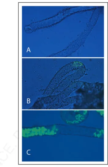
more than eight generations on a CD1 background. This transgenic line express EGFP under the control of c-kit promoter and first intron and show specific expression only in spermatogonia of the prepuberal testis.26 Figure 1 shows that EGFP expression, and thus
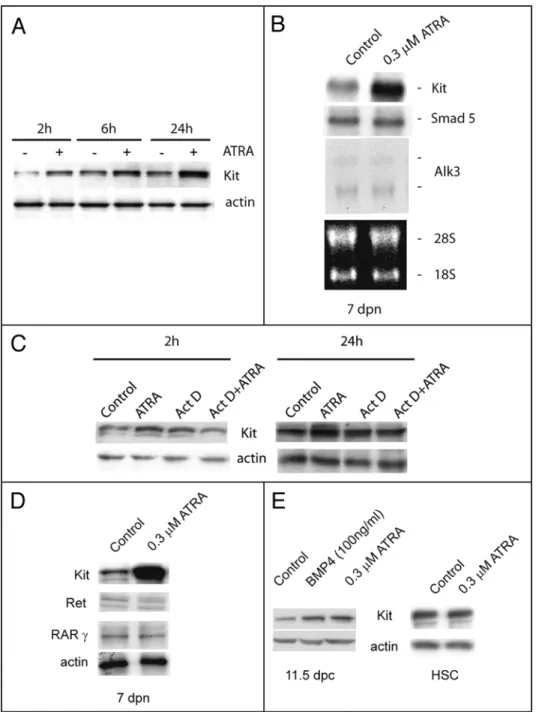
Kit, was absent at 1 dpp (Fig. 1A), appeared at 4 dpp in few small spermatogonial foci within the seminiferous tubules (Fig. 1B), and was spread at 7 dpp in discrete segments of the testicular tubules (Fig. 1C), showing the characteristic “wavy” expression pattern of mouse spermatogenesis. Having established the temporal pattern of Kit expression, we chose 7 dpp as the developmental age in which Kit was uniformly represented within all the tubules.
We isolated spermatogonia from 7 dpn animals and cultured for 24 or 48 h in the presence of increasing concentrations of ATRA. We found that at 0.3 μM concentration, ATRA has a slight negative effect on cell viability compared to the control (75 ± 3% vs 85.5 ± 6% at 24 h, and 68.8 ± 5% vs 78 ± 5% at 48 h) and no significant mitogenic effect on 7 dpn spermatogonia (data not shown). We also stimulated spermatogonia with 100 ng/ml of KL, a growth factor which stimulates proliferation and survival of spermatogonia.21,31,32
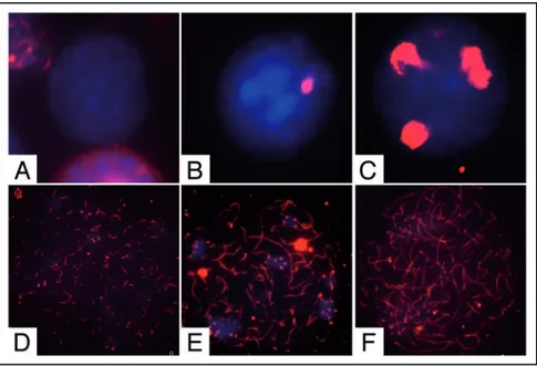
Nuclear spreads were prepared from cells at the beginning and at the end of the culture time and probed with antibodies against the synaptonemal complex protein 3 (SCP3) to detect meiotic nuclei and to evaluate the degree of meiotic progression (Figs. 2 and 3B). At the beginning of the culture we found that 4.6% showed prelep-totene morphology (Fig. 2C, in which typical SCP3 spots are found) while 1.8% of the germ cell population showed early leptotene morphology (Fig. 2D). As shown in Figure 3A and B; and Table 1, after 24 h of culture more meiotic nuclei were found in control cells and their number further increased after 48 h of culture (2.5 ± 0.12 and 7 ± 0.11 fold increase vs T0, respectively). Treatment with 0.3 μM ATRA induced a further significant increase of the percentage of meiotic nuclei after 24 and 48 h of culture (4.4 ± 0.12 and 12.8 ± 0.2
folds of increase vs T0, respectively) (Fig. 3A). We determined that this increase was essentially due to cells accumulating at leptotene stage over 48 h of culture (Fig. 3B). Similarly to ATRA, addition of KL also increases the percentage of meiotic nuclei in cultured sper-matogonia as measured by the number of nuclei in leptotene (4.5 ± 0.11 and 12.3 ± 0.6 fold of increase vs T0 after 24 and 48 h, respec-tively) (Fig. 3A and B). Simultaneous addition of both ATRA and KL did not produce any further increase of meiotic nuclei (4 ± 0.1 and 11 ± 0.8 fold of increase vs T0) either after 24 and 48 h of culture (Fig. 3A). The increased percentage of meiotic nuclei induced by ATRA or KL treatments was not associated to the increase of SCP3 protein levels, as evaluated by western blot analysis (data not shown), but rather to the typical pattern of SCP3 organization on the meiotic chromosomes (see Fig. 2). To confirm that ATRA and KL increased the number of meiotic cells in vitro, we also performed a quantitative RT-PCR using primers for Dmc1, which encodes a meiosis specific recombinase expressed during the first meiotic prophase,33 and for
the premeiotic marker Stra8 as positive control (Fig. 3C). Treatment with ATRA or KL for 24 h significantly increased Dmc1 levels (about 2 and 3.5 folds compared to the control, respectively), and as expected, ATRA caused a marked increase of Stra8 mRNA levels
Figure 1. Kit-EGFP expression in neonatal and prepuberal testis tubules. Brightfield and fluorescent merged images of testicular tubules isolated from p18 transgenic mice expressing EGFP under the control of c-kit promoter and 3.5 kb of the first kit intron. The image shows testicular tubules at 1 dpn (A), at 4 dpn (B) and at 7 dpn (C).
© 2008 LANDES BIOSCIENCE. DO NOT
DISTRIBUTE.
RA and KL regulate male meiosisincrease of Kit expression was exerted at the level of mRNA synthesis rather than of mRNA stabili-zation. Indeed, we found by western blot analysis that pre-incubation with actinomycin D, a strong inhibitor of all RNA polymerases, completely abol-ished the increase of Kit protein levels induced by ATRA (Fig. 5C). ATRA does not affect the levels of GDNF receptor Ret, a marker for undifferentiated spermatogonia,35 neither the levels of RAR
γ, the RA
receptor specifically expressed in spermatogonia10
(Fig. 5D). As in the case of postnatal spermatogonia, we found that ATRA increased Kit expression also in primordial germ cells (PGCs), harvested at 11.5 dpc, during their proliferative period,27 but not in
haematopoietic stem cells, a cell type which also express Kit19 (Fig. 5E).
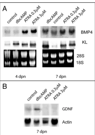
Since Sertoli cells are a known target of retinoic acid,11 we investigated whether ATRA might
regu-late in these cells the production of growth factors essential for germ cells proliferation and/or differen-tiation. Figure 6A shows that, in Sertoli cell cultures obtained from 4 dpn and 7 dpn mice, ATRA strongly upregulated the levels of KL and BPM4 mRNAs after 24 h of culture both at 0.3 and 3 μM concen-tration. As a positive control we included dbcAMP stimulation which we have previously shown is able to upregulate KL levels in Sertoli cells.21 In contrast to dbcAMP stimulation, the levels
of the stem cell growth factor GDNF were downregulated in 7 dpn cultures by ATRA treatment (Fig. 6B).
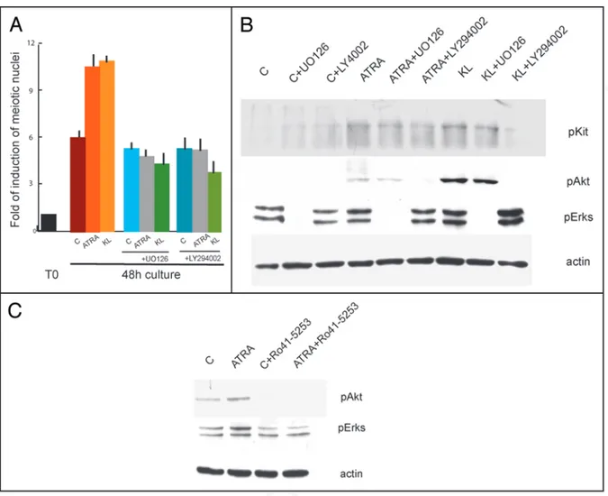
Kit-activated signal transduction pathways are required for both ATRA- and KL-induced of meiotic entry. Since Kit is induced by ATRA and is the target of KL in 7 dpn spermatogonia, we hypoth-esized that the two factors might converge on Kit signalling pathways to increase the number of meiotic cells in culture. In order to test this hypothesis, we isolated and cultured spermatogonia for 48 h in the presence or absence of 5 μM STI571, a selective inhibitor of Kit tyrosine kinase activity,36 and treated these cells with ATRA or KL.
In cells solely treated with STI571, we found the same percentage of meiotic cells as in the control cultures without the inhibitor. The drug, however, was able to completely revert the increase of meiotic nuclei induced by ATRA and KL (0.9 fold of increase compared to STI571 control sample) (Fig. 7A). Since STI571 inhibited both ATRA- and KL-mediated increase of meiotic figures, we analyzed the levels of phospho-Kit and its downstream targets phospho-Akt and phospho-Erk1/2 in spermatogonia after 15 min stimulation with ATRA and KL pretreated or not for 30 min with STI571. Kit protein levels did not change after this pulse of stimulation while, as previous reported31 we found that KL induced a significant increase of Kit,
Akt and Erk1/2 phosphorylations, completely inhibited by the pres-ence of 5 μM STI571 (Fig. 7B). Interestingly, we observed a positive effect on Kit autophosphorylation, Akt and Erk1/2 phosphorylations after only 15 min stimulation with ATRA, which were all prevented by the presence of the tyrosine kinase inhibitor. To determine which signalling pathway was preferentially activated by ATRA and KL, we cultured spermatogonia in the presence or absence of LY29406 (an inhibitor of PI3K pathways) or of U0126 (an inhibitor of the MAPK pathway). Both these inhibitors completely blocked the increase in the number of meiotic figures induced by ATRA or by KL (3.7 folds vs control). Interestingly, we found that Stra8 mRNA levels
were upregulated also by KL treatment (1.7 fold vs control), and such increase was more evident at the protein level (3.5 fold of increase with respect to control, Fig. 3D).
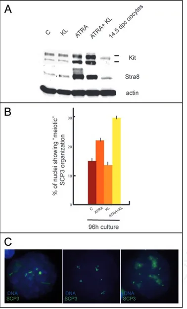
To verify whether meiotic entry could be stimulated by the two agents also in undifferentiated Kit negative spermatogonia, we treated 4 dpn spermatogonia for 48, 72 and 96 h with ATRA, KL and a combination of both. As previously described,8,30 Kit and Stra8
expression were strongly upregulated in all the culture conditions in which ATRA was present (Fig. 4A and Suppl. Fig. 1C and D), even though cell viability decreased to about 50% after 4 days of culture in all the treatments (data not shown).We did not find meiotic nuclei up to 72 h of culture in all of the conditions tested, however after 96 h of culture a significant number of cells (Fig. 4B) showed a peculiar (early leptotene-like) SCP3 staining (Fig. 4C). Such staining was different from that found in 7 dpn spermatogonia which entered meiosis in vitro (see Figs. 2D and 3B). The SCP3 pattern exhibited nuclear foci as well as short filamentous structures which were less abundant when compared to leptotene nuclei and was reminiscent of that found in ES cells differentiating into germ-cells.34 This pattern
of staining was present in control cultures (15% ± 1), its percentage increased significantly in cultures treated with ATRA (21.7% ± 1.3) but not with KL (14% ± 0.8) and it almost doubled in the presence of both ATRA and KL (30.2 % ± 0.9).
ATRA increases Kit/KL levels in isolated testicular cells. It has been reported that ATRA stimulates Kit expression in isolated spermatogonia.7,8 We confirmed these results in 4 and 7 dpn
sper-matogonia showing that Kit is induced at doses as low as 0.03 μM and that ATRA effect is not mediated by Sertoli cells (Suppl. Fig. 1). We show that Kit induction is evident as early as after 2 h of ATRA stimulation and increased linearly up to 24 h of culture (Fig. 5A) which corresponded to a significant increase at the RNA level, as shown by Northern blot analysis (Fig. 5B). ATRA-dependent
Figure 2. Pattern of SCP3 organization in male germ cells from 7 dpn mice. Representative merged immunofluorescence images showing SCP3 (red) organization on nuclear spreads (blue) in (A) undifferentiated spermatogonia, (B) differentiating spermatogonia, (C) prelep-totene spermatocytes, (D) early-lepprelep-totene spermatocytes (E) lepprelep-totene spermatocytes and (F) zygotene spermatocytes, obtained from freshly isolated or cultured spermatogonia.
© 2008 LANDES BIOSCIENCE. DO NOT
DISTRIBUTE.
RA and KL regulate male meiosiswww.landesbioscience.com Cell Cycle 3881
(Fig. 8A) and inhibited the increase in phosphorylation levels of Akt and Erk1/2 (Fig. 8B). Since ATRA was activating both PI3K and MAPK pathways, we investigated if the effect was specifically medi-ated by a RAR receptor. We incubmedi-ated spermatogonia in the presence of 20 μM Ro-41-5253, an antagonist of RARα receptor, which at this concentration is also able to block the signalling of RARγ.37 As
shown in Figure 8C, we found that pre-incubation with Ro-41-5253 prevented ATRA-mediated phosphorylation of Akt and Erk1/2.
Figure 3. ATRA and KL promote meiotic in 7 dpn differentiating spermatogonia. (A) Histogram representing the fold of increase of meiotic nuclei in control spermatogonia or in cells stimulated with ATRA, KL or ATRA and KL for 24 h or 48 h. The values were obtained as a ratio of the percentage of nuclei with meiotic SCP3 staining at the different time points with the percentage of nuclei with meiotic SCP3 staining at the beginning of the culture (see Table 1 for absolute percentage values). Bars represent SD. (B) Percentage of leptotene (light green and IF left) and lepto/zygotene (dark green and IF right) nuclei in control cultures or cells treated for 48 h with ATRA, KL or ATRA and KL. Bars represent SD. (C) qRT-PCR for Dmc1 and Stra8 in spermatogonia incubated 24 h with ATRA or KL. (D) Immunoblot analysis of Stra8 and actin expression in spermatogonia cultured for 24 h in the absence or presence of ATRA or KL.
Table 1 Percentage of leptotene and lepto/zygotene
nuclei in culture
Cont ATRA KL ATRA + KL
T0 1.8% ± 0.5 - - -
(leptotene)
24 h 4.5% ± 0.22 7.9% ± 0.25 8.1% ± 0.21 7.2% ± 0.7
© 2008 LANDES BIOSCIENCE. DO NOT
DISTRIBUTE.
RA and KL regulate male meiosispositive Intermediate and/or Type B spermatogonia) and/or that the pathways activated by the two factors might converge at some level of the pro-meiotic signalling cascade. Indeed, several evidences show that Kit positive spermatogonia are sensitive to both KL and ATRA (present results and refs. 8 and 21), which stimulate a significant increase of Stra8 (refs. 7 and 8 and present results), a fundamental regulator of both female and male meiosis.6,15,16 Importantly, ATRA
and KL similarly upregulate Dmc1, an early meiotic marker that is essential for the process of meiotic recombination.33
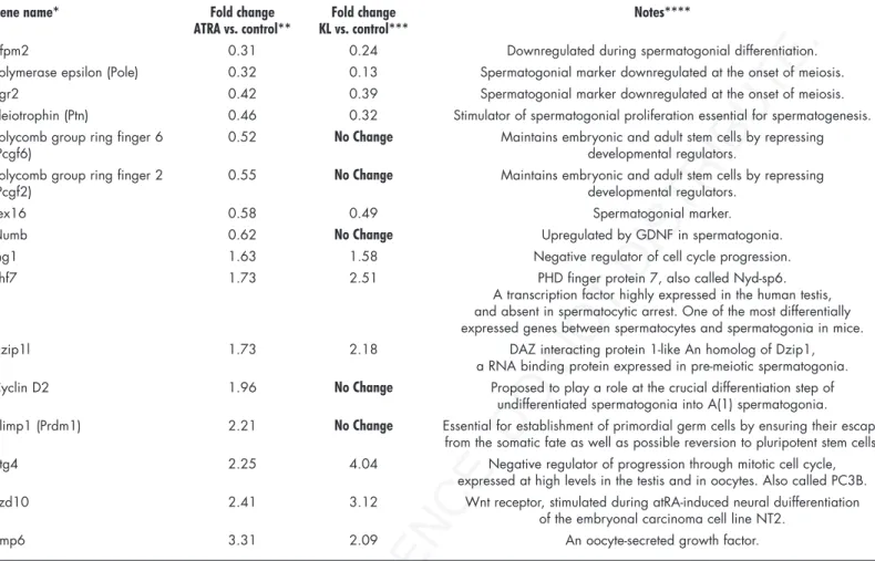
The evidence that ATRA increased the percentage of meiotic nuclei in isolated postnatal male germ cells confirms its role as a meiotic inducing substance. A microarray analysis performed on ATRA-stimulated spermatogonia (the entire set of raw and normal-ized data are available in the Array Express public repository at http:// www.ebi.ac.uk/arrayexpress, with the accession No. E-MEXP-1126) shows changes in the pattern of gene expression compatible with the ongoing spermatogonial differentiation. For instance, we found upregulation of cyclin D2, previously shown to be strongly upregu-lated in spermatogonia of the VAD testis after administration of retinoic acid,38 but also of early meiotic genes, such as Dzip1l, Phf7,
Bmp6 and Btg4 (Table 2). At the same time, ATRA downregulates the expression of stemness genes, such as Pcgf2, Pcgf6 and Numb, and of spermatogonial markers known to be turned off at the onset of meiosis, such as Zfpm2, Pole, Egr2, Ptn, Tex16 (Table 2).
The finding that KL regulates meiotic entry of spermatogonia is a novel finding and is in line with the previously reported in vivo expression pattern of KL (highly expressed in Sertoli cells when leptotene spermatocytes appear), and with the observation that male germ cells co-cultured with a KL expressing cell line can undergo transmeiotic differentiation in vitro.39 These results are also
consis-tent with the data gathered by wide genomic analysis performed with Affymetrix gene chips, which indicate that KL, similarly to ATRA, regulates spermatogonial markers and early meiotic genes.40
By comparing the effect of KL and ATRA on gene expression pattern we found that spermatogonial markers which are normally turned off at the onset of meiosis are downregulated also by KL treatment, whereas genes involved in stemness were not influenced. At the same time inhibitors of the mitotic cell cycle and early meiotic markers were all found to be equally upregulated by ATRA and by KL treat-ment40 (Table 2).
When undifferentiated Kit negative spermatogonia were stimu-lated in vitro with ATRA they underwent a partial differentiation only after 96 h, but they did not enter meiosis correctly. Although ATRA increased Kit and Stra8 levels and the percentage of SCP3-positive nuclei, these cells failed to correctly organize the synaptonemal complex and, as expected, KL did not show any effect. KL synergized with ATRA in the induction of abortive meiosis in 4 dpn sper-matogonia, probably because these cells became Kit positive during the concomitant exposure to ATRA, but they did not undergo a sufficient number of mitotic divisions necessary to become compe-tent to enter meiosis.
We also show that the action of ATRA on spermatogonia is paral-leled by a strong differentiative effect on Sertoli cells. In these cells ATRA induces expression of KL and BMP4 (known to promote proliferation and differentiation of spermatogonia29) and
down-regulation of GDNF, which is essential for stem cell renewal.35
Indeed, it is well established that RA in immature Sertoli cells is
Discussion
It has been recently proposed a model in which RA acts as a meiotic inducing substance (MIS)13 regulating, via its signalling
and metabolism, whether or not female and male germ cells initiate meiosis during embryogenesis.30 In this study we tested in vitro if
its mechanism of action might be conserved in postnatal germ cells. When spermatogonia were stimulated by ATRA, we found that the percentage of meiotic nuclei was significantly increased compared to the control. Interestingly, a similar effect was also exerted by KL, a growth factor essential for spermatogonial survival and proliferation both in vivo and in vitro.21,23,24 Simultaneous addition of ATRA and
KL did not produce any further increase in meiotic nuclei, suggesting that they might be acting on the same cell type (possibly the Kit
Figure 4. ATRA induces an abnormal SCP3 organization in cultured undif-ferentiated spermatogonia. (A) Immunoblot analysis of Kit, Stra8 and actin expression in 4 dpn spermatogonia cultured for 96 h in the absence or pres-ence of ATRA or KL. (B) Histogram representing the percentage of meiotic SCP3 organization in control spermatogonia or in cells stimulated with ATRA, KL or ATRA and KL for 96 h. (C) Representative pictures showing an abortive meiotic organization of SCP3.
© 2008 LANDES BIOSCIENCE. DO NOT
DISTRIBUTE.
RA and KL regulate male meiosiswww.landesbioscience.com Cell Cycle 3883
synthesis in differentiating spermatogonia (Pellegrini et al., unpub-lished), and because KL has been shown to stimulate DNA synthesis in type A, but not in type B spermatogonia21 which are the
presump-tive target of stimulation of meiotic entry in the present experiments. Thence, we would favour the fourth possibility, i.e., a direct effect of ATRA and KL on the induction of meiotic competence. The fact that, in vivo, Kit positive spermatogonia enter meiosis after a series of controlled Kit-dependent mitotic divisions, and the evidence that Kit requires PI3K and MAPK pathways to regulate both mitotic required to promote spermatogonia
differen-tiation during the prepubertal spermatogenic wave.11
We found that ATRA and KL share a common molecular mechanism of action in inducing meiosis, since a specific inhibitor of the Kit tyrosine kinase (STI571) was able to revert the meiotic increase induced by the two agents. Both agents activate either the PI3K or the MAPK pathways which are essential to mediate meiotic entry of sper-matogonia in vitro. We observed that ATRA not only functions as a genomic inducer of Kit synthesis, but also as a rapid non genomic agent, by inducing Kit phosphorylation and activation. Such non-genomic effect, recently described also in neuronal systems41,42 is
dependent on a RA receptor, since Kit signal-ling pathways activated by ATRA in cultured spermatogonia were prevented by the incuba-tion with a specific RAR inhibitor.
The pro-meiotic effect of ATRA and KL that we observe in vitro might be explained by at least four different possible mechanisms (i) by an increase of survival of already meiosis committed cells (pre-leptotene spermato-cytes), (ii) by an increase of survival of Kit positive spermatogonia, which then might enter meiosis in a cell-autonomous manner; (iii) by an increase of mitotic proliferation of Kit positive spermatogonia, which then might enter meiosis in a cell-autonomous manner; or (iv) by the induction of meiotic competence in Kit positive spermatogonia. We can rule out the first possibility, since we estimated that the germ cell cultures obtained from 7 dpn mice contain only 4.6% of preleptotene spermatocytes, which cannot account for the increase of lepto-tene nuclei observed after 48 h of ATRA or KL stimulation (23%). Moreover, it has been demonstrated that preleptotene sper-matocytes do not express RARs10 and we
found no preleptotene figures in Kit positive immunomagnetic sorted spermatogonia (our unpublished observation). The second possi-bility can also be ruled out by the observation that, while addition of either LY294002 or
U0126 completely abolishes meiotic differentiation, these inhibitors are not able to suppress KL anti-apoptotic effects on cultured sper-matogonia.31 Moreover, we have previously shown that the induction
of an early meiotic gene expression pattern is evident after only 4 hr of KL stimulation, a period of time at which spermatogonial survival is not affected.40 Finally, we observe that ATRA-mediated increase of
meiotic nuclei requires the activation of Kit-dependent pathways, in the absence of any pro-survival effect of ATRA. The third possibility also seems to be unlikely, because ATRA does not stimulate DNA
Figure 5. ATRA increases Kit levels in germ cells during development. (A) Time course curve of ATRA stimulated spermatogonia on Kit protein levels after ATRA treatment. Actin is shown as loading control. (B) Northern blot analysis of Kit mRNA levels in isolated germ cells cultured for 24 h with or without
ATRA stimulation. Smad5 and Alk3 were included as specific markers for spermatogonial population.29
28S and 18S are shown as loading and integrity controls. (C) Kit immunoblotting on spermatogonia treated with Actinomycin D and/or ATRA for 2 h or 24 h. Actin is shown as loading control. (D) Kit, Ret
and RARγ protein levels after 24 h of ATRA stimulation in 7 dpn spermatogonia. (E) Kit levels in PGCs
© 2008 LANDES BIOSCIENCE. DO NOT
DISTRIBUTE.
RA and KL regulate male meiosisDNA synthesis31 and meiotic entry of spermatogonia (this paper),
suggest that a link exists between Kit-mediated proliferation and the switch into the meiotic cell cycle. KL induces a gene expression pattern in differentiating spermatogonia40 that is consistent with a
progressive lengthening of the S phase and a progressive shortening of the G2/M transition, both events known to occur in vivo in male germ cells during the switch from mitosis to meiosis.43 Presumably,
during these Kit-mediated mitotic divisions, accumulation of key factors required for meiosis occur. One of these factors is certainly Stra8, which has been proposed to be essential for the last round of pre-meiotic DNA synthesis in female fetal germ cells,6 and to
regu-late the switch from a mitotic pattern of cell division to the meiotic pattern in postnatal male germ cells.15,16
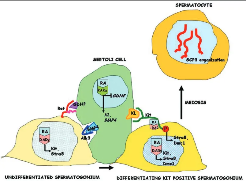
In conclusion, we propose a model (Fig. 9) in which RA acts on the somatic cell compartment of the testis, through the induction of KL and on the germ cell compartment, as a direct pro-meiotic factor sharing with KL the same signalling pathway.
Materials and Methods
Cell isolation and culture. Spermatogonia were obtained as we previously reported21 by differential enzymatic digestion of testes from
4 to 7 days post natum (dpn) CD1 albino mice. To precisely define the temporal appearance of Kit expressing spermatogonia, a transgenic line (p18) expressing EGFP under the control of the c-Kit promoter and expanded on a CD1 background,25,26 was used. Isolation of Kit
positive spermatogonia was performed by using magnetic-activated cell sorting (MACS) with CD117 conjugated microbeads (Miltenyi Biotec, Germany). Isolated germ cells were then treated with different factors and cultured for 24 h or 48 h prior to immunofluorescence, western blotting analysis or mRNA preparation. Fresh medium and growth factors were replaced after 24 h when culturing for longer times. PGCs were isolated from 11.5 days post coitum (dpc) embryos as we previously reported in ref.27 Spermatogonia were cultured in
modified Earle medium with 100 U/ml penicillin, 100 μg/ml strep-tomycin, 20 mM glutamine without serum supplementation, while
PGCs were cultured in spermatogonia medium supplemented with 10% FCS. KL-dependent SV40T-immortalized hematopoietic cells from bone marrow were cultured in RPMI supplemented with 10% FCS and 10 ng/ml KL. Sertoli cell cultures were prepared according to reference 21.
All-trans-retinoic acid (ATRA, Sigma) was dissolved in ethanol and used in a range of 0.03–3 μM. KL and BMP4 were purchased from Società Italiana Chimici (Rome, Italy) and used at 100 ng/ml. STI571, a generous gift of Dr. L. Gnessi (University of Rome “La Sapienza”), was used at 5 μM concentration and Ro 41-5253 (Biomol, DBA Italy) at 7 and 20 μM. DibutyrilcyclicAMP (Sigma) was used at 1 mM concentration. The MAPK inhibitor U0126 (Promega, Italy) and PI3K inhibitor LY294002 (Alexis, Italy) were used at 10 μM concentration.
Figure 6. ATRA regulates in Sertoli cells the expression of specific growth factors required for germ cells differentiation. Northern blot analysis (A) for BMP4 and KL, and (B) for GDNF expression in Sertoli cells. SC cultures were obtained from 4 dpn and 7 dpn testes and stimulated o/n with dibutyril-cAMP (dbdibutyril-cAMP, 1 mM), with 0.3 μM or 3 μM ATRA. 28S and 18S or actin were shown as loading and integrity controls.
Figure 7. Inhibition of Kit signalling by STI571 completely reverts meiotic progression induced by ATRA and KL. (A) Histogram representing the folds of induction of meiotic nuclei, evaluated as above, in control spermatogonia or in cells stimulated with ATRA, KL or ATRA and KL for 24 h or 48 h in the presence or absence of 5 μM STI571. (B) Immunoblot analysis of Kit activated pathways in control spermatogonia or in cells stimulated for 15 minutes with ATRA or KL, pretreated for 30 minutes with 5 μM STI571.
© 2008 LANDES BIOSCIENCE. DO NOT
DISTRIBUTE.
RA and KL regulate male meiosiswww.landesbioscience.com Cell Cycle 3885
nitrocellulose membrane (Amersham). The membrane was blocked in PBS-5% skim milk powder for 1 h. Incubation of the membrane with the primary antibody was carried out at 4°C o/n in PBS-5% BSA and then with the appropriate horseradish peroxidase-conju-gated secondary antibody (SantaCruz). Anti-Kit rabbit polyclonal (sc-6283), anti phospho-Kit (Tyr 721) (sc-23766) rabbit polyclonal, phospho Erk1/2 mouse monoclonal antibody (sc-7383), anti-actin rabbit polyclonal (sc-7210), anti-Ret rabbit polyclonal (sc-167), anti-RARγ mouse monoclonal (sc-7387) were from SantaCruz, anti-phospho Akt (Ser-473) was from New England Biolabs, anti Stra-8 antibodies were from Abcam. The horseradish peroxidase conjugate was detected by chemioluminescence with an ECL Kit (Amersham) and autofluorography. For each experiment western blotting analysis were repeated at least four times and densitometry was performed using a Molecular Dynamics Densitometer and ImageQuant soft-ware.
Northern blotting and quantitative RT PCR analyses. Spermatogonia were collected into Trizol (Invitrogen) and RNA was purified according to manufacturers suggestions. For Northern blot analysis, total RNA was run in a 1% formaldehyde-agarose gel and blotted onto a Nylon membrane (Hybond-N, Amersham, Viability of 4 and 7 dpn spermatogonia was evaluated by Trypan
blue (Sigma) exclusion.
Immunofluorescence and western blotting. For cell spreads, spermatogonia cultured for 24, 48 or 96 h were prepared and stained essentially as described in ref.28 Slides were washed twice in PBS,
and incubated o/n at 4°C with SCP3 rabbit polyclonal anti-body (Abcam, Cambridge, UK) diluted in blocking solution (10% goat serum, 3% BSA, 0.5% Triton X-100 in PBS). After washing, secondary antibody was added for 1 h at 37°C. The slides were washed and allowed to dry. Vectashield Mounting Medium with DAPI (Vector Laboratories, Burlingame, CA, USA) was added and the slides were viewed using a Leica microscope. Spreads analysis was performed in five independent experiments. A representative morphological classification of SCP3 organization in various types of germ cells obtained in vitro is reported in Figure 2. For immu-nofluorescence, spermatogonia cell suspensions were adhered onto poly-L-lysine coated slides and treated as previously described.29
For western analysis cells were lysed in 1% Triton X-100, 150 mM NaCl, 15 mM MgCl2, 15 mM EGTA, 10% Glycerol, 50 mM Hepes (pH 7.4) with protease inhibitors. Proteins were separated by SDS-10% polyacrylamide gel electrophoresis and transferred to
Figure 8. MAPK and PI3K signalling pathways activated by ATRA and KL are required for meiotic entry of spermatogonia. (A) Histogram representing the folds of induction of meiotic nuclei, evaluated as above, in control spermatogonia or in cells stimulated with ATRA and KL for 48 h in the presence or absence of UO126 or LY29406. (B) Western blot analysis of Kit activated pathways in control spermatogonia or in cells stimulated for 15 minutes with ATRA or KL, pretreated with 10 μM UO126 or 10 μM LY29406. (C) Western blot analysis of Kit activated pathways in control spermatogonia or in cells stimulated for 15 minutes with ATRA, pre-treated for 30 min with 20 μM Ro 41-5253.