ORIGINAL RESEARCH
Longitudinal Evaluation of Immune Reconstitution and B-cell
Function After Hematopoietic Cell Transplantation for Primary
Immunodeficiency
Alessia Scarselli1&Silvia Di Cesare1&Claudia Capponi2&Simona Cascioli3&
Maria L. Romiti1,6&Gigliola Di Matteo1&Alessandra Simonetti1&Paolo Palma1&
Andrea Finocchi1&Barbarella Lucarelli4&Rita M. Pinto4&Ippolita Rana4,7&
Giuseppe Palumbo1,4&Maurizio Caniglia4,8&Paolo Rossi1&Rita Carsetti2,3&
Caterina Cancrini1&Alessandro Aiuti1,5
Received: 5 December 2014 / Accepted: 16 March 2015 / Published online: 15 April 2015 # Springer Science+Business Media New York 2015
Abstract
Purpose Hematopoietic cell transplantation (HCT) pro-vides a curative therapy for severe forms of primary im-munodeficiencies (PID). While the timing and extent of T-cell reconstitution following transplant for PID has been studied in depth, less is known about the kinetics of B-cell development and long-term restoration of humoral func-tions, which been often reported to be suboptimal after HCT.
Methods We studied longitudinally B-cell development and function in a cohort of 13 PID patients transplanted between 1997 and 2010, with a follow-up ranging from 0.7 to 15 years. Flow cytometric analysis of naïve and antigen-experienced B-cell subsets and in vitro functional responses to CpG were compared with data from healthy children and correlated with the degree of B-cell chimerism and in vivo antibody production.
Results We found that total memory B-cells count remained below normal levels for the first 2 years of follow up and progressively normalized. Switched memory B-cells (CD19+ CD27+IgD-IgM-) were restored early and better than IgM memory B-cells (CD19+CD27+IgD+IgM+), which remained significantly reduced long-term. The recovery of memory B-cells correlated with good in vivo humoral function and nor-malization of CpG-response. A complete B-cell reconstitution was usually associated with donor B-cells chimerism and pre-transplant conditioning. Donor source and the underlying ge-netic defect represented also important variables.
Conclusion Monitoring of phenotypic and functional changes on B-cells following HCT may prove clinically relevant to tailor
patients’ care. In particular the analysis of IgM memory and
switched memory B-cells in addition to in vitro B-cells stimu-lation are recommended before Ig replacement therapy (IgRT) discontinuation.
Rita Carsetti, Caterina Cancrini and Alessandro Aiuti contributed equally to this manuscript. * Caterina Cancrini [email protected] * Alessandro Aiuti [email protected] 1
University Department of Pediatrics, DPUO, Unit of Immune and Infectious Diseases, Bambino Gesù Children’s Hospital, Rome, Italy
2
Department of Laboratories, Children Hospital Bambino Gesù, Rome, Italy
3 Immunology and Pharmacotherapy Area, Bambino Gesù Children’s
Hospital, Rome, Italy
4 Department of Pediatric Hematology and Oncology Bambino Gesù
Children’s Hospital, Rome, Italy
5 San Raffaele Telethon Institute for Gene Therapy (HSR-TIGET),
Milan, Italy
6 Servizio Prevenzione e Protezione, University of Rome Tor Vergata,
Rome, Italy
7 General Pediatrics and Infectious Diseases, Bambino Gesù
Children’s Hospital, Rome, Italy
8 Pediatric Hematology and Oncology, S. Maria della Misericordia,
Keywords
Primary immunodeficiency . hematopoietic stem cell transplantation . immune reconstitution .B-Lymphocytes . conditioning . follow up studies . child
Introduction
Hematopoietic cell transplantation (HCT) provides a curative therapy for primary immunodeficiencies (PID), a heteroge-neous group of inherited disorders affecting the immune sys-tem and leading to recurrent severe infections and
predisposi-tion to autoimmunity and cancer [1–3]. The outcome of HCT
has improved considerably due to better supportive care, graft versus host disease (GVHD) prevention and use of the re-duced intensity conditioning regimen and novel strategies
for stem cell manipulation and purification [4–8]. While the
timing and extent of T-cell reconstitution has been studied in
depth [9–14], less is known about B-cell development and
functions after transplant for PID. Previous studies suggested that B-cell recovery can be slower than T-cell reconstitution and often suboptimal, and in these patients immunoglobulin
replacement therapy (IgRT) is still required [15–18]. It has
been suggested that delayed or partial humoral reconstitution is often associated to the persistence of autologous defective B
lymphocyte [19–21]. The use of myeloablative conditioning
facilitates establishment of donor B lymphocyte chimerism
[22–24], but this benefit may be outweighed by the increased
toxicity and some patients may require IgRT even after
pre-transplantation conditioning [25,26]. Moreover, humoral
re-constitution is also influenced by the molecular type of PID, degree of HLA compatibility, source of grafts, and
post-transplant course (e.g. infections and GVHD) [25,27–29].
Here, we studied long-term B-cell reconstitution in a small cohort of PID patients who received different sources of HCT. We found that the kinetic of recovery of naïve and antigen-experienced B cells was variable and mainly influenced by the degree of donor chimerism, type of donor source and the underlying disease. Our data suggest that monitoring of phe-notypic and functional changes on B-cells following HCT may allow to identify early predictors of efficient humoral function.
Methods
Patients’ Characteristics and Data Collection
We collected data on immune reconstitution and clinical status after HCT of 16 patients who consecutively underwent HCT at
Bambino Gesù Children’s Hospital (OPBG) in Rome between
2002 and 2010 and one patient who was transplanted in 1997 at the Necker-Enfants Malades Hospital and followed at OPBG. Data on immune reconstitution and clinical status
after HCT were collected until the end of 2012. PID was diagnosed based on clinical findings, immune phenotype and function, and confirmed by genetic analysis. In 2 patients no mutations were identified. In vivo T cell depletion has been
used in 12 procedures (all but one mismatched related donor–
MMRD-, all matched unrelated donor- MUD- and two
matched sibling donor –MSD- transplants) by
anti-thymocyte globulin or Alemtuzumab or anti-CD2 anti-leuko-cyte function atigen-1. In addition, Pt 11 received a donor graft enriched for CD34+ cells. Patients underwent HCT from dif-ferent donor sources (BM, n=14, CB, n=3), with (n=11) or without (n =6) conditioning treatment. The median age at transplantation was 11 months (range 5–144 months), pa-tients’ follow-up ranged from 0,7 to 15 years, with an overall survival of 77 %. Two SCID patients (Pt 2, 3) died early post HCT due to disseminated infections present at time of HCT, a patient affected by Wiskott-Aldrich syndrome (WAS) (Pt 13) died 9 months after HCT due to a EBV-positive diffuse large B-cell lymphoma, whereas one patient with Familial Haemophagocytic Lymphohistiocytosis (FHLH) (Pt 15) died 6 months after the rejection of the third HCT transplant
(Table1). Patients’ family signed informed consent for
immu-nological studies under a research protocol approved by Ethic Committee of OPBG.
Chimerism and T Cell Function
Whole blood and sorted peripheral cell subsets (CD3+, CD19+, CD14+, CD16+56+) were studied for chimerism at 1, 3, and 6 months after HCT and then yearly. Chimerism was analyzed by fluorescence in situ hybridization for donor–re-cipient sex mismatches or by polymerase chain reaction am-plification of short tandem repeats in case of donor-recipient sex-matching. Immunological reconstitution of T, B and NK populations were monitored by flow cytometry on peripheral blood mononuclear cells (PBMCs) according to standard pro-tocols with a FACS CANTO II flow cytometer and monoclo-nal murine antibodies conjugated to FITC or PE specific for CD3, CD4, CD8, CD16, CD19, CD45RO, and CD45RA (30). Pt17 was analyzed at 0.7 years before he returned to his country, and data were grouped with the 1 year follow up. The function of T lymphocytes was assessed by lympho-cyte proliferation upon mitogens, T-cell repertoire and thymic
activity (TREC) as previously described [30,31].
B Cell Function
Reconstitution of B-cell compartment was analyzed through multiple approaches including the analysis of naïve and antigen-experienced B-cell subsets (anti-CD27, anti-IgD and anti- IgM were used to identify different stages of B-cell mat-uration), the determination of immunoglobulin isotype, the IgG subclasses and the ability to generate antibodies to protein
Ta b le 1 C h ar act er isti cs of pa tie nts and HCT Pt Gend er Diagnosis Age at HC T Con d itio ning Regi me n G vHD p rophylax is Don o r type Source Cl inic al ev ents Follow u p 1M S C ID γ -chain 7 m onths nd CsA/Methyl-pred/A T G M MR D (1AgMM) BM warts 7 years post H CT 10 year 2M S C ID γ -chain 7 m onths nd CsA/Methyl-pred/A T G M MRD (1A gMM) BM CMV encephalitis Died at 1 7 days post H CT 3M S C ID γ -chain 6 m onths nd nd MMRD BM fungal sepsis D ied at 5 weeks post H C T 4 M SCID Jak3 13 month s BU/Cy C sA/Methyl-p red/A T G M MRD B M G VHD III gut; V OD 8 y ears 5 F SCID Jak3 7 m onths nd nd MS D B M 8 years 6 M ADA-S C ID 5 m onths nd nd MS D B M H HV -6 viremia 6 years 7 F SCID T -B+ 1 1 months nd nd MS D B M BCG reactivation, immu ne re constit ution in fla mmat ory sy ndrome 1.2 y ears 12 month s nd nd MS D B M 8 F OS (RAG1) 7 m onths BU/FLU/ T hiotepa C sA/M ethyl-pred/A T G M UD UCB G VHD III skin 3 y ears 9 F OS (RAG1) 8 m onths T reo/FLU/Thiotepa C sA/My coph/A T G M UD BM GVHD II skin 2 y ears 10 M O S (RAG2) 7 months Thiotepa/T reo /FLU C sA/M ethyl-pred/A T G M UD UCB G VHD III/IV skin and gut; cGVHD, haemolytic syndrome; PC J p n eumonia 2 y ear s 11 M E D A -I D (I k B α ) 1 2 m onth s BU/Cy A nti C D2 ant i-L F A 1 MMRD BM seizures ; P CJ pneumonia; chronic broncho-pneu m opathy 15 years 12 F D CML d eficiency 8 years T re o/ FLU/Thio tepa CsA/My coph/A T G M UD BM Gv HD I skin 3 years 13 M W AS 3 y ea rs T h iote pa/B U/ FL U CsA/My coph/P DN/A T G M UD UCB E BV -positive d if fus e lar g e B-cell lymphoma Died at 9 m onths after H CT 14 M X L P 7 y ea rs BU /Cy C sA /MT X / Methylpred/ AT G M U D B M G VH D I ski n ; E B V re ac ti va ti on controlled b y R ituximab 3 y ear s 15 M F HLH (M UN C 1 3– 4) 12 y ears F LU/Melphalan/ Al em tu zum ab CsA /Me thyl -pr ed M S D BM he mol y tic an emia; H CT re jec tion D ied 6 months af te r the 3 rd H CT 12 y ear s T hiote p a/T reo /FL U CsA /MT X M S D BM 13 y ear s n d A TG MS D B M 16 M C GD 4 y ears BU/Cy C sA MS D B M m ild hemorrhagic cys titis 7 y ears 17 M C GD 6 y ears Thiotepa/T reo / FLU C sA/M TX MS D B M P se udomonas A . sepsis 0 .7 years nd not done, SCID Sev ere C o mbined Immunodeficiency , ADA Adenos ine d eaminase, OS Omenn S yndrome, ED A-ID anhidrotic ectodermal d y spla si a w ith immunodeficiency , DC ML D C ,m o n o cy te ,B and N K lympho id cells, WA S W isko tt-Aldrich syndro m e; XL P X-linked lymphoprolif erative d isease, FH LH Familial h emophagocytic ly mphohistiocytosis; CG D Chronic g ranulomatous disease, BU Bus u lphan, Cy Cyclophosphamide, Fl u F ludarabine, Tr eo Tr eo su lf an ,M ycoph My cophenolate m ofetil, anti-L F A1 anti-leukocyte function atigen-1; PD N prednis one; Methylpr ed methy lprednisolone; MTX Methotrexate, 1AgM M = 1 -a nti g en mismatc h ,MMRD mismatched related donor; MSD Matched S ibling Donor ,MU D Match ed U nrelated Donor; BM bone marrow; UCB umbilical cord blood; VO D veno-occlus ive d isease, PC J pneumocystis jiroveci; GV H D g raf t v er sus host d is ea se, sdr syn d rome
and polysaccharide antigens after tetanus toxoid, hepatitis B, Haemophilus influenzae and pneumococcal immunization. B-cell subsets references for controls were obtained from 81 healthy children at different age being referred to our hospital for elective procedures (14 children of 1 year, 7 of 2 years, 14 between 3 and 5 years, 46 between 6 and 15 years). We cal-culated the 5° and 95° percentile for each group of age and compared them to the values of patients according to the years following HCT. The ability of B-cells to differentiate, prolif-erate and produce immunoglobulins was tested by stimulation with bacterial DNA (CpG). Briefly, PBMC obtained through ficoll from peripheral blood of patients, were labeled at the
f i n a l c o n c e n t r a t i o n o f 0 . 1 μg/ml CMFDA (
5-chloromethylfluorescein diacetate, CellTracker; Molecular
Probes) and cultured at 5×10⁵cells per well in 96-well plates
in complete RPMI 1640 (InvivoGen) supplemented with 1 0 % FB S ( H y C l o ne L a b o r at o r i e s ) . Hu m a n C pG oligodeoxynucleotides (Hycult Biotechnology) was used at
the optimal concentration of 2.5μg/ml. Cell proliferation
and differentiation into plasma cells, were measured on day 7 by FACSCanto II flow cytometer (BD Biosciences).
Secret-ed Igs were detectSecret-ed at by ELISA [32].
Results
Patients’ Treatment and Donor Cell Engraftment
We studied longitudinally the immune reconstitution in 13 surviving PID patients in a cohort of 17 patients who received allogeneic HCT. Among them 6 patients had long-term fol-low-up (>5 years). Characteristics of patients and details about
transplants are summarized in Table1. Conditioning regimens
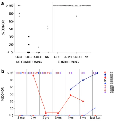
varied based on patient’s underlying disease and clinical con-dition at transplantation. Successful hematopoietic engraft-ment was observed in all 13 survivors with a median time of 17 days (range 12–29 days) for neutrophils (ANC>500/ul) and 20 days (range 12–55 days) for platelets (>50,000/ul). Four patients developed grade II-III acute Graft versus Host Disease (GvHD), and one experienced chronic GvHD. In un-conditioned SCID patients, nearly full donor chimerism was detected in the T-cell lineage whereas B-cell and monocytes remained mostly or totally of recipient origin. Specifically, 2 out of 4 non-conditioned patients had an absent donor B,
monocyte and NK-cell engraftment (Fig.1a). In contrast, all
patients who received cytoreductive conditioning regimens achieved stable full donor chimerism in all lineages, except one patient (Pt 8) with mixed chimerism on CD14+ cells. Three patients showed substantial fluctuations in donor B-cell contribution over time and in two of them an increase of donor B-cells occurred very late (>4 years) during follow up
(Fig. 1b). Interestingly, Pt5 (JAK-3 deficiency) and Pt6
(ADA-SCID) who received unconditioned transplants from
siblings, showed normal humoral function despite the preva-lence of host B-cells. When switched memory B-cells were sorted from these patients, molecular analyses of chimerism revealed that the majority of them were of donor origin (60 and 95 %, respectively, data not shown).
Effective Long-Term T-Cell Reconstitution
T-cell immune reconstitution was evaluated at yearly follow up in 13 surviving patients. Complete T-cell count recovery was obtained in all except two patients who experienced se-vere clinical complications (GvHD, immune reconstitution syndrome associated to BCG). One year after HCT total CD3+, CD4+ and CD8+ cell counts were normal in the ma-jority of patients. NK cell count was normal in all patient
except one affected byγ-chain SCID and treated with HCT
from unconditioned mismatched related donor. Evaluation of thymic function revealed normal CD4+CD45RA+ naïve T-cell counts and T-T-cell receptor excision circle (TREC) copies, in a high proportion of patients starting the first year after
transplantation (Table2) [33]. Of note, patients with reduced
TREC values had received unconditioned transplants (data not shown). T-cell diversity analysis by T-cell repertoire spectratyping showed a polyclonal distribution in the CD4+ compartment and to a lower extent in CD8+ T-cells 12 months after transplantation, which was maintained or improved dur-ing follow up. T-cell proliferation in responses to both OKT3 and PHA achieved normal levels in 54 % of patients within 1 year after HCT. In 5 out of 6 long term-survivors (follow up >5 years) T-cell reconstitution was sustained over time. In Pt1 a decline in T cell count and functionality with skewed TCR profile, low response to PHA, undetectable TRECs, and ap-pearance of face warts was observed 7 years after HCT.
B-Cell Development After HCT
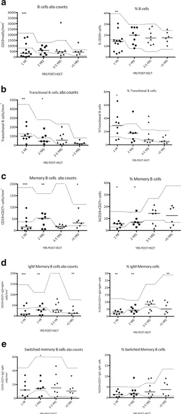
One year after HCT proportions and absolute B-cell counts were significantly reduced as compared to age matched
con-trols (Fig.2a); they reached normal value for age after 2 years,
remaining in the normal range long-term (>5 years). B-cell subsets were examined longitudinally in depth by multiparam-eter flow cytometry in 11 evaluable patients after transplant. We found a significant increase in the proportion of transition-al B-cells (CD19+CD38++IgM++) at 1 year follow up, which
normalized over time (Fig.2b). Total memory B-cells (CD19+
CD27+) were significantly below normal at 1 and 2 years and
increased starting 3–5 years of follow up (Fig.2c).
Remark-ably, switched memory B-cells (CD19+CD27+IgD-IgM-) were restored earlier and better then IgM memory B-cells (CD19+CD27+IgD+IgM+), which remained significantly
re-duced in the long-term cohort (Fig. 2d, e). B-cell absolute
counts and percentage did not differ between MSD and MMRD transplant in long-term surviving patients, but the
latter group displayed a reduced frequency of memory B-cells (data not shown).
In Vivo Antibody Production
The majority of patients discontinued IgRT between the first and second year after HCT and maintained normal values of
IgG, IgA, IgM and IgG subclasses during the follow up [34]
(Fig.3). Four patients required long-term IgRT substitution
ther-apy. Pt1 (SCIDγ-chain deficiency) did not attain donor B-cell
engraftment whereas Pt11 (EDA-ID patient) was maintained on IgRT in spite of B-cell engraftment due to very low antibody response for S. pneumoniae antigens. Both patients showed very low frequency of memory B-cells and almost absent Fig. 1 a Donor chimerism in
different lineages Percentage of donor cells in peripheral blood CD3+,CD19+, CD14+ and NK analyzed by in situ hybridization or STR analysis in unconditioned and conditioned patients at latest follow up. b Percentage of donor cells in peripheral blood CD19+ during the follow up. Donor chimerism was >95 % in patients 8,9,10,11,12,14,16, <5 % in Patients 1, 7. Patients 4,5,6 had intermediate chimerism and changed over time. Chimerism on sorted cells were not performed in Pt 17, chimerism on PBMC was 100 % donor
Table 2 Proportion of
transplanted patients with normal immune parameters after transplant 1 year post HCT % n 2 years post HCT % n Long-term follow-up (>5 years) % n Normal CD3+ 76 % (10/13) 80 % (8/10) 100 % (6/6) Normal CD4+ 76 % (10/13) 80 % (8/10) 83 % (5/6) Normal CD8+ 76 % (10/13) 80 % (8/10) 100 % (6/6) Normal CD4+CD45RA+ 69 % (9/13) 80 % (8/10) 60 % (4/6) Normal NK cell 92 % (12/13) 90 % (9/10) 83 % (5/6) Normal T-cell function 54 % (6/11) 66 % (6/9) 83 % (5/6) Polyclonal TCRVβ CD4+ Polyclonal TCRVβ CD8+ 72 % (8/11) 54 % (6/11) 83 % (5/6) 83 % (5/6) 83 % (5/6) 66 % (4/6) Normal CD3+ cell count at different years post HCT: 1 year>1000/mcl, 2 years>1400/mcl, >5 years>1000/mcl; Normal CD4+ cell count at different years post HCT: 1 year>500/mcl, 2 years>650/mcl, >5 years>450/mcl; Normal CD8+ cell count at different years post HCT: 1 year>300/mcl, 2 years>400/mcl, >5 y>300/mcl; Normal CD4+CD45RA+cell count>200/mcl; Normal NK cell count >50 /mcl.Normal reference values according to (33). Normal T cell function measured by proliferative response to mitogens (PHA>30.000 cpm and OKT3> 20.000 cpm, according to internal reference range)
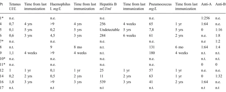
switched B-cells for several years after HCT. Two other patients (Pt 7 and Pt 10) experienced severe clinical complications and required IgRT longer than 2 years after HCT. The nine patients in which IgRT was discontinued received vaccinations accord-ing to European Blood Marrow Transplantation (EBMT)
rec-ommendations [35]. Protective antibody titers developed
fol-lowing vaccination in almost all tested patients. One patient (Pt 4) required an additional hepatitis B booster three years after initial doses, one patient (Pt 14) required a fourth dose of con-jugate pneumococcal vaccine and one patient (Pt 5) showed low titer anti-hepatitis B and anti-Pneumococcus 5 years after the last vaccinations. Isohemagglutinins appropriate for the blood group were found at significant titers in 7 patients evaluated
beyond 1 year post HCT (Table3).
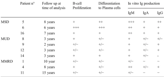
In Vitro B-cell Functions After HCT
To better study restoration of B-cell functions in transplanted patients we studied B-cell proliferation, mat-uration and Ig production in vitro. B cells isolated from peripheral blood were stimulated with human CpG oligodeoxynucleotides (unmethylated bacterial DNA), an agonist of TLR9 expressed in human on plasmocitoid dendritic and cells and at high levels on memory
B-cells [32, 36]. Under these conditions memory B-cells
proliferate and differentiate to immunoglobulin–secreting cells in response to CpG through T-independent signal-ling. All 3 patients who underwent transplantation from a MSD showed an optimal and complete B-cell response. In the MUD and MMRD cohort, B-cell proliferation, dif-ferentiation and antibody production were present in most patients but overall reduced as compared to MSD patients. In particular, 2 MMRD still requiring IgRT, failed to
pro-duce IgG and IgA in response to CpG (Table 4).
Discussion
HCT is an established curative treatment for the most severe PID variants. However, reduced or poor humoral immune function can persist in a significant proportion of patients re-quiring lifelong IgRT and leading to a higher frequency of clinical complications such as infections, autoimmunity and
chronic-GVHD [33, 37, 38]. In our cohort of long-term
surving patients, the majority of patients were clinically well and did not experience severe late HCT-related complications.
In patients with long-term follow up (6–15 years) T-cell
func-tion was retained in all but oneγ-chain patient who received a
non-conditioned mismatched related HCT. In this case we observed a gradual decline of T-cell counts and functionality. Four patients experienced main clinical events including chronic GvHD requiring immunosuppressive therapy, chronic bronchopneumopathy, BCG reactivation, and warts. Of
a
b
c
d
e
Fig. 2 a–e B-cell subsets at different time points after HCT Lines repre-sent median values. B-cell are defined as CD19+ (A), transitional as CD19+38++24++ (b), memory B-cell as CD19+27+ (c), IgM memory as CD19+27+IgD+IgM+ (d), switched B-cell as CD19+27+IgD-IgM-(E). Relative numbers in percentage (right) and absolute counts in cells/ mm3(left). Significant differences between median values at different
time points after HCT are indicated by asterisks: * p < 0,01, ** p<0,001,***s <0,0001. Dotted lines represent 5° percentile and 95° per-centile of healthy controls as specified in the methods
notice, all these patients required IgRT supplementation and 3 of them displayed low T-cell numbers and functions, indicating that long-term complications were associated with suboptimal immune reconstitution. We found that a good humoral function was usually associated with the presence of donor B-cell chi-merism and promoted by myeloablative conditioning regimen. Indeed, all patients who received conditioning (n=9) showed a
full donor B-cell chimerism and the majority (n=7; 78 %) discontinued IgRT 1–2 years after HCT maintaining protective
levels of serum Igs and specific antibody titers (Table5).
Our data indicate that the development of the B-cell pool is a slow process after HCT for PID and that quantitative defects may persist in the long-term. The relative expansion of the transitional B-cell compartment in the first year after HCT is
l (9/9) (7/7) (6/6) (9/9) (7/7) (6/6) (8/9) (6/7) (5/6) (8/8) (6/6) (5/5) (5/5) (6/6) (8/8) m g /dl m g /dl m g /dl mg/ d l mg /d l
Fig. 3 Serum Ig level in transplanted patients Levels of IgA, IgM, IgG, IgG1, IgG2 are reported at 1 year, 2 years and at latest follow up (>2 years) after IgRT discontinuation. Subjects with normal values according to age are also indicated between brackets
Table 3 Antibody titers to vaccination and isohemagglutinins at last follow up Pt Tetanus
UI/L
Time from last immunization
Haemophilus I. mg/L
Time from last immunization
Hepatitis B mUI/ml
Time from last immunization
Pneumococcus mg/L
Time from last immunization
Anti-A Anti-B
1* n.e. n.e. n.e. n.e. 1:256 n.e.
4 0,7 4 yrs >9 4 yrs 256 4 weeks 65 1 yr 1:64 n.e.
5 0,1 5 yrs 0,2 5 yrs Undetectable 5 yrs 7,8 5 yrs 0 1:16
6 0,6 3 yrs 4,5 3 yrs 284 6 weeks 61 2 yrs n.e. 1:8
7* n.e. n.e. n.e. n.e. n.e 1:2
8 n.t. 9 8 mo n.t. 131 6 mo 1:64 1:4
9 1,1 4 weeks >9 4 weeks n.t. 180 4 weeks n.t. n.t.
10* n.e. n.e. n.e. n.e. n.t. n.t.
11* n.e. n.e. n.e. n.e. 0 0
12 1 1 yr 0,1 1 yr 25 1 yr 57 1 yr n.e. n.e.
14 0,2 2 yrs 0,5 2 yrs 11 2 yrs 63 1 yr 0 1:32
16 1,8 3 yrs >9 3 yrs 539 3 yrs 41 2 yrs 1:64 n.e.
17 n.t. n.t n.t n.t. n.t n.t
Patients with * are on IgRT; Haemophilus I. = Haemophilus Influenzae. The following range are reported for antibodies responses to vaccination: Tetanus: <0,03 absent; 0,03-0,1 not warranted; 0,1-0,5 present; 0,6-1 sufficient; 1,1-5,5 long-term; >5 very high; Haemophilus Influenzae: <0,03 absent; 0,15 present; 0,15-1,0 sufficient; 1,1-5,5 long-term; >9 very high; Pneumococcus>35 mg/l present; Hepatitis B≥11 mUI/ml present; n.e. not evaluable; n.t. not tested; yrs years; mo months
in line with previous reports showing that post-HCT the ma-jority of cells are immature and develop recapitulating
B-cell ontogeny [39–43]. Patients who achieved a good humoral
function in vivo and in vitro showed a progressive increase in switched memory B cells, which was paralleled by a recovery of CpG response. Despite the normalization of the in vitro functional response we noticed a delay in the recovery of the IgM memory subset. The reduced number of IgM memory B-cells, that are thought to be generated in a T-independent man-ner, could reflect the inability of host defective B-cells or even donor B-cells to properly differentiate in the host environ-ment. This defect in T-cell-independent antibody response could be responsible of the recurrent infections with encapsu-lated bacteria and poor responses to polysaccharides vaccines
which occur even in presence of antibody production (41, 42) and remain relevant late complications after HCT. The estab-lishment of the switched B-cell pool, generated in germinal center (GC)-dependent manner, reflects the T-cell reconstitu-tion occurring from the first year post HCT and mirrors the ability to effectively respond to antigen and develop
serolog-ical memory [44,45]. Thus, a complete T-cell reconstitution is
necessary for an appropriate B-cell function in the germinal center. Indeed, in the 9 patients who had discontinued IgRT and had a relevant specific antibody after vaccination, a pro-gressive increase of both IgM and switched B-cell occurred and response to CpG normalized twelve months after HCT. In contrast, within the group of patients still requiring IgRT, two patients were severely lymphopenic (Pt7, Pt10) and two had Table 4 Proliferation,
differentiation and in vitro Ig production after CpG stimulation
Patient n° Follow up at time of analysis B-cell Proliferation Differentiation to Plasma cells In vitro Ig production IgM IgA IgG
MSD 5 8 years + ++ +++ + ++ 6 6 years +++ +++ ++ + + 16 7 years + + ++ + + MUD 8 3 years + +/− + +/− +/− 9 2 years +/− +/− + +/− + 12 3 years +/− +/− + +/− + 14 3 years + + + +/− -MMRD 1 10 year +/− +/− +/− − − 4 8 years + +/− ++ +/− + 11 15 years +/− +/− +/− − −
Data form patients’ at latest follow-up are shown. The degree of functional responses are indicated by a scale from negative (−) to highly positive (+++)
Table 5 Summary of immune recovery at last follow up in surviving patients Pt Immunophenotype Chimerism (% of donor cells) IgRT CD3 (/mm3) CD4 (/mm3) CD8 (/mm3) CD19 (/mm3) CD16/56 (/mm3) CD3 CD19 CD14 1 1277 531 740 246 20 >95 <5 <5 Yes 4 2198 1156 678 508 315 >95 >95 >95 No 5 2584 1139 1212 434 110 80 20 15 No 6 1396 613 629 160 238 75 35 75 No 7 155 109 43 36 166 >95 <5 <5 Yes 8 4190 2648 1033 607 798 >95 >95 75 No 9 2555 1480 810 730 314 >95 >95 >95 No 10 173 160 13 32 63 >95 >95 >95 Yes 11 3357 1683 1603 1272 326 >95 >95 >95 Yes 12 2588 1255 1290 1006 185 >95 >95 >95 No 14 2206 1064 896 574 240 >95 >95 >95 No 16 1671 945 525 462 95 >95 >95 >95 No 17 730 283 436 340 436 >95 >95 >95 No
almost undetectable switched B-cells (Pt1 0.6 %, Pt 14 0.9 %) and poor response to CpG despite the normalization of T-cell counts. B-cell reconstitution was also dependent on the nature of the underlying genetic defect. Pt11, affected by EDA-ID, despite a complete full donor chimerism including T, mono-cytes and B-cells showed an altered B-cell phenotype and mostly IgM production after CpG stimulation without switched B-cells. Since B-cells derive from healthy transplanted maternal stem and progenitor cells, the absence of memory cells is not due to an intrinsic alteration of the B-cells. We hypothesize that developmental abnormalities of the secondary lymphoid organs, caused by the IkBα genetic de-fect, are responsible for the altered B-cell development and
function in vivo [46]. The use of pre-conditioning was
gener-ally associated with a better humoral reconstitution with ade-quate production of immunoglobulins. On the other hand, humoral reconstitution was achieved in 2 patients (ADA-SCID and JAK3) who did not receive pre-conditioning regi-men and, despite low proportion of donor B-cell engraftregi-ment, showed the presence of normal values of switched B-cell and appropriate in vitro and in vivo B-cell functions. The obser-vation in the ADA-SCID patient (Pt6) is in agreement with the recent retrospective analyses on HCT in ADA-SCID patients showing that humoral immunity was present even after
un-conditioned transplant [47]. On the other hand, a side by side
comparison with a relative of Pt6 who underwent HCT fol-lowing reduced chemotherapy showed better thymopoiesis and faster B-cell and metabolic reconstitution following
conditioning [48]. In the JAK3-deficient (Pt5) patient most
B-cells remained of host origin and thus non-functional. In
the small fraction of donor B-cellsγc-dependent cytokine
signaling was intact and the cells were perfectly functional, able to differentiate in switched B-cell and produce immu-noglobulin. Indeed, in both patients purified switched mem-ory B-cell were predominant of donor origin indicating a strong selective pressure for normal cells during final stages of B-development. According to our experience, if the meth-od of detecting donor chimerism is not sufficiently sensitive
[21], it could understimate the presence of chimerism in
small subsets. Thus, for genetic defects expected to lead to selective advantage, it is important to investigate the per-centage of donor-derived B-cells in sorted switched cell sub-sets. In addition to the underlying genetic disease, the donor source has been an additional factor associated to humoral recovery that is influenced by the use of non-HLA-identical donors. We did not find major difference between MSD and MUD in terms of B-cell recovery kinetic, although the two group are too small to draw definitive conclusions. The worse outcome was detected in 2 patients who underwent transplant from a MMRD and showed persisting low mem-ory B-cell (both IgM memmem-ory and switched B-cell), did not respond properly to CpG and needed IgRT. In this regard, the use of alternative methods of T-cell depletion has greatly
improved the outcome of MMRD transplant and kinetics of T-cell reconstitution and could provide also enhanced
humoral immunity long-term [6].
Conclusions
B-cell reconstitution remains a significant issue after HCT for PID with important implications. Since the recovery of switched B-cells and their capacity to produce Ig in vitro corresponds to ability to mount an effective antibody response to antigens and to vaccinations, in patients with absent memory B-cells or abnor-mal functional in vitro tests would be safer postpone the IgRT discontinuation. Even if our cohort is too small to give general reccomandations more thorough analyses of B cell function and distinct B-cell phenotype (IgM memory and switched memory) are recommended. Moreover, due to the delay in IgM memory recovery, the patients could benefit from long-term antibiotic prophylaxis and a careful anti-pneumococcus immunization. Multicenter studies exploiting standardised methodology seem therefore necessary to better define the determinants of B-cell recovery in different form of PID and allow the development of new algorithms for management and follow-up of these critical children.
Acknowledgments This work was supported by the European Com-mission: Advanced Cell–based Therapies for the treatment of Primary Immuno-Deficiency (HEALTH-F5-2010-261387, CELL-PID); Fondazione Roma and Italian Ministero della Salute (Ricerca corrente). Authorship and Disclosures AS recruited the patients, collected and analysed data and wrote the paper; SDC, MLR, FC, SC, GDM performed the laboratory analysis and contributed to data collection, interpretation and revision of the manuscript; AF, BL, RMP, IR, GP, MC, PP, PR participated in the clinical care of patients, discussed data and critically revised the paper; CC, RC and AA designed the study, supervised the project and wrote the paper. All authors have approved the final version of the manuscript. The authors report no potential conflicts of interest.
References
1. Fischer A. Human primary immunodeficiency diseases. Immunity. 2007;27(6):835–45.
2. Porta F, Forino C, De Martiis D, Soncini E, Notarangelo L, Tettoni K, et al. Stem cell transplantation for primary immunodeficiencies. Bone Marrow Transplant. 2008;41 Suppl 2:S83–6.
3. Rappeport JM, O’Reilly RJ, Kapoor N, Parkman R. Hematopoietic stem cell transplantation for severe combined immune deficiency or what the children have taught us. Hematology/Oncol Clin North America. 2011;25(1):17–30.
4. Gennery AR, Slatter MA, Grandin L, Taupin P, Cant AJ, Veys P, et al. Transplantation of hematopoietic stem cells and long-term survival for primary immunodeficiencies in Europe: entering a new century, do we do better? J Allergy Clin Immunol. 2010;126(3):602–10 e1-11.
5. Veys P. Reduced intensity transplantation for primary immunodefi-ciency disorders. Immunol Allergy Clin North Am. 2010;30(1): 103–24.
6. Bertaina A, Merli P, Rutella S, Pagliara D, Bernardo ME, Masetti R, et al. HLA-haploidentical stem cell transplantation after removal of alphabeta+T and B cells in children with nonmalignant disorders. Blood. 2014;124(5):822–6.
7. Rao K, Amrolia PJ, Jones A, Cale CM, Naik P, King D, et al. Improved survival after unrelated donor bone marrow transplanta-tion in children with primary immunodeficiency using a reduced-intensity conditioning regimen. Blood. 2005;105(2):879–85. 8. Antoine C, Muller S, Cant A, Cavazzana-Calvo M, Veys P, Vossen
J, et al. Long-term survival and transplantation of haemopoietic stem cells for immunodeficiencies: report of the European experi-ence 1968–99. Lancet. 2003;361(9357):553–60.
9. Burroughs L, Woolfrey A. Hematopoietic cell transplantation for treatment of primary immune deficiencies. Cell Ther Transplant. 2010;2(8) : [Author Manuscript 1–22].
10. Friedrich W, Honig M, Muller SM. Long-term follow-up in patients with severe combined immunodeficiency treated by bone marrow transplantation. Immunol Res. 2007;38(1–3):165–73.
11. Slatter MA, Brigham K, Dickinson AM, Harvey HL, Barge D, Jackson A, et al. Long-term immune reconstitution after anti-CD52-treated or anti-CD34-treated hematopoietic stem cell trans-plantation for severe T-lymphocyte immunodeficiency. J Allergy Clin Immunol. 2008;121(2):361–7.
12. Borghans JA, Bredius RG, Hazenberg MD, Roelofs H, Jol-van der Zijde EC, Heidt J, et al. Early determinants of long-term T-cell reconstitution after hematopoietic stem cell transplantation for se-vere combined immunodeficiency. Blood. 2006;108(2):763–9. 13. Cavazzana-Calvo M, Carlier F, Le Deist F, Morillon E, Taupin P,
Gautier D, et al. Long-term T-cell reconstitution after hematopoietic stem-cell transplantation in primary T-cell-immunodeficient pa-tients is associated with myeloid chimerism and possibly the prima-ry disease phenotype. Blood. 2007;109(10):4575–81.
14. Patel DD, Gooding ME, Parrott RE, Curtis KM, Haynes BF, Buckley RH. Thymic function after hematopoietic stem-cell trans-plantation for the treatment of severe combined immunodeficiency. N Engl J Med. 2000;342(18):1325–32.
15. Avanzini MA, Locatelli F, Dos Santos C, Maccario R, Lenta E, Oliveri M, et al. B lymphocyte reconstitution after hematopoietic stem cell transplantation: functional immaturity and slow recovery of memory CD27+ B cells. Exp Hematol. 2005;33(4):480–6. 16. D’Orsogna LJ, Wright MP, Krueger RG, McKinnon EJ, Buffery SI,
Witt CS, et al. Allogeneic hematopoietic stem cell transplantation recipients have defects of both switched and igm memory B cells. Biol Blood Marrow Transplant. 2009;15(7):795–803.
17. Griffith LM, Cowan MJ, Kohn DB, Notarangelo LD, Puck JM, Schultz KR, et al. Allogeneic hematopoietic cell transplantation for primary immune deficiency diseases: current status and critical needs. J Allergy Clin Immunol. 2008;122(6):1087–96.
18. Griffith LM, Cowan MJ, Notarangelo LD, Kohn DB, Puck JM, Pai SY, et al. Primary Immune Deficiency Treatment Consortium (PIDTC) report. J Allergy Clin Immunol. 2014;133(2):335–47. 19. Liu A, Vosshenrich CA, Lagresle-Peyrou C, Malassis-Seris M, Hue
C, Fischer A, et al. Competition within the early B-cell compart-ment conditions B-cell reconstitution after hematopoietic stem cell transplantation in nonirradiated recipients. Blood. 2006;108(4): 1123–8.
20. White H, Thrasher A, Veys P, Kinnon C, Gaspar HB. Intrinsic defects of B cell function in X-linked severe combined immunode-ficiency. Eur J Immunol. 2000;30(3):732–7.
21. Recher M, Berglund LJ, Avery DT, Cowan MJ, Gennery AR, Smart J, et al. IL-21 is the primary common gamma chain-binding cyto-kine required for human B-cell differentiation in vivo. Blood. 2011;118(26):6824–35.
22. Haddad E, Le Deist F, Aucouturier P, Cavazzana-Calvo M, Blanche S, De Saint BG, et al. Long-term chimerism and B-cell function after bone marrow transplantation in patients with severe combined immunodeficiency with B cells: A single-center study of 22 pa-tients. Blood. 1999;94(8):2923–30.
23. Mazzolari E, Forino C, Guerci S, Imberti L, Lanfranchi A, Porta F, et al. Long-term immune reconstitution and clinical outcome after stem cell transplantation for severe T-cell immunodeficiency. J Allergy Clin Immunol. 2007;120(4):892–9.
24. Haddad E, Leroy S, Buckley RH. B-cell reconstitution for SCID: should a conditioning regimen be used in SCID treatment? J Allergy Clin Immunol. 2013;131(4):994–1000.
25. Buckley RH. B-cell function in severe combined immunodeficien-cy after stem cell or gene therapy: a review. J Allergy Clin Immunol. 2010;125(4):790–7.
26. Railey MD, Lokhnygina Y, Buckley RH. Long-term clinical out-come of patients with severe combined immunodeficiency who received related donor bone marrow transplants without pretransplant chemotherapy or post-transplant GVHD prophylaxis. J Pediatr. 2009;155D6]:834–40. 6.
27. Xie M, Fu HX, Chang YJ, Xu LP, Liu DH, Zhang XH, et al. Characteristics and influencing factors of CD19+ B cell reconstitu-tion in patients following haploidentical/mismatched hematopoietic stem cell transplantation. Int J Hematol. 2012;96(1):109–21. 28. Chan WY, Roberts RL, Moore TB, Stiehm ER. Cord blood
trans-plants for SCID: better B-cell engraftment? J Pediatr Hematol Oncol. 2013;35(1):e14–8.
29. Buckley RH, Win CM, Moser BK, Parrott RE, Sajaroff E, Sarzotti-Kelsoe M. Post-transplantation B cell function in different molecu-lar types of SCID. J Clin Immunol. 2013;33(1):96–110.
30. Finocchi A, Romiti ML, Di Cesare S, Puliafito P, Pensieroso S, Rana I, et al. Rapid T-cell receptor CD4+ repertoire reconstitution and immune recovery in unrelated umbilical cord blood transplanted pediatric leukemia patients. J Pediatr Hematol Oncol. 2006;28(7):403–11.
31. Aiuti A, Cattaneo F, Galimberti S, Benninghoff U, Cassani B, Callegaro L, et al. Gene therapy for immunodeficiency due to aden-osine deaminase deficiency. N Engl J Med. 2009;360(5):447–58. 32. Capolunghi F, Cascioli S, Giorda E, Rosado MM, Plebani A, Auriti
C, et al. CpG drives human transitional B cells to terminal differ-entiation and production of natural antibodies. J Immunol. 2008;180(2):800–8.
33. Neven B, Leroy S, Decaluwe H, Le Deist F, Picard C, Moshous D, et al. Long-term outcome after hematopoietic stem cell transplanta-tion of a single-center cohort of 90 patients with severe combined immunodeficiency. Blood. 2009;113(17):4114–24.
34. Ugazio AGDM, Notarangelo LD, Plebani A, Porta F. Il bambino immunodepresso. Perché lo è e come va difeso. 2nd ed. Casa Editrice Ambrosiana: Milan; 1995.
35. Ljungman P, Engelhard D, de la Camara R, Einsele H, Locasciulli A, Martino R, et al. Vaccination of stem cell transplant recipients: recommendations of the Infectious Diseases Working Party of the EBMT. Bone Marrow Transplant. 2005;35(8):737–46.
36. Bernasconi NL, Onai N, Lanzavecchia A. A role for Toll-like re-ceptors in acquired immunity: up-regulation of TLR9 by BCR trig-gering in naive B cells and constitutive expression in memory B cells. Blood. 2003;101(11):4500–4.
37. Corre E, Carmagnat M, Busson M, de Latour RP, Robin M, Ribaud P, et al. Long-term immune deficiency after allogeneic stem cell transplantation: B-cell deficiency is associated with late infections. Haematologica. 2010;95(6):1025–9.
38. Greinix HT, Pohlreich D, Kouba M, Kormoczi U, Lohmann I, Feldmann K, et al. Elevated numbers of immature/transitional CD21- B lymphocytes and deficiency of memory CD27+ B cells identify patients with active chronic graft-versus-host disease. Biol Blood Marrow Transplant. 2008;14(2):208–19.
39. Brigida I, Sauer AV, Ferrua F, Giannelli S, Scaramuzza S, Pistoia V, et al. B-cell development and functions and therapeutic options in adenosine deaminase-deficient patients. J Allergy Clin Immunol. 2014;133D3]:799–806. 3.
40. Marie-Cardine A, Divay F, Dutot I, Green A, Perdrix A, Boyer O, et al. Transitional B cells in humans: characterization and insight from B lymphocyte reconstitution after hematopoietic stem cell transplantation. Clin Immunol. 2008;127(1):14–25.
41. Suryani S, Tangye SG. Therapeutic implications of advances in our understanding of transitional B-cell development in humans. Expert Rev Clin Immunol. 2010;6(5):765–75.
42. Bemark M, Holmqvist J, Abrahamsson J, Mellgren K. Translational Mini-Review Series on B cell subsets in disease. Reconstitution after haematopoietic stem cell transplantation - revelation of B cell developmental pathways and lineage phenotypes. Clin Exp Immunol. 2012;167(1):15–25.
43. Storek J, Ferrara S, Ku N, Giorgi JV, Champlin RE, Saxon A. B cell reconstitution after human bone marrow transplantation: recapitu-lation of ontogeny? Bone Marrow Transplant. 1993;12(4):387–98.
44. Kruetzmann S, Rosado MM, Weber H, Germing U, Tournilhac O, Peter HH, et al. Human immunoglobulin M memory B cells con-trolling Streptococcus pneumoniae infections are generated in the spleen. J Exp Med. 2003;197(7):939–45.
45. Tangye SG, Tarlinton DM. Memory B cells: effectors of long-lived immune responses. Eur J Immunol. 2009;39(8):2065–75. 46. Mooster JL, Le Bras S, Massaad MJ, Jabara H, Yoon J, Galand C,
et al. Defective lymphoid organogenesis underlies the immune de-ficiency caused by a heterozygous S32I mutation in IkappaBalpha. J Exp Med. 2015.
47. Hassan A, Booth C, Brightwell A, Allwood Z, Veys P, Rao K, et al. Outcome of hematopoietic stem cell transplantation for adenosine deaminase-deficient severe combined immunodeficiency. Blood. 2012;120D17]:3615–24. quiz 26.
48. Cancrini C, Ferrua F, Scarselli A, Brigida I, Romiti ML, Barera G, et al. Role of reduced intensity conditioning in T-cell and B-cell immune reconstitution after HLA-identical bone marrow transplantation in ADA-SCID. Haematologica. 2010;95(10): 1778–82.