R E V I E W A R T I C L E
O p e n A c c e s s
Redox metals homeostasis in multiple
sclerosis and amyotrophic lateral sclerosis:
a review
Sahar Sheykhansari
1, Kristen Kozielski
1, Joachim Bill
2, Metin Sitti
1, Donato Gemmati
3, Paolo Zamboni
4and
Ajay Vikram Singh
1Abstract
The effect of redox metals such as iron and copper on multiple sclerosis and amyotrophic lateral sclerosis has been
intensively studied. However, the origin of these disorders remains uncertain. This review article critically describes the
physiology of redox metals that produce oxidative stress, which in turn leads to cascades of immunomodulatory
alteration of neurons in multiple sclerosis and amyotrophic lateral sclerosis. Iron and copper overload has been well
established in motor neurons of these diseases’ lesions. On the other hand, the role of other metals like cadmium
participating indirectly in the redox cascade of neurobiological mechanism is less studied. In the second part of this
review, we focus on this less conspicuous correlation between cadmium as an inactive-redox metal and multiple
sclerosis and amyotrophic lateral sclerosis, providing novel treatment modalities and approaches as future prospects.
Facts
●
Essential metals (e.g., iron and copper) regulate gene
expression, maintain cell structure, conduct
neurotransmission, and are involved in homeostasis
of antioxidant response.
●
Transmembrane proteins, including Ctr 1, DMT1,
ATPases (ATP7A and ATP7B) play crucial roles in
the intracellular copper regulation related to ALS
pathophysiology.
●
Cadmium is known to in
fluence multiple
sclerosis-related motor speed, attention, memory and its
turnover in
fluences Polyneuropathy.
Open questions
●
What is the correlation between metalomics and
MS/ALS?
●
What is the impact of crosstalk between molecular
machinery regulating lesser known trace reactive
metals into the MS/ALS metalomics profile?
●
How does CCSVI-related extra-cranial venous
strictures in MS patients influence the homeostasis
of the reactive metal via local related arterial and
venous circulation?
Introduction
Multiple sclerosis (MS) is a demyelinating disease of the
central nervous system (CNS). Its resultant in
flammation
causes oligodendrocyte degeneration, myelin sheath
destruction, and neurodegeneration
1–4. Although the
origin of MS is still unknown, genetic predisposition and
environmental toxicity activate the immune system
against neural cells
5,6. The onset of MS is often in young
people aged between 20 and 40 years
7,8. Caucasians race,
particularly those of northern European descent and
© The Author(s) 2018
Open Access This article is licensed under a Creative Commons Attribution 4.0 International License, which permits use, sharing, adaptation, distribution and reproduction in any medium or format, as long as you give appropriate credit to the original author(s) and the source, provide a link to the Creative Commons license, and indicate if changes were made. The images or other third party material in this article are included in the article’s Creative Commons license, unless indicated otherwise in a credit line to the material. If material is not included in the article’s Creative Commons license and your intended use is not permitted by statutory regulation or exceeds the permitted use, you will need to obtain permission directly from the copyright holder. To view a copy of this license, visithttp://creativecommons.org/licenses/by/4.0/.
Correspondence: Paolo Zamboni ([email protected]) or Ajay Vikram. Singh ([email protected])
1Max Planck Institute for Intelligent Systems, Heisenbergstr. 3, Stuttgart 70569,
Germany
2Institute for Materials Science, University of Stuttgart, Heisenbergstr. 3,
Stuttgart 70569, Germany
Full list of author information is available at the end of the article Edited by A. Verkhratsky
1234567890()
:,;
1234567890(
white women living in cold and humid areas are more
affected by MS
9.
Amyotrophic lateral sclerosis (ALS) is an adult onset,
fatal, and quick destructive CNS disease, one of the most
common neurodegenerative disorders with incidence
around 1/100,000 with growing population in many
countries
10,11. 90% of ALS cases are sporadic and the rest
are linked to genetics (familial), but their manifestations
and pathological mechanisms are similar. ALS is clinically
categorized as a heterogeneous disease, where the onset
age, area and initial of symptoms, and speed of
progres-sion are varied among patients. Upper and lower motor
neurons in the brain stem, cerebral cortex, and spinal cord
are the most regions attacked by ALS. Considering clinical
heterogeneity, ALS often manifests with progressive
muscles atrophy, causing paralysis and death in 2–5 years
after symptom onset in most patients. Respiratory failure
resulted from neuronal and skeletal injury is typically
identified as the primary cause of death. Despite the
unknown and complex origin of ALS, numerous reasons,
including redox metals dys-homeostasis, overproduced
oxidative
stress,
mitochondrial
dysfunction,
neuro-in
flammation, and glutamate excitotoxicity are
respon-sible for motor neuron loss
10–13.
Metals are essential cofactors for enzymes and
struc-tural elements for stabilizing static biomolecules
14. They
also participate in principal biological metabolisms of the
brain, including neurotransmitter synthesis, nerve
transi-tion, and oxygen transport
15. Recently, notable attention
has been paid to redox-active metals such as iron (Fe) and
copper (Cu), and redox-inactive metals like cadmium (Cd)
due to their inevitable capacity in neurodegeneration.
Oxidative stress (overproduction of reactive oxygen
spe-cies (ROS) and reactive nitrogen spespe-cies (RNS)) is
pro-duced by either metals dys-homeostasis or an imbalance
between the formation of free radicals and their
destruction by antioxidants, leading to cellular damage,
aging, and apoptosis through oxidation of principal
cel-lular components (i.e., lipids, proteins, and DNA). It is
obscure that metals interaction is initial or secondary
factors, or outcome of the neurodegeneration
11,15–19.
Mitochondria are the main site of ROS production and
cells apoptosis (see Fig.
1
for details). They are vulnerable
to ROS and it has been confirmed that mitochondrial
injury intensi
fies ROS and oxidative damage in MS
20,21.
Herein, we review the role of redox-active metals (iron
and copper) and redox-inactive metal (cadmium) in MS/
ALS. First, we brie
fly explain the iron and copper
Fig. 1 ROS and anti-ROS cellular machinery involved intracellular homeostasis of protein/lipid/DNA . ROS is formed by complex I and III in the electron transport chain in the inner layer of mitochondria through oxidative phosphorylation process, consuming oxidation of NADH or FADH to generate potential energy for protons19,211,212.homeostasis in the human body and the cell biology of
these metals in MS/ALS. Second, we summarize recent
progress on the role of iron and copper in MS/ALS. Third,
we emphasize the effect of cadmium on these diseases.
The last section provides our future perspectives and
conclusions.
Iron homeostasis
Iron is a redox-active metal circulating between Fe
2+and Fe
3+ionic states. Cellular iron homeostasis is tuned
by iron-responsive/regulatory element proteins (IRE and
IRP), adjusting the process of iron uptake and storage to
maintain iron balance in different cells
17. Brain cells
synthesize several receptors (e.g., transferrin receptor
(TfR), divalent metal transporter 1 (DMT1), amyloid
precursor protein (APP), ferroportin 1 (FPN1),
cer-uloplasmin (CP), and ferritin) and handle iron traf
ficking
by many ways depending on functions and requirements
(see Figs.
2
and
3
for details)
22,23.
Cell biology of Iron and its role in MS/ALS
Iron is accommodated in different parts of the human
body. Sixty-
five percent of iron exists in hemoglobin, 25%
of the total iron is bound to storage proteins (ferritin and
hemosiderin), and 10% of the iron concentration
partici-pates in the structure of myoglobin, cytochromes, and
enzymes. Only 0.1% of iron binds to Tf and circulates in
plasma
17,24. In the brain, the majority of iron components
is stored as non-heme iron in oligodendrocytes and
myelin. The accumulation of iron is elevated by increase
in age in the normal human brain, particularly after
40–50, which is the time of onset of two forms of MS
known as primary progressive MS (PPMS) and secondary
progressive MS (SPMS)
1. Basal ganglia are known as the
high iron content region in the brain
25, and the elevation
of iron deposition in basal ganglia is related to the normal
aging process
26.
Iron is a cofactor in the catalytic center of various
enzymes
for
normal
brain
metabolism,
including
Fig. 2 Iron metabolism in the brain. Astrocytes express CP to oxidize Fe2+. Oligodendrocytes, a primary target in inflammatory attack, and synthesize Tf that controls intracellular iron transport. Microglia represent DMT1, APP, and ferritin, assisting neurons to maintain iron hemostasis in the brain environment. They also protect normal neuron function by iron regulation. The ferric iron (Fe3+) derived from diet, excreted enterocytes, and reticulocytes binds to transferrin (Tf). This combination uptake in the endothelial surfaces in the BBB is mediated by TfR. Fe3+releases from Tf-TFR complex in the endosome and is catalyzed to ferrous iron (Fe2+). Thereby, TfR is recycled to bind to the iron Tf complex in the plasma. Alternatively, Fe2+is transported to cytosol of endothelial cells and extracellularfluid by DMT1 and FPN1, respectively. In addition, released Fe2+is quickly converted to Fe3+by CP, expressed by both astrocytes and endothelial cells followed by bonding to Tf or low molecular weight molecules (e.g., citrate and ATP). Non-Tf-bound iron (NTBI) synthesized in the cytosol is the iron source for oligodendrocytes and astrocytes where Tf is highly saturated by iron22,23oxidative phosphorylation, myelination, neurotransmitter
formation, etc. Moreover, iron participates in normal
physiological processes within oligodendrocytes for
con-struction of myelin where enzymes machinery utilizes
iron
1. Tight homeostasis of cellular iron is required to
maintain the normal level of iron, since its excessive
concentration can become deleterious for cells
func-tion
27–30. Iron mismanagement can cause microglia
acti-vation, induction of mitochondria dysfunction, generation
of free radicals in the brain
22,31–34. Indeed, the redox
capacity of free iron to carry out one-electron reactions,
catalyzing the formation of ROS, is proposed as the key
factor in MS/ALS
1. It remains unclear that iron
deposi-tion is an epiphenomenon or an initializer in the MS
development
32.
Superoxide dismutase (SOD1) mutation gene is the
most frequent mutation in ALS
35, detected in 20% of
familial cases and in about 2% of cases overall. SOD1
mutation participates in pathogenesis of ALS through
generation of oxidative stress, cytoskeletal abnormality,
glutamate
toxicity,
mitochondrial
dysfunction,
and
extracellular toxicity. It is unclear whether an altered
enzyme activity or indirectly a disturbance in transition
metal homeostasis is involved in the disease
pathogen-esis
36,37. Under the normal condition, mitochondria
convert 1–3% of oxygen molecules to superoxide radicals,
which are later removed by SOD1. In the absence of
SOD1, slow dysmutation process causes oxidative stress.
In fact, superoxide radicals release iron from
iron-containing proteins (ferritin) in vivo stress conditions.
They
also
partake
in
Haber
–Weiss reaction, the
combination of Fenton reaction and the reduction of Fe
3+by superoxide and formation of Fe
2+. The liberated iron,
Fe
2+, participates in a Fenton reaction and produces free
radicals (e.g., reactive hydroxyl radicals) that damage
cells
24,36.
Anomalous iron handling may be exacerbated by
genetic predisposition due to particular gene variants in
different iron homeostasis genes
27,38–41. The mutation of
Hfe is proposed as a risk factor of MS
40and ALS
36,42–44. It
leads to Hemochromatosis and decreases the expression
of Cu/Zn SOD1
36,42–44.
Iron and MS
As mentioned above, iron dys-homeostasis can lead to
neurodegeneration, which is relevant to MS pathology.
The relationship between abnormal iron content and an
abundance of oxidative damage is reported in MS
1,45–47.
Hametner et al. observed the elevation of iron
accumu-lation with age increase in the white matter (WM)
1.
Iron-rich oligodendrocytes, myelin, and microglia were also
destroyed in the active MS lesion, increasing the liberated
iron, thereby intensifying oxidative damage and
neuro-degeneration
4,48,49. Conversely, considerable reduction of
iron and elevation of oxidative stress are found in MS
patients
16,50.
Numerous experimental animal models have been
employed to investigate mechanisms of MS pathology.
Experimental allergic encephalomyelitis (EAE) is one of
the most commonly used models. Iron accumulation,
which presumably comes from myelin, oligodendrocytes,
Fig. 3 Neuronal iron homeostasisand the breakdown of the blood–brain barrier (BBB), was
found during active and recovery phases of EAE
47.
Magnetic Resonance Imaging (MRI) phase have widely
been used to illustrate the iron accumulation in MS
lesions. Moreover, increased local
field in basal ganglia
can be interpreted as pathological iron accumulation and
peripheral phase rings display iron-positive macrophages
at the edge of lesions
51,52. Mehta et al. showed the
dis-tribution of iron in non-phagocytosing microglia at the
periphery of demyelinated sections with Perls
’ stain and
immunohistochemistry
53. Iron-containing macrophages
represented markers of proin
flammatory (M1) contrast.
Likewise, iron was preferentially taken up by
non-pha-gocytosing, M1-polarized macrophages, and induced M1
(super) polarization in human macrophage cultures.
Bagnato et al. also observed oligodendrocytes in normal
WM and microglia at the edges of WM lesions as iron
origins in gradient echo MRI
54. In another example,
susceptibility weighted imaging-filtered phase images
demonstrated high iron content in phase MS lesions
55.
Interestingly, Adams et al. found additional source of iron
in the CNS and showed that chronic in
flammation and
vein walls damage lead to the deposition of hemosiderin
within and outside of lesions
56. The histologic evidence of
red blood cells extravasation through the BBB is currently
mirrored by the frequent MRI of micro-bleedings around
the brain venules
54,56,57. Moreover, differently from the
other neurodegenerative disorders, each MS lesion is
crossed by a central venule, which may exhibit increased
pressure as a consequence of restricted outflow in the
jugular system. Therefore, chronic cerebrospinal venous
insufficiency (CCSVI) may favorite the BBB leakage and
iron loading of heme origin
58.
Iron deposition can be represented by T
2hypointensity
or black T
2in MRI. Gray matter (GM) T
2hypointensity in
MS patients is related to physical disability, brain atrophy,
and disease course
59,60. Bakshi et al. reported black T
2in
subcortical nuclei in wheelchair-bound and SPMS
patients prominently correlated to longer disease course
and ambulatory disorders
59. Additionally, the role of iron
is emphasized in cognitive disorders of MS patients
61,62. A
study by Brass et al. revealed the relationship between T
2hypointensity, cognitive impairments, and the effect of
iron content in basal ganglia in neuropsychological
disorders
61.
The demyelination of WM has long been considered as
a significant sign of MS, though, the importance of the
GM demyelination is emphasized
63,64. Indeed, high iron
deposition in deep GM is shown in MS cases
34,65,66. It has
been proposed that WM damage disturbs the axonal iron
transition and elevates the iron deposition in deep GM
34.
Haider et al. suggested the correlation between high iron
bulk, deep GM demyelination, and clinical disability
65.
Iron and ALS
Since iron is known to promote the motor neuron
degeneration, some trials have examined the iron state in
ALS patients. It has been demonstrated that the excess
ferritin level can worsen muscle degeneration and shorten
patients
’ survival
10,67–69. High iron concentrations is also
reported in the spinal cord of ALS patients
70–73. Hozumi
et al. reported an increase of the iron content in the
cerebrospinal
fluid of ALS cases
74. Interestingly, Mizuno
et al. noticed the presence of Tf (an iron regulator) in
Bunina bodies and some of basophilic inclusions, where
the pathogenesis of ALS occurs
75.
Similar to MS, T2 shortening reveals iron deposition in
the brains of ALS patients and iron is considered as a
biomarker for ALS
76–78. Nevertheless, Hecht et al.
declared that hypointensities are not due to iron
accu-mulation in ALS patients and offered alternative sources
like free oxygen radicals
79.
Animal models have been adopted to demonstrate the
association between iron and ALS. The disturbance of
iron homeostasis was observed in a murine model and it
has since been suggested that the inhibition of axonal
transport iron, perturbation of proteins adjusting the iron
in
flux, and elevation of mitochondrial iron storage in
neurons and glia lead to iron accumulation in SOD1
transgenic mice
37. The blood accumulation in damaged
blood vessels and iron deposition created motor neuron
degeneration in SOD1 transgenic mice
80. .Also, the
ele-vation of iron-related mRNA expression increased iron
and subsequent oxidative damage in SOD1-G93A mice
12.
In some trials, iron chelator therapy has been
adminis-tered in a G93A-SOD1 murine model of ALS, resulting in
neuroprotective effects and increased lifespan
37,81,82.
Copper homeostasis
The majority of dietary copper is absorbed by the small
intestine and stored in the liver. The biliary pathway is
responsible for 80% of copper excretion through the
liver
24. In the blood stream, around 65
–90% of serum
copper is attached to Ceruloplasmin, while remaining is
bound to serum albumin, transcurein, and amino acids for
delivery to tissues
24,83. Only unbound copper ions can
pass through the BBB
84. The basal ganglia, cerebellar
granular neurons, neuropil of the cerebral cortex,
astro-cytes, and hippocampus can accommodate high
con-centrations of copper
15. Astrocytes are the primary cells
regulate extracellular ions in the brain
85,86. Moreover,
astrocytes are considered as major contributors to copper
homeostasis and high storage region of copper (see Fig.
4
for details)
86,87.
Cell biology of copper and its role in MS/ALS
Copper participates in the structure and function of
several brain enzymes, regulating neurotransmitters
synthesis,
iron metabolism,
oxidative
defense,
etc.
Since most of the copper is presented as cuprous (Cu
+)
and cupric (Cu
2+) ions in biological systems, it mediates
electron
transfer
in
redox
reactions
88.
Therefore,
cellular copper availability should be precisely adjusted
for essential enzyme activity and prevention of oxidative
damage
15,24,83,89–96. The measurements showed abnormal
copper level in ALS patients
97,98. The high concentration
of copper generates oxidative stress through two
mechanisms, that is, catalyzing the production of hydroxyl
radicals in a Fenton-like reaction as redox-active
form and reducing the GSH (an antioxidant that
removes ROS).
Copper and MS
The role of copper in MS pathology is proposed to be
via
excessive
copper
and
subsequent
oxidative
damage
9,99–102. The injury of mitochondrial electron
transport system, cytochrome oxidase, and activated glia
increase copper contents
103. However, conflicting findings
have also been reported
104–111.
Cuprizone drug administration (a copper chelator) in
animals is a method to model toxic de/remyelination of
the CNS. This model supports the idea that copper
dys-homeostasis can mediate ROS. Furthermore, Cuprizone
carries copper into the CNS and induces prominent
demyelination lesions through oxidative stress and
oli-godendrocytes toxicity
112–123. Animal studies have also
Fig. 4 Copper homeostasis in the brain. A group of transmembrane proteins including Ctr 1, DMT1, ATPases (ATP7A and ATP7B) play crucial roles for intracellular copper regulation. Ctr1 is an essential copper transporter expressed in intestinal and brain cells to handle copper influx. DMT1 is also expressed in brain tissues and may contribute to copper uptake86,88. ATP7a acts as a critical source of brain copper and mediates copper movementacross the basolateral membrane into the extra-vascular space of the brain. It also exports copper for subsequent incorporation into Cu-dependent enzymes15,86,96. ATP7b also transfers copper across membranes, however, the function of ATP7b is less clear compared to ATP7a86,88. Copper
chaperone proteins control copper traffic and delivery into specific cellular targets. Moreover, chaperone for SOD1 (CCS), chaperone for cytochrome C oxygenase (Cox17), anti-oxidant protein 1 (Atox 1) deliver copper to SOD1, cytochrome oxidase, and ATP7a, respectively15,86,88. MTs are low
molecular weight proteins with neuroprotective roles and a high number of cysteine residues for metal binding such as copper and zinc86,88,154.
There are four types of MTs in mammals. MT1 and MT2 are expressed in all tissues, MT3 exists in CNS, and MT4 is found in the stratified squamous epithelia. MTs are known as copper buffers in the glutamatergic synapse where excess copper induces a high level of MTs86,88.
demonstrated the positive effect of Clioquinol (CQ) as
another metal chelator on suppression of different
neu-rodegenerative diseases (i.e., Parkinson’s and
Alzhei-mer’s)
124,125. Choi et al. found that CQ can diminish the
activation of microglia in the spinal cord of EAE and
improve clinical symptoms
126.
Copper and ALS
SOD1 genetic mutation has been extensively
investi-gated in animal models of ALS that express human
mutant SOD1 protein. SOD1 is a major copper-binding
protein with the highest af
finity to copper and scavenger
function of free radicals
127. In addition to redox
home-ostasis, SOD1 plays a critical role on intracellular copper
buffering
128. Evidence from clinical trials confirmed that
the elevation of SOD1 (D90A and G93A) levels lead to the
disruption of copper metabolism and its accumulation in
the spinal cord of mice, the most common region injured
by ALS
129–133. Although wild-type SOD1 is considered
non-pathogenic, the overexpression of wild-type human
SOD1 enhances the amount of copper in an age-related
manner
130, and causes neurotoxicity in the spinal cord
134.
Interestingly, Tokuda et al. observed high copper content
in the outside SOD1 active region (non-SOD1 Cu level),
which promotes disease progression in transgenic
mice
130. Regardless of SOD1 copper-binding ability,
copper-regulating proteins are also affected by SOD1
mutant expression, leading to abnormal copper
accumu-lation. Indeed, mutant SOD1 alters the expression of
CTR1 and ATP7a as copper importer and exporter,
respectively
129,130. Additional trials on the spinal cord of
transgenic mice revealed low copper concentration in
mutant SOD1
133,135,136, and a correlation between the
degree of low copper content and ALS manifestations
137.
There are three forms of SOD1, varying by the metal
content, which are as follows: (a) fully metalated form
(Holo) SOD1 that bind to one copper and one zinc, (b)
demetalated (apo) SOD1, and (c) metal-de
ficient SOD1
that bind to one metal, copper or zinc. The fully metalated
form has high stability and half-life time
138. Enormous
ALS-associated SOD1 mutations alter metal binding
af
finity and protein structure. Copper deficiency leads to
improper hydrophobicity in wild and mutant types of
SOD1, while adding copper can improve this defect
139,140.
Moreover, copper deficiency has been shown in
recom-binant SOD1 mutant and >50% of SOD1–G37R proteins
in the spinal cord of transgenic mice
141–144. The copper
treatment could increase the concentration of the fully
metalated form
144. Nevertheless, a study claimed that a
G37R mutation is inert to the SOD1 structure in its
metals binding region
145.
A trial in mice with genetically modi
fied
copper-regulating enzymes demonstrated the role of copper
homeostasis in SOD1 toxicity. The CCS overexpression in
SOD1-G93A mice can worsen the disease progression,
though, it promotes the holo-SOD1 form
146. This defect
can be treated by copper delivery with a drug known as
CuATSM
147. Also, the deletion of CCS in SOD1
trans-genic mice caused reduction of copper loaded SOD1.
Interestingly, other copper-dependent enzymes and the
disease progression are not affected by this procedure
148.
These suggest that copper traf
ficking by CCS is not
related to SOD1 toxicity
149.
Metallothioneins (MTs) are also crucial for balancing
the intracellular copper content
150, and provide copper
for SOD1 and other enzymes
151,152. The use of MTs in
copper dys-homeostasis confirmed the important role of
MTs in pathological signs without altering the SOD1
function
153–157. The delivery of MT-III improved motor
neurons loss
155, whereas the reduction of MTs expression
prominently enhanced disease onset and progression in
SOD1 mice
156. The diminution of MT-III mRNA was
confirmed in the sporadic ALS
158. Additionally, the effect
of Dexamethasone on the elevation of MTs and decrease
of the disease progression was proved in G93A-SOD1
mice
153,159. Conversely, the elevation of astrocytic MTs
immunoreactivity was observed in the spinal cord of ALS
patients
160.
Copper delivery therapy with CuATSM that releases
copper into oxidative tissues could promote the survival
of
animal
ALS
models
144,147,161–163.
Utilization
of
CuATSM in SOD & CCS mice reduced mortality and
motor neuron deficit in symptomatic cases
147. Also,
CuATSM is suggested to help familial SOD1 ALS
patients, representing human CCS. On the other hand,
various copper chelators, including Ammonium
tetra-thiomolybdate (TTM)
130,164, d-penicillamine
165,
Trien-tine
166–168, and lipophilic metal chelator were examined
in SOD1 mice
169,170. They could remove copper
accu-mulation, inhibit peroxidase action of SOD1, and
post-pone disease progression
130,165–171.
Pyrrolidine dithiocarbamate (PDTC) (an antioxidant
drug) regulates proin
flammatory and apoptosis genes.
Despite the bene
ficial effect of PDTC in animals models of
diseases like Alzheimer
’s
172, it decreased the survival of
G93A-SOD1 ALS rat models
173. Additionally, the copper
distribution was also elevated in the spinal cord of these
rats, suggesting that the excess copper may increase
neurotoxicity of mutant SOD1. Similarly, oral
adminis-tration of PDTC enhanced the copper concenadminis-tration and
the level of lipid peroxidation products due to the
oxi-dative stress in the rat peripheral nerve
174.
Cadmium
Cadmium is categorized as a redox-inactive metal that
cannot generate free radicals directly. It is rapidly
absor-bed by vegetables and grains, the main sources of dietary
cadmium. Cadmium oxide produced during smoking is
Table
1
Literature
review
on
Cd
neurotoxicity
in
humans
and
rats
Year Stu dy de sign Age group E/C (n ) Expo sure to C d Expose pathway s Effe cts Refere nces 196 1 Cros s-secti onal Male work er 106 E/84C — Occupationalexpo sure A nosmia 216 197 7 Cros s-secti onal Chi ldren 31E/22 C C dH Daily life Neuro logical disorders, such as learn ing disabilities and hyperactivi ty 217 198 1 Cros s-secti onal Chi ldren 73E/44 C C dH Daily life Dysle xic, learn ing diso rder 218 198 1 Cros s-secti onal Work ers 49E C d U Occupationalexpo sure Pol yneuropathy 219 198 2 Cros s-secti onal Chi ldren 149 C d H Daily life Ef fect on verbal I.Q 220 198 5 C ase-control You ng men 40 C d H Daily life Beh avioral dif ficulty 221 198 5 Cros s-secti onal Chi ldren 69 C d H Daily life No nadapt ive classroom beh avior,affected behav ioral devel opm entvisuomotor ski lls ↓ 222 198 9 Cros s-secti onal Male work ers 31E C d U Occupationalexpo sure ↓ Attentio n, memo ry, andps ychom otor spee d 223 199 2 Cros s-secti onal Work er 38E C d U Occupationalexpo sure 90% head ache; 42% diz zy spells 21% weakness; 16% bra in atrop hy 224 199 2 Cros s-secti onal Work er 55E/16 C C dU Occupationalexpo sure Hypo smia 225 199 7 C as e report Ol d man 1 Mult iple organ failure Occupationalexpo sure, acut e Par kinsonism 226 199 9 Cros s-secti onal Work er 13E/19 C C dU Occupationalexpo sure Pol yneuropathy 227 200 0 Cros s-secti onal A dult work er 42E/47 C C dU Occupationalexpo sure ↓ Moto r speed , atte ntion, m emory ↑ equi librium, PNP, and co ncentrat ioncom plaints 228 200 6 C as e report A dult work er 1 C dU Inhale th e fumes Perip her al neurop athy 229 200 9 Cros s-secti onal Chi ldren 549 C d H Daily life Withdr awal, social problems andat tenti on problems asso ciated 230 201 2 Wist er rats Male 20E/20 C Intr atracheal instilletio n Experimen t exposure Dose -and time -dependent shi f fromslowe r to fast er waves 231 E exposed subjects, C control subjects; CdU urinary cadmium concentration; CdH concentration of cadmium in hair, IQ Intelligence Quotientmainly responsible for cadmium exposure and deposition
in lungs or other organs
17. Cadmium may not behave like
a metal ion, which participates in routine metabolism.
Thus, human physiology is not evolved to handle
cad-mium metal tolerance and resistance (Table
1
)
175. Several
decades ago, the toxic effect of cadmium on industrial
workers was confirmed
176,177. Its accumulation in tissues
creates severe organ damage in the brain, kidney, lung,
and testis
24. . A combination of Cd and MT generates
Cd–MT complex in the human body as reported in other
divalent metal ions
6,178. Importantly, cadmium in
fluences
the intra
–extra neuronal homeostasis (see Fig.
5
for
details).
Some studies demonstrated the direct relationship
between cadmium and MS/ALS. An earlier study proved
that the cadmium exposure can lead to retrograde axonal
transport and neurotoxicity in rat motor neurons
179. In
other reports, cadmium injected to animals produced
ROS, which in turn changed membrane
fluidity,
intra-cellular calcium levels
180, lipid peroxidation, and protein
carbonylation
181. Also, cadmium induced neurotoxicity
through decrease of GSH level in hippocampus and
midbrain
182, increase of free radicals, mitochondrial
membrane dysfunction, and cell apoptosis in brain
tissues
183.
Fig. 5 Cadmium influence on intra- and extracellular neuronal homeostasis, contributing to CNS pathophysiology. Extracellular cadmium has an estrogen-like effect, disturbing hormonal balance via the hypothalamic-pituitary-gonadal pathway. Intracellular cadmium disturbs neurogenesis and leads to neuronal apoptosis and ROS by impairing mitochondria signaling and inhibition of Jak/Stat signaling. The cadmium accumulation in the brain alters gene expression and causes epigenetic effects through DNA binding175. Moreover, it leads to oxidative stress via inhibition of antioxidant enzymes, depletion of antioxidants, dislocation of redox active metals, and suppression of the mitochondrial electron transport chain17,212. The replacement of iron and copper by cadmium, and thereby the increase of free iron and copper content, generate hydroxyl
radicals and promote oxidative stress via Fenton’s reaction6,17,24,212. Additionally, the activity of different antioxidant enzymes, including Cu/Zn SOD1,
glutathione peroxidase, glutathione reductase, and catalase is altered by cadmium intoxication213. Cadmium-induced selenium deficiency causes
depletion of glutathione peroxidase17. Also, cellular antioxidant GSH is disrupted by cadmium and results in the elevation of ROS214. The excess of
Cadmium and MS
Compared to iron and copper, few studies are available
about cadmium toxicity in neurodegenerative disease like
MS. Some reports showed high levels of cadmium in MS
patients
5,6,184. Aliomrani et al. found high concentration
of cadmium in MS patients and its relationship with
glutation-S-transferase (i.e., an oxidative stress correlated
gene and metals biotransformation regulator)
184.
Environmental factors are the key reasons for increasing
the cadmium level in the human body. Excess cadmium
contents are found either in MS patients living in
indus-trial areas or in foods such as corn, rice, and wheat in
industrial areas
185. Also, the long-term exposure to air
pollutions consisting of cadmium has potential roles in
pathogenesis of MS
186.
Cadmium and ALS
Cadmium mediates neurotoxicity and motor neuron
disease by reducing Cu/Zn SOD1 enzyme, disrupting the
BBB, and glutamate toxicity via upregulation of glutamate
dehydrogenase and downregulation of glutamate uptake
in glial cells (Fig.
5
). These effects support the hypothesis
about the relationship between cadmium and ALS
pathogenesis
97,187–190. The high concentration of
cad-mium is reported in CNS, blood, WM, and GM of the
brain in ALS patients
97,189,190. Wu-Tao et al. showed
disruption of motor neuron conduction, Cu/Zn SOD1
activity, and spinal motor-neurons function in the
cadmium-treated rat
188. Interestingly, symptoms of
dis-ease and lab data from electromyography in battery
fac-tory workers showed that ALS onset was due to cadmium
neurotoxicity through the reduction of SOD1 activity.
Also, high levels of MTs and MT-bound cadmium in the
liver and kidney, a sign of exposure to heavy metals, was
found in patients
187,191. Similarly, experiment on
Escher-ichia coli
showed the effect of cadmium on the reduction
of SOD1 activity by misfolding of Cu/Zn SOD1 protein
and elevation of MTs expression
192. Conversely, another
study reported no relationship between cadmium and
ALS
193.
Outlook and future prospects
New candidate metals (e.g., cadmium, arsenic, and
nickel), which are indirectly involved in the ROS
pro-duction, generally have long biological half-life due to the
lack of a bodily recognition system. Therefore, these
metals need extensive screening, particularly due to
recent evidence of physiological correlations to
cardio-vascular diseases and CCSVI-MS. Recent developments in
imaging techniques for metal detection may contribute
signi
ficantly to elucidate the role of iron in MS/ALS
pathology, and to develop metal-based disease
bio-markers. Advanced MRI techniques and susceptibility
weighted neuroimaging can detect metal deposition in the
brain with high sensitivity
194–196. Ultra-small
super-paramagnetic iron oxide (USPIO) nanoparticles can
visualize pluriformity and cellular infiltration in MS
pre-cisely. Therefore, USPIO-enhanced MRI can be a new
marker for observing WM inflammation, which cannot be
visualized by routine techniques
197. One important
con-sideration is early exposure of these metals during
preg-nancy and childhood, and their late onset and association
with MS/ALS. In this context, heritable changes without
substantial DNA changes and their epigenetic correlations
could shed new evidences about metal-induced
biomo-lecular alterations in MS/ALS. Unlike mutations,
epige-netic changes can be reversible and responsive to
environmental influences, but can also have a profound
impact
on
genetic
expression.
Formation
of
5-methylcytosine through DNA methylation at the 5′
position of the cytosine ring of CpG Island to could be an
epigenetic marker that regulates gene silencing/activation,
as shown in few reports in response to metal exposure to
biological systems
198. ROS/RNS-mediated DNA damage
can cause an imbalance in normal methyltransferase
activity, leading to dysregulation. Aberrant gene
expres-sion due to gene-speci
fic hypo/hyper-DNA methylation
may lead to diminished glutathione activity, making
neuronal systems prone to oxidative stress.
Although the involvement of iron dysregulation in MS
seems apparent, the disease mechanism has yet to be
clearly delineated. Many clinicians argue that there is
inadequate evidence to support the iron-related
hypoth-esis in MS/ALS. The limited efficacy of current therapies
in the prevention of relapse/disability warrants
explora-tion of alternative possibilities. MS patients differ widely
in clinical outcome and symptoms as evident from
plethora of literature, except those clinically definitive MS
diagnosed patients. These uncertainties make MS disease
pathogenesis
worse.
Integrating
iron
homeostatic
biochemical markers with genetic screening could be used
as novel MS treatment. It could tremendously help to
identify MS cohorts requiring nontraditional treatment
plans, to target MS pathogenesis aggravated by iron
deposition in the brain. It may also help such patients to
regulate their dietary iron supplement to avoid
uncer-tainty related with iron in MS/ALS. If disease-related
peripheral blood iron level and molecular regulators
such as ferritin, transferrin saturation are known among
such subgroup, clinical improvement can be achieved via
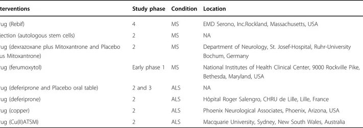
dietary iron supplementation. Several clinical trials
are underway to create a systematic approach to
identify individualized therapy (Table
2
). An interesting
future approach to MS diagnostics could include
correlating metal concentrations in blood with positive
MRI-based diagnoses of MS. It could lead to earlier
and more routine detection of MS. This may
fill the
gap created due to the lack of satisfactory treatments
and will put forth a better prospect of obtaining definitive
clinical evidence for ef
ficacy/fail-safe therapeutic design.
Last but not the least, the use of untethered, wirelessly
controlled, mobile, milli/microrobots as unconventional
approaches can be utilized to enhance the quality of life in
post-treatment of MS/ALS while reducing emotional
load, recovery time, and cost, which in recent times
became a topic of choice among clinicians for future
prospects
199–204. Collaborative efforts between robotic
researchers, clinicians, biomedical engineers, materials
scientists and chemists have led to superior
biomaterial-based design of daily need utilities to reduce the toxic
metals exposure
199–201,206–208. Plaques and implants
could be future blockbuster therapeutic procedure to cure
MS/ALS
209.
Conclusion
We highlighted the recent progress in the role of redox
metals in MS/ALS. The redox capacity of iron and copper,
their contribution to Fenton reactions, and production of
oxidative stress are identi
fied as prominent factors for
neurodegeneration in MS/ALS. Moreover, metals
dys-homeostasis is reported to cause oxidative damage. The
role of SOD1 mutation on copper dys-regulation in
neurodegenerative disorders, particularly in ALS has been
noticed by some researchers. In addition, metal chelator
therapy in animal models of these disorders rescue
neu-ronal degeneration and increased survival. On the other
hand, there have been few studies on the effect of
cad-mium on MS/ALS; its neurotoxicity has proved through
oxidative stress formation, reduction of antioxidant
enzymes activity, and cellular antioxidants. Despite
extensive studies to date, the main cause of these
neuro-degenerative disorders is still unknown and requires
fur-ther investigation.
Acknowledgements
We thank Morteza Amjadi for proofreading, Yang Xiao and Shuo Wang for their assistance with the graphic designs. A.V.S. thanks Max Planck Society for the grass root project grant 2017 (M10335) and 2018 (M10338).
Author details
1
Max Planck Institute for Intelligent Systems, Heisenbergstr. 3, Stuttgart 70569, Germany.2Institute for Materials Science, University of Stuttgart, Heisenbergstr.
3, Stuttgart 70569, Germany.3Hemostasis & Thrombosis Center - Azienda Ospedaliera-Universitaria di Ferrara, Ferrara, Italy.4Translational Surgery Unit,
Azienda Ospedaliera Universitaria di Ferrara, via Aldo Moro 8, 44124 Ferrara, Italy
Competing interests
The authors declare no competingfinancial interests. Publisher's note
Springer Nature remains neutral with regard to jurisdictional claims in published maps and institutional affiliations.
Received: 15 October 2017 Revised: 13 December 2017 Accepted: 27 December 2017
References
1. Hametner, S. et al. Iron and neurodegeneration in the multiple sclerosis brain. Ann. Neurol. 74, 848–861 (2013).
2. Singh, A. V. & Zamboni, P. Anomalous venous blood flow and iron deposition in multiple sclerosis. J. Cereb. Blood. Flow. Metab. 29, 1867–1878 (2009).
3. Singh, A. V. Multiple sclerosis takes venous route: CCSVI and liberation therapy. Indian. J. Med. Sci. 64, 337–340 (2010).
4. Raymond, M. J. et al. Multiaxial polarity determines individual cellular and nuclear chirality. Cell. Mol. Bioeng. 10, 63–74 (2017).
5. Ghoreishi, A., Mohseni, M., Amraei, R., Alizadeh, A. & Mazloomzadeh, S. Investigation the amount of copper, lead, zinc and cadmium levels in serum of Iranian multiple sclerosis patients. J. Chem. Pharm. Sci. 8, 40–45 (2015). 6. Aliomrani, M. et al. Blood concentrations of cadmium and lead in multiple
sclerosis patients from Iran. Iran. J. Pharm. Res. 15, 825 (2016).
7. Ibrahim, S. M. & Gold, R. Genomics, proteomics, metabolomics: what is in a word for multiple sclerosis? Curr. Opin. Neurol. 18, 231–235 (2005). 8. Reiber, H., Teut, M., Pohl, D., Rostasy, K. & Hanefeld, F. Paediatric and adult
multiple sclerosis: age-related differences and time course of the
Table 2
Clinical Trials in Iron and copper related with MS and ALS pathology (Data from clinicaltrials.gov)
Interventions Study phase Condition Location
Drug (Rebif) 4 MS EMD Serono, Inc.Rockland, Massachusetts, USA
Injection (autologous stem cells) 2 MS NA
Drug (dexrazoxane plus Mitoxantrone and Placebo plus Mitoxantrone)
2 MS Department of Neurology, St. Josef-Hospital, Ruhr-University Bochum, Germany
Drug (ferumoxytol) Early phase 1 MS National Institutes of Health Clinical Center, 9000 Rockville Pike,
Bethesda, Maryland, USA
Drug (deferiprone and Placebo oral table) 2 and 3 ALS NA
Drug (deferiprone) 2 ALS Hôpital Roger Salengro, CHRU de Lille, Lille, France
Drug (copper) 2 ALS Phoenix Neurological Associates, Phoenix, Arizona, USA
neuroimmunological response in cerebrospinal fluid. Mult. Scler. 15, 1466–1480 (2009).
9. Johnson, S. The possible role of gradual accumulation of copper, cadmium, lead and iron and gradual depletion of zinc, magnesium, selenium, vitamins B2, B6, D, and E and essential fatty acids in multiple sclerosis. Med. Hypotheses 55, 239–241 (2000).
10. Goodall, E. F., Haque, M. S. & Morrison, K. E. Increased serum ferritin levels in amyotrophic lateral sclerosis (ALS) patients. J. Neurol. 255, 1652–1656 (2008). 11. Carrı ̀, M. T., Ferri, A., Cozzolino, M., Calabrese, L. & Rotilio, G. Neurodegen-eration in amyotrophic lateral sclerosis: the role of oxidative stress and altered homeostasis of metals. Brain Res. Bull. 61, 365–374 (2003). 12. Hadzhieva, M. et al. Dysregulation of iron protein expression in the G93A
model of amyotrophic lateral sclerosis. Neuroscience 230, 94–101 (2013). 13. Tokuda, E. & Furukawa, Y. Copper homeostasis as a therapeutic target in
amyotrophic lateral sclerosis with SOD1 mutations. Int. J. Mol. Sci. 17, 636 (2016).
14. Que, E. L., Domaille, D. W. & Chang, C. J. Metals in neurobiology: probing their chemistry and biology with molecular imaging. Chem. Rev. 108, 1517–1549 (2008).
15. Crichton, R. R., Dexter, D. & Ward, R. J. Metal based neurodegenerative diseases—from molecular mechanisms to therapeutic strategies. Coord. Chem. Rev. 252, 1189–1199 (2008).
16. Visconti, A. et al. Concentration of elements in serum of patients affected by multiple sclerosis withfirst demyelinating episode: a six-month longitudinal follow-up study. Ann. Ist. Super. Sanita 41, 217–222 (2004).
17. Valko, M., Jomova, K., Rhodes, C. J., Kuča, K. & Musílek, K. Redox-and non-redox-metal-induced formation of free radicals and their role in human disease. Arch. Toxicol. 90, 1–37 (2016).
18. Gilgun-Sherki, Y., Melamed, E. & Offen, D. Oxidative stress induced-neurodegenerative diseases: the need for antioxidants that penetrate the blood brain barrier. Neuropharmacology 40, 959–975 (2001).
19. Chen, Q., Vazquez, E. J., Moghaddas, S., Hoppel, C. L. & Lesnefsky, E. J. Pro-duction of reactive oxygen species by mitochondria central role of complex III. J. Biol. Chem. 278, 36027–36031 (2003).
20. Mahad, D., Ziabreva, I., Lassmann, H. & Turnbull, D. Mitochondrial defects in acute multiple sclerosis lesions. Brain 131, 1722–1735 (2008).
21. Campbell, G. R. et al. Mitochondrial DNA deletions and neurodegeneration in multiple sclerosis. Ann. Neurol. 69, 481–492 (2011).
22. Oshiro, S., Morioka, M. S. & Kikuchi, M. Dysregulation of iron metabolism in Alzheimer’s disease, Parkinson’s disease, and amyotrophic lateral sclerosis. Adv. Pharmacol. Sci. 2011, 378278 (2011).
23. Biasiotto, G., Di Lorenzo, D., Archetti, S. & Zanella, I. Iron and neurodegen-eration: is ferritinophagy the link? Mol. Neurobiol. 53, 5542–5574 (2016). 24. Jomova, K. & Valko, M. Advances in metal-induced oxidative stress and
human disease. Toxicology 283, 65–87 (2011).
25. Hallgren, B. & Sourander, P. The effect of age on the non‐haemin iron in the human brain. J. Neurochem. 3, 41–51 (1958).
26. Aquino, D. et al. Age-related iron deposition in the basal ganglia: quantitative analysis in healthy subjects 1. Radiology 252, 165–172 (2009).
27. Zamboni, P. et al. Serum iron and matrix metalloproteinase‐9 variations in limbs affected by chronic venous disease and venous leg ulcers. Dermatol. Surg. 31, 644–649 (2005).
28. Singh, A. V. et al. Investigation of in vitro cytotoxicity of the redox state of ionic iron in neuroblastoma cells. J. Neurosci. Rural Pract. 3, 301 (2012). 29. Zamboni, P. et al. The overlapping of local iron overload and HFE mutation
in venous leg ulcer pathogenesis. Free. Radic. Biol. Med. 40, 1869–1873 (2006). 30. Zamboni, P. The big idea: iron-dependent inflammation in venous disease and proposed parallels in multiple sclerosis. J. R. Soc. Med. 99, 589–593 (2006). 31. Haacke, E. M. et al. Characterizing iron deposition in multiple sclerosis lesions using susceptibility weighted imaging. J. Magn. Reson. Imaging 29, 537–544 (2009).
32. Stankiewicz, J. et al. Iron in chronic brain disorders: imaging and neu-rotherapeutic implications. Neuneu-rotherapeutics 4, 371–386 (2007).
33. LeVine, S. M. & Chakrabarty, A. The role of iron in the pathogenesis of experimental allergic encephalomyelitis and multiple sclerosis. Ann. N. Y. Acad. Sci. 1012, 252–266 (2004).
34. Bergsland, N. et al. White matter tract injury is associated with deep gray matter iron deposition in multiple sclerosis. J. Neuroimaging. 27, 107–113 (2017).
35. Yoshida, M. et al. A mutation database for amyotrophic lateral sclerosis. Hum. Mutat. 31, 1003–1010 (2010).
36. Wang, X.-S. et al. Increased incidence of the Hfe mutation in amyotrophic lateral sclerosis and related cellular consequences. J. Neurol. Sci. 227, 27–33 (2004).
37. Jeong, S. Y. et al. Dysregulation of iron homeostasis in the CNS contributes to disease progression in a mouse model of amyotrophic lateral sclerosis. J. Neurosci. 29, 610–619 (2009).
38. Singh, A. V., Subhashree, L., Milani, P., Gemmati, D. & Zamboni, P. Interplay of iron metallobiology, metalloproteinases, and FXIII, and role of their gene variants in venous leg ulcer. Int. J. Low. Extrem. Wounds 9, 166–179 (2010). 39. Gemmati, D. et al. Influence of gene polymorphisms in ulcer healing process
after superficial venous surgery. J. Vasc. Surg. 44, 554–562 (2006). 40. Gemmati, D. et al. Polymorphisms in the genes coding for iron binding and
transporting proteins are associated with disability, severity, and early pro-gression in multiple sclerosis. BMC Med. Genet. 13, 70 (2012).
41. Zamboni, P. et al. Hemochromatosis C282Y gene mutation increases the risk of venous leg ulceration. J. Vasc. Surg. 42, 309–314 (2005).
42. Restagno, G. et al. HFE H63D polymorphism is increased in patients with amyotrophic lateral sclerosis of Italian origin. J. Neurol. Neurosurg. Psychiatry 78, 327–327 (2007).
43. Goodall, E. et al. Association of the H63D polymorphism in the hemochro-matosis gene with sporadic ALS. Neurology 65, 934–937 (2005). 44. Sutedja, N. A. et al The association between H63D mutations in HFE and
amyotrophic lateral sclerosis in a Dutch population. Arch. Neurol. 64, 63–67 (2007).
45. Ferreira, K. P. Z. et al. Disease progression and oxidative stress are associated with higher serum ferritin levels in patients with multiple sclerosis. J. Neurol. Sci. 373, 236–241 (2017).
46. Iranmanesh, M., Iranmanesh, F. & Sadeghi, H. Serum level of iron, zinc and copper in patients with multiple sclerosis. J. Jahr. Uni. Med. Sci. 10, 1–5 (2013). 47. Forge, J. K., Pedchenko, T. V. & Le Vine, S. M. Iron deposits in the central nervous system of SJL mice with experimental allergic encephalomyelitis. Life. Sci. 63, 2271–2284 (1998).
48. Khare, M., Singh, A. & Zamboni, P. Prospect of brain machine interface in motor disabilities: the future support for multiple sclerosis patient to improve quality of life. Ann. Med. Health Sci. Res. 4, 305–312 (2014).
49. Uttara, B., Singh, A. V., Zamboni, P. & Mahajan, R. Oxidative stress and neu-rodegenerative diseases: a review of upstream and downstream antioxidant therapeutic options. Curr. Neuropharmacol. 7, 65–74 (2009).
50. Alimonti, A. et al. Serum chemical elements and oxidative status in Alzhei-mer’s disease, Parkinson disease and multiple sclerosis. Neurotoxicology 28, 450–456 (2007).
51. Hammond, K. E. et al. Quantitative in vivo magnetic resonance imaging of multiple sclerosis at 7 Tesla with sensitivity to iron. Ann. Neurol. 64, 707–713 (2008).
52. Bian, W. et al. A serial in vivo 7T magnetic resonance phase imaging study of white matter lesions in multiple sclerosis. Mult. Scler. 19, 69–75 (2013). 53. Mehta, V. et al. Iron is a sensitive biomarker for inflammation in multiple
sclerosis lesions. PLoS ONE 8, e57573 (2013).
54. Bagnato, F. et al. Tracking iron in multiple sclerosis: a combined imaging and histopathological study at 7 Tesla. Brain 134, 3602–3615 (2011). 55. Hagemeier, J. et al. Iron deposition in multiple sclerosis lesions measured by
susceptibility‐weighted imaging filtered phase: A case control study. J. Magn. Reson. Imaging 36, 73–83 (2012).
56. Adams, C. Perivascular iron deposition and other vascular damage in mul-tiple sclerosis. J. Neurol. Neurosurg. Psychiatry 51, 260–265 (1988). 57. Zivadinov, R. et al. Cerebral microbleeds in multiple sclerosis evaluated on
susceptibility-weighted images and quantitative susceptibility maps: a case-control study. Radiology 281, 884–895 (2016).
58. Zamboni, P. The contribution of extra cranial venous drainage to neuro-inflammation in multiple sclerosis. Neuroinflammation (2018).
59. Bakshi, R., Shaikh, Z. A. & Janardhan, V. MRI T2 shortening (‘black T2’) in multiple sclerosis: frequency, location, and clinical correlation. Neuroreport 11, 15–21 (2000).
60. Tjoa, C., Benedict, R., Weinstock-Guttman, B., Fabiano, A. & Bakshi, R. MRI T2 hypointensity of the dentate nucleus is related to ambulatory impairment in multiple sclerosis. J. Neurol. Sci. 234, 17–24 (2005).
61. Brass, S. D., Benedict, R. H., Weinstock-Guttman, B., Munschauer, F. & Bakshi, R. Cognitive impairment is associated with subcortical magnetic resonance imaging grey matter T2 hypointensity in multiple sclerosis. Mult. Scler. 12, 437–444 (2006).
62. Modica, C. et al. Iron and volume in the deep gray matter: association with cognitive impairment in multiple sclerosis. Am. J. Neuroradiol. 36, 57–62 (2015).
63. Bø, L., Vedeler, C. A., Nyland, H., Trapp, B. D. & Mørk, S. J. Intracortical multiple sclerosis lesions are not associated with increased lymphocyte infiltration. Mult. Scler. 9, 323–331 (2003).
64. Peterson, J. W., Bö, L., Mörk, S., Chang, A. & Trapp, B. D. Transected neurites, apoptotic neurons, and reduced inflammation in cortical multiple sclerosis lesions. Ann. Neurol. 50, 389–400 (2001).
65. Haider, L. et al Multiple sclerosis deep grey matter: the relation between demyelination, neurodegeneration, inflammation and iron. J. Neurol. Neuro-surg. Psychiatry 85, 1386–1395 (2014).
66. Ge, Y. et al. Quantitative assessment of iron accumulation in the deep gray matter of multiple sclerosis by magneticfield correlation imaging. Am. J. Neuroradiol. 28, 1639–1644 (2007).
67. Veyrat-Durebex, C. et al. Iron metabolism disturbance in a French cohort of ALS patients. BioMed. Res. Int. 2014, 485723 (2014).
68. Nadjar, Y. et al. Elevated serum ferritin is associated with reduced survival in amyotrophic lateral sclerosis. PLoS ONE 7, e45034 (2012).
69. Ikeda, K., Hirayama, T., Takazawa, T., Kawabe, K. & Iwasaki, Y. Relationships between disease progression and serum levels of lipid, urate, creatinine and ferritin in Japanese patients with amyotrophic lateral sclerosis: a cross-sectional study. Intern. Med. 51, 1501–1508 (2012).
70. Ince, P. et al. Iron, selenium and glutathione peroxidase activity are elevated in sporadic motor neuron disease. Neurosci. Lett. 182, 87–90 (1994). 71. Kasarskis, E. J., Tandon, L., Lovell, M. A. & Ehmann, W. D. Aluminum, calcium,
and iron in the spinal cord of patients with sporadic amyotrophic lateral sclerosis using laser microprobe mass spectroscopy: a preliminary study. J. Neurol. Sci. 130, 203–208 (1995).
72. Markesbery, W. R. et al. Neutron activation analysis of trace elements in motor neuron disease spinal cord. Neurodegeneration 4, 383–390 (1995). 73. Yasui, M., Ota, K. & Garruto, R. M. Concentrations of zinc and iron in the brains
of Guamanian patients with amyotrophic lateral sclerosis and parkinsonism-dementia. Neurotoxicology 14, 445–450 (1993).
74. Hozumi, I. et al. Patterns of levels of biological metals in CSF differ among neurodegenerative diseases. J. Neurol. Sci. 303, 95–99 (2011).
75. Mizuno, Y. et al. Transferrin localizes in Bunina bodies in amyotrophic lateral sclerosis. Acta Neuropathol. 112, 597–603 (2006).
76. Ignjatović, A., Stević, Z., Lavrnić, S., Daković, M. & Bačić, G. Brain iron MRI: a biomarker for amyotrophic lateral sclerosis. J. Magn. Reson. Imaging 38, 1472–1479 (2013).
77. Oba, H. et al. Amyotrophic lateral sclerosis: T2 shortening in motor cortex at MR imaging. Radiology 189, 843–846 (1993).
78. Imon, Y. et al. A decrease in cerebral cortex intensity on T2-weighted with ageing images of normal subjects. Neuroradiology 40, 76–80 (1998). 79. Hecht, M., Fellner, C., Schmid, A., Neundörfer, B. & Fellner, F. Cortical T2 signal
shortening in amyotrophic lateral sclerosis is not due to iron deposits. Neuroradiology 47, 805–808 (2005).
80. Winkler, E. A. et al. Blood–spinal cord barrier disruption contributes to early motor-neuron degeneration in ALS-model mice. Proc. Natl Acad. Sci. 111, E1035–E1042 (2014).
81. Wang, Q. et al. Prevention of motor neuron degeneration by novel iron chelators in SOD1G93A transgenic mice of amyotrophic lateral sclerosis. Neurodegener. Dis. 8, 310–321 (2011).
82. Kupershmidt, L. et al. Neuroprotective and neuritogenic activities of novel multimodal iron-chelating drugs in motor-neuron-like NSC-34 cells and transgenic mouse model of amyotrophic lateral sclerosis. Faseb. J. 23, 3766–3779 (2009).
83. Zheng, W. & Monnot, A. D. Regulation of brain iron and copper homeostasis by brain barrier systems: implication in neurodegenerative diseases. Phar-macol. Ther. 133, 177–188 (2012).
84. Choi, B.-S. & Zheng, W. Copper transport to the brain by the blood–brain barrier and blood-CSF barrier. Brain Res. 1248, 14–21 (2009).
85. Scheiber, I. F. & Dringen, R. Astrocyte functions in the copper homeostasis of the brain. Neurochem. Int. 62, 556–565 (2013).
86. Scheiber, I. F., Mercer, J. F. & Dringen, R. Metabolism and functions of copper in brain. Prog. Neurobiol. 116, 33–57 (2014).
87. West, A. K., Hidalgo, J., Eddins, D., Levin, E. D. & Aschner, M. Metallothionein in the central nervous system: roles in protection, regeneration and cognition. Neurotoxicology 29, 489–503 (2008).
88. Manto, M. Abnormal copper homeostasis: mechanisms and roles in neuro-degeneration. Toxics 2, 327–345 (2014).
89. Lutsenko, S., Gupta, A., Burkhead, J. L. & Zuzel, V. Cellular multitasking: the dual role of human Cu-ATPases in cofactor delivery and intracellular copper balance. Arch. Biochem. Biophys. 476, 22–32 (2008).
90. Lutsenko, S., Barnes, N. L., Bartee, M. Y. & Dmitriev, O. Y. Function and regulation of human copper-transporting ATPases. Physiol. Rev. 87, 1011–1046 (2007).
91. Marikovsky, M., Ziv, V., Nevo, N., Harris-Cerruti, C. & Mahler, O. Cu/Zn super-oxide dismutase plays important role in immune response. J. Immunol. 170, 2993–3001 (2003).
92. Tapiero, H., Townsend, D. & Tew, K. Trace elements in human physiology and pathology. Copp. Biomed. Pharmacother. 57, 386–398 (2003).
93. Konstantinova, S. G., Russanova, I. E. & Russanov, E. M. Do the copper complexes of histamine, histidine and of two H2-antagonists react with O− 2? Free. Radic. Res. Commun. 12, 215–220 (1991).
94. Socha, K. et al. Dietary habits; concentration of copper, zinc, and Cu-to-Zn ratio in serum and ability status of patients with relapsing-remitting multiple sclerosis. Nutrition 39, 76–81 (2017).
95. Banci, L. et al. Affinity gradients drive copper to cellular destinations. Nature 465, 645 (2010).
96. Dringen, R., Scheiber, I. F. & Mercer, J. F. Copper metabolism of astrocytes. Front. Aging Neurosci. 5, 9 (2013).
97. Roos, P. M., Vesterberg, O., Syversen, T., Flaten, T. P. & Nordberg, M. Metal concentrations in cerebrospinalfluid and blood plasma from patients with amyotrophic lateral sclerosis. Biol. Trace Elem. Res. 151, 159–170 (2013). 98. Ibrahimagić, O., Sinanović, O., Zonić, L. & Hudić, J. Amyotrophic lateral
sclerosis in younger age associated with abnormality of copper level. Med. Arh. 60, 108–109 (2006).
99. Van Horssen, J., Witte, M. E., Schreibelt, G. & De Vries, H. E. Radical changes in multiple sclerosis pathogenesis. Biochim. Biophys. Acta 1812, 141–150 (2011). 100. Aspli, K. T. et al. Iron and copper in progressive demyelination–New lessons
from Skogholt’s disease. J. Trace Elem. Med. Biol. 31, 183–187 (2015). 101. Ghazavi, A., Kianbakht, S., Ghasami, K. & Mosayebi, G. High copper and low
zinc serum levels in Iranian patients with multiple sclerosis: a case control study. Clin. Lab. 58, 161–164 (2012).
102. Sedighi, B., Ebrahimi, H. A., Haghdoost, A. A. & Abotorabi, M. Comparison of serum levels of copper and zinc among multiple sclerosis patients and control group. Iran J. Neurol. 12, 125 (2013).
103. Melø, T. M. et al. Manganese, copper, and zinc in cerebrospinalfluid from patients with multiple sclerosis. Biol. Trace Elem. Res. 93, 1–8 (2003). 104. Mandelbrote, B., Stanier, M., Thompson, R. & THBUSTON, M. Studies on
copper metabolism in demyelinating diseases of the central nervous system. Brain 71, 212–228 (1948).
105. Plum, C. M. & Hansen, S. E. Studies on variations in serum copper and serum copper oxidase activity, together with studies on the copper content of the cerebrospinalfluid, with particular reference to the variations in multiple sclerosis. Acta Psychiatr. Scand. 35, 41–78 (1960).
106. Wikström, J., Westermarcik, T. & Palo, J. Selenium, vitamin E and copper in multiple sclerosis. Acta Neurol. Scand. 54, 287–290 (1976).
107. Bammer, H. Caeruloplasmin-und kupferstoffwechsel bei multipler sklerose. J. Neurol. 189, 312–329 (1966).
108. Giacoppo, S. et al. Heavy metals and neurodegenerative diseases: an observational study. Biol. Trace Elem. Res. 161, 151–160 (2014).
109. Tamburo, E., Varrica, D., Dongarrà, G. & Grimaldi, L. M. E. Trace elements in scalp hair samples from patients with relapsing-remitting multiple sclerosis. PLoS ONE 10, e0122142 (2015).
110. Palm, R. & Hallmans, G. Zinc and copper in multiple sclerosis. J. Neurol. Neurosurg. Psychiatry 45, 691–698 (1982).
111. Kapaki, E., Segditsa, J. & Papageorgiou, C. Zinc, copper and magnesium concentration in serum and CSF of patients with neurological disorders. Acta Neurol. Scand. 79, 373–378 (1989).
112. Norkute, A. et al. Cuprizone treatment induces demyelination and astro-cytosis in the mouse hippocampus. J. Neurosci. Res. 87, 1343–1355 (2009). 113. Torkildsen, Ø., Brunborg, L., Myhr, K. M. & Bø, L. The cuprizone model for
demyelination. Acta Neurol. Scand. 117, 72–76 (2008).
114. Matsushima, G. K. & Morell, P. The neurotoxicant, cuprizone, as a model to study demyelination and remyelination in the central nervous system. Brain Pathol. 11, 107–116 (2001).
115. Herring, N. R. & Konradi, C. Myelin, copper, and the cuprizone model of schizophrenia. Front. Biosci. (Sch. Ed.) 3, 23 (2011).