Printed in U.S.A.
Ryanodine Receptors of Striated Muscles: a Complex Channel
Capable of Multiple Interactions
CLARA FRANZINI-ARMSTRONG
AND FELICIANO
PROTASI
Department
of CeU and Developmental
Biology, University
of
Pennsylvania,
Philadelphia,
Pennsylvania
I. Introduction
II. Ryanodine
Receptor: the Molecule
A. Isolation
and characterization
of ryanodine
receptors
B. Ryanodine
receptor
isoforms: tissue distribution
C. Sequence and primary structure
of ryanodine
receptors
D. Ryanodine
receptor
channel: its function
and modulation
E. Developmental
regulation
F. Functional
domains
G. Three-dimensional
structure
of ryanodine
receptors
III. Protein-Protein
Interactions
of Ryanodine
Receptors
A. FKBP12: a stabilizing
factor
B. Interactions
of ryanodine
receptors
with other junctional
sarcoplasmic
reticulum
proteins
IV. Ryanodine
Receptor-Dihydropyridine
Receptor
Interactions
in Excitation-Contraction
Coupling
A. Architecture
of junctional
sarcoplasmic
reticulum
and its relationship
to exterior
membranes
B. Dihydropyridine
receptors
and excitation-contraction
coupling
C. Spatial relationship
between
ryanodine
receptors
and dihydropyridine
receptors:
clues to excitation-contraction
coupling
D. Functional
dihydropyridine
receptor-ryanodine
receptor
interactions
are isoform dependent
V. Conclusions
699
700
700
701
705
705
708
708
710
710
710
711
714
714
714
716
718
720
Franzini-Armstrong,
Clara, and Feliciano
Protasi.
Ryanodine Receptors of Striated Muscles: a Complex Channel
Capable of Multiple
Interactions.
Physiol. Rev. 77: 699-729,
1997. -The
ryanodine
receptor
(RyR) is a high-conduc-
tance Ca2’ channel of the sarcoplasmic
reticulum
in muscle and of the endoplasmic
reticulum
in other cells. In
striated muscle fibers, RyRs are responsible
for the rapid release of Ca2’ that activates contraction.
Ryanodine
receptors are complex molecules,
with unusually large cytoplasmic
domains containing
numerous binding
sites for
agents that control the state of activity of the channel-forming
domain of the molecule.
Structural
considerations
indicate
that long-range
interactions
between
cytoplasmic
and intramembrane
domains control
channel function.
Ryanodine
receptors
are located
in specialized
regions of the SR, where they are structurally
and functionally
associated
with other intrinsic
proteins
and, indirectly,
also with the luminal
Ca2’-binding
protein
calsequestrin.
Activation
of RyRs during the early part of the excitation-contraction
coupling
cascade is initiated
by the activity
of surface-membrane
Ca2’ channels, the dihydropyridine
receptors
(DHPRs). Skeletal and cardiac muscles contain
different RyR and DHPR isoforms and both contribute
to the diversity in cardiac and skeletal excitation-contraction
coupling
mechanisms.
The architecture
of the sarcoplasmic
reticulum-surface
junctions
determines
the types of
RyR-DHPR interactions
in the two muscle types.
I. INTRODUCTION
The ryanodine receptor (RyR) is a Ca2’ channel of the
endoplasmic reticulum [or sarcoplasmic reticulum (SR) in
the case of muscle cells] with a very large cytoplasmic
domain, with high affinity for ryanodine, a neutral plant
alkaloid. Ryanodine receptors have considerable se-
quence and general structure similarities with the other
intracellular
channels, the inositol 1,4,5-trisphosphate
(InsP3) receptors, with which they share the task of releas-
ing Ca2’ from the internal stores (231). Ryanodine recep-
tors, however, have higher conductivity than InsP3 recep-
tors, and thus they are employed in situations that need
fast release of large quantities of Ca2’, such as during
excitation-contraction
(e-c) coupling in muscle.
In striated muscles, RyRs interact with several other
proteins, and thus both their structure and their function
must be understood within this complex set of interac-
tions. Ryanodine receptors are located at high density in
a special domain of the SR membrane, the junctional face
membrane, belonging to the junctional SR (jSR). Within
this domain, RyRs are associated, either directly or indi-
700
CLARA FRANZINI-ARMSTRONG AND FELICIANO PROTASI Volume 776 PM
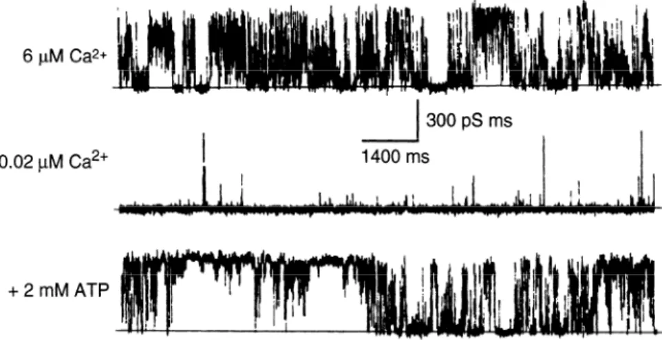
Ca*+
FIG. 1. Reconstitution of purified ryanodine re-
0.02 PM Ca*+
300 pS ms
1400 ms
ceptor (RyR) isoform RyRl into a planar lipid bi-
layer reveals a high-conductance channel with prop-
erties essentially identical to those of Ca2’ release
channel of heavy sarcoplasmic reticulum (SR). Cur-
rents were recorded in presence of 6 PM Ca2’ (top),
0.02 ,uM Ca”’ (middle), and 0.02 PM Ca2’ plus 2
mM ATP (bottom). Ca2+ and adenine nucleotides
greatly increase channel’s open probability. [From
Lai et al. (160).]
+2mMATP
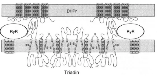
rectly, with other structural components of the jSR,
calsequestrin, triadin, and junctin, with which they may
functionally
interact. The junctional face membrane is
closely apposed to, and forms a specialized junction with,
the external cell membranes [the surface membrane and
its invaginations, the transverse (t) tubules]. The junctions
between SR and external membranes allow interactions
between RyRs and proteins of the surface membrane, the
dihydropyridine receptors (DHPRs). Finally, RyRs are sta-
bly bound to one small protein, FKBP12 (FK506 binding
protein, see sect.
IIIA),and they are modulated by calmod-
ulin (CaM) and cytoplasmic solutes. Several reviews (15,
59,201,206,236,304) thoroughly cover the properties and
pharmacology of RyRs; a collection of monographs deals
with properties and distribution of RyRs (298); and the
proceedings of a recent symposium on channels include
RyRs and InsP3 receptors (56).
tionship between a Ca”+ release channel of heavy SR and
the fast Ca”’ release that plays a role in the activation of
muscle contraction in skeletal and cardiac muscle.
Large structural components of the SR, the feet, are
located in the junctional face membrane of skeletal (36,
60, 92, 93, 216) and cardiac (295, 296) muscle, facing the
external membranes. A high-molecular-weight component
of the jSR (or perhaps a doublet of proteins) was isolated
and identified as the spanning protein by immunoelectron
microscopy (33, 147, 148). Thus evidence for the location
of Ca2’ release sites in the jSR and for a high-molecular-
weight component playing a major role in the junction
was already in place before RyRs were isolated.
II. RYANODINE
RECEPTOR:
THE MOLECULE
A. Isolation and Characterization
of Ryanodine Receptors
Intact RyR molecules were first isolated and purified
in skeletal (127, 130, 159, 160) and cardiac (9, 129, 131,
132, 267) muscles, after mild 3-[ (3-cholamidopropyl)di-
methylammoniol-propanesulfonate
(CHAPS) solubiliza-
tion of the heavy SR and using [3H]ryanodine as a selective
marker. They were subsequently found in smooth muscle
(116, 170, 346; see Ref. 186 for a review) and are now
known to be present in a large variety of cell types (see
Refs. 297-299 for reviews). They are very large (30s) com-
plexes constituted by homotetramers of -560-kDa poly-
peptides. The large size of the molecule allows single-
step purification by sucrose density gradient (160), but
The way for the isolation of the RyR and its identifi-
cation as the SR Ca2’ release channel was paved by sev- combined procedures have been used. These include se-
era1 key observations on the properties of the heavy SR. quential column chromatography on heparin-agarose and
The heavy SR is the higher density fraction of the reticu-
hydroxyapatite
(130); immunoaffinity
purification (127)
lum containing the jSR and the internal Ca2’ binding pro-
and either ion-exchange chromatography or heparin-agar-
tein calsequestrin (36, 203) and also intact triads (41).
ose column chromatography combined with density gradi-
Ryanodine and doxorubicin, agents that have profound
effects on muscle contraction by causing and/or inhibiting
liberation of Ca2’ from the SR, bind with high affinity to
a high-molecular-weight component of the heavy SR (85,
253, 255, 354, 356), which also binds CaM (279). Heavy
SR, but not light SR, rapidly releases Ca2+ (203), due to
the presence of channels with high permeability for mono-
valent and divalent cations (289) that are activated by
adenine nucleotides and Ca2’ and inhibited by Mg2+ (274,
288; Fig. 1). These experiments established a direct rela-
ent centrifugation (114,128) have also been used. A simple
one-step procedure (286) and an affinity purification pro-
cedure based on the strong binding between RyR and
FKBP12 have been devised (344).
The purified receptor, incorporated in a planar lipid
bilayer functions as a Ca2’ channel (9, 20, 37, 121, 127,
160,275, see 162 for review), with characteristics identical
to the Ca”+ release channels previously identified in the
heavy SR (288,289) and in SR in situ (300). The full-length
rabbit skeletal RyR cDNA has been functionally expressed
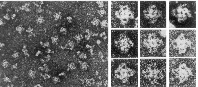
FIG. 2. Negatively stained purified RyRl from rabbit skeletal muscle. Protein has 4 equal large subunits in form
of a quatrefoil, identical in size and shape to feet of junctional SR. A smaller central region, also composed of 4 equal
subunits, is intramembrane channel-forming domain. [From Lai et al. (160).]
in COS and Chinese hamster
ovary (CHO)
cells (48, 251,
310) and shown to form a channel with the appropriate
properties
(48). Identification
of RyR as Ca” release chan-
nels relies on the assumption
that the channel activity is
due to the major protein composing
the purified fraction
(the RyR) and not to a minor contaminant.
Reconstitution
of the purified RyR into proteoliposomes
allowed observa-
tion of channel function on a macroscopic
scale, conflrm-
ing the channel identity of RyRs (169). The presence of a
single high-affinity
site for ryanodine
within
a tetramer
identifies
the tetramer
as a single functional
entity or
channel (160). The hereditary
defect in malignant hyper-
thermia has been traced to the RyR (184, 213).
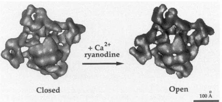
The isolated RyR is a large molecule,
-29
X29
X12
nm, with four equal subunits
roughly approximated
by
four spheres
that are closely associated
to form a four-
leaf clover or quatrefoil
(21, 130, 131, 160, 332; Fig. 2).
Structural
comparison
of the isolated channel and of the
feet allowed
direct identification.
The overall size of the
feet and their quatrefoil structure
(82, 174,216) are identi-
cal to those of the isolated RyR. In addition to the large
domain, corresponding
to the feet, the isolated molecule
has a smaller
central region constituted
of four equal
lobes, rotated by -45”
relative to the larger domain. Be-
cause the small domain is not visible in the in situ mole-
cule, it is logical to assume that it represents
the intra-
membrane,
channel-forming,
portion of the RyR. Indeed,
a small quatrefoil,
with four equal lobes, is visible within
the jSR membrane
after freeze fracturing
(21). Because
ryanodine
binds close to, but not within
the presumed
transmembrane
domain, the molecule is often called RyRI
Ca2’ release channel complex.
Foot protein is another
name that identifies
the molecule based on the structure
of its cytoplasmic
domain.
With the use of the quatrefoil
shape and the charac-
teristic large size of the feet as unique identification
mark-
ers for RyRs, the molecule has been identified in a large
variety
of muscles
from vertebrates
and invertebrates
(174). A RyR has also been identified in two muscles from
crustaceans
(91, 239, ZSO), characterized
in Caenorhab-
ditis elegans (153), and identified and sequenced in Dro-
sophila
(113, 312). Ryanodine
receptors
have been de-
tected in almost all cell types, although at variable levels
of expression
(see sect. Iti).
One question remains unsolved: What is the relation-
ship between the RyR and the 106-kDa protein that can be
isolated by ryanodine-affinity
chromatography
and forms
channels with conductance
and pharmacology
compara-
ble to those of the RyR (118, 276, 349)?
Identification
of the cytoplasmic
domains of RyRs as
feet has a very important
implication.
Feet are located in
regions of the heavy SR that form junctions
with surface
membrane and t tubules. Therefore,
the cytoplasmic
do-
mains of RyRs, constituting
the feet, allow
a direct con-
nection between the Ca2’ release channel and the exterior
membranes.
The latter part of the review
covers the role
that this connection
plays in e-c coupling,
the series of
steps that link depolarization
of the surface membrane to
contraction
of the myofibrils,
via release of Ca2’ by RyRs.
B. Ryanodine
Receptor
Isoforms:
Tissue
Distribution
1. Three ryanodine
receptor isoforms
in a variety
of tissues
Three types of RyR with specific tissue distribution
are now recognized (104; see Refs. 297, 299 for reviews).
702
CLARA FRANZINI-ARMSTRONG AND FELICIANO PROTASI Volume 77The currently
accepted terminology
is based on the timing
of purification
of the RyRs from various
tissues
and the
identification
of the three isoforms
by molecular
probes.
Thus RyRl, also called the skeletal type, is the isoform
first detected (189) and then fully sequenced
(313, 353)
in skeletal muscle; RyR2, the dominant
form in cardiac
muscle,
was
subsequently
sequenced
(228, 244); and
RyR3, sometimes
called the brain isoform,
was first de-
tected (228) and later fully sequenced in brain (112) and
epithelial cells (103). Sequence comparison
of the tree
isoforms
in a single species reveals a homology of 67, 67,
and 70% between RyRlLRyR2,
RyRlLRyR3,
and RyR2/RyR3
(112), respectively,
but a phylogenetic
tree generated from
the optimal alignment of full-length
RyR sequences
avail-
able in 1996 indicates overall closer relationships
between
RyRl and RyR3 than RyRl and RyR2 (326).
The RyRl isoform is primarily
expressed
in all skele-
tal muscles
(5, 161, 237, 313, 326, 352) and in some parts
of the brain, most prominently
in Purkinje
cells of the
cerebellum
(99, 104, 157, 168, 245), and is also present
in some smooth muscle (230). The RyR2 isoform
is the
predominant
form in cardiac muscle (228, 244) and is also
the most widely distributed
isoform in the brain (99, 104,
157, 158, 168). Indeed, the major form of RyR purified
from brain is identified
as RyR2 immunologically
and by
analysis
of proteolytic
products
(200). Some expression
of RyR2 in smooth muscle has also been found (230). The
RyR3 isoform is a minor component
of the brain, where
it is the least widely distributed
of the three isoforms.
The
RyR3 isoform is immunologically
distinct from RyRl and
RyR2 (143). RyR3 is present in skeletal muscle, but it is
particularly
abundant in selected skeletal muscles
from
some species (see sect.
IIBZ).Smooth muscle expresses
RyR3 (230), but in minor amounts
(189).
The dominant form of RyR in cardiac muscle is RyR2
(69, 228, 244). Both RyRl and RyR3 are at such extremely
low levels (112) that they can be truly detected only by
ribonuclease
protection
assays
(103) and reverse
tran-
scription-polymerase
chain reaction
amplification
(168).
Much of the RyR3 is attributable
to smooth muscle cells
of intracardiac
coronary
vessels, which
are quite numer-
ous, but weak hybridization
of the working
myocardium
and of the conducting
Purkinje
system for cRNA probes
for RyR3 indicates
a minor presence
of this isoform
in
myocardial
cells (232). A detailed search for functional
variations
in RyR composition
of SR isolated from various
regions
of the heart has shown
channels
with indistin-
guishable conductance
and pharmacology,
indicating that
no region of the heart has a noticeably
high presence of
channels other than RyR2 (345). Thus myocardium
is the
muscle with the purest RyR composition,
despite the fact
that feet are located in two distinct regions of the SR: the
jSR and corbular
SR (see sect. IvC).
So far, few variations
from the three isoforms
have
been found. First. a gene encoding a partial sequence of
RyR3 expresses
a protein that does not respond to caf-
feine (103). Second, a transcript
from the COOH-terminal
region of the RyR gene has been identified in brain (314).
This shortened
version
of the molecule would
lack the
receptor
regions for agents that affect channel activity.
Third, alternative
splicing has been found to introduce
complexity
in the RyR family. Two alternately spliced RyR
transcripts
were
found in embryonic,
slow-
and fast-
twitch
rabbit skeletal muscle, regardless
of the develop-
mental stage (355). The difference
between
the two is
very small: one of the transcripts
lacks five amino acids,
and it is not yet known
if both mRNAs
are expressed.
Tissue-specific
and developmentally
regulated
splicing
have been detected for RyR3 (193, 219) and RyRl (101).
The latter
involves
modulatory
segments
with
binding
sites for Ca2+, CaM, and ATP.
It is likely that further
variations
exist but have not
yet been detected, due to the difficulty
of fully sequencing
this very large molecule.
However,
the positive
results
with hybridization
analysis
and immunolabeling
across
tissue types certainly
indicate that RyR types are highly
conserved.
In this respect, it is interesting to contrast RyR
with another large protein, myosin, which spans at least
11 families, each comprising
a large number of isoforms.
2. One versus
two ryanodine
receptors
in skeletal muscle
Muscles in nonmammalian
vertebrate,
bird, amphib-
ian, reptile, and fish muscle contain two RyR isoforms,
initially called cy and p (5, 161, 221, 233, 240). Molecular
approaches
have shown that the skeletal muscle @iso-
form of chicken
and frog is actually RyR3, the isoform
initially identified in the brain, whereas
the cu-isoform is
recognized as homologous
to RyRl (58, 104, 237, 246). In
frog and bullfrog, the cu-isoform has 80% sequence similar-
ity to rabbit skeletal
RyRl, and the P-isoform
has 85-
86% similarity
to rabbit brain RyR3, whereas
they diverge
considerably
from cardiac RyR2 (237, 246). Use of the
appropriate
antibodies
confirms
that CY- and p-isoforms
of
chicken
differ from RyR2 (7), replacing initial evidence
to the contrary.
In fish, the homology
of cu-isoform with
RyRl has also been confirmed,
but the /3-isoform has not
been characterized
at the molecular
level (234). The pres-
ence of RyR3 instead of RyR2 in skeletal muscle came as
a surprise,
in view
of the fact that developing
skeletal
muscle transiently
expresses
cardiac type DHPR and myo-
fibrillar
proteins.
Diversity
of RyRs in muscle fibers and
its significance
are well covered in a recent review
(304).
The RyRl and RyR3 isoforms
(cw and ,O) are present in
approximately
equal amounts in nonmammalian
skeletal
muscle. The two isoforms
are clearly located within
the
same muscle fiber in a position corresponding
to that of
the triads
(5, 161, 240; Fig. 3), and both are equally en-
riched in the triad fraction (221, 233). However,
although
FIG. 3. Two ryanodine binding proteins coexist in same muscle fiber in bullfrog skeletal muscle. Consecutive,
longitudinal section of muscle was stained with monoclonal antibodies (monoclonal antibody 32E in A and monoclonal
antibody 26G in B) recognizing specific regions of (Y- and fl-RyR isoforms, or RyRl and RyR3. [Prom Olivares et al.
isoforms, the resolution
is not sufficient to detect whether
they are actually located within the same triad. Given the
approximately
equal amounts
of the two proteins,
and
the fact that all triads appear structurally
equal in these
muscles, it is logical to assume that cx and p coexist within
the same junction.
So far, no evidence for heterotetramers
were found by immunoprecipitation
studies with specific
antibodies
(see Ref. 304 for a review).
Indeed, two types
of channels with distinct properties
are present in muscles
that express
the two isoforms
(see sect.
1rB3),and this
would not happen if the two isoforms
were freely mixing.
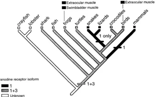
Interestingly,
although most muscles
in fish, reptiles,
and birds express
RyRl and RyR3, major groups of rep-
tiles and some fast muscles
in fish have a single isoform,
the CY or RyRl (22; Fig. 4). The muscle-derived
heater
tissue has a single isoform, predicting a fast muscle origin
for these cells (23).
IExtraocular muscle
ISwimbladder muscle
Mammalian
skeletal muscles
also contain a second
(RyR3) isoform
in addition to the dominating
RyRl type,
but at a 20- to 50-fold less concentration
(104, 168, 315;
see Ref. 297 for a review).
It is not clear whether
the
two isoforms
are present in the same muscle fibers, but
location
of both in the triadic microsomal
fraction
has
been shown
(58). For the moment, we assume that the
two isoforms
are intercalated
in the junctions.
However,
it is clearly not excluded
that RyR3 is segregated
over
some small regions of the SR. Indeed, a small percentage
of RyRs are located at a small distance from the junctional
face in mammalian muscle, although within the triad (68).
The relative amounts of RyR3 vary widely between
mam-
malian skeletal muscles, being higher in diaphragm
and
soleus and lower
in the abdominal
and tibialis
anterior
muscles
of a variety of mammals
(58). Interestingly,
the
fast-twitch
extensor
digitorum
longus
muscle
has no
IExtraocular muscle
FIG. 4. Phylogenetic distribution of RyR
expression patterns in skeletal muscle. Mus-
cles with a predominant expression of RyRl
(or o) are present in more advanced phyla,
but also in some very fast-acting muscles of
fish. Isoform distribution in invertebrates has
704
CLARA FRANZINI-ARMSTRONG AND FELICIANO PROTASI Volume 77RyR3, and this compares
with the fact that in fish the fast
muscles lack RyR3 (233). Immunologic
cross-reactivities
and mobility in sodium dodecyl sulfate-polyacrylamide
gel
electrophoresis
gels indicate more interspecies
variations
in the RyRl than in the RyR3 isoforms.
The latter indeed
seems to be very highly conserved
(222).
So far, only one type of RyR has been detected in
muscles
of invertebrates
(91, 239, 280). The invertebrate
RyR has properties
closer to RyR2 and RyR3 than RyRl.
3. Speci@c properties
of ryanodine
receptor
isoforms
Both RyRl and RyR2 can be isolated in large quanti-
ties from skeletal and cardiac muscle, thus allowing stud-
ies of their individual and ensemble properties
and a rigor-
ous definition
of their characteristics.
Properties
of RyR2
from different species are more similar to each other than
those of RyRl and RyR2 from the same species, indicating
that general channel behavior
is truly significant
in the
function of skeletal and cardiac muscle (see Ref. 304 for
a review).
The RyR2 channels
are more sensitive to activation
by micromolar
Ca”+ and less sensitive to inactivation
by
millimolar
Ca2 + than RyRl (54, 166, 209, 211, 258), and
this also results in differential
effect of Ca2’ on ryanodine
binding to the two channels
(see Ref. 206 for a review).
Skeletal muscle channels,
on the other hand, are more
effectively
activated
by adenine nucleotides
and more
readily inhibited
by Mg”+ (204, 207, 209). Responses
to
pharmacological
agents also differ, with RyRl being more
easily blocked
by ruthenium
red whereas
RyR2 is more
readily activated by danorubicin
and caffeine (211, 352).
However,
caffeine at low
concentration
activates
Ca”+-
induced Ca”
release (CICR)
in all three types of RyRs
(see Ref. 59 for a review).
Overall, the cardiac channel is
more readily activated and more reluctant
to close than
skeletal muscle. It is likely that RyR isolated from the
brain (202) is RyR2, due to the low content of RyRl and
RyR3, relative to the RyR2 in this organ. It is interesting,
however,
that the channel purified from brain seems to
have unusual properties,
such as the requirement
for caf-
feine and the single effect of ryanodine,
which acts only
as a blocker
(202). Single channels
mildly sensitive
to
InsP3 have also been detected (11). These are not types
of behavior
that one would
expect from a cardiac-type
channel.
Because RyR3s are more highly expressed
in some
skeletal muscles than in the brain, most of their properties
are deduced from muscle studies.
Differences
between
RyR3 and RyRl can be indirectly
detected in studies
of
single-channel
properties
of muscle fibers that contain the
A+ and p-isoforms.
Avian and amphibian skeletal muscles
have two distinct sets of Ca”+ release channels that differ
in Ca”+ sensitivity
(31, 221, 252). One type, presumably
the cy or RyRl, has properties
closely related to those of
the rabbit RvRl: it is activated
bv relativelv
high Ca2’
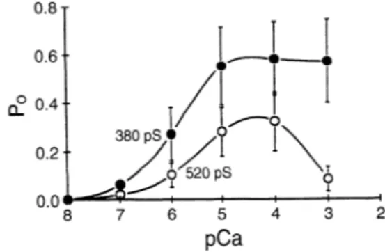
0.8
0.6
O 0.4 a,
FIG. 5. Open probability (P,) of RyR channels from toadfish white
swimming muscle shows 2 types of channels with different conductance
(520 and 380 pS) and Ca2’ dependence, as expected from a muscle
containing both cy- and P-RyR isoforms. Comparison with channels from
fast extraocular muscles, containing only cy-isoform, identifies this as a
channel that has a higher conductance and a narrower bell-shaped re-
sponse to Ca2+. a-Isoform in fish has been identified with RyRl. [From
O’Brien et al. (234).]
concentrations
and is inhibited at higher Ca”+ concentra-
tions. The other type, presumably
the p or RyR3, has a
wider
sensitivity
to Ca”‘. Thus 0 or RyR3 channels
of
skeletal muscle are functionally
more similar to RyR2 than
RyRl, i.e., a- and P-RyRs in fish also differ in their Ca”+
sensitivities,
indicating
a similarity
between
P-RyR and
RyR3 in these animals
(234; see Ref. 22 for a review)
(Fig. 5). Excitation-contraction
coupling
in en/en chick
embryos,
which lack RyRl as a result of a null mutation
but express
low levels of RyR3, is dependent on extracel-
lular Ca2+, confirming
the RyRl is needed for skeletal
type e-c coupling (133). An initial characterization
of RyR3
isolated from brain, taking advantage of its cross-reactiv-
ity for antibodies
against the skeletal p-isoform,
indicates
properties
similar to those of the muscle RyR3 isoform
(224).
However,
some recent data do not agree with the
assessment
of the differences
between skeletal cy - and p-
isoforms
given above. In bullfrog skeletal muscle, the two
types of RyRs seem to have similar
Ca2’ dependence,
although sensitivity
of the ,&isoform
was increased in the
presence
of 1 M NaCl (223). In dyspedic myotubes
from
mice carrying a null mutation
of the gene for RyRl (315),
RyR3 channels
respond
to caffeine,
Ca2’, and adenine
nucleotides,
but contrary to the fish RyR3 (234), they have
a much lower sensitivity
to Ca”’ than RyRl. One possibil-
ity for the difference
is that mouse muscle RyR3 were
tested in situ, where their properties
may be affected by
intrinsic
modulators.
-4. Why skeletal muscle has two
ryanodine
receptor isoforms
What is the meaning of the coexistence
of two RyR
isoforms
in approximately
equal amounts in some skeletal
muscle fibers? The phylogenetic
distribution
of RyR iso-
forms in vertebrates
indicates that this condition is “primi-
tive,” whereas
a dominant isoform is present in more ad-
vanced organisms and/or in the fast-acting muscles of
some of the lower vertebrates (233; see Ref. 22 for a re-
view) (Fig. 4). The RyRl isoform is a channel readily inac-
tivated by Ca2+ and thus more likely to turn itself off
during a cycle of activity, whereas RyR3, with its extended
sensitivity to Ca”, may be more readily activated and
more capable of a sustained activity. On the basis of these
observations and on the different sensitivity to Ca”+ of
the CY-
and P-isoforms, it is proposed that the RyRl isoform
evolved to allow rapid cycles of muscle activity (234). It
will be interesting to see whether a continued exploration
of functional characteristics and phylogenetic relation-
ships of RyR channels in invertebrate muscles dedicated
to rapid activity will confirm this intriguing hypothesis.
C . Sequence and Primary Structure
of Ryanodine Receptors
Primary sequences of the three known RyR isoforms
predict molecules of +OOO amino acids and -560 kDa
(112, 228, 244, 313, 326, 353). This is slightly larger than
the mass of the peptides constituting the purified mole-
cule (127, 130, 131, 160), and definitely larger that the
doublet of -350 kDa that was obtained in early studies
(148). The latter is clearly due to proteolysis, to which
the molecule is particularly prone.
Hydropathy plots indicate a large hydrophilic NH2-
terminal region, thought to constitute the cytoplasmic do-
main of the molecule or “foot” and a smaller, mostly hy-
drophobic COOH-terminal region, predicted to form the
intramembrane channel. This is in good agreement with
the general structure of the molecule. The membrane-
crossing region is the most highly conserved domain of
the molecule (112, 228, 244, 313, 326, 353) and has strong
similarity with the same region of the InsP3 receptor (100,
214). In the foot region, four repeated motifs of - 120
residues, occurring in 2 tandem pairs, are located in corre-
sponding positions in the 3 isoforms (112, 228, 244, 353).
These four stretches are missing in the InsP3 receptor,
suggesting that their insertion (followed by duplication)
in the RyR sequence contributed to the evolutionary diver-
gence between the two molecules.
There is some disagreement on the number of mem-
brane crossings of the COOH-terminal region of the mole-
cule. Model 1 (313) predicts a molecule with 4 intramem-
brane domains of -20 amino acids each (Ml-M4) located
in the COOH-terminal “tenth” with the Ml between amino
acids 4564 and 4580. Model 2 (324,353) proposes 10 mem-
brane crossings in the COOH-terminal “fifth” (Ml-Ml0 be-
tween amino acids 3978 and 4932) and 2 additional ones
in the middle of the molecule (M’ and M’ ‘, respectively,
at amino acids 3123-3143 and 3187-3205). Model 3 (24)
proposes six membrane crossings in the COOH-terminal
regions and four more in the foot region, but at locations
different from model 2. Model 4 (326) supports the pres-
ence of at least six membrane-spanning regions, based on
the alignment of the human RyR2 and on the cytoplasmic
position of the epitope to a monoclonal antibody. All mod-
els generally agree on the position of the most hydropho-
bic domains closest to the COOH-terminal.
Predictions of the three models have been put to the
test, and the results are more often in favor of model 1.
Experimental evidence supports the commonly predicted
cytoplasmic location of NH2- and COOH-terminals (105,
192). Thus the molecule requires an even number of mem-
brane crossings.
Extensive proteolysis of the isolated RyR shows nu-
merous cleavage sites in the proposed foot region of the
molecule, whereas the highly hydrophobic region of the
molecule constitutes a large fragment (46, 55, 187). The
Ml-M2 and M3-M4 loops of model 1 contain several nega-
tively charged amino acids (187), consistent with their
proposed luminal location in that model. It is suggested,
however, that the Ml segment of model 1 may need to be
five amino acids longer (4559-4580) to fully cross the
membrane (187). Binding of a hydrophobic probe after
calpain hydrolysis confirms that the majority of the trans-
membrane segments are located in large fragments at the
COOH-terminal, but also indicates a weak hydrophobic
segment in the middle of the molecule, in partial support
of model 2.
Antibodies against specific amino acids sequences in-
dicate two luminal regions (amino acids 4581-4640 and
4860-4886) (105). The luminal position at amino acids
4860-4886 discriminates between models 1 and 2, fa-
voring the former. In addition, an antibody assigns a lumi-
nal location to amino acids 4879-4898, which constitute
one of the putative membrane-spanning regions in model
2. This also implies that a second putative membrane seg-
ment (amino acids 4789-4820) should not exist, since the
total number of membrane crossings should be even and
also because this crossing would result in a luminal loca-
tion of Arg-4756, which is a cytoplasmic tryptic cleavage
site (34).
D. Ryanodine Receptor Channel: Its Function
and Modulation
1. Agents affecting
channel activity
ryanodine receptor
Activity of the SR Ca2+ release channel is modulated
by a variety of agents (see Refs. 59, 206, 236 for reviews).
Calcium in the micromolar range and adenine nucleotides
at millimolar concentrations are activators, acting equally
to increase the channel open probability and to induce
rapid Ca2+ release from the SR, whereas Mg2+, also in
millimolar concentrations, is an inhibitor (124, 204, 205,
207,209,225,257,288,289; see Ref. 84 for a review). These
experiments provide a direct link between the activity of
706
CLARA FRANZINI-ARMSTRONG AND FELICIANO PROTASI Volume 77the Ca2+ release channel and the role of the SR in rapid
Ca2+ release. Under appropriate,
not necessarily
physio-
logical, conditions,
Ca2’ triggers
a sudden, massive
re-
lease of Ca2’ from the SR both in skinned fibers (see Refs.
75, 76 for reviews)
and in the isolated SR (204, 238). This
CICR is a property
of the SR Ca2’ channels, as well as of
InsP3 receptors.
Several pharmacological
agents have become tools
for studying the function
of RyR channels,
and also for
locating their presence
in cells: caffeine, an activator
of
the channel; ruthenium
red, a blocker; ryanodine and dox-
orubicin, agents with dual effects.
Caffeine at concentrations
in the millimolar
range
induces muscle contracture
and reduces the Ca2+ accumu-
lation ability of the SR. The effect is stronger
in the heavier
SR fraction
and is seen as a reduced coupling
between
ATP hydrolysis
and Ca2’ accumulation,
indicating
leaky
vesicles
(338, 339). Caffeine action on the muscle fiber is
also due to its effect on the SR, since caffeine does not
change membrane polarization
(65) and the drug can act
directly on skinned muscle fibers. Indeed, caffeine allows
CICR to occur in skinned
skeletal fibers even at Mg2+
concentrations
that would normally inhibit this phenome-
non, by acting directly
on the Ca2+ release channels
of
the heavy SR (124, 209, 274; see Refs. 75, 76 for reviews).
This is confirmed
by the Ca2’ dependence (272, 273) and
ryanodine inhibition
of caffeine action (78). However,
al-
though there is agreement that caffeine at low Ca2’ con-
centrations
increases
frequency
of open-channel
events
(242, 272, 273, 287), there is some disagreement
on
whether
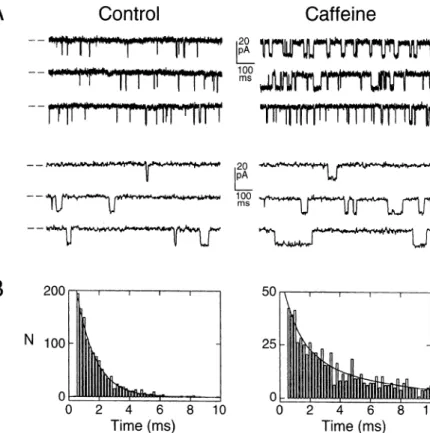
the duration of the events is also affected. Figure
6 illustrates
the effect of caffeine on the single-channel
properties
of the cloned expressed
RyR, showing
a defi-
nite increase in mean open time. The effect of caffeine
on binding of ryanodine is related to its effect on channel
opening (see sect.
11D2).
However,
caffeine may have two
modes of action (287). At relatively
low
concentrations
(<2 mM), the effect of caffeine is dependent on the pres-
ence of Ca2+, and it may simply be due to an increased
sensitivity
of the Ca2’ activation
site. At higher caffeine
levels, the channels open in the absence of Ca”+ and with
different kinetics,
while maintaining
the same permeabil-
ity, indicating
a more direct effect of caffeine.
Ruthenium
red is an agent that completely
blocks
CICR (178, 204, 218, 238, 289) and is often used as a tool
to check for RyR-dependent
Ca2’ leaks from the SR.
Calmodulin
is a cytoplasmic,
Ca2+-dependent
enzyme
regulator.
Millimolar
CaM inhibits
Ca2’ release from the
heavy SR of cardiac and skeletal muscle (209), and it
partially reduces single-channel
open time without
affect-
ing the unitary conductance
(291). The effect occurs
in
the absence of ATP, and thus it is not mediated by channel
phosphorylation
(98). Recent experiments,
however,
indi-
cate a more complex
response
of the channels
to CaM
and a possible role of this protein in modulating
channel
activity during contraction.
The effect of CaM on the chan-
nels is Ca2+ concentration
dependent;
at the concentra-
tions prevalent in a relaxed muscle, CaM increases
open
probability
of the RyR channel and SR Ca2’ release by
severalfold,
whereas
at the higher Ca2’ concentrations
to
be expected during muscle activation,
it has the opposite
effect (32,106,325).
This dual mode of action is confirmed
in skinned
muscle fibers, where
CaM enhances
CICR at
low Ca2’ concentrations
and inhibits it at high concentra-
tions (126). Calmodulin
binding to the channel is also Ca”+
dependent. It is estimated that at low Ca2+ concentration
(<O. 1 PM), 16 CaMs bind with high affinity to one tetra-
mer, whereas
at higher Ca2’ concentrations,
4 CaMs bind
(325). However,
under conditions
similar to those during
contraction
and relaxation, the half time of CaM dissocia-
tion is very slow
so that during a contraction-relaxation
cycle most of the CaM remains bound (325). However,
CaM rate of activity is not known,
and thus it is not clear
whether
it may exert its inhibitory
modulation
during a
short contraction.
Thus the effect of CaM on possible
positive or negative feedback
of Ca2’ on the RyRs during
contraction
(see Ref. 277 for a review)
cannot be pre-
dicted.
In addition to its direct action, CaM may affect chan-
nel activity through
Ca2+/CaM-dependent
protein kinase
(CaMKII).
Phosphorylation
by CaMKII affects channel ac-
tivity
and the effect of CaM on it (111, 117, 119, 175,
291, 335). Opposite
effects of phosphorylation
reported
in these papers may have to do with the site of phosphory-
lation and/or ionic conditions
of experiments.
Both car-
diac and skeletal channels need to be phosphorylated
to
be active under physiological
Mg2+ concentrations
(111,
196), but it is not known
whether
phosphorylation
plays
a modulatory
role during e-c coupling in either muscle.
Because phosphorylation
of the cardiac channel seems to
play a role opposite to the direct action of CaM at high
Ca2’ concentration,
the exact interplay
of these effects
during muscle activity remains to be ascertained.
Doxorubicin
is a widely
used chemotherapeutic
agent, which
can cause a cardiomyopathy,
possibly
due
to sensitization
of RyRs to activation by two physiological
agents, Ca2+ and ATP (254, 256, 356), followed
by an ac-
tual decline in RyR density (66). Doxorubicin
has been
used as a high-affinity
label for the RyR (354). The effect
on the isolated channel is an initial activation,
followed
by an irreversible
inhibition,
which
occurs
with a time
delay, but not in a concentration-dependent
manner (241).
Dithiothreitol
protects
against the final inhibitory
action,
indicating
importance
of sulfhydryl
groups for the func-
tional integrity of the channel (241).
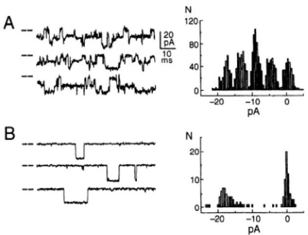
2. Complex action of ryanodine
Ryanodine
is a neutral plant alkaloid
derived from
the stem and root of Ryana speciosa,
a plant native of
Trinidad.
Its action is complex; in the whole animal and
A
Control
--/
-- fl
I3
200N 100
0
0
2 4 68 10
Time (ms)
Caffeine
FIG. 6. A: single-channel properties of cloned ex-
pressed RyRl (left) and its activation by 5 mM caffeine
(right). Caffeine increases open probability as well as du-
ration of events. B: mean open time before caffeine is 1.2
ms (Left), and after caffeine is 9.0 ms (right). [From On-
drias et al. (242).]
25
0
0 2 4 6 81
Time (ms)
when applied to an intact muscle, ryanodine can produce
either flaccidity
or an intense contracture,
whose onset is
accelerated
by activity (see Ref. 136 for a review).
The
reason for these apparently
contradictory
effects is the
direct, dual action of ryanodine
on the heavy SR (85)
and on the SR Ca”+ release channels (290). Low and high
concentrations
of ryanodine have opposite effects on Ca’+
retention by the heavy SR; at low
concentrations,
ryano-
dine results
in Ca”
loss, whereas
at higher concentra-
tions, it has a blocking action similar to that of ruthenium
red (79,85,306,351).
Corresponding
to these two actions,
heavy SR has high-affinity
(85, 163, 258) and low-affinity
(198) sites for ryanodine.
The above observations,
and
particularly
the high affinity for ryanodine,
provided
the
basis for the identification
and purification
of the RyR.
At low concentrations
(< 10 PM), ryanodine locks the
channels in a partially
open subconductance
state (275,
290). This effect is strongly dependent on the channel condi-
tion at the time of ryanodine binding. Ligands known
to
open channels and stimulate Ca2’ release from the SR (PM
Ca2’ or mM ATP, caffeine) stimulate ryanodine binding to
the high-affinity
site (53; see Ref. 59 for a review).
Calcium,
which has both activating and inactivating
effect on RyRs,
also dually affects ryanodine binding (162, 211, 258). This
is thought to be due to better accessibility
of the high-
affinity ryanodine binding site in the open channel. Thus
ryanodine binding can be used as a probe of channel confor-
mation and properties
(352; see Ref. 208 for a review).
As the concentration
of ryanodine is increased, the
affinity of RyRs for ryanodine
decreases
(198, 211, 258).
The effect has been described as an allosteric negative inter-
action between four initially identical binding sites (one on
each monomer).
The first ryanodine molecule binds with
high affinity to the open channel, blocking it into the par-
tially open configuration
(see above), and reducing binding
to the other sites. Three more binding steps follow,
each
with increasingly
lower affinity, indicating a different con-
formational
state, until one ryanodine
per monomer
is
bound and a long-lived state is reached, in which ryanodine
is occluded and the channel is totally blocked (30, 39, 259).
The effect occurs equally well after cross-linking
with bi-
functional reagents, indicating that the decreased ryanodine
affinity is the result of interactions
within the tetrameric
molecule (39). However,
the action of some cross-linkers
results in a tetramer
that is capable of binding ryanodine
at high affinity and of occluding it, but has lost the low-
affinity sites (284). Pretreatment
with 100 mM ryanodine
decreases
maximum
binding of high-affinity
sites and in-
duces loss of the low-affinity
ones, perhaps by uncoupling
the four negatively cooperative binding sites. Oxidation
of
critical receptor thiols is implicated in the process
(351). A
closer look at dissociation
constants
implies the presence
of two distinct binding sites, which
are allosterically
or
sterically coupled (336, 337).
3. Ryanodine
recep tom and inositol
l&5-
trisphosphate
receptors
have
different
phamnacological
procfiles
Inositol 1,4,5-trisphosphate
is a specific activator
of
a class of widely
distributed
intracellular
Ca2* release
708
CLARA FRANZINI-ARMSTRONG AND FELICIANO PROTASI Volume 77channels
closely related to the RyRs (16, 72, 100). The
InsP3 receptors
are tetramers
with a general configuration
similar to that of RyRs, including
the large cytoplasmic
domains
(43). Some cells (most notably smooth muscle
and Purkinje cells) express both RyRs and InsPB receptors
at high levels, but the two receptors
are located in differ-
ent areas of the cell and with different distributions
(150,
334). Most cells have a prevalence
of either one or the
other of the two Ca2+ release channels (see Refs. 17, 122
for reviews).
The pharmacological
profiles
of RyRs and InsP3 re-
ceptors
are quite different
(70, 71). For example, ruthe-
nium red and relatively
high concentrations
of ryanodine
totally block the RyR channel, but neither has an effect
on the InsP3 receptor
(72, 247). Caffeine, the facilitator
of
CICR in the RyR, is an inhibitor
of the InsP3 receptor
activity (19). Heparin is a competitive
inhibitor
of InsP3
on the InsP3 receptor
(195), but it activates isolated RyRs
in a Ca2+-dependent
manner (20).
After the observation
that InsP3 induces Ca2+ release
from the isolated SR (329) and in skinned
muscle fibers
(328), it was proposed
that InsP, may directly affect the
SR Ca2+ release channels and play a role in e-c coupling.
The evidence for and against this hypothesis
has been
well reviewed
(see Ref. 134). The hypothesis,
however,
has fallen into disfavor, following
inability of other labora-
tories to confirm a definite effect of InsP3, particularly
on
frog fibers (167) and on single SR channels (72). However,
large-conductance
SR channels
(presumably
RyRs)
of
Chilean frogs seem to respond quite readily to InsPs (302).
Heparin microinjected
into single intact skeletal muscle
fibers has no effect on depolarization-activated
contrac-
tions (249), but it does block InsP3-induced
Ca2’ release
in smooth muscle (156), a tissue rich in InsP3 receptors
(195). Some effect of heparin on e-c coupling may depend
on its action on the t-tubule DHPR, rather than on the
RyR (165).
E. Developmental
Regulation
Ryanodine receptors
are expressed
early during mus-
cle differentiation,
and they are regulated by growth
fac-
tors, similar to myofibrillar
proteins
(4, 189, 188). Ryano-
dine receptors
are detected in a tetrameric
form as early
as embryonal day -4 (E4) in chick myocardium
(69), and
feet are visible as early as E2.5 by electron microscopy
(263).
Differential
regulation
of a- and ,&RyR isoforms
in
skeletal muscle has been shown in the chicken (305). The
cu-isoform is expressed
first in breast muscle, and it is the
only form detected between
El0 and E15. Around E15,
the ,&isoform
first appears, and both are then synthesized
at increasingly
higher levels while the fibers mature. Note
that the appearance
of the ,&isoform
coincides
approxi-
matelv with the transition
between
myotubes
and muscle
(313) - 0 a (353) - a.0 u, (167) - 0 0 0 0 a> (244) - l mm 2
(228)
- n n a, (112) - A ii!PW
- 0 0 0 Tij (47) - 0 = (106) - (326) - mmm I I I I N-terminal 1,000 2,000 3,000 4,000 C-terminalAmino Acid Residues
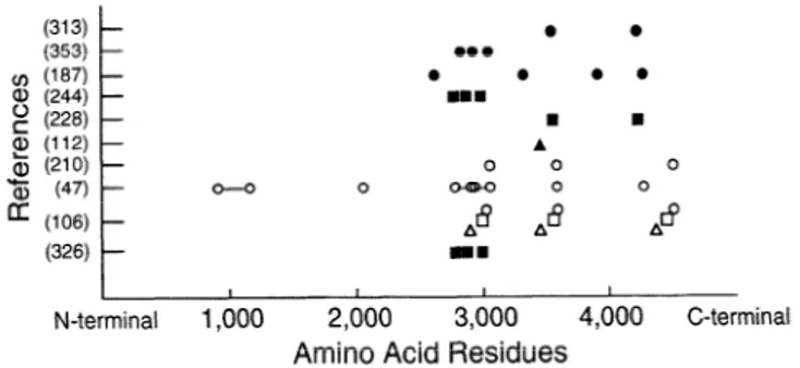
FIG. 7. Locations of proposed (solid syn-tbols) and experimentally
demonstrated (open symbols) calmodulin (CaM) binding sites. Circles,
RyRl; squares, RyR2; triangles, RyR3. Consensus has been reached on
general location of 3 CaM binding sites (CaMl-CaM3), located in region
preceding most hydrophobic domain of molecule, which starts at amino
acid 4564 for RyRl. Some disagreement exists on details, and also on
total number of binding sites.