Innovative superparamagnetic iron-oxide nanoparticles coated with
silica and conjugated with linoleic acid: Effect on tumor cell growth
and viability
Giuliana Muzio
a, Marta Miola
b,d,1, Sara Ferraris
b,1, Marina Maggiora
a, Elisa Bertone
b, Maria Paola Puccinelli
c,
Marina Ricci
a, Ester Borroni
d, Rosa Angela Canuto
a,1, Enrica Verné
b,⁎
,1, Antonia Follenzi
d,⁎⁎
,1a
Department of Clinical and Biological Sciences, University of Turin, Corso Raffaello 30, 10125 Turin, Italy
b
Department of Applied Science and Technology, Politecnico di Torino, Corso Duca degli Abruzzi 24, 10129 Turin, Italy
cDepartment of Laboratory Medicine, Azienda Ospedaliera Universitaria, Città della Salute e della Scienza, Corso Bramante 88/90, 10126 Turin, Italy dDepartment of Health Sciences, University "Amedeo Avogadro" of East Piedmont, Novara, Italy
a b s t r a c t
a r t i c l e i n f o
Article history:
Received 18 October 2016
Received in revised form 14 February 2017 Accepted 9 March 2017
Available online 10 March 2017
One of the goals for the development of more effective cancer therapies with reduced toxic side effects is the op-timization of innovative treatments to selectively kill tumor cells. The use of nanovectors loaded with targeted therapeutic payloads is one of the most investigated strategies. In this paper superparamagnetic iron oxide nano-particles (SPIONs) coated by a silica shell or uncoated, were functionalized with single-layer and bi-layer conju-gated linoleic acid (CLA). Silica was used to protect the magnetic core from oxidation, improve the stability of SPIONs and tailor their surface reactivity. CLA was used as novel grafting biomolecule for its anti-tumor activity and to improve particle dispersibility. Mouse breast cancer 4T1 cells were treated with these different SPIONs. SPIONs functionalized with the highest quantity of CLA and coated with silica shell were the most dispersed. Cell viability was reduced by SPIONs functionalized with CLA in comparison with cells which were untreated or treat-ed with SPIONs without CLA. As regards the types of SPIONs functionaliztreat-ed with CLA, the lowest viability was ob-served in cells treated with uncoated SPIONs with the highest quantity of CLA.
In conclusion, the silica shell free SPIONs functionalized with the highest amount of CLA can be suggested as ther-apeutic carriers because they have the best dispersion and ability to decrease 4T1 cell viability.
© 2017 Elsevier B.V. All rights reserved.
1. Introduction
Some progress has been made in thefield of anticancer therapies, al-though the research on new strategies tofind even more effective ther-apies and to reduce their toxic side effects is still needed. The development of innovative treatments to kill tumor or metastatic cells is a challenging goal and for this reason, several possible approaches are under investigation. Today nanomedicine-based therapies are used in cancer research because they can bypass cancer cell multi-drug resis-tance, poor solubility of hydrophobic anti-cancer drugs, and the use of
dangerous radiations[1]. Cancer nanomedicine pays particular atten-tion to superparamagnetic iron oxide nanoparticles (SPIONs), that can reach the tumor sites carrying chemotherapeutic drugs, nucleic acids, monoclonal antibodies, viral vectors engineered with therapeutic sui-cide genes or shRNAs[1,2,3]. SPIONs are also used for diagnostic assays, generation of local hyperthermia for tumor therapy or tissue repair by delivering stem cells[4,5]. In fact, SPIONs can be combined with contrast fluorescent agents to improve cancer cell imaging[1]. In hyperthermia therapy, SPIONs are localized near the cancer site by magnetic driving and can induce localized heating by an external alternating magnetic field. For example in “in vitro” experiments, 14 nm magnetic nanoclusters killed about 74% of MCF-7 cancer cells, by applying tem-perature of 45 °C for 1 h[6]. Similarly, a temperature of 43 °C for about 17 min reduced the viability of HeLa cells exposed to an alternat-ing magneticfield in the presence of silica coated iron oxide nanoparti-cles (NPs)[7]. SPIONs can be internalized in human mesenchymal stem cells without affecting viability and structure. Following this, the SPION-loaded stem cells can be attracted to specific sites by applying an exter-nal magneticfield[8].
⁎ Corresponding author.
⁎⁎ Correspondence to: A. Follenzi, Department of Health Sciences, University "Amedeo Avogadro" of East Piedmont, Via Solaroli 17, 28100 Novara, Italy.
E-mail addresses:[email protected](G. Muzio),[email protected]
(M. Miola),[email protected](S. Ferraris),[email protected](M. Maggiora),
[email protected](E. Bertone),[email protected](M.P. Puccinelli),
[email protected](M. Ricci),[email protected](R.A. Canuto),
[email protected](E. Verné),[email protected](A. Follenzi).
1
Co-shared authorship.
http://dx.doi.org/10.1016/j.msec.2017.03.063
0928-4931/© 2017 Elsevier B.V. All rights reserved.
Contents lists available atScienceDirect
Materials Science and Engineering C
When preparing SPIONs for the delivery of chemotherapeutic agents it is important to control their size, shape and surface properties, in order to assure their stability in solution and maintain super-paramagnetic properties also in the presence of a variety of molecules. SPIONs which are smaller than 100 nm can easily circulate in the blood without being captured by reticulo-endothelial cells and can therefore selectively accumulate in the tumor microenvironment[3]. On the contrary unmodified SPIONs tend to aggregate into large clusters and several studies suggest surface modification to avoid NP aggrega-tion. A widely explored solution is surface coverage by polymers that has the unavoidable disadvantage of causing an increase in particle size and a potential loss in magnetic response[1]. Alternatively, small intermediary molecules can be used, acting as stabilizing agents and for the subsequent coupling of functional molecules. Fatty acids, in par-ticular oleic acid (OA), have been used as capping agent to obtain mono-disperse SPIONs suspensions[9].
This study aimed to prepare functionalized SPIONs able to affect tumor cell viability. SPIONs, either coated with silica shell or uncoated, were prepared and functionalized with conjugated linoleic acid (CLA) in single- and bi-layer configuration. Silica was used for its ability to pro-tect the magnetic core from oxidation, to improve the magnetite stabil-ity and to tailor the surface reactivstabil-ity by improving biomolecule grafting
[10]. CLA was chosen to improve SPION dispersion and to add an anti-cancer potential[11–13]. Fatty acids are crucial structural and function-al cell components, and contribute to the functionfunction-al/physicfunction-al/chemicfunction-al features of membranes[11–13]. Concerning cancer, it is well known that carcinogenesis is characterized by changes in fatty acid composi-tion of membrane phospholipids.
2. Materials and methods
2.1. Synthesis of superparamagnetic iron-oxide nanoparticles (Fe3O4)
Among the different methods for the SPIONs production, the co-pre-cipitation process was selected since it is simple, rapid and allows a high yield. An aqueous solution of Fe2+and Fe3+salts in a 1:2 M ratio was
prepared by mixing FeCl2 ∗ 4H2O and FeCl3∗ 6H2O in bi-distilled
water; subsequently the mixture was mechanically mixed at 300 rpm and Fe3O4precipitation occurred by adding NH4OH drop by drop, until
the pH of the mixture reached about 10. The suspension was put in ul-trasound for 20 min; subsequently an aliquot was washed twice with bi-distilled water to remove the unreacted reagents (Fe3O4), while the
other aliquot was not washed (Fe3O4-NW).
2.2. One-step synthesis of conjugated linoleic acid-capped Fe3O4
In this procedure conjugated linoleic acid (CLA) was added in a single-step to the NP suspensions. Afirst synthesis of CLA-capped Fe3O4NPs was carried out by adding drop by drop 3.0μl of CLA/ml
of NP suspension, to both washed (Fe3O4+ CLA1) or not washed
(Fe3O4-NW + CLA1), NP under mechanical mixing. A second synthesis
(Fe3O4+ CLA2) was performed using a higher amount of CLA (4.5μl
CLA/ml of NPs suspension). All suspensions were heated at 80 °C with stirring at 150 rpm for half hour. At the end of the functionalization pro-cess NP suspensions were washed twice with ethanol and re-suspended in bi-distilled water.
2.3. Two-step synthesis of conjugated linoleic acid-capped Fe3O4
In this procedure CLA was added in two steps to the NP suspensions. The two-step CLA-capped Fe3O4NPs were synthesized by adding, in a
second step, a further aliquot of CLA (3.0μl CLA/ml of NP suspension) in the suspension of the one-step CLA-coated Fe3O4NPs, both washed
(Fe3O4+ CLA1-TS) and not washed (Fe3O4-NW + CLA1-TS). The
sus-pension was placed in orbital shaker at 80 °C at 150 rpm for half hour. Subsequently, NPs were washed twice with ethanol and re-suspended
in bi-distilled water, adjusting the pH at 12 by adding NH4OH drop by
drop[14].
2.4. Synthesis of silica-coated magnetite nanoparticles (Fe3O4-SiO2)
In order to promote the single nanoparticles coating in a uniform way and obtain a stable suspension, Fe3O4NPs were stabilized with
0.05 M citric acid. The pH was adjusted to 5.2 by dropwise NH4OH and
the suspension was placed 90 min at room temperature in orbital shak-er (KS 4000i control, IKA®) at 150 rpm allowing the deprotonation of two carboxylic groups of citric acid and the bond to the OH groups ex-posed by Fe3O4NPs[15]. Subsequently, citric acid-functionalized NPs
were washed with bi-distilled water using an ultrafiltration device (Solvent Resistant Stirred Cells - Merck Millipore) and re-suspended in bi-distilled water, adjusting the pH at about 10.1 to induce the depro-tonation of the third carboxylic group, which allow an optimal NP dis-persion. Then, magnetite NPs functionalized with citric acid were coated with a silica shell (Fe3O4-SiO2) by sol-gel method, by adding
TEOS (tetraethoxysilane) as silica precursor, ethanol as catalyst and water as solvent, to Fe3O4NPs suspended in a water and ethanol
solu-tion (water: ethanol 1:4)[16]. The pH of suspension was adjusted at 10 (NH4OH); the suspension was placed in orbital shaker at room
tem-perature for 3 h at 150 rpm. Subsequently, the Fe3O4-SiO2NPs were
washed with bi-distilled water using an ultrafiltration device (Solvent Resistant Stirred Cells - Merck Millipore) and re-dispersed in water.
2.5. One-step synthesis of conjugated linoleic acid-capped Fe3O4-SiO2
On the base of the results obtained for CLA coated Fe3O4, Fe3O4-SiO2
were functionalized with the higher CLA amount (4.5μl CLA/ml of NPs) with one-step procedure (Fe3O4-SiO2+ CLA2). The suspension was
heated at 80 °C and stirred at 150 rpm for half hour. At the end of the functionalization process CLA capped Fe3O4-SiO2NPs were washed
twice with ethanol and re-suspended in bi-distilled water.
Table 1resumes the name and samples composition. All chemicals were purchased from Sigma-Aldrich Co. (St Louis, MO, USA).
2.6. Nanoparticles characterization
All synthesized NPs were subjected to morphological characteriza-tion by scanning transmission electron microscopy (STEM, MERLIN Zeiss– Germany). For STEM observation, a drop of diluted NP suspen-sion was deposited on a copper TEM grid with carbonfilm (SPI Sup-plies® Brand Lacey Carbon Coated 200 Mesh Copper Grids– JEOL S.p.A.). Fourier transformation infrared spectroscopy (FT-IR) was used to evidence the effective grafting of CLA and to confirm the presence of silica shell. FT-IR spectra were acquired in a Hyperion 2000 FT/IR (Tensor 27, Bruker Optics S.p.A, Ettlingen, Germany) from 4000 to 400 cm−1and with 2 cm−1resolution. OPUS software (v. 6.5, Bruker S.p.A) was used for instrumental control and spectral acquisition. Sus-pension stability and dispersion were evaluated in a semi-quantitative way by measuring the time required for particle precipitation.
Table 1
Name and composition of the samples.
Acronym NPs Washing CLA CLA [μl] per 1 ml of NPs suspension Fe3O4+ CLA1 Fe3O4 Yes one-step 3.0
Fe3O4-NW + CLA1 Fe3O4 No one-step 3.0
Fe3O4+ CLA1-TS Fe3O4 Yes two-step 3.0
Fe3O4-NW + CLA1-TS Fe3O4 No two-step 3.0
Fe3O4+ CLA2 Fe3O4 Yes one-step 4.5
2.7. Cell cultures: treatment of 4T1 with Fe3O4NPs capped or not with CLA,
or with CLA alone, and of MS1 cells with Fe3O4NPs capped or not with CLA
Mouse breast cancer 4T1 cells were seeded (12,500 cells/cm2) in
DMEM/F-12 medium supplemented with 2 mM glutamine, 1% (v/v) antibiotic/antimycotic solution, 10% (v/v) fetal bovine serum (FBS) and maintained at 37 °C in a humidified atmosphere of 5% CO2
in air. Mouse pancreatic islet endothelial MS1 cells were seeded (12,500 cells/cm2) in DMEM medium supplemented with 1% glutamine, 1% (v/v) antibiotic solution, 10% (v/v) fetal bovine serum (FBS). Both cell lines were maintained at 37 °C in a humidified atmosphere of 5% CO2in
air and 24 h after seeding, culture medium was removed and replaced by the same medium not supplemented or supplemented with Fe3O4,
Fe3O4-SiO2, Fe3O4 + CLA1, Fe3O4-NW + CLA1, Fe3O4 + CLA1-TS,
Fe3O4-NW + CLA1-TS, Fe3O4+ CLA2 and Fe3O4-SiO2+ CLA2 at the
concentrations 8μg or 16 μg of NPs/100,000 cells. Twenty-four hours after seeding, the 4T1 cells were also treated with CLA alone at 10 and 25μM concentrations. The experimental times were 24, 48 and 72 h. 2.7.1. Cell viability
Cell viability was evaluated by means of the MTT assay after 24, 48 and 72 h of treatment. 4T1 or MS1 cells grown in multiwells were added with 30μl of 4.5-dimethylthiazol-2-yl-,5-diphenyltetrazolium bromide (MTT, 5 mg/ml) in PBS solution, and incubated for 3 h at 37 °C in a humidified atmosphere of 5% CO2in air. After the supernatants
were removed and 150μl of DMSO was added to each well. After 20 min of incubation, the absorbance was measured at 590 nm using a microplate reader (Dynatech MR580 microElisa, USA).
2.7.2. Iron staining
After 24 and 72 h of treatment in chamber slides, the iron stain KIT HT20 (Sigma-Aldrich Co., St Louis, MO, USA) was used to evidence the SPIONs functionalized or not with CLA internalized by 4T1 and MS1 cells.
2.7.3. CLA percentage content in incubated cells
As indication of CLA-capped NP internalization, the CLA percentage content was determined in lipids extracted from 6 × 10⁶cells previously
incubated with Fe3O4, Fe3O4+ CLA1, Fe3O4+ CLA2 or Fe3O4-SiO2
+ CLA2 NPs for 72 h. After treatment, the cells were washed with PBS + EDTA (0.53 mM), detached with trypsin/EDTA (0.25%/0.3%), and cen-trifuged at 600 g for 10 min at 4 °C.
Lipids, isolated by Folch et al. method[17], were suspended in 0.5 ml of methanol containing internal standard 1,2-dihenarachidoyl-sn-glycero-phosphocholine. The method of Klem et al.[18]for analysis of red blood cell fatty acid composition was adapted for the determination of CLA in 4T1 cell lipids. The extracts were evaporated under nitrogen flow, dissolved with chilled methanol containing 2,6 di-ter-butyl-4-methyl-phenol (BHT) as antioxidant, treated for 15 min in an ultra-sound bath and then centrifuged. The supernatants were treated with a methanol solution of sodium methoxide to synthetize CLA methyl es-ters, and after 5 min a hydrochloric acid solution was added. CLA methyl esters were extracted twice with hexane, evaporated under nitrogen flow at 35 °C, and re-dissolved in hexane containing BHT for injection in gas chromatograph–mass spectrometer. CLA were identified and quantified with a mass spectrometer operating in electron impact ioni-zation (EI) mode. The selection of ions for selective ion monitoring (SIM) was based on comparison with standards and those reported in the literature[19,20].
2.8. Statistical analysis
All data are expressed as means ± SD. Differences between group means were assessed by analysis of variance followed by a post hoc Newman–Keuls test.
3. Results
3.1. Nanoparticles synthesis and characterization
The superparamagnetic properties and the XRD pattern of Fe3O4NPs
obtained with analogue procedure have been previously assessed[21].
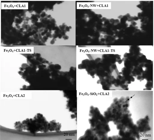
Fig. 1shows STEM analyses of Fe3O4+ CLA1, Fe3O4-NW + CLA1,
Fe3O4+ CLA1-TS, Fe3O4-NW + CLA1-TS, Fe3O4+ CLA2 and Fe3O4
-SiO2+ CLA2 NPs. All NPs present a spherical shape with a dimensional
range of about 5–15 nm, with the exception of silica coated NPs, seem-ing slightly bigger (up to 20 nm). A silica shell (of about 1–2 nm) is well visible in the micrograph (last panel ofFig. 1) and is evidenced by arrows.
FT-IR spectra of pure CLA, Fe3O4+ CLA1, Fe3O4-NW + CLA1,
Fe3O4+ CLA1-TS, Fe3O4-NW + CLA1-TS are reported inFig. 2.Fig. 2a
shows the spectrum of CLA, in which two peaks at about 2852 e 2922 cm−1are assigned respectively to the asymmetric and symmetric stretching of CH2; the peak at about 1710 cm−1can be attributed to the
C_O stretch vibration, the peak at 1460 cm−1to the\\COO− asymmet-ric stretch vibration[14], the peak at 1408 to the“umbrella” bending mode of CH3group[14], the band between 1250 and 1285 cm−1can
be associated to the presence of the C\\O stretch or more generally to the vibration of the COOH group in oleic acid[22,23], the peaks at 981 and 945 cm−1are characteristic of the cis,trans conjugated dienes[24]
and the peak at about 720 cm−1can be assigned both to CH2bending
or rocking vibration and CH_CH vibration[24,23]. No significant differ-ences can be observed in the FT-IR spectrum of pure CLA treated at 80 °C (not reported), as a confirmation that the functionalization procedure does not alter the molecule.Fig. 2b shows the whole spectrum for all NPs, showing peaks at about 560 cm−1, ascribable to Fe\\O stretching vibrational mode of Fe3O4, peaks at about 2852 e 2922 cm−1that can
be assigned respectively to the asymmetric and symmetric stretching of CH2group of CLA, and a peak at about 1408 cm−1which can be
as-cribed to the“umbrella” bending mode of CH3group[14]. Focusing
the analysis between 1000 and 4000 cm−1(Fig. 2c), it is possible to no-tice also the presence of a broad band at about 1500–1640 cm−1that
can be attributed to the asymmetric and symmetric stretch vibration of COO−group reported in literature for fatty acids adsorbed on Fe3O4
[14,22]. Moreover, in the Fe3O4+ CLA1-TS and Fe3O4-NW + CLA1-TS
curves, the band at about 1250–1285 cm−1and the peak at about
1710 cm−1appeared (thefirst one ascribed to the C\\O stretch vibra-tion, or to the vibration of the COOH group in oleic acid[22,23], the sec-ond one to the C_O stretch vibration[14]).
The FT-IR spectra of Fe3O4+ CLA2 and Fe3O4-SiO2+ CLA2 NPs are
re-ported inFig. 3. The silica presence is evidenced by: a new peak at about 1060 cm−1, attributed to the asymmetric stretching of the Si\\O\\Si group, two peaks at 960 and 780 cm−1, attributed Si\\OH stretching vi-bration and Si\\O\\Si symmetric stretching[25,26], and a band at about 3000–3300 cm−1, ascribable to OH groups. The peaks at 2852 e
2922 cm−1can be assigned respectively to the asymmetric and symmet-ric stretching of CH2group of CLA, the band at about 1500–1640 cm−1,
at-tributed to the asymmetric and symmetric stretch vibration of adsorbed COO−group[14,22], and the peak at about 1408 cm−1ascribed to the “umbrella” bending mode of CH3group. Also in this samples the peak at
about 1710 cm−1, ascribable to the C_O vibration of CLA, appears.
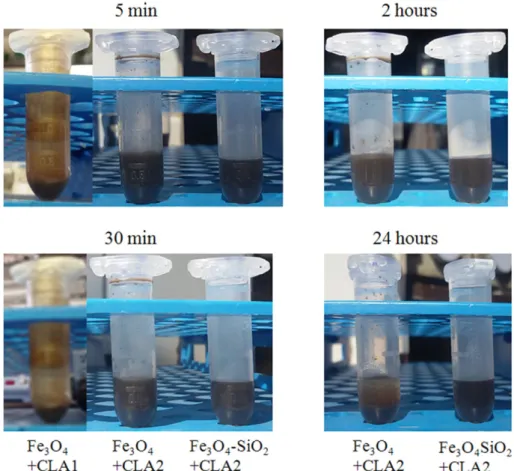
Fig. 4reports the behavior of the various NP suspensions after different times of sedimentation. The precipitation of Fe3O4+ CLA1
NPs starts after 5 min and is already complete after 30 min. The Fe3O4+ CLA2 and Fe3O4-SiO2+ CLA2 solutions are both still stable
after 2 h, and only Fe3O4-SiO2+ CLA2 also after 24 h.
3.2. The effect of SPIONs and CLA alone on mouse breast cancer 4T1 cells and on mouse pancreatic islet endothelial MS1 cells
SPIONs, washed or not, and SPIONs functionalized with CLA (low and high amount, one- or two-steps) were tested to choose the preparation
showing the highest inhibition of 4T1 cell proliferation. All functionalized particles decrease cell number in comparison with SPIONs not functional-ized with CLA (Fe3O4), being the major inhibition obtained by SPIONs
washed and capped with CLA with one-step procedure (data not
shown). Therefore, in the following experiments, only Fe3O4,Fe3O4+
CLA1 and Fe3O4+ CLA2, with or without silica shell, were compared.
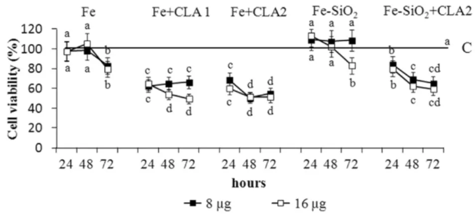
Fig. 5shows the percentages of viability in cells treated with various SPIONs in comparison with the control cells, set to 100%. The viability in
Fig. 1. STEM images of SPIONs. Fe3O4+ CLA1, washed CLA-capped Fe3O4NPs; Fe3O4-NW + CLA1, not washed CLA-capped Fe3O4NPs; Fe3O4+ CLA1-TS, washed two step CLA-capped
Fe3O4NPs; Fe3O4-NW + CLA1-TS NPs, not washed two step CLA-capped Fe3O4NPs; Fe3O4+ CLA2; Fe3O4-SiO2+ CLA2, silica shell-coated, washed CLA-capped Fe3O4NPs. CLA1, 3.0μl
of CLA/ml of NPs; CLA2, 4.5μl of CLA/ml of NPs.
Fig. 2. FT-IR spectra. (a) FT-IR spectra of pure CLA; (b) whole spectrum of Fe3O4-NW + CLA1 (curve I), Fe3O4-NW + CLA1-TS (curve II), Fe3O4+ CLA1 (curve III), Fe3O4+ CLA1-TS
cells treated with Fe3O4+ CLA1, Fe3O4+ CLA2 or Fe3O4-SiO2+ CLA2
is lower than in control cells and in cells treated with Fe3O4 and
Fe3O4-SiO2.
When the cells were treated with SPIONs coated or not with silica and capped or not with CLA, the reduction of viability is higher in cells treated with Fe3O4+ CLA2 than in the cells treated with other ones.
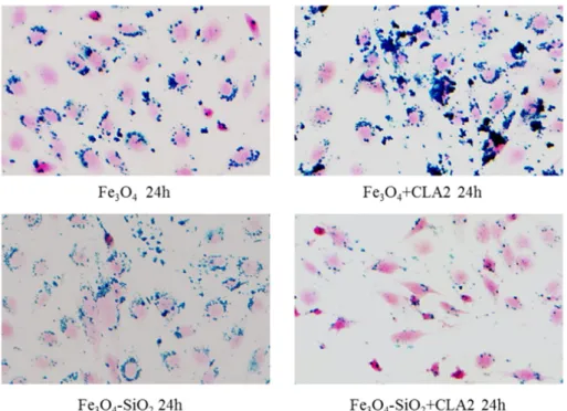
No significant difference was found between 48 and 72 h for cells treat-ed with SPIONs + CLA for both quantity of NPs. The NP internalization by cells was evidenced with iron staining (Fig. 6). NPs functionalized or not with CLA are present in the cells, whereas no staining is evident in cells untreated with NPs. The analysis by gas chromatography-mass
spectrometry of lipids extracted from 4T1 cells incubated with SPIONs coated or not with silica and capped or not with CLA shows that CLA is incorporated in lipids, and it is higher in cells treated with Fe3O4+ CLA2 in comparison with cells treated with Fe3O4+ CLA1
NPs (Table 2). With regards the incorporation of CLA in lipids extracted from 4T1 cells incubated with Fe3O4-SiO2+ CLA2, theTable 2showed a
lesser incorporation of CLA for isomer 1 and 2 and a slight increase for the isomer 3.
The effect of CLA alone on 4T1 cell viability is reported inTable 3. Two concentrations of CLA similar to the quantity used for the prepara-tion of SPIONs were added to the 4T1 cells. A time-dependent reducprepara-tion of cell viability was observed.
To verify whether the different types of SPIONs affected in different way normal and cancer cells, pancreatic islet endothelial MS1 cells were treated. For these experiments, only CLA2 (4.5μl CLA/ml of NPs) was used.Table 4shows that no decrease of viability was induced by all types of SPIONs in comparison with control cells. In particular there were no differences between SPIONs capped or not with CLA.
Fig. 7reports that NPs coated or not with silica and capped or not with CLA are present in the MS1 cells.
Prussian blue staining was used to evidence the presence of SPIONs with and without silica capped or not with CLA in pancreatic islet endo-thelial MS1 cells. As shown in the panels, all the SPIONs highly interact with MS1 cells, with the exception of silica coated with CLA, that are less internalized in cells. It was reported only the iron staining relative to 16 μg/100,000 cells of various SPIONs after 24 h of treatment. SeeTable 1
for NP acronyms. 4. Discussion
In recent years, NPs have been proposed for biomedical utilization
[27–31]. This approach implies the synthesis of biocompatible NPs, which are also stable and easily manageable in biological media. For
Fig. 3. FT-IR spectra. FT-IR spectra of Fe3O4+ CLA2 (curve I) and Fe3O4-SiO2+ CLA2
(curve II) NPs. See legend inFig. 1for NP acronyms.
this reason, several coatings have been proposed to improve colloidal stability, prevent oxidation, and improve biocompatibility of NPs[10]. Among them, fatty acids have been also investigated.
Some authors have used OA, which belongs to unsaturated monocarboxylic acids, to functionalize Fe3O4NPs; in particular the
study reported by Hemei Chen et al.[32]showed that OA capped Fe3O4NPs possessed high magnetization and were useful to further
adsorb other biomacromolecules. Other studies, instead of using a single adsorption layer of OA, stabilized magnetic NPs by short-chain-length monocarboxylic acids, such as lauric acid and myristic acid
[33], but the magnetic NPs were in part aggregated[34]. However, mag-netic NPs coated with myristic or lauric acids were cytotoxic for glio-blastoma cells, showing low toxicity for normal astrocytes [33]. Starting from the evidence that fatty acids seem biocompatible for healthy cells and toxic for cancer cells, the rationale of their use as cap-ping agent for magnetic NPs should be adjusted, trying to optimize their role as NPs driven therapeutic agents, not only as biocompatible surfactants.
In this study, CLA was chosen as capping agent for magnetic NPs for its antitumor properties[11–13]; its effect on colloidal stability of NPs suspensions and its potential therapeutic effect, when grafted on NPs, were evaluated. The effect of silica coating was also investigated. The superparamagnetic properties of pure Fe3O4NPs were previously
assessed[21]. The size and morphology of the NP batch prepared for this research were investigated and a good reproducibility of the results was observed in respect of previous preparations, both for uncoated and silica coated magnetic NPs. The presence of CLA on the magnetic NPs was evidenced by the FT-IR analysis. The results showed the presence of iron oxide and of organic species ascribable to CLA on all samples. It can therefore be assumed that all the functionalization methods were successful. Interestingly it can be evidenced that different signals ascrib-able to CLA, which could be related to the presence of a single- or bi-lay-ered configuration, were present. All functionalized particles showed the signals related to the CLA main chain (2852 e 2922 cm−1of CH2
group and 1408 cm−1of CH3group[14]). Moreover, in all
functional-ized NPs also the band ascribed to asymmetric and symmetric stretch
Fig. 5. Viability of mouse breast cancer 4T1 cells exposed to Fe3O4NPs coated or not with silica and capped or not with CLA. The values are means ± S.D. of 4 experiments and are expressed
as % of control cells set equal to 100%. The absorbance values of control are 0.591 ± 0.192 for 24 h, 1.457 ± 0.151 for 48 h and 1.596 ± 0.168 for 72 h. For each NP quantity (8 or 16μg), means with different letters are significantly different from one another (p b 0.05) as determined by analysis of variance followed by a post-hoc Newman-Keuls test. SeeTable 1for NPs acronyms. C, control cells (black line); 8μg, 16 μg, were the quantity of various NPs added to 100,000 cells.
Fig. 6. Iron staining. Prussian blue staining was used to evidence the presence of SPIONs with and without silica capped or not with CLA in the mouse breast cancer 4T1 cells. It was reported only the iron staining relative to 16μg/100,000 cells of various SPIONs after 72 h of treatment. C, control cells without SPIONs; seeTable 1for NPs acronyms.
vibration of COO−group (1500–1640 cm−1) appeared[14,22]. This
band was not present in the FT-IR spectrum of pure CLA (i.e. not adsorbed on any support) since it can be related to the covalent interac-tion between COO−groups and Fe atoms in a chelating bidentate inter-action with iron oxide surfaces[22]. The patterns of the Fe3O4+
CLA1-TS and Fe3O4-NW + CLA1-TS revealed signals also present in not
adsorbed CLA (1250–1285 cm−1for C\\O[22,23]and 1710 cm−1
C_O[14]), but absent in the patterns of CLA adsorbed on NPs with a single-step procedure and low CLA content (see for example the pat-terns of Fe3O4+ CLA1, Fe3O4-NW + CLA1 and[23]), as a demonstration
of the presence of free\\COOH groups exposed on the NP surface, typical of a bi-layer configuration[14]. The functionalization with highest CLA amount, even in a single step (Fe3O4 + CLA2 and
Fe3O4-SiO2+ CLA2), gave FT-IR results similar to Fe3O4+ CLA1-TS
and Fe3O4-NW + CLA1-TS NPs both with and without the presence of
silica shell. In this case the excess of CLA probably produced a secondary layer, with a configuration similar to the one obtained by the two-step synthesis procedure[14].
The NPs functionalized with CLA in a one step were chosen, among different preparations of SPIONs, because they reduced cell via-bility more than NPs coated with CLA with two-step procedure (data not shown). Two amounts of CLA (one step) were used: Fe3O4+ CLA1
(3μl CLA/ml of NPs) and Fe3O4+ CLA2 (4.5μl of CLA/ml of NPs). The
re-sults obtained showed that both Fe3O4+ CLA1 and Fe3O4+ CLA2
re-duced the cell viability of 4 T1 cells treated for 24 and 72 h, respect to control cells not incubated with SPIONs and to 4 T1 cells incubated with SPIONs without CLA, but Fe3O4+ CLA2 was more effective in the
reduction than Fe3O4+ CLA1. For Fe3O4+ CLA2, the viability values
were not significantly different between 48 and 72 h. A similar trend was observed in 4 T1 cells treated with CLA alone.
To be highlighted that the experiments, carried out to evaluate the effect of various SPIONs on normal cells, showed that SPIONs capped or not with CLA2 and coated or not with silica did not decrease the via-bility of the normal MS1 cells.
The NP precipitation time with both CLA amounts was also deter-mined. Although all the particles seem aggregated in STEM images of
Fig. 1suspensions stable up to 24 h were obtained (Fig. 4), as discussed below. This observation may be explained by the fact that STEM images of the particles were obtained after drying a drop (5μl) of the suspen-sion onto a carbon coated copper TEM grid. Upon drying onto a
substrate particles obviously aggregates and appear as agglomerates in the images.
As shown inFig. 4the precipitation time was shorter for Fe3O4+ CLA1
than for Fe3O4+ CLA2: at 30 min Fe3O4+ CLA1 NPs were all precipitated,
while Fe3O4+ CLA2 NPs remained in suspension up to 2 h. This difference
can be explained taking into account that Fe3O4+ CLA2, even if
synthe-sized with one step procedure, were covered by a secondary layer of CLA (see FT-IR results). It can be supposed that the highest amount of CLA caused a condition similar to that of bilayer-coated NPs (even if the bilayer was probably not continuous). It is reasonable that Fe3O4+ CLA2 NP surface is more hydrophilic in comparison to
that of Fe3O4 + CLA1 ones (which do not expose free\\COOH
groups) and, in turn, NPs show a better stability in aqueous medium. The determination, by gas chromatography-mass spectrophotome-try, of CLA isomers in the lipids extracted from cells incubated with SPIONs functionalized or not with CLA, showed that this fatty acid was more incorporated in the cells treated with Fe3O4+ CLA2 NPs than in
the cells treated with Fe3O4+ CLA1 NPs. The results of this study
indi-cate that the use of the highest amount of CLA (4.5μl/1 ml of NPs) im-proves the suspension stability, the CLA incorporation into the cells and the antitumor activity. It worth of mentioning that it is very impor-tant to avoid the formation of clusters, because they partially lose the magnetic properties, and, when injected intravascularly, show a shorter circulation time due to rapid clearance by monocytes, this decreasing the reaching of target cells or tissues.
Interestingly the NPs coated with a thin layer of silica and function-alized with CLA showed a further increase of time required to have the precipitation, in fact at 24 h Fe3O4-SiO2+ CLA2 NPs were all
dis-persed in solution. This effect can be mainly attributed to the citric acid functionalization needed for silica coating, and cannot be ascribed exclusively to CLA amount. Fe3O4-SiO2+ CLA2 NPs also cause a
reduc-tion of cell viability in comparison with control cells and CLA-free Fe3O4-SiO2, but the inhibition was lower than that obtained with
Fe3O4+ CLA2. In the same way, the incorporation of CLA in lipid
ex-tracted from the cells treated with Fe3O4-SiO2+ CLA2 NPs was lower
for the isomers 1 and 2 than that obtained with Fe3O4+ CLA2.
This difference could be ascribed to the role of the silica shell avoiding the direct contact between the magnetite NPs and the biolog-ical environment, which in turn reduces their potential toxicity.
Moreover, according to the reported data, we can infer that fatty acids may improve the affinity between NPs and cell membrane and favor particle internalization in tumor cells. This is an important issue for future research, for example for studies with other cancer cell lines as well as for magnetic NP-assisted gene therapy, to improve the use of viral vectors to transfer therapeutic genes to target cells[35–37]. 5. Conclusions
In this research colloidal suspensions of Fe3O4NPs, both uncoated or
coated by silica shell, of about 5–20 nm in diameter, were successfully prepared and functionalized with different amounts of CLA both in
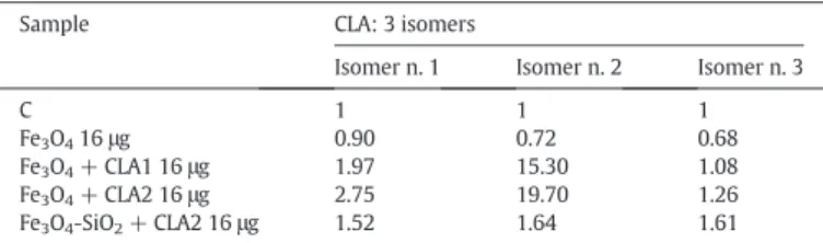
Table 2
Conjugated linoleic acid (CLA) in lipids extracted from 4T1 cells treated for 72 h with SPIONs coated or not with silica and capped or not with CLA. The values are means of two experiments and are expressed as number of times the control cells (C) set equal to 1. See legend inTable 1for NPs acronyms. 16μg were the quantity of NPs added to 100,000 cells.
Sample CLA: 3 isomers
Isomer n. 1 Isomer n. 2 Isomer n. 3
C 1 1 1 Fe3O416μg 0.90 0.72 0.68 Fe3O4+ CLA1 16μg 1.97 15.30 1.08 Fe3O4+ CLA2 16μg 2.75 19.70 1.26 Fe3O4-SiO2+ CLA2 16μg 1.52 1.64 1.61 Table 3
Viability of mouse breast cancer 4T1 cells exposed to CLA.
Sample 24 h 48 h 72 h
C 100 100 100
CLA 10μM 91.7 69.1 72.0
CLA 25μM 86.5 69.2 69.5
The values are means of 2 experiments and are expressed as % of control cells set equal to 100%. The absorbance values of control are 0.591 ± 0.192 for 24 h, 1.457 ± 0.151 for 48 h and 1.596 ± 0.168 for 72 h.
C, control cells.
Table 4
Viability of pancreatic islet endothelial MS1 cells exposed to Fe3O4NPs coated or not with
silica and capped or not with CLA. The values are means of 2 experiments and are expressed as % of control cells set equal to 100%. The absorbance values of control are 0.256 for 24 h, 0.771 for 48 h and 0.601 for 72 h.
Sample 24 h 48 h 72 h 8μg 16μg 8μg 16μg 8μg 16μg C 100 100 100 100 100 100 Fe3O4 97.09 103.41 111.47 103.24 151.99 140.76 Fe3O4+ CLA2 94.80 104.86 125.76 103.63 146.53 133.11 Fe3O4-SiO2 101.56 110.86 114.02 107.13 127.03 131.44 Fe3O4-SiO2+ CLA2 102.94 114.79 118.96 94.16 136.76 148.91
SeeTable 1for NPs acronyms. C, control cells; 8μg, 16 μg, were the quantity of various NPs added to 100,000 cells.
single- and bi-layer configurations. Both the silica shell and CLA layer played a role in determining suspension stability, especially when CLA was grafted on the NP surface in a bi-layer configuration. The results ev-idenced that the viability of mouse breast cancer 4T1 cells was reduced in presence of Fe3O4NPs functionalized with CLA in comparison with
both untreated control cells and cells treated with CLA-free NPs. The presence of the silica coating in CLA capped NPs caused a lesser inhibi-tion of cell viability. Since it is known that the colloidal stability must be assured to avoid uncontrolled NPs clusterization, loosening of mag-netic properties and short circulation time, control of this parameter is crucial, even if it is not directly related to the antitumoral effect ob-served in this work. Based on these results, silica shell free Fe3O4NPs
functionalized with high amount of CLA (with bi-layered configuration) can be suggested as therapeutic carriers for their good dispersion and ability to decrease mouse breast cancer 4T1 cell viability.
Acknowledgments
This work was supported by grants from AIRC, Italy: (IG n. 13166), “Development of engineered magnetic nanoparticles for cancer therapy”, http://www.airc.itto A.F., from Compagnia di San Paolo (CSP-Torino-Piemonte: 12-CSP-C04-018), Italy to AF, and from the Uni-versity of Turin (Local Research Funding ex-60%), Italy to RAC. References
[1] G. Kandasamy, D. Maity, Recent advances in superparamagnetic iron oxide nanopar-ticles (SPIONs) for in vitro and in vivo cancer nanotheranostics, Int. J. Pharm. 496 (2015) 191–218.
[2] A. Schroeder, M.M. Winslow, J.E. Dahlman, G.W. Pratt, R. Langer, T. Jacks, D.G. Anderson, Treating metastatic cancer with nanotechnology, Nat. Rev. Cancer 12 (2012) 39–50.
[3] S. Pietronave, D. Locarno, L. Rimondini, M. Prat, Functionalized nanomaterials for di-agnosis and therapy of cancer, J. Appl. Biomater. Biomech. 7 (2009) 77–89.
[4] B.J. Tefft, S. Uthamaraj, J.J. Harburn, M. Klabusay, D. Dragomir-Daescu, G.S. Sandhu, Cell labeling and targeting with superparamagnetic iron oxide nanoparticles, J. Vis. Exp. (2015)http://dx.doi.org/10.3791/53099.
[5] B.D. Brown, L. Naldini, Exploiting and antagonizing microRNA regulation for thera-peutic and experimental applications, Nat. Rev. Genet. 10 (2009) 578–585.
[6] D. Maity, P. Chandrasekharan, P. Pradhan, K.H. Chuang, J.M. Xue, S.S. Feng, J. Ding, Novel synthesis of superparamagnetic magnetite nanoclusters for biomedical appli-cations, J. Mater. Chem. 21 (2011) 14717–14724.
[7] J. Majeed, L. Pradhan, R.S. Ningthoujam, R.K. Vatsa, D. Bahadur, A.K. Tyagi, Enhanced specific absorption rate in silanol functionalized Fe3O4 core-shell nanoparticles: study of Fe leaching in Fe3O4 and hyperthermia in L929 and HeLa cells, Colloids Surf. B: Biointerfaces 122 (2014) 396–403.
[8] N. Landázuri, S. Tong, J. Suo, G. Joseph, D. Weiss, D.J. Sutcliffe, D.P. Giddens, G. Bao, W.R. Taylor, Magnetic targeting of human mesenchymal stem cells with internal-ized superparamagnetic iron oxide nanoparticles, Small 9 (2013) 4017–4026.
[9] J. Park, K. An, Y. Hwang, J.G. Park, H.J. Noh, J.Y. Kim, J.H. Park, N.M. Hwang, T. Hyeon, Ultra-large-scale syntheses of monodisperse nanocrystals, Nat. Mater. 3 (2004) 891–895.
[10]W. Wu, Z. Wu, T. Yu, C. Jiang, W.S. Kim, Recent progress on magnetic iron oxide nanoparticles: synthesis, surface functional strategies and biomedical applications, Sci. Technol. Adv. Mater. 16 (2015) 023501.
[11] M.A. Belury, Inhibition of carcinogenesis by conjugated linoleic acid: potential mechanisms of action, J. Nutr. 132 (2002) 2995–2998.
[12]M. Maggiora, et al., An overview of the effect of linoleic and conjugated-linoleic acids on the growth of several human tumor cell lines, Int. J. Cancer 112 (2004) 909–919.
[13] G. Muzio, M. Oraldi, A. Trombetta, R.A. Canuto, PARalpha and PP2A are involved in the proapoptotic effect of conjugated linoleic acid on human hepatoma cell line SK-HEP-1, Int. J. Cancer 121 (2007) 2395–2401.
[14]K. Yang, H. Peng, Y. Wen, N. Li, Re-examination of characteristic FTIR spectrum of secondary layer in bilayer oleic acid coated Fe3O4 nanoparticles, Appl. Surf. Sci. 256 (2010) 3093–3097.
[15] S. Campelj, D. Makovec, M. Drofenik, Preparation and properties of water-based magneticfluids, J. Phys. Condens. Matter 20 (2008) 204101 (5pp) 10.1088/0953-8984/20/20/204101.
[16] W. Stöber, A. Fink, Controlled growth of monodisperse silica spheres in the micron size range, J. Colloid Interface Sci. 26 (1968) 62–69.
[17] J. Folch, M. Lees, G.H. Sloane Stanley, A simple method for the isolation and purifica-tion of total lipides from animal tissues, J. Biol. Chem. 226 (1957) 497–509.
[18] S. Klem, M. Klinger, H. Demmelmair, B. Koletzko, Efficient and specific analysis of red blood cell glycerophospholid fatty acid composition, PLoS One 7 (2012) e33874.
[19] T.Y. Yen, B. Stephen Inbaraj, J.T. Chien, B.H. Chen, Determination of conjugated linoleic acids and cholesterol oxides and their stability in a model system, Anal. Biochem. 400 (2010) 130–138.
[20] N. Sánchez-Ávila, J.M. Mata-Granadosa, J. Ruiz-Jiménez, M.D. Luque de Castro, Fast, sensitive and highly discriminant gas chromatography–mass spectrometry method for profiling analysis of fatty acids in serum, J. Chromatogr. A 1216 (2009) 6864–6872.
[21] E. Verné, et al., Iron-oxide nanoparticles used for target cancer gene therapy and hy-perthermia, Proceedings of the 29th Annual Meeting of the European Society for Hy-perthermic Oncology, 56, Minerva Med 2014, p. 16.
[22] L. Zhang, R. He, H.C. Gu, Oleic acid coating on the monodisperse magnetite nanopar-ticles, Appl. Surf. Sci. 253 (2006) 2611–2617.
[23]V.V. Korolev, A.G. Ramazanova, A.V. Blinov, Adsorption of surfactants on superfine magnetite, Russ. Chem. Bull. 51 (2002) 2044–2049 (Int. Ed.).
[24] J.V. Kadamne, C.L. Castrodale, A. Proctor, Measurement of Conjugated Linoleic Acid (CLA) in CLA-Rich Potato Chips by ATR-FTIR Spectroscopy, J. Agric. Food Chem. 59 (2011) 2190–2196.
[25] B. Mojic, K.P. Giannakopoulos, Z. Cvejic, V.V. Srdic, Silica coated ferrite nanoparticles: influence of citrate functionalization procedure on final particle morphology, Ceram. Int. 38 (2012) 6635–6641.
[26] A. Fatemeh, H. Ali, N. Sirous, Surface modification of Fe3O4@SiO2 microsphere by si-lane coupling agent, Int. Nano Lett. 3 (2013) 23.
[27] S. Mornet, S. Vasseur, F. Grasset, P. Veverka, G. Goglio, A. Demourgues, J. Portier, E. Pollert, E. Duguet, Magnetic nanoparticles design for medical applications, Prog. Solid State Chem. 34 (2006) 237–247.
[28]S.C. Wuang, K.G. Neoh, E.T. Kang, D.W. Pack, D.E. Leckband, Heparinized magnetic nanoparticles: in-vitro assessment for biomedical applications, Adv. Funct. Mater. 16 (2006) 1723–1730.
[29] E. Duguet, S. Vasseur, S. Mornet, J.M. Devoisselle, Magnetic nanoparticles and their applications in medicine, Nanomedicine 1 (2006) 157–168.
[30] J.P. Fortin, C. Wilhelm, J. Servais, C. Ménager, J.C. Bacri, F. Gazeau, Size-sorted anionic iron oxide Nanomagnets as colloidal mediators for magnetic hyperthermia, J. Am. Chem. Soc. 129 (2007) 2628–2635.
[31]N.A. Brusentsov, L.V. Nikitin, T.N. Brusentsova, A.A. Kuznetsov, F.S. Bayburtskiy, L.I. Shumakov, N.Y. Jurchenko, Magneticfield hyperthermia of the mouse experimental tumor, J. Magn. Magn. Mater. 252 (2002) 378–380.
[32] H. Chen, S. Liu, Y. Li, C. Deng, X. Zhang, P. Yang, Development of oleic acid-function-alized magnetite nanoparticles as hydrophobic probes for concentrating peptides with MALDI-TOF-MS analysis, Proteomics 11 (2011) 890–897.
[33]M.V. Avdeev, K. Lamszus, L. Vekas, V.M. Garamus, A.V. Feoktystov, O. Marinica, R. Turcu, R. Willumeit, Structure and in vitro biological testing of water-based ferrofluids stabilized by monocarboxylic acids, Langmuir 26 (2010) 8503–8509.
[34]D. Bica, L. Vekas, M.V. Avdeev, O. Marinic, V. Socoliuc, M. Balasoiu, V.M. Garamus, Sterically stabilized water based magneticfluids: synthesis, structure and proper-ties, J. Magn. Magn. Mater. 311 (2007) 17–21.
[35] M. Wiznerowicz, T.D. Harnessing, HIV for therapy, basic research and biotechnology, Trends Biotechnol. 23 (2005) 42–47.
[36]A. Follenzi, L.E. Ailles, S. Bakovic, M. Geuna, L. Naldini, Gene transfer by lentiviral vectors is limited by nuclear translocation and rescued by HIV-1 pol sequences, Nat. Genet. 25 (2000) 217–222.
[37]A. Follenzi, L. Naldini, HIV-based vectors. Preparation and use, Methods Mol. Med. 69 (2002) 259–274.