UNIVERSITY OF MOLISE
DEPARTMENT OF BIOSCIENCES AND TERRITORY
DOCTOR OF PHILOSOPHY IN
BIOLOGICAL AND ENVIRONMENTAL SCIENCE AND TECHNOLOGY XXVII CYCLE
“MOLECULAR STUDIES ON A LARGE MULTI-GENE FAMILY OF
POLYGALACTURONASES IN TWO DIFFERENT
PHYTOPHTHORA SPECIES.”
SCIENTIFIC-DISCIPLINARY SECTOR BIO/04
TUTOR/Coordinator of PhD
PhD STUDENT:
Distinguished Prof
Claudio Caprari
Maria Maistrou
146251
ACADEMIC YEAR 2013-2014
2
3 CONTENTS
Abstract ... 4
1. INTRODUCTION ... 6
1.1 Oomycetes and fungi ... 7
1.2 Oomycetes evolutionary relationship ... 9
1.3 Morphological and physiological differences between oomycetes and fungi. ... 10
1.4 Economically relevant Oomycete species ... 11
1.5 Genus Phytophthora ... 13
1.6 Oomycetes-plant communication ... 15
1.7 Plant Immunity... 19
1.8 Plant Basal Defences ... 24
1.9 The plant cell wall is the first barrier against pathogens ... 28
1.10 Cell Wall Degrading Enzymes (CWDEs) as apoplastic effectors ... 29
1.11 Polygalacturonase (PG) ... 31
1.12 Polygalacturonase inhibiting proteins (PGIPs) and their role in plant defense ... 35
1.13 PG-PGIP interaction ... 36
1.14 Oligogalacturonides ... 37
1.15 Reverse Genetics ... 40
2. AIM OF THE THESIS... 44
3. MATERIALS AND METHODS ... 47
3.1 Used Organisms ... 48
3.2 Culture media for Escherichia coli ... 48
3.3 Culture media for Pichia pastoris... 49
3.4 Culture media for Phytophthora species ... 49
3.5 RNA extraction ... 51
3.6 DNA Methods ... 52
3.7 Cloning Vectors ... 56
3.8 Expression of polygalacturonase genes ... 61
3.9 Vector construction for transformation P.capsici ... 62
3.10 Protein Methods ... 64
3.11 Characterization of P. nicotianae polygalacturonases ... 69
3.12 In silico analysis of Polygalacturonase ... 70
4. RESULTS ... 71
4.1 In silico analysis of the Polygalacturonase: Identification and Phylogenetic analysis ... 72
4.2 Polygalacturonase from P.nicotianae: Expression and Biochemical characterization .... 80
4.3 In vivo Analysis of the Polygalacturonase expression from P.capsici ... 88
4.4 Renewed method for knock out/down genes in P. capsici ... 95
4.5 Silencing of PGs with Hairpin construct in P.capsici. ... 98
5. DISCUSSION ... 102
4
Abstract
MOLECULAR STUDIES ON A LARGE MULTI-GENE FAMILY OF POLYGALACTURONASES IN TWO DIFFERENT PHYTOPHTHORA SPECIES. The plant cell wall is a structural barrier to pathogens, composed of a network of polysaccharides such as cellulose, hemicellulose and pectin. The majority of pathogenic microorganisms produce cell wall degrading enzymes (CWDEs) that are essential for the invasion process. Among the different CWDEs, polygalacturonases (PGs) play a critical role since their action on pectin makes other cell wall components more accessible to other CWDEs and causes tissue maceration. PGIPs (polygalacturonase-inhibiting proteins) are plant cell wall proteins that specifically modulate the activity of the PGs, and hamper the invasion process by limiting the host tissue colonization. The PG–PGIP interaction retards pectin hydrolysis and favors oligogalacturonide (OGs) accumulation and leading to plant defense activation. This work wants to contribute to study the role of the PGs in P. nicotianae and P. capsici, among the most dangerous pathogens for many plant species: Specific points of this thesis are: 1) Identification of the whole set of the PGs from well-known oomycetes, which present different lifestyles. 2) Comparison of large PG families found in the oomycetes species using phylogenetic analysis for tracking evolutionary relationships. 3) Analysis of amino acid sequences on identified PGs to detect domains and/or amino acids involved in PG-PGIP interaction. 4) Characterization of PGs from P. nicotianae and P. capsici. 5) Construction of P. capsici mutants for investigate the role of PGs in the pathogenesis, using different approach of reverse genetics.
The results from this thesis enhances the hypothesis that the multiplicity of PGs may give flexibility to the pathogen, with each enzyme having its own unique properties to contribute to the performance of all the enzymes to successfully colonize plants.
5 MOLECULAR STUDIES ON A LARGE MULTI-GENE FAMILY OF
POLYGALACTURONASES IN TWO DIFFERENT PHYTOPHTHORA SPECIES. Nei primi stadi dell’infezione, i microrganismi fitopatogeni producono un arsenale di enzimi che depolimerizzano in maniera ordinata e sequenziale i componenti della parete cellulare vegetale (CWDEs - Cell Wall Degrading Enzymes). Le poligalatturonasi (PG) sono tra i primi enzimi pectici ad essere prodotti e favoriscono la macerazione del tessuto vegetale rendendo accessibili a cellulasi ed emicellulasi gli altri componenti della parete. I carboidrati rilasciati dal processo degradativo della parete cellulare vengono utilizzati dal patogeno per il sostentamento e per la crescita, conferendo al processo di degradazione un ulteriore significato biologico oltre quello di distruzione fisica della parete. Le PGIP (PolyGalacturonase Inhibiting Proteins) presenti nella parete cellulare vegetale, sono delle glicoproteine in grado di inibire e/o modulare in maniera specifica l’attività delle PG. La formazione del complesso PG-PGIP rallenta la capacità delle PG di degradare l’omogalatturonano della parete cellulare, favorendo l’accumulo di oligogalatturonidi in grado di attivare le risposte di difesa della pianta. Questo lavoro vuole contribuire allo studio del ruolo delle PG in Phytophthora nicotianae e Phytophthora capsici, che sono ritenuti tra gli agenti patogeni più pericolosi per molte specie vegetali. Argomenti specifici che vengono affrontati in questa tesi riguardano: 1) l’identificazione di PG da alcune specie di oomiceti considerate tra le più pericolose per le piante (generi Phytophthora, Pythium ed Aphanomycetes); 2) l'analisi filogenetica delle famiglie geniche PG; 3) l’analisi di sequenze proteiche delle PG identificate allo scopo di rilevare domini e/o amminoacidi responsabili dell'interazione PG-PGIP; 4) la caratterizzazione delle PG da P. nicotianae e P. capsici; 5) la costruzione di mutanti di P. capsici per indagare il ruolo di PG nella patogenesi, utilizzando diversi approcci di genetica “reverse” I risultati ottenuti in questa tesi, potenziano l'ipotesi che la molteplicità delle PG può dare flessibilità al patogeno e ogni PG, con le proprie caratteristiche uniche, contribuisce alla performance del patogeno per colonizzare le piante con successo
6
7
1.1
Oomycetes and fungi
Fungi and oomycetes are the causal agents of many of the world’s most serious plant diseases and are unique among the microbial pathogens, are being able to breach the intact surfaces of host plants, and quickly establish infections that can have disastrous consequences for the environment and large-scale agricultural production. Fungi and oomycetes can also cause disease in animals and humans, and in certain cases mortality (Soanes, Richards, & Talbot, 2007). Through convergent evolution, oomycetes and fungi have acquired striking similarities in their mechanisms of host colonization, including physiological adaptations, mechanisms of adhesion, modulation of host defenses, and strategies of nutrient acquisition (Kale, 2011). Pathogenesis by a fungus or oomycete is a complex process and include the following steps (Meng et al., 2009) (Figure 1.1):
dispersal and arrival of an infectious particle (usually a spore) near the host, adhesion to the host, recognition of the host (which may occur prior to adhesion), penetration into the host,
invasive growth within the host, lesion development in the host,
production of additional infectious particles.
Figure 1.1: Generalized diagram displaying infection and disease cycle caused by fungi and oomycetes. Adapted by Meng et al.,2009.
Plant pathogens can be divided into groups based on the different strategies they employ to colonize plants. (Figure 1.2) (Latijnhouwers, Wit, & Govers, 2003).
1.1.2 Biotrophs
Biotrophy is a lifestyle generally associated with a narrow host range, requiring living host tissue, and involving the establishment of intimate interactions with living plant cells for the
8 exchange of nutrients and signals. They possess haustoria for retrieval of nutrients from plants. Secrete limited amounts of lytic enzymes and cause little damage to the host plant. Their sporangia ripen simultaneously, giving these pathogens a defined infection, proliferation and reproduction phase. (Oliver & Ipcho, 2004).Biotrophic pathogens evade or suppress defense responses (Vleeshouwers & Oliver, 2014).
1.1.3 Necrotrophs
Necrotrophy is a lifestyle generally associated with a wide host range (Lévesque et al., 2010), in which the pathogen invades the host tissue, immediately kills host cells and lives on dead plant material. They have very destructive pathogenesis strategies resulting in extensive necrosis, tissue maceration and plants rots. Necrotrophs secrete disease agents including cell wall degrading enzymes and toxins both prior and during colonization (Laluk & Mengiste, 2010; Oliver & Ipcho, 2004). Cochliobolus and Botrytis species are examples of fungal necrotrophs.
1.1.4 Hemibiotrophs
Hemibiotrophy is a lifestyle that is generally associated with a more limited host range. Hemibiotrophic pathogens initially establish a relationship with living host cells, extracting nutrients from them, yet subsequently kill these host cells as the infection proceeds. Spores are formed while at the same time new plant tissues are being infected. During the biotrophic stage, these pathogens form invasive structures called haustoria. These specialized structures breach the plant cell wall, yet do not penetrate the host cell membrane. Haustoria are thought to serve both in nutrient uptake and delivery of factors for manipulate living host cells (Jonge, 2013; Oliver & Ipcho, 2004).
Plant-pathogenic oomycetes include species with diverse life styles. Downy mildews (Peronosporales) are biotrophic pathogens. Albugo spp. that causes white rust on Arabidopsis are obligate biotrophs (Links et al., 2011). The biotrophic downy mildew pathogen Hyaloperonospora arabidopsidis also infects Arabidopsis and, this plant– oomycete interaction is well characterized (Coates & Beynon, 2010).
Phytophthora spp. include mostly hemibiotrophic pathogens and cause devastating diseases on a variety of staple crops, fruit, ornamentals, and trees (Erwin & Ribiero 1996).
Pythium spp. inhabit water and soil habitats and display necrotrophic growth. On a broad range of plants, Pythium spp. can cause root rot and damping off on seedlings (Laluk & Mengiste, 2010).
9 Figure 1.2: Disease symptoms on Arabidopsis leaves caused by the necrotrophic fungus Botrytis
cinerea, the biotrophic oomycete H. arabidopsidis and the hemibiotrophic bacterium Pseudomonas syringae. Adapted from Corné M J P. et al., 2009
1.2
Oomycetes evolutionary relationship
Currently, at least 800 oomycete species are known, but depending on the definition of a species, this number might actually reach 1500. Nevertheless, the species richness seems low when compared to the number of fungal species known to date: 30000 basidiomycete species have been described and ascomycetes reach a similar number It is, however, likely that there are many oomycetes out there yet to be discovered (Bouwmeester, Poppel, & Govers, 2009).Due to their shared morphology (filamentous, branched somatic structures that bear spores), oomycetes and fungi were traditionally classified in the same kingdom, the Fungi (Erwin & Ribeiro, 1996). For this reason, oomycetes were for a long time considered a class within the kingdom fungi. Oomycetes, also known as water molds, resemble fungi in many ways (Sleigh, M.A. 1989). Like fungi, oomycetes have a global distribution and prosper in quite diverse environments. Both, show filamentous growth in their vegetative stage, produce mycelia and form spores for asexual and sexual reproduction. In recent years, barcode sequences as rRNA internal transcribed spacer (ITS) or cytochrome oxidase (COX) regions have been used for the identification of the species of the oomycetes. This new insights based on molecular phylogeny and comparative genomics reshaped again the tree of life (Cooke, et al., 2000). In this classification, the ascomycete and basidiomycete fungi are grouped together with animals in the supergroup Unikonts (Burki, 2014). Modern molecular and biochemical analyses suggest that oomycetes have taxonomic affinity with to brown algae (heterokonts) in the Stramenopiles, one of several major eukaryotic kingdoms (Figure 1.3) (Sophien Kamoun, 2003).
10 Figure 1.3: Phylogenetic tree showing the evolutionary relationships between the major eukaryotic groups. The Oomycetes and the Ascomycetous and Basidiomy fungi are highlighted in green. To be note the evolutionary distance between the Oomycetes and the fungi. Adapted from Latijnhouwers, Wit, & Govers, 2003
1.3
Morphological and physiological differences between oomycetes and
fungi.
There are important differences, which can justify the phylogenetic distance observed among fungi and oomycetes, including both, morphological and physiological characteristics.
1. Fungi are haploid or dikaryotic during the major part of their lifecycle, whereas oomycetes are diploid and homologous recombination has not been found to occur. Oomycetes are far less tractable to genetic manipulation than many fungi.
2. Fungal hyphae are septate, whereas Oomycete hyphae are non-septate also called coenocytic mycelium.
3. Many Oomycetes are (partial) sterol auxotrophs. Their membranes contain lipids with unusual structures and long-chain fatty acids that presumably replace sterols in mycelial membranes. Stacked Golgi cisternae (versus unstacked in fungi) and, tubular mitochondrial cisternae (versus disclike in fungi).
4. Fungi and Oomycetes synthesize lysine by different pathways. The Oomycetes use the a, ε-diaminopimelic acid pathway, whereas fungi synthesize this amino acid by the α-aminoadipic acid pathway.
5. Oomycetes cell walls lack chitin but are composed of a mix of cellulosic compounds and glycans. In detail the cell wall consists mainly of 1,3-β-glucans, some 1,6-β-glucans and
11 1,4-β glucans (cellulose). Chitin, which is a major constituent of fungal cell walls, is detected in small amounts only in a few oomycetes (Bouwmeester et al., 2009; Latijnhouwers et al., 2003).
6. Most characteristic for oomycetes are the zoospores, the free-swimming asexual spores that are propelled by two unequal flagellae and explain why a moist environment is most favorable for these water molds. One of the flagella has lateral hairlike structures called mastigonemes that contain the β-1,3-glucan mycolaminarin, an energy storage molecule that is also found in brown algae and diatoms (Feofilova, 2001). Literally, oomycetes means ‘egg fungi’, a name based on the egg-shaped resting spores, named oospores. Oomycetes can be either homothallic or heterothallic. Sexual reproduction is initiated upon release of hormones that trigger the formation of gametangia (♀ oogonium and ♂ antheridium) in which meiosis takes place. The diploid oospores are produced as a result of oogamous fertilization when a haploid oosphere fuses to a haploid gamete. Thick-walled oospores are most durable propagules that can survive harsh environmental conditions and are important for the generation and maintenance of genetic variation in a population (Bouwmeester et al., 2009). The role of the zoospores is the transmission of the pathogen from host to host. Zoospores are chemotactically and electrotactically attracted to the surface of potential host plants (Jung, 2007). Spores can also be moved to adjacent places by the wind, explaining their fast dissemination. They are also essential for targeting the site of infection (Figure 1.4-1.5) (Walker & West, 2007).
1.4 Economically relevant Oomycete species
Oomycete diseases occur on nearly every agricultural crop across the globe. Oomycetes include both saprophytes and pathogens of plants, insects, crustaceans, fish, vertebrate animals, and various microorganisms. Some of the most damaging oomycete genera are Aphanomyces (Diéguez-Uribeondo et al., 2009), Peronospora (Cooke et al., 2000), Phytophthora (Bouwmeester et al., 2009), Plasmopara (Göker M et al., 2006), Pseudoperonospora (Runge et al., 2011), and Pythium (André LéVesque & De Cock, 2004). In particular, severe damages are provoked from the obligate biotrophs P, viticola (the agent of downy mildew of grapevine), Albugo, Bremia, and Peronospora species, which cause white rust and downy mildew on several crops. Other important pathogens include more than a hundred species of the genus Pythium, which are abundantly present in water and soil habitats and cause a diversity of plant diseases, mainly in root tissue.
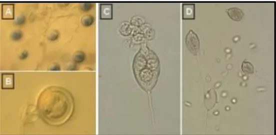
12 Figure 1.4: A) oospores; B) an oospore; C) a sporangium releasing zoospores; D) sporangia and zoospores.
Figure 1.5: Mycelia disks (5mm diameter) of P. nicotianae were cut from the periphery of 4 to 6 days old cultures on V8 agar were transferred individually to the center of new plates. After different periods of incubation the plates were flooded with distilled water to induce the formation of sporangia (under microscope Primo Star Zeiss 100X). M. Maistrou observation
Pythium infections are usually limited to the meristematic tips, epidermis, cortex of roots, and fruits, but occasionally, severe Pythium infections occur when the pathogen moves deeper into plant tissue and reaches the vascular system. Some Pythium species, such as Pythium oligandrum, are essentially beneficial and reduce infections caused by more-severe pathogenic microbes. This can occur directly, through antagonistic effects or mycoparasitism, or indirectly, by induction of defense responses in plants (Sophien Kamoun, 2000, 2003). Most dangerous Phytophthora spp will be analyzed next paragraph. Animal-pathogenic oomycetes, such as species in the genus Saprolegnia, can cause severe losses in aquaculture and fisheries (Bruno, D. W, & B. P. Wood. 1999). At least one
13 species, Pythium insidiosum, is known to infect various mammals, including humans, horses, and dogs. P. insidiosum colonizes cutaneous and subcutaneous tissues and can invade blood vessels and bones, resulting in fatal lesions (Ravishankar, J. P et al., 2001). One genus, Aphanomyces, includes both plant and animal pathogenic species. A facultatively parasitic oomycete, Lagenidium giganteum, infects the larval stage of many mosquito species, and spore formulations of this oomycete have been used for biocontrol of mosquitoes (Woodring et al., 1995).
1.5
Genus Phytophthora
There are more than 60 species of the genus of Phytophthora that are arguably the most devastating pathogens of dicotyledonous plants and cause enormous economic damage to important crop species such as potatoes, tomatoes, peppers, soybeans, and alfalfa, as well as environmental damage in natural ecosystems. Virtually every dicot plant is affected by one or more species of Phytophthora, and several monocot species are infected as well. (Erwin DC & Ribeiro OK,1996; Adhikari et al., 2013).
The most notable pathogenic oomycete is P. infestans,well known for causing the disease that triggered the Irish potato famine in the mid-nineteenth century resulted in the potato blight famine (Figure 1.6) with death and displacement of millions of people (Sophien Kamoun, 2003). Today, P. infestans remains a devastating pathogen, causing losses as high as $5 billion in potato production worldwide through losses in potato and increased fungicide costs. The appearance of highly aggressive and fungicide- insensitive strains in North America and Europe in the 1990s resulted in a new wave of severe and destructive potato and tomato late-blight epidemics (Sophien Kamoun & Smart, 2005).
Other economically important Phytophthora diseases include root rot of soybean, caused by P. sojae; black pod of cocoa, a recurring threat to worldwide chocolate production, caused by P. palmivora and P. megakarya; dieback and related root rot diseases in crops and native plant communities, caused by P. cinnamomi; and sudden oak death, caused by P. ramorum (Sophien Kamoun, 2003).
1.5.1 Life cycle and reproduction of Phytophthora species
Phytophthora species are able to survive unsuitable environmental conditions over several years as dormant resting spores (oospores or chlamydospores) in the soil or in infected tissue. When environmental conditions become suitable (high soil moisture, soil
14 temperature > 10 °C) the resting spores germinate by forming sporangia which release motile, biflagellate (Jung, 2007; Hardham, 2007) In Phytophthora, infection generally starts when motile zoospores released from sporangia reach a leaf or root surface, encyst, and germinate (Sophien Kamoun, 2003).
Figure 1.6: Late blight infection on tomato and potato plants. Lesion can be seen on the stem, on the foliage as well as the fruits.
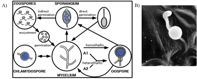
The life cycle of Phytophthora includes sexual and asexual reproduction, with flagellated free-swimming spores. The mycelium of Phytophthora produces branched sporangiophores containing asexual sporangia at their tips. When the sporangium bursts, releases motile zoospores with two flagella, which is typical for heterokonts. Upon the contact with the host, zoospores become immotile and encyst, which leads to the formation of a cell wall. A germ tube protrudes from the cyst forming an appressorium that resembles a swollen tip of the germ tube. The appressorium penetrates the host tissue directly or enters via stomata in the leaf (Figure 1.7_B), and forms an infection vesicle. The mycelium then grows from the infection vesicle in between the plant cells, sometimes with haustoria, which is a mycelium that has penetrated the host cell wall and invaginated the plasma membrane (Grenville-Briggs et al., 2008). Older infected plant cells die while the mycelium continues to spread into the host tissue (Agrios, 2005; Hardham, 2007). The sexual reproduction of Oomycetes results in thick walled resistant oospores (Figure 1.4_A). Oospores are formed when the male reproductive organ, the antheridium, fertilizes the female reproductive organ, the oogonium. For oospore formation in P. infestans to take place, strains of two mating types are needed, A1 and A2. It was not until recently that the A2 mating type emerged from
15 Mexico and spread in the rest of the world, thereby making possible the sexual reproduction of Phytophthora. This has lead to the emergence of more virulent strains due to genetic recombinations of pathogenic characteristics of the mating strains (Figure 1.7_A,) (Agrios, 2005).
Figure 1.7: A) Life cycle of P. infestans with sexual and asexual sporulation. B) Germinated cyst of P. infestans with its appressorium on a potato leaf and (from Grenville‐Briggs et al., 2008)
1.6
Oomycetes-plant communication
Like many other species that survive via dependent interaction with other organisms, oomycetes secrete a suite of proteins with functions including basic metabolic processes, nutrient acquisition, cell wall manufacture/adhesion/digestion, and virulence (Misner et al., 2014). The definition of “pathogen effectors” is: molecules that manipulate host cell structure and function, thereby facilitating infection (virulence factors or toxins) and/or triggering defence responses (avirulence factors or elicitors) (Kamoun 2006). Over recent years, a number of oomycete genomes have been sequenced (Table 1.1), providing many information for studies of oomycete effectors, with important roles in pathogenicity mechanisms.
16
Phytophthora sojae (Tyler et al. 2006)
Phytophthora ramorum (Tyler et al. 2006)
Phytophthora infestans (Haas et al. 2009)
Phytophthora capsici (Lamour et al. 2012)
Pythium ultimum (Lévesque et al. 2010)
Albugo laibachii (Kemen et al. 2011)
Hyaloperonospora arabidopsidis (Baxter et al. 2010),
Saprolegnia parasitica (Jiang et al. 2013) Table 1.1: oomycete sequenced genomes.
The secretome is defined as the sum of all proteins secreted by an organism often reflecting the niche that these microbes reside in, rather than there phylogenetic affinities (Soanes et al., 2008). Analyses of the genomes identified a large repertoire of secreted proteins, known as effectors, during plant infection (Haas et al., 2009; Lamour et al., 2012; Lévesque et al., 2010) such as pectinases, cutinases, protease inhibitors, Nep1-Like Proteins (NLPs), Crinklers (CRNs), elicitins, RXLR-effectors and many more (Haas et al., 2009; Tyler et al., 2006) associated with plant pathogenicity.
Effectors are secreted by the invading pathogen but their site of action in the host differentiates them into two main categories. Extracellular effectors remain in the apoplastic space where they interact with extracellular host molecules. Cytoplasmic effectors, on the other hand, move across the host cell’s plasma membrane and function inside the plant cell (Figure 1.8) (Hardham & Cahill 2010).
Effectors are often under diversifying selection, as they are in a continuous co-evolution with factors from the host (Stahl & Bishop, 2000). Many oomycete effectors have been found in families with extensive gene duplication and gene loss, speeding up the process of diversifying selection. In this category, we can include: cell wall degrading enzymes, trypsin-like serine proteases, berberine-bridge enzymes, carbonic anhydrases, small cysteine-rich proteins, and repeat-containing proteins (Seidl, et al.,2012).
17 Figure 1.8: Role of effectors in fungal and oomycete colonization. Oomycete secrete effectors to facilitate host colonization. A subset of effectors localize and function in the apoplast. Other effectors are able to translocate into host cells and localize in diverse host compartments. These effectors have a variety of cellular targets and many contribute to colonization by modulating host defense machinery. Some effectors are delivered through the haustorium, a site of intimate interaction between pathogen and host formed by certain pathogens, while other effectors enter directly from the apoplast. Adapted from Kale, 2011 .
Despite considerable progress in the control strategies of plant disease, our global food supply is still threatened by a multitude of pathogens and pests. Plant diseases can dramatically reduce crop yield and the impact of disease outbreaks is particularly acute in developing nations. Pesticides provide effective protection but their applicability can be compromised by adverse environmental effects and by the emergence of resistant pathogen strains. For these reasons, much effort has been invested to understand innate resistance mechanisms in plants. Plants can activate a very effective arsenal of inducible defense responses, comprised of genetically programmed suicide of infected cells (the hypersensitive response, HR), as well as tissue reinforcement and toxin production at the site of infection (McDowell & Woffenden, 2003). Plants do not have the benefit of a circulating antibody system so plant cells autonomously maintain constant vigilance against pathogens by expressing large arrays of ‘R genes’ (R, resistance). R genes encode putative receptors that interact to the products of ‘Avr genes’ (Avr = avirulence) expressed by the pathogen during infection (Friedman & Baker, 2007; Jones & Dangl, 2006). In many cases, a single R gene can provide complete resistance to one or more strains of particular pathogen, when transferred to a previously susceptible plant of the same species (Figure 1.9).
18 Figure 1.9: Interactions between pathogen Avr proteins and plant R proteins. A hypothetical pathogen (grey) has attached to a plant cell and is expressing virulence proteins (red). These proteins are translocate into the plant and once inside, they target host proteins (green) that control defense responses, metabolism or other plant process that affect pathogen virulence. (a) In this panel, the plant cell does not express an R protein that is capable of recognizing any virulence protein. Thus, the plant cannot detect the pathogen efficiently and defenses are weakly induced. We have the development of the disease. (b) This panel figure the classic receptor–elicitor hypothesis, in which an R protein directly binds a virulence protein. This recognition event activates a complex signal transduction network, which in turn triggers defense responses. Adapted by McDowell & Woffenden, 2003.
1.6.1 Cytoplasmic effectors
There are two groups of oomycete proteins that they could be categorised as cytoplasmic effectors. The first group is the RXLR effectors, which possess an RXLR motif (arginine–any amino acid–leucine–arginine) near their N-terminus that is involved in effector translocation into the host cytoplasm (Dou et al.2008; Whisson et al., 2007). The second group is the CRN effectors (also known as the Crinkler effectors), so named because they cause leaf crinkling and necrosis (Stam, Jupe, et al., 2013). It has been reported that CRN deletion mutants identify a C-terminal domain that is sufficient to induce cell death when expressed in the plant cytoplasm (Haas et al., 2009; Sophien Kamoun, 2006; Oliva et al., 2010). These proteins have the LXLFLAK motif (leucine–any amino acid– phenylalanine–leucine–alanine–lysine) that is proposed to be involved in their movement into the host cytoplasm (Schornack et al., 2010). Recent sequencing of Phytophthora genomes has revealed that both categories of effectors include large numbers of genes.
Analysis of the genomes of P. infestans, P. sojae and P. ramorum predict that they encode 563, 350 and 350 RXLR, and 196, 100 and 19 CRN effectors, respectively (Haas et al., 2009; Tyler et al., 2006). The large repertoires of predicted cytoplasmic effectors are indicative of their important roles during host infection. Precisely how oomycete cytoplasmic effectors are translocated into the host cytoplasm from the apoplast after
19 secretion by the pathogen is currently not known, although a mechanism involving plant endocytosis has been suggested (Birch et al., 2009; Dou et al., 2008). Both categories of effectors are modular proteins containing a conserved N-terminal region that includes a signal peptide that directs protein secretion and the RXLR or putative LXLFLAK motifs that direct uptake into the host cytoplasm, and a variable C-terminal domain (Haas et al., 2009). However, although there are similarities in the uptake motifs, RXLR-mediated entry of oomycete effectors into plant cells is independent of any other pathogen component (Dou et al., 2008) but the uptake of malarial parasite effectors is not.
1.7
Plant Immunity
The environment is full of dangerous microorganisms, the survival of higher eukaryotic organisms depends on efficient pathogen sensing and rapidly mounted defence responses. Such protective mechanisms are found in all multicellular organisms and are collectively referred as innate immunity. Though plants, in contrast to vertebrates, do not possess an adaptive immune system, their innate immune system effectively protects them from a wide range of different phytopathogenic microorganisms such as bacteria, viruses, fungi, oomycetes (Akira et al.,2006; Mazzotta & Kemmerling, 2011). Pathogenic microbes must access to plant tissues either, by penetrating the leaf or root surface directly or by entering through wounds or natural openings such as stomata, pores in the underside of the leaf used for gas exchange. Once the plant interior has been breached, microbes are faced with another obstacle: the plant cell wall, a rigid, cellulose-based support surrounding every cell. Penetration of the cell wall exposes the host plasma membrane to the microbe, where they encounter extracellular surface receptors that recognize “Pathogens or Microbe-Associated Molecular Patterns” (PAMPs or MAMPs) (Chisholm et al., 2006)
Plants have evolved strategies for the perception of pathogens. First, on the cell surface, conserved microbial elicitors PAMPs, are recognized by receptor proteins called pattern recognition receptors (PRRs)(Goss et al.,2013). Plant PRRs are receptor-like kinases (RLKs) or receptor-like proteins (RLPs), which are localized at the plasma membrane and possess extracellular domain for ligand recognition. The major PRR types carry leucine rich repeats (LRR) or lysine motifs (LysM), while others can carry C-type lectin or EGF-like ectodomain (Trouvelot et al., 2014).
Plants also respond to endogenous molecules released by pathogen invasion, such as cell wall or cuticular fragments called damage-associated molecular patterns (DAMPs). Stimulation of PRRs leads to PAMP-triggered immunity (PTI). The second class of
20 perception involves recognition by intracellular receptors of pathogen virulence molecules called effectors; this recognition induces effector-triggered immunity (ETI) (Figure 1.10) (Dodds & Rathjen, 2010).
1.7.1 Zig-Zag Model
For many years, view of the plant immune system was represented as a four-phased 'zigzag' model (Figure 1.10).
Phase 1: Plants recognize chemical elicitors, Microbe-Associated Molecular Patterns (MAMPS) derived from non-pathogenic microbes, Pathogen-Associated Molecular Patterns (PAMPS) derived from pathogens and Damage-Associated Molecular Patterns (DAMPS) that are produced by plants upon insect, herbivore or pathogen attack, via transmembrane Pattern Recognition Receptors (PRRs). The recognition leads to the onset of defense mechanisms referred to as pattern-triggered immunity (PTI).
Phase 2: successful pathogens deploy effectors that contribute to pathogen virulence. Effectors can interfere with PTI. This results in effector-triggered susceptibility (ETS). Phase 3: a given effector is 'specifically recognized' by one of the NB-LRR proteins, resulting in effector-triggered immunity (ETI). Recognition is either indirect, or through direct NB-LRR recognition of an effector. ETI is an accelerated and amplified PTI response, resulting in disease resistance and, usually, a hypersensitive cell death response (HR) at the infection site.
Phase 4: natural selection drives pathogens to avoid ETI either by shedding or diversifying the recognized effector gene, or by acquiring additional effectors that suppress ETI. Natural selection results in new R specificities so that ETI can be triggered again (Jones & Dangl, 2006).
21 Figure 1.10:A zigzag model in oomycete plant interactions. Treatment of plants with elicitor compounds (chemicals, MAMPs, DAMPs, or PAMPs) in the absence of adapted pathogen leads to priming and/or PTI-based immunity that put plants into an alerted stage of defense that provides some enhanced resistance toward otherwise virulent pathogens via PRRs. The recognition leads to the onset of defense mechanisms referred to as pattern-triggered immunity (PTI). Adapted pathogens secrete effectors that disturb plant defense mechanisms leading to effector-triggered susceptibility (ETS). Plant resistance (R) proteins recognize pathogen effectors and induce effector-triggered immunity (ETI). Adapted from Wiesel et al., 2014.
22
1.7.2 Pathogens Elicitors
Three well-known PAMPs from bacteria are flagellin (Felix et al., 1999), EF-Tu (Kunze et al., 2004) and peptidoglycan (Willmann et al., 2011). Flagellin is one of the 6 different proteins making up the flagellum, a long thin rotating helical filament used by mobile eubacteria for movement (Chevance & Hughes, 2008). A highly conserved N-terminal fragment of 22-amino acid, named flg22 (Felix et al., 1999), is sufficient to trigger PTI in A. thaliana and other plant species. EF-Tu (elongation factor thermo unstable) is the most abundant bacterial protein, which is highly conserved and plays a central role in the elongation phase of protein synthesis in bacteria (Fu et al., 2012). An N-terminal (N-acetylated) 18-amino acid domain of EF-Tu, named elf18, is recognized as a MAMP in Brassicaceae spp, but not in other tested plant families (Kunze et al., 2004). Peptidoglycans are made up of strands of alternating N-acetylglucosamine and N-acetylmuramic acid residues linked by β-1-4 bonds (Vollmer et al., 2008), and are only found in bacteria in which they are a major structural component of most bacterial cell walls. The best studied example of a fungal PAMP is chitin, a long linear homopolymer of β-1,4- linked N-acetylglucosamine, which is an essential structural component of the fungal cell wall (Sharp, 2013). Plants are able to recognize chitin, and fragments of four to ten N-acetylglucosamine residues are the most potent inducers of defense (Trouvelot et al., 2014).
One of the first proteinaceous oomycete PAMPs to be identified is a Glycoprotein 42 (GP42) calcium dependent transglutaminase that functions in irreversible protein cross-linking and is abundant in the cell wall of the genus Phytophthora (Brunner et al., 2002). Heptaglucoside fragments derived from branched 1,3-1,6-β-glucans, the main polysaccharide components of oomycete cell walls, which trigger defense responses in many Fabaceous plants (Hein, Gilroy, Armstrong, & Birch, 2009).
23 Figure 1.11: The plant immunity. Recognition of pathogen-associated molecular patterns (such as bacterial flagellin) by cell surface pattern recognition receptors (PRRs) promptly triggers PTI leading to basal immunity. Many PRRs interact with the related protein BRASSINOSTEROID INSENSITIVE 1-ASSOCIATED KINASE 1 (BAK1) to initiate the PTI signalling pathway. Pathogenic bacteria use the type III secretion system to deliver effector proteins that target multiple host proteins to suppress basal immune responses. Plant resistance proteins (such as NB-LRR) recognize effector activity and restore resistance through effector-triggered immune responses (ETI). Adapted from (Dodds et al. 2010).
1.7.3 Phytophthora Elicitors
The Pep-13 domain from the cell wall elicitor GP42 of Phytophthora sojae is required and sufficient to elicit MTI responses in parsley and potato (Brunner et al., 2002). Elicitins, proteins with a sterol binding activity , which are able to elicits necrosis in the genus Nicotiana through induction of an HR (Takemoto, Hardham, & Jones, 2005). The Phytophthora cellulose-binding elicitor lectin (CBEL), which is thought to cause perturbation of the cell wall cellulose status, there by triggering necrosis and PTI in tobacco and A. thaliana (Dumas, Bottin, & Gaulin, 2008). Necrosis and ethylene-inducing peptide 1 (Nep1)-like proteins (NLP) are recognized in dicots and it has been shown that these proteins trigger a variety of defense responses in A. thaliana (Qutob et al., 2006). Similarly, P. infestans INF1 elicitin causes an HR response in Nicotiana benthamiana (Sophien Kamoun, Huitema, & Vleeshouwers, 1999) that is dependent on the receptor-like kinase SERK3/BAK1 which, as a central regulator of innate immunity in plants, is required for
24 multiple resistance responses, including those mediated through FLS2 (Chaparro-garcia et al., 2011).
1.7.4 Elicitors suppressing PTI
To suppress PTI during infection, Phytophthora, like other plant pathogens, secretes extracellular and intracellular effectors into plants. Some extracellular effectors encode protease or glucanase inhibitors to prevent, respectively, host protease or glucanase activity in the apoplast (Wiesel et al., 2014). Intracellular effectors contain the canonical RXLR motif and contain an N-terminal signal peptide and a C-terminal effector activity site (Birch et al., 2009). The modes of action of RXLR effectors in promoting virulence are diverse. For example, it has recently been shown that the P. infestans RXLR effector PexRD2 interacts with the kinase domain of the host MAPKKKε to perturb PTI signaling pathways and to yield ETS responses (King et al., 2014) . The RXLR effector PITG_03192, on the other hand, targets two membrane- associated NAC transcription factors that rapidly accumulate following PTI elicitation (Zheng et al., 2014). The effector prevents the release of these NAC transcription factors from the endoplasmic reticulum and subsequent accumulation in the plant nucleus that is typically observed as part of a PTI response. In contrast, the P. infestans RXLR effectorAvrblb2 prevents secretion of an immune-associated protease (Bozkurt et al., 2011), whereas two P. sojae RXLRs have been shown to act as silencing suppressors (Qiao et al., 2014). One of the best-characterized intracellular RXLR effectors is Avr3a from P. infestans. Avr3a interacts with, stabilizes the potato E3 ubiquitin ligase CMPG1, and thus perturbs cell death responses triggered by INF1 and a range of other pathogen elicitors. Avr3a exists in two forms that both suppress INF1 responses but differ in two amino acids that determine recognition by the potato R gene R3 that subsequently triggers ETI (Bos et al., 2010). Finally, several RXLRs from P. infestans act redundantly to suppress flg22-mediated signal transduction and early transcriptional changes (Zheng et al., 2014).
1.8
Plant Basal Defences
Disease is actually a relatively rare phenomenon in plants; the majority of plant species are resistant to infection by all isolates of any given microbial species. The ability of an entire plant species to resist infection by all isolates of a pathogen species is termed non-host (or species) resistance. This is the commonest form of disease resistance in plants, and the infrequent change in the range of host species colonised by plant pathogens is indicative of its stability (Ingle, Carstens, & Denby, 2006). Non-host resistance is thought to rely on both preformed barriers, such as the waxy cuticle and cell wall, which physically
25 impede the growth and spread of the potential pathogen, and on the induction of the basal defence system mounted in response to the recognition of non-self by the plant (Qutob et al., 2006)
Induction of PTI in response to PAMPs or DAMPs occurs in both host and non-host plant species and is based on basal defense mechanisms. Studies of the effects of PAMPs and DAMPs point to a stereotypical response, indicating that signaling converges to a common defense response. This is exerted trough a time course of events following PRR activation.
1.8.1 Very Early Responses
Among the earliest and most easily recordable physiological responses to MAMPs and DAMPs in plant cell cultures, starting after a lag phase of ∼0.5–2 min, is an alkalinization of the growth medium due to changes of ion fluxes across the plasma membrane (Boller & Felix, 2009; Chisholm et al., 2006). These changes include increased influx of H+ and Ca2+ and a concomitant efflux of K+; an efflux of anions, in particular of nitrate, has also been observed (Wendehenne et al. 2002). The ion fluxes lead to membrane depolarization. PAMPs and DAMPs are known to stimulate an influx of Ca2+ from the apoplast and cause a rapid increase in cytoplasmic Ca2+ concentrations, which might serve as second messenger to promote the opening of other membrane channels (Blume et al. 2000;Lecourieux et al. 2002), or to activate calcium-dependent protein kinases (Boudsocq et al. 2010).
Another very early response to PAMPs and DAMPs, with a lag phase of ∼2 min, is the oxidative burst (Mersmann et al.,2010). Reactive oxygen species can act as antibiotic agents directly or they may contribute indirectly to defense by causing cell wall crosslinking; in addition, reactive oxygen species may act as secondary stress signals to induce various defense responses. The oxidative burst is an immediate and localized reaction that is believed to have several roles in plant defense (Sharma et al., 2012). The quantities of reactive oxygen species produced can be cytotoxic and thus are expected to be antimicrobial. Reactive oxygen species are thought to have direct (through cytotoxicity) and indirect (through signaling) roles in the plant cell death required for the HR. Reactive oxygen species induce the expression of defense related genes, and are implicated as second messengers that elicit other defense responses, including systemic acquired resistance (SAR) and the HR (Boller & Felix, 2009). SAR is the induction of defense mechanisms at locations remote from the original wound or infection site that serve to prepare the plant to defend itself against new attacks by pathogens. In addition, reactive oxygen species drive the rapid
26 peroxidase-mediated oxidative cross-linking of cell wall lignins, proteins, and Reactive oxygen species: metabolism, oxidative stress, and signal transduction, thereby reinforcing the wall against enzymatic maceration by the pathogen (Sharma et al., 2012).
1.8.2 Early Responses
An early response to PAMP and DAMP signals is an activation of Mitogen-Activated Protein Kinase (MAPK) cascades . The MAPK phosphorylation cascade is a highly conserved signal transduction mechanism that plays a key role in regulating many aspects of growth and development in eukaryotes. In plants, MAPK cascades have been associated with hormonal, abiotic stress, and disease defense responses and with the regulation of the cell cycle (Tena et al. 2001). A MAPK cascade consists of a core module of three kinases that act in sequence: a MAPK kinase kinase (MAPKKK) that activates, via phosphorylation, a MAPK kinase (MAPKK), which activates a MAPK (Sheen et al., 2007). Activated MAPKs phosphorylate a number of different target proteins; the majority of targets appear to be transcription factors, but other targets include various protein kinases, phospholipases, and cytoskeletal proteins, all of which effect changes in gene expression and/or physiological responses appropriate to the stimulus in question (King et al., 2014).
Activation of MAPK is accompanied by changes in protein phosphorylation. Pulse-labeling of Arabidopsis cells with radioactive phosphate, followed by two-dimensional gel electrophoresis, revealed dozens of proteins that showed increased phosphorylation within minutes of flg22 stimulation (Boller & Felix, 2009). With the advent of technologies that allow large-scale analysis of phosphopeptides, a number of proteins showing elicitor-responsive phosphorylation could be directly identified and their phosphorylation sites determined (de la Fuente van Bentem et al., 2006).
Gene activation. Treatment of Arabidopsis plants with flg22 and elf26 caused the induction of almost 1000 genes within 30 min and the downregulation of approximately 200 genes. The pattern of gene regulation in response to flg22 and elf26 is almost identical, indicating that signaling through FLS2 and EFR converges at an early step (Zipfel et al., 2004, 2006). In fact, other MAMPs such as fungal chitin and endogenous elicitors such as OGA seem to induce a similar set of genes (Feng et al., 2011; Ferrari et al., 2007; Ramonell et al., 2002), which suggests a stereotypical gene activation response to all MAMPs and DAMPs. Interestingly, among the induced genes, RLKs are overrepresented. FLS2 and EFR are included in the induced genes, indicating that one role of early gene induction is a positive feed- back to increase PRR perception capabilities (Zipfel et al., 2006).
27
1.8.3 Late Responses
Callose deposition. Arabidopsis leaves treated with flg22 and fixed and stained with aniline blue, display a strong accumulation of fluorescent spots thought to represent callose deposits (Ellinger et al., 2013). Although the biological foundation of this response is not clear, it has been used frequently, particularly to characterize pathogen effectors that interfere with MAMP signaling (Figure 1.12) (Chisholm et al., 2006; Jones & Dangl, 2006).
Figure 1.12: Plant immune responses. Perception of flagellin (flg22) by FLS2 elicits ion fluxes, the generation of reactive oxygen species (ROS), production of the stress hormone ethylene and activates a MAP kinase cascade. WRKY transcription factors mediate numerous changes in gene expression, including those encoding components of the flg22/FLS2 pathway itself. Phytopathogenic bacteria inject effector proteins via their type-III-secretion system (TTSS) into the cell where they exert suppressive functions on PAMP- triggered immunity. Additionally, plants use intracellular immune receptors (CC-NB-LRR or TIR-NB-LRR type) that recognize bacterial effector proteins in a plant-cultivar/pathogen-strain specific manner. Effector- triggered immunity appears to be a potentiation of PAMP-triggered responses leading to a rapid localized cell death (HR). Adapted from (Chinchilla et al., 2007).
28
1.9 The plant cell wall is the first barrier against pathogens
The plant cell wall is an exoskeleton surrounding the external face of the plasma membrane that controls cell shape and allows high turgor pressures to develop. It is responsible for the shape of the cell, it plays a role as a reserve storage and in the intercellular transport control but also has a defence function (Carpita & Gibeaut, 1993).
It is composed of a highly integrated and structurally complex network rich of polysaccharides, including cellulose, hemicelluloses and pectin (Figure 1.13) (Tomassini, et al.,2009). It is the first barrier to invading organisms and many of the recognition events of the plant pathogen interactions occur at the cell wall level. Plants pathogens extensively invade the plant tissue cause the degradation of plant cell wall.
The plant cell wall is composed of cellulose fibrils which form an insoluble and inelastic crystalline material, interconnected with high molecular weight hemicellulose (typically xyloglucan or arabinoxylan) molecules.
Figure 1. 13: Representation of the plant cell wall structure
These are embedded in a matrix of pectin, a term to indicate the galacturonic acid-rich fraction of the cell wall. The most abundant component of pectin is rhamnogalacturonan I (RGI), which consists of a backbone of alternating rhamnose and galacturonic acid with various side groups,principally galactans and arabinans. The three major components are homogalacturonan (HG), xylogalacturonan (XGA) and rhamnogalacturonan II (RGII) (Figure 1.14_A).Homogalacturonans are linear 1,4-linked α-D-galactopyranosyluronic acid chains, in which some of the residues carry methyl or acetyl groups (Peaucelle et al., 2012). Portions of carboxylic acid of pectins confer them the ability to bind calcium and other divalent cations by lateral association of two different chains in a structure known as
29 “egg- box” (Figure 1.14_B) (Peaucelle et al., 2012). This characteristic structure is responsible for gel formation in the presence of calcium and gives to pectins a specific defence function in the cell wall.
Many pathogens release enzymes such as polygalacturonases and pectate lyases that degrade cell wall polysaccharides; some of their degradation products elicit defensive responses by plants (see below).
Figure 1.14: A) Schematic diagram of the major structural components of the primary cell wall and their likely arrangement. Cellulose microfibrils are coated with hemi- celluloses (such as xyloglucan), which may also cross-link the microfibrils to one another. Pectins form an interlocking matrix gel, perhaps interacting with structural proteins. Adapted from(Taiz 2010), B) Egg-box model. The “glue” effect of pectin is due to the presence of non-esterified homogalacturonan that forms calcium-mediated cross-links.
1.10 Cell Wall Degrading Enzymes (CWDEs) as apoplastic effectors
Recent research has focused on the elucidation of genomic DNA sequences of various species including animals, plants and microbes, which will contribute to genetic healing, increases in crop yields, fermentation, production of valuable substances and many other proposed benefits. The amount of data in fungal DNA databases is increasing, but the databases are sufficiently detailed at present to be utilized. DNA and amino acid sequences of CWDEs are extractable from published genomic DNA sequences by in silico comparisons with identified DNA and amino acid sequences.The protein databases associated with cell wall modification, glycoside hydrolases (GHs), glycosyl transferases (GTs), polysaccharide lyases (PLs) and carbohydrate esterases (CEs), are organized and can be accessed at the Carbohydrate-Active Enzymes database
30 (CAZY) website (http://www.cazy.org/). Out of more than 100,000 non-redundant entries in the database, GHs are at present classified into 133 families based on amino acid similarity (Takeda, 2014).
The ability to penetrate the formidable physical barrier of the plant cell wall is fundamental to successful pathogen invasion of plants and is facilitated by the secretion of CWDEs by the pathogen. Pathogen-encoded cell wall degrading enzymes may be considered to be effectors because they clearly manipulate plant structure and function to aid infection (Adrienne R. HardhamA, 2010). These extracellular effectors degrade a wide range of complex and cross-linked polysaccharides and glycoproteins. Pathogen CWDEs function not only in plant penetration but also in the release of nutrients for pathogen use (Blackman et al., 2014). Many different microbial enzymes that catalyse the degradation of the cell wall components have been described (Donèche, 2002). In addition, some species produce different isozymes with specific activities. To degrade pectin, plant pathogens produce different types of pectinases during the infection process that are classified by their substrates and mode of action on the pectin polymer such as polygalacturonases (PGs), pectate lyases (PLs), pectin methylesterases (PMEs), and cellulases (Bellincampi, Cervone, & Lionetti, 2014). The first proof that CWDEs can be involved in pathogenesis was reported for the bacterium E. chrysanthemi. A directed mutation in pelB gene, encoding a pectate lyase resulted in a strain with reduced macerating capability. (Ried and Collmer, 1988).
1.10.1 Pectin Methyl Esterases (PMEs)
The activities of pectin methyl esterases from both plants and pathogens and the degree and pattern of pectin methyl esterification are critical for the outcome of plant– pathogen infections (Lionetti et al., 2010). PMEs, which remove methyl esters from pectin (Figure 1.15), are controlled by PME inhibitor proteins (PMEIs) either during growth and development (Bellincampi et al., 2014) and during plant–pathogen interactions (Lionetti, Cervone, & Bellincampi, 2012). The biochemical and structural bases of the enzyme/inhibitor interaction have been elucidated (A. Di Matteo et al., 2005). A. thaliana over expressing PMEIs have a lower level of PME activity, a higher degree of pectin esterification and a concomitant reduced susceptibility to B. cinerea and Pectobacterium carotovorum (Lionetti et al., 2007; Raiola et al., 2011).
31 Figure 1.15: The methyl esterification of pectin is mainly controlled by pectin methyl esterases (PME). PME activity, which catalyzes the de-methyl esterification of the C6 linked methyl ester group of HG.
1.10.2 Pectate Lyase (PL)
Pectate Lyase, catalyse the eliminate cleavage of the de-esterified pectin, which is the major component of the primary cell wall of many plants. The backbone of the pectic polysaccharides is built of blocks of α-1,4 linked polygalactosyluronic acid and rhamnosyl residues. Cleavage by PL requires the presence of calcium ions and generates oligosaccharides with unsaturated galacturonosyl residues at their non-reducing ends (Figure 1.16) (Rondriguez et al., 2002).
1.11 Polygalacturonase (PG)
Polygalacturonases play a critical role since their action on pectin makes other cell wall components accessible to other CWDEs. Consequently, as a strategy to optimize the action of CWDEs, PGs are often the first enzymes secreted by pathogens growing on the plant cell walls (Desiderio et al., 1997). PGs in plants play important roles in processes such as growth, fruit softening, root formation, organ abscission and pollen development. They are poly [1,4-α-D-galacturonide] glycanohydrolases (EC 3.2.1.15) and they have the ability to hydrolyse the α-1,4-glycosidic linkages between galacturonic acids in homogalacturonans. Plant pathogens, produce two types of PGs ExoPGs and EndoPGs (Cook et al., 1999). ExoPGs release monomers in a processive fashion from the nonreducing end of the substrate polymer. EndoPGs cleaves homogalacturonan by binding at random sites along the length of the substrate, catalysing the hydrolysis of a glycosidic linkage (Figure 1.16). The PGs then dissociates from the products of the reaction and is then available for another random cleavage of the substrate. This mode of cleavage results in the production of a mixture of oligomers, ranging in degree of polymerisation (DP) from monomers to the maximum DP of the starting polymer (Cervone et al., 1989). Polygalacturonase genes and enzymes have been isolated and characterized from a number of prokaryotic and eukaryotic species. PGs exist in a variety of isoenzymatic forms that
32 differ in their stability, specific activity, pH optimum, substrate preference and types of oligosaccharides released such as molecular mass, pI, kinetic constants, mode of action, catalytic conserved residues, structural aspects and their interactions with inhibitors. (Niture, 2008).
Figure 1.16: The enzymes hydrolyzing the pectic substancesare pectinases, including EndoPolygalacturonase (endoPG) that cleaves homogalacturonan by binding at random sites along the length of the substrate, ExoPolygalacturonase (exoPG) release monomers in a processive fashion from the nonreducing end and Pectate Lyase catalyse the eliminate cleavage of the de-esterified pectin.
1.11.1 Structure of Polygalacturonase
During the last few years, the structures of several polygalacturonase enzymes have been determined by x-ray crystallography. From the fungus Aspergillus niger (AnPGII) (van Santen et al., 1999), from the bacterium Erwinia carotovora (Pickersgill et al.,1998) and the PG from Fusarium moniliforme (FmPG) (L Federici et al., 2001). The sequence identity between FmPG and AnPGII is 43.5%, and the two proteins maintain a β-helix fold with the same number of turns, the same length and position of β-strands, and the same number and position of disulphide bridges. The two proteins are almost completely superimposable. The architecture consists of a right-handed parallel β-helix, resulting in the tandem repetition of 10 coils, each formed by three or four β-strands (Figure 1.17) (Federici et al., 2001).
Sequence alignment analysis of more than 100 PGs analyzed including bacterial, fungal, oomycetes and plant enzymes confirmed that there are four strictly conserved sequence segments 178NTD, 201DD, 222GHG and 256RIK (according to the A. niger PG II sequence numbering). In the 178NTD conserved segment, Asn178 and Thr179 are strictly
33 amino acid residues neighbouring at both sides of this dipeptide. The third segment 222GHG is also highly conserved: 95% conserved Gly222 followed by two very conserved His223 and Gly224. The forth conserved segment 256RIK contains a highly conserved (87%) Ile257 in addition to the strictly conserved Lys258 and Arg256(Federici et al., 2001; Götesson et al.,2002; Palanivelu, 2006).
Figure 1.17: Structure of FmPG. (A) MOLSCRIPT representation of the right-handed parallel β -helix, consisting of 10 coils each made up of three or four β–strands.(B) Electrostatic potential surface representation. The model is oriented to highlight the putative active site. Negative charges are shown in red, positive charges in blue. C) Overview of the active site of FmPG. According to the proposed mechanism of action, three aspartic acids catalyze the reaction; D212 D191 and D213. K269 and R267 are necessary for substrate binding and are involved in binding to PGIP-2 together with H188.Adapted from Federici et al., 2001.
1.11.2 Polygalacturonase as virulence factor
Targeted mutagenesis of PG-encoding genes has demonstrated that PG is a virulence factor of several plant pathogenic fungi. Disruption of BcPG1 (Botrytis cinerea polygalacturonase 1) reduces the virulence of B. cinerea on host plants (Have et al., 1998) Mutation in the PG gene reduced the ability of Alternaria citri to cause black rot symptoms in citrus and the maceration of potato tissues (Isshiki et al., 2001). Gene replacement mutants of Claviceps purpurea, which lack PG activity, are nearly nonpathogenic in rye (Volpi et al., 2013) .
However, genetic evidence obtained by targeted inactivation does not always support an essential role of PG in fungal pathogenicity. Disruption of enpg-1, which encodes the major extracellular PG produced by Cryphonectria parasitica in culture, demonstrated that this endoPG is not required for the expression of C. parasitica virulence in American chestnut stems (Gao, Choi, & Shain, 1996). In Cochliobolus carbonum, targeted inactivation of both the Exo- and EndoPG genes resulted in a nearly complete loss of PG activity.
34 However, the double mutant was still pathogenic in maize (Scott-craig et al., 1998). Disruption of pg5, which encodes EndoPG, has no detectable effect on the virulence of Fusarium oxysporum f. sp. lycopersici in tomato (García-Maceira et al., 2001). A possible explanation for these results is the presence of multigene families of EndoPG in fungi, each gene performing defined biological tasks in the infection process. Studies indicated that B. cinerea, that cause gray mold, contains a family of 6 PG genes that are differentially expressed during interaction with plants and when grown in different carbon sources (Donèche, 2002). BcPG recombinant proteins heterologously expressed by Pichia pastoris differ in several aspects, including specific activity, substrate preference, and mode of action. In addition, analysis by infiltration of recombinant proteins indicated the differential activity of these BcPG in causing necrosis in plants (Kars et al., 2005). Sclerotinia sclerotiorum secrets a set of endoPG that differ in their catalytic properties and expression patterns. Of these endoPG, sspg1 (S. sclerotiorum polygalacturonase 1), sspg2, and sspg3 are up-regulated during colonization in healthy plant tissues, sspg5 was induced in the final phase of maceration, and sspg6 and sspg7 were expressed constitutively throughout the infection process (Kasza et al., 2004; Li et al., 2004). As well, Chondrostereum purpureum, which causes silvery leaf disease on apple, contains a multigene family of endoPG and is the first basidiomycete reported to have an endoPG gene family (Williams & Benen, 2002).
1.11.3 Role of PGs
The ability of a plant to respond defensively against an invading pathogen depends on its perception (recognition) of the pathogen. This information then must be transmitted from the infected cells to adjacent plant cells. PGs are thought to be important during the early stages of plant pathogenesis, thus acting as pathogenicity factors, is that these enzymes spread through the host tissue in advance of the invading fungal mycelia. This results in hydrolysis of the pectic components present in the primary plant cell walls and in the middle lamellas, leading to cell wall degradation and tissue maceration (Mertens & Bowman, 2011). The degradation of the plant cell wall also provides the fungus with a nutrient source needed for growth of the invading fungus (Talbot, 2010). EndoPGs initiate the production of elicitors for signal transduction known as oligogalacturonides (OG) from degradation of homogalacturonan polymer of pectin (Cervone et al., 1987, 1989). Degradation of pectin by endoPGs in the presence of PGIPs gives rise to the production of elicitor-active OGAs of 10-15 residues in size. EndoPGs are potential signalling molecules that elicit plant defense responses. Phytoalexin and proteinase inhibitor production β-1,3- glucanase production,
35 synthesis of PR proteins, lignin synthesis and necrosis are examples of defense responses elicited in plant tissue by fungal PGs (Ferrari et al., 2013).
1.11.4 PGs in Phytophthora spp.
Phytophthora genomes typically contain large multigene families encoding Polygalacturonase (Götesson et al., 2002; Wu et al., 2008). P. infestans encodes an endopolygalacturonase gene and it was one of the first putative virulence genes of Phytophthora identified using EST databases.(Torto, Rauser, & Kamoun, 2002). Phytophthora cinnamomi contains a large endoPG gene family with 19 members(Götesson, et al. 2002). Not much is known, however, about the proteins encoded by each of these genes. Also Phytophthora parasitica contain a family of 10 members of PGs which they have been cloned and analyzed finding that they encode functional endoPG and they have distinctbiological functions in planta and some are induced during plant infection (Wu et al., 2008; Yan & Liou, 2005).
1.12 Polygalacturonase inhibiting proteins (PGIPs) and their role in
plant defense
Plants have developed different systems to defend themselves from pathogens. When phytopatogenic fungi secrete PG to breech the cell wall during the early stages of infection, in the wall is present a leucine rich repeat protein (LRR) which inhibits PG activity. This protein, called polygalacturonase inhibiting protein (PGIP), interacts with PG, slows down its pectin degrading activity and promotes the formation of active elicitors (OGs) (Aziz, Heyraud, & Lambert, 2004). The occurrence of PGIP has been reported in a variety of dicotyledonous plants and in the pectin–rich monocotyledonous plants such as onion and leek (Lorenzo, Ovidio, & Cervone, 2001).
The structure of the isoform 2 of Phaseolus vulgaris PGIP (PvPGIP2) has been solved (Di Matteo et al., 2003) showing that the central LRR domain consists of a set of 10 tandemly repeating units, each derived from modification of a 24-amino acids leucine-rich peptide. The LRR element matches the consensus GxIPxxLxxLxxLxxLxLxxNxLx and has regularly spaced Leu residues and conserved Gly, Pro, and Asn in the 1st, 4th and 21st position (De Lorenzo et al., 1994). A long parallel β- sheet (B1) occupies the concave inner side of the structure. The β-sheet B1 corresponds to the predicted β-sheet where the residues