proto-oncogene BCL3 in atherosclerosis
Article in Thrombosis and Haemostasis · November 2014 DOI: 10.1160/TH14-05-0466 CITATIONS 10 READS 191 18 authors, including:
Some of the authors of this publication are also working on these related projects:
Development of LMTK3 inhibitorsView project
carotid diseaseView project Giovanna Marchetti University of Ferrara 143PUBLICATIONS 2,332CITATIONS SEE PROFILE Domenico Girelli University of Verona 406PUBLICATIONS 20,968CITATIONS SEE PROFILE Carlotta Zerbinati University of Ferrara 36PUBLICATIONS 647CITATIONS SEE PROFILE Barbara Lunghi University of Ferrara 70PUBLICATIONS 2,178CITATIONS SEE PROFILE
1
An integrated genomic-transcriptomic approach supports a role for the
proto-oncogene BCL3 in atherosclerosis
Giovanna Marchetti1; Domenico Girelli2; Carlotta Zerbinati1; Barbara Lunghi3; Simonetta Friso2;Silvia Meneghetti1; Matteo Coen4;
Teresa Gagliano5;Giuseppe Guastella1; Marie-Luce Bochaton-Piallat4; Francesca Pizzolo2; Francesco Mascoli6; Giovanni Malerba7;
Matteo Bovolenta3; Manuela Ferracin8,9; Oliviero Olivieri2 ; Francesco Bernardi3; Nicola Martinelli2
1Department of Biomedical and Specialty Surgical Sciences, University of Ferrara, Ferrara, Italy; 2 Department of Medicine, University of Verona, Verona, Italy; 3Department of Life
Sciences and Biotechnology, University of Ferrara, Ferrara, Italy; 4Department of Pathology and Immunology, University of Geneva, Geneva, Switzerland; 5Department of Medical
Sciences, University of Ferrara, Ferrara, Italy; 6Unit of Vascular and Endovascular Surgery, S. Anna University-Hospital, Ferrara, Italy; 7Department of Life and Reproduction Sciences,
Section of Biology and Genetics, University of Verona, Verona, Italy; 8Department of Morphology, Surgery and Experimental Medicine, University of Ferrara, Ferrara, Italy; 9Laboratory for Technologies of Advanced Therapies (LTTA), University of Ferrara, Ferrara, Italy
Summary
Data with border-line statistical significance, copiously generated in genome-wide association studies of coronary artery disease (CAD), could include functionally relevant associations. We propose an inte-grated genomic and transcriptomic approach for unravelling new po-tential genetic signatures of atherosclerosis. Fifteen among 91 single nucleotide polymorphisms (SNPs) were first selected for association in a sex- and age-adjusted model by examining 510 patients with CAD and myocardial infarction and 388 subjects with normal coronary ar-teries (CAD-free) in the replication stages of a genome-wide associ-ation study. We investigated the expression of 71 genes proximal to the 15 tag-SNPs by two subsequent steps of microarray-based mRNA profiling, the former in vascular smooth muscle cell populations, iso-lated from non-atherosclerotic and atherosclerotic human carotid por-tions, and the latter in whole carotid specimens. BCL3 and PVRL2, con-tiguously located on chromosome 19, and ABCA1, extensively investi-gated before, were found to be differentially expressed. BCL3 and
PVRL2 SNPs were genotyped within a second population of CAD pa-tients (n=442) and compared with CAD-free subjects (n=393). The car-riership of the BCL3 rs2965169 G allele was more represented among CAD patients and remained independently associated with CAD after adjustment for all the traditional cardiovascular risk factors (odds ratio=1.70 with 95% confidence interval 1.07–2.71), while the BCL3 rs8100239 A allele correlated with metabolic abnormalities. The up-regulation of BCL3 mRNA levels in atherosclerotic tissue samples was consistent with BCL3 protein expression, which was detected by im-munostaining in the intima-media of atherosclerotic specimens, but not within non-atherosclerotic ones. Our integrated approach sug-gests a role for BCL3 in cardiovascular diseases.
Keywords
BCL3, genomics, transcriptomics, coronary artery disease, metabolic disorders
Correspondence to:
Nicola Martinelli
Department of Medicine, University of Verona 37134 Verona, Italy
E-mail: [email protected]
Financial support:
This study was supported by grants from the Italian Ministry of University and Research; the Veneto Region; the Cariverona Foundation, Verona; the Carife Foundation, Ferrara; the Italian Ministry of Health (Finalized Research “Emilia Romagna Region”).
Received: May 26, 2014
Accepted after major revision: October 8, 2010 Epub ahead of print: November 6, 2014
http://dx.doi.org/10.1160/TH14-05-0466
Thromb Haemost 2015; 113: ■■■
Cardiovascular Biology and Cell Signalling
Introduction
Cardiovascular disease (CVD) is a leading health problem in the world, and inherited DNA sequence variants are well recognised to influence CVD risk (1, 2). Genome-wide association studies (GWAS) have identified a large number of CVD-associated loci (3, 4) with several of them containing genes not previously implicated in the traditional pathways of CVD. Functional explanations are still lacking for the majority of gene variants identified by GWAS, but required to elucidate the molecular mechanisms with potential application in clinical practice (5-11). Moreover, it is worthy to
note that in spite of the abundance of results obtained by GWAS the identified loci explain only a limited proportion of the whole CVD heritability (1). The stringent level of statistical significance required in GWAS may lead to discard genetic variants that could contribute to disease risk (12). Recent observations suggest that GWAS databases likely include a number of “hidden” effective variants that up to now have only suggestive statistical evidence of association (13, 14).
Atherosclerosis is the common ground of several clinical mani-festations of CVD, including coronary artery disease (CAD), myo-cardial infarction (MI), peripheral artery occlusive diseases, and
stroke (15, 16). Vascular smooth muscle cells (VSMCs), which are the only cell type of the media layer of normal vessel wall, play also a pivotal role in development and progression of atherosclerotic plaques by virtue of their remarkable plasticity and phenotypic modulation (17-22). VSMCs are recognised as one of the suitable cellular models to detect gene expression signatures underlying molecular processes in atherosclerosis, as well as for functional validation of genes identified/refined through GWAS (23, 24).
With the aim to unravel new potential genetic signatures of the atherosclerotic phenotype, we used an integrated approach, join-ing information from i) gene polymorphisms analysis in a case-control study of subjects with or without angiographically demon-strated CAD, ii) microarray-based gene expression analysis on human cultured VSMCs and on carotid artery specimens, and iii) immunohistochemical analysis in carotid artery specimens.
Methods
A schematic representation of the study design is summarised in Suppl. Figure 1 (available online at www.thrombosis-online.com). Study population
This study was performed within the VHS framework, a regional survey designed for identification of new risk factors for CAD in subjects with objective angiographic documentation of their coron-ary vessels. Details about enrolment criteria have been previously described (25, 26). Subjects with proven CAD had at least one of the main epicardial coronary arteries affected (left anterior de-scending, circumflex, or right) with ( 1 significant stenosis ((50%). CAD patients were classified into MI and non-MI groups on the
basis of a thorough review of medical records including history, electrocardiogram, enzyme changes, and/or the typical sequelae of MI on ventricular angiography. CAD-free subjects had completely normal coronary arteries, being submitted to coronary angi-ography for reasons other than CAD, mainly valvular heart disease. These controls were also required to have neither history nor clini-cal or instrumental evidence of atherosclerosis in vascular districts beyond the coronary bed. All participants came from the same geo-graphical area (North-East Italy). At the time of blood sampling, a complete clinical history was collected, including the assessment of cardiovascular risk factors such as obesity, smoking, hypertension and diabetes. As regards biochemical analysis, samples of venous blood were withdrawn from each subject, after an overnight fast. A detailed description of laboratory methods is provided in the Suppl. Methods (available online at www.thrombosis-online.com).
The first study population was represented by 510 patients with a history of MI before the age of 65 years and 388 CAD-free sub-jects who were included in the MIGen Consortium as replication population of a GWAS (27). The second study population analysis, addressing specifically the atherosclerotic (more than thrombotic) phenotype, included 442 CAD patients without MI history and 393 CAD-free subjects.
Within the second study population in a subgroup of non-dia-betic subjects (n=395) with available data for fasting insulin con-centration, the HOMA (homeostasis model assessment) score was calculated as surrogate of insulin resistance, using the following formula: [fasting glucose (mmol/l)*fasting insulin (mIU/l)/22.5] (28).
The study was approved by the Ethic Committee of Azienda Ospedaliera Universitaria Integrata, Verona, Italy. Written in-formed consent was obtained from all the participants after a full explanation of the study.
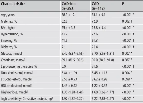
Table 1: General characteristics of the study populations, with or without coronary ar-tery disease (CAD).
Characteristics Age, years Male sex, % BMI, kg/m² Hypertension, % Smoking, % Diabetes, % Glucose, mmol/l Creatinine, mmol/l Lipid-lowering therapies, % Total cholesterol, mmol/l LDL-cholesterol, mmol/l HDL-cholesterol, mmol/l Triglycerides, mmol/l
high sensitivity- C-reactive protein, mg/l
CAD patients were required to have no history of previous myocardial infarction. * by t-test. † by Chi2 test.
CAD-free (n=393) 58.9 ± 12.1 62.8 25.4 ± 3.5 41.2 41.9 7.1 5.47 (5.37–5.58) 89.1 (86.5–90.9) 5.9 5.44 ± 1.09 3.50 ± 0.93 1.43 ± 0.42 1.35 (1.28–1.40) 1.97 (1.72–2.27) CAD (n=442) 63.1 ± 9.1 72.9 26.8 ± 3.4 72.6 61.3 20.4 5.70 (5.58–5.81) 90.0 (88.2–91.8) 31.6 5.45 ± 1.15 3.62 ± 0.98 1.22 ± 0.32 1.68 (1.62–1.77) 3.22 (2.83–3.67) P <0.001 * 0.002 † <0.001 * <0.001 † <0.001 † <0.001 † 0.007 * 0.587 * <0.001 † 0.904 * 0.098 * <0.001 * <0.001 * <0.001 *
3
Marchetti et al. A genomic-transcriptomic study in atherosclerosis
SNPs and genotyping
Genomic DNA was prepared from whole blood samples by phen-ol-chloroform extraction. The 91 SNPs investigated as replication stage of MIGen study, including intergenic rs10402271 polymor-phism (BCL3/PVRL2 locus), were genotyped using the iPLEX MassARRAY platform (Mass Array, Sequenom). PCR primers were designed by Sequenom Mass-Array-Assay-Design program, as previously described (27). The BCL3 polymorphisms, rs2965169 (5’ UTR, NM_005178.4: c.-892T>G ) and rs8100239 (intron 1, NM_005178.4: c.256+801T>A) and the PVRL2 poly-morphisms, rs3810143 (5’ UTR, NM_001042724.1: c.-381T>C or NM_002856.2: c.-381T>C) and rs1871047 (intron 1,
NM_001042724.1: c.88+1876A>G or NM_002856.2:
c.88+1876A>G) were genotyped by allele-specific real-time PCR (TaqMan SNP Genotyping Assays, Applied Biosystems, Foster City, CA, USA).
Collection of carotid specimens and histological- immunohistochemical analysis
Carotid artery specimens were obtained from 58 patients who underwent carotid endarterectomy (CEA) at the Unit of Vascular and Endovascular Surgery of S. Anna University-Hospital (Ferra-ra, Italy) for extracranial high-grade (>70%) internal carotid artery stenosis (29) by the same surgeon (FM). Restenotic lesions were
excluded. The study was approved by the Ethic Committee of the University-Hospital and written informed consent was obtained from all patients.
Arteriotomy was performed on the common carotid artery and extended to the internal carotid artery (Suppl. Figure 2, available online at www.thrombosis-online.com). The CEA specimen was removed as a single piece and included the atheromatous area and two small adjacent areas, proximal and cranial, respectively, with-out clear evidence of atherosclerotic lesions. CEA specimen was cut transversally at the bifurcation and the portion towards the aortic arch was used. This segment, consisting of the common ca-rotid artery, was further cut into: a proximal small portion without evidence of atherosclerotic lesions (grossly non-atherosclerotic portion, NP), and the distal one (cephalad), characterised by dif-fuse atherosclerosis (diseased portion, DP).
Although the NP showed some histological alterations, like a thin thickened intima, this portion was the available and suitable endogenous control.
NP and DP portions were processed for i) histology-immunoh-istochemistry analysis, ii) total RNA extraction, and iii) VSMC cultures. Portions for total RNA isolation were immediately placed into RNAlater (Ambion Inc., Austin, TX, USA) and then stored at -80°C.
Sections of formaldehyde-fixed paraffin-embedded NP and DP were stained with Masson’s trichrome for histological analysis (Suppl. Figure 2, available online at www.thrombosis-online.com). Figure 1: Tissue mRNA expression levels by microarray profiling of atherosclerotic diseased (DP) and non-atherosclerotic (NP) portions of
carotid artery. The expression levels of the seven candidate genes, stemming from the preliminary screen in VSMC populations, are reported as log2 value.
Data obtained with two probes, present in the Agilent microarray, are reported for ABCA1, SARS, GATAD2A and ALS2CR13. NP: grossly non-atherosclerotic carotid portion; DP: atherosclerotic diseased portion; NS: not significant.
Immunostaining on paraffin sections was performed as previously described (30) by using the following primary mouse monoclonal antibodies: IgG2a for (-smooth muscle actin ((-SMA, clone 1A4) (31), IgG1 for CD68 (clone KP1; Dako, Glostrup, Denmark) and IgG2a for BCL3 (clone1E8, Abcam, Cambridge, UK). Before using the first antibody, immunoreactivity was intensified by pressure cooker (3 minutes [min]) for BCL3 or microwave treatment (750 W, 5 min) for α-SMA and CD68.
VSMC culture
Primary VSMC cultures were obtained from NP and DP carotid specimens collected at surgery, as previously reported (28). After luminal gentle scraping to remove endothelial tissue, the NP (inti-mal thickening and media) and the DP (plaque with underlying media) were cut into 3x3-mm pieces and plated on separate dishes with the abluminal side in contact with the culture dish. Tissue pieces were cultured in RPMI 1640 medium supplemented with 10% fetal bovine serum (Invitrogen, Carlsbad, CA, USA), 100 U/ml penicillin, 100 µg/ml streptomycin and 2 mM L-glutamine, at 37°C with 5% CO2 . Explanted tissues were removed 7-10 days after the first VSMCs appeared. VSMC populations from both NP
and DP were maintained in identical baseline culture conditions and studied at the third passage. SMC lineage was confirmed by immunofluorescence staining using a mouse IgG2a recognizing (-smooth muscle actin ((-SMA, clone 1A4) (31) and rabbit poly-clonal IgGs recognizing both SMMHC types 1 and 2 (BT-562, Bio-medical Technologies Inc, Stoughton, MA, USA).
RNA expression study
Total RNA was isolated either from confluent cultured VSMCs or homogenised NP and DP portions with TRIzol Reagent (Invit-rogen) according to the manufacturer’s instructions and treated with RNase-free DNase (New England Biolabs, Ipswich, MA, USA). RNA quality was assessed on an Agilent 2100 Bioanalyzer (Agilent Technologies, Palo Alto, CA, USA) and low-quality RNA (RNA integrity number below 7) was excluded from further ana-lyses.
Microarray profiling of cultured VSMCs
Labelled cRNA was synthesised from 500 ng of total RNA isolated from cultured VSMCs using the Low RNA Input Linear Amplifi-cation Kit (Agilent Technologies) in the presence of cyanine 3-CTP (Perkin-Elmer Life Sciences, Boston, MA, USA). Global gene expression was detected using Agilent Whole Human Ge-nome microarray (Cat.No. G4112F, Agilent Technologies), which represents 41,000 unique human transcripts. RNA labelling and hybridisation were performed in accordance to manufacturer’s in-dications. Feature Extraction 10.7 software (Agilent Technologies) was used to obtain the microarray raw-data. Microarray results were analysed using GeneSpring GX 12 software (Agilent Technol-ogies). Data transformation was applied to set all negative raw values at 1.0, followed by a normalisation on 75th percentile. Microarray profiling of whole carotid artery specimens RNA from six specimens was hybridised on Agilent whole human genome oligo microarray (Cat.No. G4851A, Agilent Technologies) which represents 60,000 unique human transcripts. RNA labelling and hybridisation were performed in accordance to manufac-turer’s indications. Feature Extraction software v.10.7 (Agilent Technologies) was used to obtain the microarray raw-data. Micro-array results were analysed using the GeneSpring GX 12 software (Agilent Technologies). Data transformation was applied to set all negative raw values at 1.0, followed by a quantile normalisation. cDNA preparation and real-time quantitative polyme-rase chain reaction (qPCR)
cDNA was obtained from 1 μg (cultured cells) or 0.5 μg (carotid tissues) of total RNA by reverse transcription using SuperScript VILO cDNA Synthesis Kit (Invitrogen) according to the manufac-turer’s recommendations. Aliquots of diluted first-strand cDNA were amplified on CFX96 Real-Time PCR Detection System Rad, Hercules, CA, USA) using SsoFast EvaGreen Supermix (Bio-Table 2: Distribution of BCL3 and PVRL2 genotypes in the study
populations, with or without coronary artery disease (CAD). Genotypes rs2965169 (BCL3) TT TG GG rs8100239 (BCL3) TT TA AA rs10402271 (intergenic BCL3-PVRL2) TT TG GG rs3810143 (PVRL2) TT TC CC rs1871047 (PVRL2) AA AG GG
* by Chi2 test for linear trend.
CAD-free (n=393) 29.3 49.4 21.4 33.1 45.8 21.1 50.1 41.3 8.6 32.5 46.1 21.4 36.6 45.3 18.1 CAD (n=442) 21.8 53.1 25.2 27.1 50.5 22.4 46.7 41.1 12.2 31.7 49.4 18.9 33.9 46.2 19.9 P 0.020 * 0.146 * 0.168 * 0.753 * 0.372 *
5
Marchetti et al. A genomic-transcriptomic study in atherosclerosis
Rad). PCR protocol was: 95°C for 30 seconds (sec), then 40 cycles of 5 secat 95°C and 10 sec at 60°C.
Forward and reverse primers are reported in the Suppl. Table 1 (available online at www.thrombosis-online.com). Each reaction was performed in triplicate. The relative levels of mRNAs were cal-culated by the comparative CT method by using 18S rRNA as en-dogenous control in cell populations or ACTB in carotid speci-mens. Values were expressed as mean fold change ± standard error of the mean (SEM).
Statistical analysis
Differences in mRNA expression levels between VSMC popu-lations by microarray-based transcriptome analysis were evaluated by using the log-transformed (log2) ratio values (DP/NP) and as-suming the log ratio equal to 0 when there is no difference of ex-pression (32).A p-value <0.05 was considered significant without correction for multiple testing. Relationships between the fold-change (log2 DP/NP ratio)and the p-value (negative log10) were re-ported in a volcano plot.
Genes differentially expressed between three atherosclerotic and three non-atherosclerotic carotid specimens in microarray ex-periment were identified using GeneSpring GX software. A filter on fold change (1.5 was applied to the list of probes expressed in at least one sample. Then, a moderated t-test was used to identify the significantly modulated genes with p<0.05 and false discovery rate (FDR) =10%. Gene expression levels between VSMC populations or carotid portions in q-PCR analysis were compared by means of paired or unpaired t-test.
As regards population data, distributions of continuous vari-ables in groups were expressed as means ± standard deviation (SD). Statistical analysis on skewed variables, like glucose, creati-nine, triglyceride, and high-sensitivity C-reactive protein (hs-CRP), was computed on the corresponding log-transformed valu-es. Thus, results are reported as geometric mean with 95% confi-dence interval (95% CI). Quantitative data were assessed using the Student’s t-test or analysis of variance (ANOVA), with polynomial
contrast for linear trend when indicated. Qualitative data were analysed with either Chi2-test or Chi2 for linear trend analysis when indicated. Within each group examined, the frequencies of the genotypes associated with each of the polymorphisms were compared by the Chi2-test with the values predicted on the basis of the Hardy-Weinberg equilibrium.
In the first case-control population the strength of association with CAD/MI was assessed by means of a sex- and age-adjusted model. In the second case-control population the strength of as-sociation with CAD was estimated calculating the odds ratios (ORs) with 95% CIs by multiple logistic regression after sex- and age-adjustment and then after adjustment for all the traditional cardiovascular risk factors (i.e. sex, age, body mass index [BMI], smoking, hypertension, diabetes, LDL- and HDL-cholesterol, trig-lycerides, creatinine, and hs-CRP). A value of p<0.05 was con-sidered statistically significant. Statistical power was estimated by Altman nomogram (33). Calculations were performed with IBM SPSS 20.0 statistical package (IBM Inc., Armonk, NY, USA).
Haplotype frequencies were estimated using the R software with haplo.stats package (R Foundation for Statistical Computing, Vienna, Austria; http: //www.R-project.org). The associations be-tween haplotypes and laboratory and clinical outcomes were examined using a generalised linear model regression of a trait on haplotype effects, allowing for ambiguous haplotypes (haplo.glm function) (34). Statistical significance of associations was ascer-tained by randomly permuting the disease status in 1,000 rep-licates by Monte Carlo method.
Results
SNPs selectionStarting from a previous analysis of 91 SNPsin 898 subjects of the VHS population (388 CAD-free and 510 CAD with MI) within stages of replication of GWAS – MIGen – for cardiovascular risk (27), we selected 15 SNPs showing nominally an association with MI by means of a sex- and age-adjusted model with P values arbit-Figure 2: Association between the
carrier-ship of BCL3 rs2965169 minor allele G and coronary artery disease (CAD). The strength
of the association was estimated by means of ORs with 95% CI at univariate, sex- and age-ad-justed, and full adjusted analysis. The homozy-gotes for the major allele (TT) were considered as reference. * by logistic regression analysis ad-justed for sex, age, BMI, smoking, hypertension, diabetes, LDL- and HDL- cholesterol, triglycerides, creatinine, and hs-CRP.
rarily set at <0.10 (Suppl. Table 2, available online at www.thrombo sis-online.com).
As more than half (8/15) of the selected SNPs localise within in-tergenic regions, the genomic regions tagged by the 15 SNPs were examined in the NCBI database for the presence of validated or putative coding gene sequences within a region of 400 kb centred on each SNP. This search identified 77 genes, listed in Suppl. Table 3 (available online at www.thrombosis-online.com).
Transcriptome analysis of cultured VSMCs and of carotid specimens
As a preliminary screen to identify, among the 77 genes, candi-dates for further evaluation, we performed a microarray-based transcriptome analysis of VSMC populations, cultured from NP: DP couples collected from three patients. The Agilent whole human genome microarray (44k) included probes for 71 of the 77 neighbouring genes. The analysis revealed significant differences (P<0.05) in the expression levels for seven genes (ABCA1,
ALS2CR13, PVRL2, BCL3, HAX1, SARS, GATAD2A, Suppl.Table
4 and Suppl. Figure 3, both available online at www.thrombosis-on line.com). Validation was conducted by qPCR analyses in inde-pendent cell populations (see Suppl. Results, available online at www.thrombosis-online.com).
To explore also in the vessel wall the differences in the ex-pression levels detected in cultured VSMCs, we performed a sec-ond microarray experiment of whole atherosclerotic and non-atherosclerotic specimens, collected from four additional patients. The comparison (fold change (1.5, p<0.05, FDR 10%) of DP and NP tissues showed that three (ABCA1, BCL3 and PVRL2) out the seven genes, identified in the first transcriptome screen, were sig-nificantly up regulated in DP (
▶
Figure 1). This analysis revealed the most significant differences for the ATP-binding membrane cassette transporter A1 (ABCA1), whose role in lipid metabolism and in atherosclerosis pathways is well recognised.We addressed then our attention to the BCL3 and PVRL2 genes, contiguously located on the chromosome region (19q13) marked
by the intergenic rs10402271. With the aim to validate findings from microarray profiling of specimens, qPCR analyses were per-formed on carotid tissue samples from independent patients.
BCL3 mRNA levels were higher in DP than in NP specimens by
comparison of i) DP: NP couples (five patients), with a mean fold change difference of 10.07 ± 2.87 (p=0.034) as well as ii) non-coupled DP and NP (14 patients), with a mean fold change differ-ence of 6.81 ± 3.16 (p=0.013). PVRL2 expression levels were sig-nificantly higher in DP by comparison of couples (mean fold change 2.60 ± 0.46, p=0.026) and, as a trend, in non- coupled DP and NP (mean fold change 2.52 ± 0.80; p=0.060).
BCL3 SNPs genotyping for association with CAD
As the originally investigated rs10402271 maps in the intergenic 19q13 region between BCL3 and PVRL2, intragenic SNPs, two for
BCL3 (rs2965169 and rs8100239, previously associated to oral
clefts) (35), and two for PVRL2 (rs3810143 and rs1871047 ), were selected for the second genetic association analysis. It is worthy to note that the low MAF value (<0.02, by NCBI) of cDNA SNPs, both in BCL3 and PVRL2, makes them not eligible for this study.
We compared CAD-free controls (n=393) with a second popu-lation of CAD patients without previous MI (n=442). This cohort enabled us to preferentially investigate the atherosclerotic pheno-type, and to avoid the potential biases due to selection/survival re-lated to MI history.
The clinical and laboratory characteristics of the two groups are summarised in
▶
Table 1, while the data about genotype preva-lence of the five SNPs are reported in▶
Table 2. Genotypes had a distribution consistent with Hardy-Weinberg equilibrium in both cases and controls and showed a low or moderate degree of linkage disequilibrium (Suppl. Figure 4, available online at www.thrombo sis-online.com), which were in accordance with Hap Map data.While the PVRL2 rs3810143 and rs1871047 and the intergenic rs10402271 did not show a significant association with CAD, the
BCL3 rs2965169 genotype distribution differed significantly
be-tween cases and controls (
▶
Table 2). The carriership of the minorFigure 3: BCL3 protein expression in atherosclerotic (DP) carotid specimens. Representative immunohistochemical staining of the DP plaque for
α-SMA (A), CD68 (B) and BCL3 (C). BCL3 is expressed by plaque foam cells and rare VSMCs (arrows). The lumen is located on the right side of the pictures. Bar = 200 μm.
7
Marchetti et al. A genomic-transcriptomic study in atherosclerosis
allele G was more frequent in CAD patients (
▶
Table 2) and re-mained independently associated with CAD in different adjusted logistic regression models (▶
Figure 2).The carriership of the BCL3 rs8100239 minor allele A appeared to be more represented in CAD patients, but did not reach a statis-tically significant difference (72.9% vs 66.9%, p=0.062). Further analyses showed association between rs8100239 and some inter-mediate metabolic phenotypes, with the A allele linked with insu-lin resistance-related conditions (see Suppl. Results and Suppl. Tables 5A and 6, all available online at www.thrombosis-online. com). On the other hand, associations of BCL3 rs2965169 (Suppl. Table 5B, available online at www.thrombosis-online.com), PVRL2 rs3810143 and rs1871047, and intergenic rs10402271 genotypes (data not shown) with lipid profile and metabolic phenotypes were not detected.The haplotype analysis based on BCL3 rs2965169 and rs8100239 showed that the G-A haplotype, containing the minor alleles, was more represented in patients with CAD than in CAD-free (
▶
Table 3, OR 1.25 with 95%CI 1.02-1.53 as compared with the most frequent T-T haplotype; p=0.035).Expression of the BCL3 protein in carotid specimens The expression of BCL3 protein was investigated in five NP and 10 DP specimens by immunohistochemical analysis. A positive stain-ing for BCL3 (
▶
Figure 3) was found in 20-40% of plaque foam cells and in rare intimal and medial VSMCs in nine of 10 DP samples. We did not detect any BCL3-expressing cells in the inti-ma-media of all the NP samples (data not shown).Discussion
GWAS are powerful tools for unveiling the genetic components of CAD and MI, but their rigorous approach may hide some poten-tial limitations which should be taken into account. The extensive multiple testing correction in GWAS, necessary to exclude false-positive results, may simultaneously discard genetic loci which could contribute to pathophysiology (12-14). Moreover, the con-siderable overlap among clinical phenotypes of CVD, leads several GWAS to cumulate indifferently CAD and MI in their analysis (3).
Recent studies have consistently demonstrated that some SNPs are associated more with CAD than with a specific predisposition to MI (36). Combined approaches exploiting complementary methodologies may help to overcome these limitations and lead to significant advances in understanding the mechanisms of multi-factorial diseases, like CVD (37-39).
Our experimental design integrates analyses at DNA, RNA and protein levels.The GWAS-related tag SNPs and two tran-scriptome analyses, in both cultured cells and tissue, enabled us to propose three candidate genes, BCL3, PVRL2 and ABCA1. The last one has been extensively investigated in relation to the pathogenetic mechanisms of atherosclerotic vascular diseases (40).The recognised role of ABCA1 in CVD, strengthened by the observation that ABCA1 mutations cause Tangier disease and premature atherosclerosis (41), supports our experimental ap-proach aimed at detecting genetic components of atherosclerotic vascular diseases.
The two other candidate genes (BCL3 and PVRL2) were con-tiguously located on chromosome 19, in a region previously as-sociated to plasma lipid traits (42), thus increasing the interest for this genomic region. This prompted us to further investigate spe-cific SNPs for BCL3 and the adjacent PVRL2, in a second case-control analysis considering CAD patients without MI history. This step produced coherent results: the carriership of BCL3 rs2965169 G allele was associated to an increased risk of CAD in-dependently from traditional atherosclerosis risk factors and the
BCL3 rs8100239 A allele, in moderate linkage, tended also to be
more represented among CAD patients. Consistently the rs2965169 G - rs8100239 A haplotype was more frequent in CAD patients than in CAD-free subjects.
Although until now classical GWAS approaches have failed to identify BCL3 as risk gene for CAD (1), a recent study combining GWAS results with expression quantitative trait loci (eQTLs) analysis identified BCL3 as a potential new risk gene for Crohn’s disease (43), a chronic inflammatory disease associated with an in-creased atherosclerotic risk unexplained by traditional cardiovas-cular risk factors (44). With regard to the results at RNA level, our findings are also consistent with data from a transcriptome analy-sis by microarray on human carotid plaques (GEO database, GSE 43292), which shows higher BCL3 mRNA levels in the majority of Table 3: Distribution of BCL3 haplotypes in
the whole study population and in subjects with or without coronary artery disease (CAD).
Haplotype 1 Haplotype 2 * Haplotype 3 Haplotype 4
* subjects with G-A haplotype carried a greater risk of CAD than subjects with T-T haplotype (OR 1.25 with 95%CI 1.02–1.53, p=0.035; p=0.026 in a sex- and age-adjusted model after 1,000 permutations by the Monte Carlo method).
BCL3 polymorphisms rs2965169 T G G T rs8100239 T A T A Haplotype frequency (%) Whole study population (n=835) 46.9 41.9 7.1 4.1 CAD-free subjects (n=393) 49.3 39.2 6.7 4.8 CAD patients (n=442) 44.9 44.3 7.4 3.4
atheroma plaques as compared to the macroscopically intact tis-sues.
Evidence from our study for BCL3 protein expression in atherosclerotic vascular vessel wall, not reported so far, further supports the hypothesis of a role of BCL3 in CVD and fit with RNA data in carotid specimens. We found BCL3 positivity only in atherosclerotic portions and in cells, like foam cells and VSMCs, which are well known to play crucial role in atherogenesis. Ac-tually, we observed very few positive VSMCs, but this result is not surprising taking into account the lack of BCL3 positivity in most of tissues as reported in the Human Protein Atlas database. On the other hand, the very low BCL3 protein expression levels in dis-eased tissue might not favour to disentangle the interpretation of BCL3 role in atherosclerosis.
BCL3 is a transcriptional coregulator and member of the in-hibitor of nuclear factor-κB (IκB) family, which takes part to both positive and negative modulation of genes belonging to several pathways, including those of cell death/proliferation, inflamma-tory and stress responses (45, 46). Biological information from Gene Ontology database locates BCL3 in multiple processes which are known to be relevant in atherosclerosis (Suppl. Figure 5, avail-able online at www.thrombosis-online.com).
BCL3 was also proposed as a key interface for regulatory cross-talk between inflammation and cellular energy metabolism (47). According with this hypothesis, in our study the BCL3 rs8100239 A allele correlated with traits usually characterizing the so-called metabolic syndrome, a still debated cluster of risk factors including altered energy metabolism and whose core is thought to be insulin resistance (48, 49). The lack of waist circumference data in our population does not allow to define the conventionally stated diag-nosis of metabolic syndrome (50). Nonetheless, we observed sig-nificant associations with both unfavorable lipid profile and over-weight. Although assessed only in a relatively small group of sub-jects, the A allele was associated also with both hyperinsulinaemia and higher HOMA score, which is considered a surrogate measure of insulin resistance.
Our study has some limitations that warrant discussion. The al-lele specific contribution to BCL3 mRNA expression level, which could provide insight for a functional effect and for the associated risk, is difficult to determine because of the SNPs localisation (5’ UTR and intron 1) and the virtual absence of in linkage coding SNPs. On the other hand, at inspection (UCSC Genome Browser, hg19, ENCODE data) for functional elements by H3K27 histone acetylation mark and by chromatin state segmentation analysis, both BCL3 rs2965169 and rs8100239 polymorphisms have been shown to lie within potentially active regulatory elements in en-dothelial-, B-lymphocyte- and fibroblast cell lines, which are of in-terest for atherosclerosis pathways.
The transcriptional analysis was obtained in VSMC popu-lations or in whole specimens from a small number of patients. This limitation has been tackled by i) the study of couples of speci-mens from the same donor, which would reduce variability in the gene expression profile; ii) the combination of subsequent and in-dependent transcriptome analyses, expected to increase robustness of the approach and to reveal hits of “in vivo” relevance; and iii) the validation of transcriptome changes by qPCR. We also disclose that the complex transcriptional and post-transcriptional regu-lation of BCL3 (45) could be perturbed by in vitro culture state and thus differ from the in vivo condition.
Regarding the statistical analysis, the lack of adjustment for multiple comparisons is a further limitation of our work. We at-tempted to overcome this drawback not only by requiring case-control analyses in two CAD populations (with or without MI), but also by showing additional support for the association between BCL3 and atherosclerosis at mRNA (in cultured VSMC popu-lations and in carotid tissues) and protein levels.
Other significant caveats of this study are related to the popu-lation analysis, including the retrospective case-control design, the relatively low number of subjects, and the lack of some clinical data. On the other hand, a remarkable strength of this study is rep-resented by the angiography-evaluation of the coronary artery bed, which allows a clear-cut definition of the clinical phenotype and avoids the possibility to include in the control group subjects with subclinical, but significant CAD. Finally, considering the dif-ferent prevalence of the carriership of rs2965169 G allele among subjects with or without CAD in the second case-control analysis, the statistical power of our study was about 80% by Altman nomo-gram with a significance level of 0.05 (33).
In summary, our integrated approach suggests for the first time the involvement of BCL3 in CVD, which could be partly mediated through the influence on metabolic phenotypes. Our results should be considered as hypothesis-generating and further epi-demiological studies on larger samples, as well as basic investi-gations about BCL3 expression and function, are needed to con-firm the role of BCL3 in the pathways of atherosclerosis.
Acknowledgements
We are very grateful to Prof. Sekar Kathiresan (Cardiovascular Re-search Center and Cardiology Division and Center for Human Genetic Research, Massachusetts General Hospital, Boston, Pro-gram in Medical and Population Genetics, Broad Institute of MIT What is known about this topic?
•
The stringent level of statistical significance required in genome-wide association studies may lead to discard genetic variants that could contribute to disease risk.•
Integrated genomic and transcriptomic investigations might be a tool to unravel new genetic signatures for cardiovascular disease.•
The expression of the proto-oncogene BCL3 in human athero-sclerotic lesions has not previously been reported.What does this paper add?
•
BCL3 mRNA and protein were differently expressed inathero-sclerotic versus non-atheroathero-sclerotic portions of carotid artery.
•
BCL3 genotypes were associated with both coronary arterydis-ease and metabolic derangements.
9
Marchetti et al. A genomic-transcriptomic study in atherosclerosis and Harvard, Cambridge, Massachusetts 02142, USA) for the
on-going cooperation and for providing the data about SNPs of MIGen Consortium. We wish to thank Mrs. Maria Zoppi for her invaluable secretary help, and Dr. Patrizia Guarini, Diego Minguz-zi and PatriMinguz-zia Pattini for their excellent technical help.
Conflicts of interest None declared.
References
1. Kathiresan S, Srivastava D. Genetics of human cardiovascular disease. Cell 2012; 148: 1242-1257.
2. Roger VL, Go AS, Lloyd-Jones DM, et al. American Heart Association Statistics Committee and Stroke Statistics Subcommittee. Executive summary: heart dis-ease and stroke statistics-2012 update: a report from the American Heart As-sociation. Circulation 2012; 125: 188-197.
3. Ding K, Kullo IJ. Genome-wide association studies for atherosclerotic vascular disease and its risk factors. Circ Cardiovasc Genet 2009; 2: 63-72.
4. O'Donnell CJ, Nabel EG. Genomics of cardiovascular disease. N Engl J Med 2011; 365: 2098-2109.
5. Bijnens APJJ, Lutgens E, Ayoubi T, et al. Genome-wide expression studies of atherosclerosis-Critical issue in methodology, analysis, interpretation of tran-scriptomics data. Arterioscler Thromb Vasc Biol 2006; 26: 1226-1235. 6. Miller DT, Ridker PM, Libby P, et al. Atherosclerosis: the path from genomics to
therapeutics. J Am Coll Cardiol 2007; 49: 1589-1599.
7. Cappola TP, Margulies KB. Functional genomics applied to cardiovascular medicine. Circulation 2011; 124: 87-94.
8. Park SW, Moon YA, Horton JD. Post-transcriptional regulation of low density li-poprotein receptor protein by proprotein convertase subtilisin/kexin type 9a in mouse liver. J Biol Chem 2004; 279: 50630–50638.
9. Lambert G, Charlton F, Rye KA, et al. Molecular basis of PCSK9 function. Atherosclerosis 2009; 203: 1-7.
10. Musunuru K, Strong A, Frank-Kamenetsky M, et al. From noncoding variant to phenotype via SORT1 at the 1p13 cholesterol locus. Nature 2010; 466: 714-719. 11. Yang X. Use of functional genomics to identify candidate genes underlying
human genetic association studies of vascular diseases. Arterioscler Thromb Vasc Biol 2012; 32: 216-222.
12. Marian AJ, Belmont J. Strategic approaches to unravelling genetic causes of car-diovascular diseases. Circ Res 2011; 108: 1252-1269.
13. Stahl EA, Wegmann D, Trynka G, et al. Bayesian inference analyses of the poly-genic architecture of rheumatoid arthritis. Nat Genet 2012; 44: 483-489. 14. Park JH, Wacholder S, Gail MH, et al. Estimation of effect size distribution from
genome-wide association studies and implications for future discoveries. Nat Genet 2010; 42: 570-575.
15. Ross R. Mechanisms of disease: Atherosclerosis-An inflammatory disease. N Engl J Med 1999; 340: 115–126.
16. Lusis AJ. Atherosclerosis. Nature 2000; 407: 233-241.
17. Campbell JH, Campbell GR. Smooth Muscle Phenotypic Modulation—A Per-sonal Experience. Arterioscler Thromb Vasc Biol 2012; 32: 1784-1789. 18. Gomez D, Owens GK. Smooth muscle cell phenotypic switching in
athero-sclerosis. Cardiovasc Res 2012; 95: 156-164.
19. Hao H, Gabbiani G, Bochaton-Piallat ML. Arterial smooth muscle cell het-erogeneity: implications for atherosclerosis and restenosis development. Arte-rioscler Thromb Vasc Biol 2003; 23: 1510-1520.
20. Doran AC, Meller N, McNamara CA. Role of smooth muscle cells in the initi-ation and early progression of atherosclerosis. Arterioscler Thromb Vasc Biol 2008; 28: 812-819.
21. Lacolley P, Regnault V, Nicoletti A, et al. The vascular smooth muscle cell in ar-terial pathology: a cell that can take on multiple roles. Cardiovasc Res 2012; 95: 194-204.
22. Johnson JL. Emerging regulators of vascular smooth muscle cell function in the development and progression of atherosclerosis. Cardiovasc Res 2014; Epub ahead of print.
23. Motterle A, Pu X, Wood H, et al. Functional analyses of coronary artery disease associated variation on chromosome 9p21 in vascular smooth muscle cells. Hum Mol Genet 2012; 21: 4021-4029.
24. Perisic L, Hedin E, Razuvaev A, et al. Profiling of atherosclerotic lesions by gene and tissue microarrays reveals PCSK6 as a novel protease in unstable carotid atherosclerosis. Arterioscler Thromb Vasc Biol 2013; 33: 2432-2443.
25. Bozzini C, Girelli D, Bernardi F, et al. Influence of polymorphisms in the factor VII gene promoter on activated factor VII levels and on the risk of myocardial infarction in advanced coronary atherosclerosis. Thromb Haemost 2004; 92: 541-549.
26. Martinelli N, Girelli D, Lunghi B, et al. Polymorphisms at LDLR locus may be associated with coronary artery disease through modulation of coagulation fac-tor VIII activity and independently from lipid profile. Blood 2010; 116: 5688-5697.
27. Kathiresan S, Myocardial Infarction Genetics Consortium. Genome-wide as-sociation of early-onset myocardial infarction with single nucleotide polymor-phisms and copy number variants. Nat Genet 2009; 41: 334-341.
28. Matthews DR, Hosker JP, Rudenski AS, et al. Homeostasis model assessment: insulin resistance and beta-cell function from fasting plasma glucose and insu-lin concentrations in man. Diabetologia 1985; 28: 412-419.
29. North American Symptomatic Carotid EndarterectomyTrial Collaborators. Beneficial effect of carotid endarterectomy in symptomatic patients with high-grade carotid stenosis. N Engl J Med 1991; 325: 445-453.
30. Coen M, Marchetti G, Palagi PM, et al. Calmodulin expression distinguishes the smooth muscle cell population of human carotid plaque. Am J Pathol 2013; 183: 996-1009.
31. Skalli O, Ropraz P, Trzeciak A, et al. A monoclonal antibody against alpha-smooth muscle actin: a new probe for alpha-smooth muscle differentiation. J Cell Biol 1986; 103: 2787-2796.
32. Cui X, Churchill GA. Statistical tests for differential expression in cDNA micro-array experiments. Genome Biol 2003; 4: 210-219.
33. Altman DG. Statistics and ethics in medical research. How large a sample? Br Med J 1980; 281: 1336-1338.
34. Lake SL, Lyon H, Tantisira K, et al. Estimation and tests of haplotype-environ-ment interaction when linkage phase is ambiguous. Hum Hered 2003; 55: 56-65. 35. Park BY, Sull JW, Park JY, et al. Differential parental transmission of markers in
BCL3 among Korean cleft case-parent trios. J Prev Med Public Health 2009; 42: 1-4.
36. Reilly MP, Li M, He J, et al. Myocardial Infarction Genetics Consortium; Well-come Trust Case Control Consortium. Identification of ADAMTS7 as a novel locus for coronary atherosclerosis and association of ABO with myocardial in-farction in the presence of coronary atherosclerosis: two genome-wide associ-ation studies. Lancet 2011; 377: 383-392.
37. Folkersen L, van't Hooft F, Chernogubova E, et al. BiKE and ASAP study groups. Association of genetic risk variants with expression of proximal genes identifies novel susceptibility genes for cardiovascular disease. Circ Cardiovasc Genet 2010; 3: 365-373.
38. Ramsey SA, Gold ES, Aderem A. A systems biology approach to understanding atherosclerosis. EMBO Mol Med 2010; 2: 79-89.
39. Maouche S, Schunkert H. Strategies beyond genome-wide association studies for atherosclerosis. Arterioscler Thromb Vasc Biol 2012; 32: 170-181.
40. Oram JF, Vaughan AM. ATP-Binding cassette cholesterol transporters and car-diovascular disease. Circ Res 2006; 99: 1031-1043.
41. Fitzgerald ML, Mujawar Z, Tamehiro N. ABC transporters, atherosclerosis and inflammation. Atherosclerosis 2010; 211: 361-370.
42. Talmud PJ, Drenos F, Shah S, et al. Gene-centric association signals for lipids and apolipoproteins identified via the Human CVD BeadChip. Am J Hum Genet 2009; 85: 628-642.
43. Fransen K, Visschedijk MC, van Sommeren S, et al. Analysis of SNPs with an ef-fect on gene expression identifies UBE2L3 and BCL3 as potential new risk genes for Crohn's disease. Hum Mol Genet 2010; 19: 3482-3488.
44. Miller AM, McInnes IB. Cytokines as therapeutic targets to reduce cardiovascu-lar risk in chronic inflammation. Curr Pharm Des 2011; 17: 1-8.
45. Palmer S, Chen YH. Bcl-3, a multifaced modulator of NF-kB-mediated gene transcription. Immunol Res 2008; 42: 210-218.
46. Hinz M, Arslan SÇ, Scheidereit C. It takes two to tango: IκBs, the multifunc-tional partners of NF-κB. Immunol Rev 2012; 246: 59-76.
47. Yang J, Williams RS, Kelly DP. BCL3 interacts cooperatively with peroxisome proliferator-activated receptor gamma (PPARgamma) coactivator 1alpha to
coactivate nuclear receptors estrogen-related receptor alpha and PPARalpha. Mol Cell Biol 2009; 29: 4091-4102.
48. Alberti KG, Eckel RH, Grundy SM, et al. Harmonizing the metabolic syndrome: a joint interim statement of the International Diabetes Federation Task Force on Epidemiology and Prevention; National Heart, Lung, and Blood Institute; American Heart Association; World Heart Federation; International Athero-sclerosis Society; and International Association for the Study of Obesity. Circu-lation 2009; 120: 1640-1645.
49. Grundy SM. Metabolic syndrome: a multiplex cardiovascular risk factor. J Clin Endocrinol Metab 2007; 92: 399-404.
50. Executive Summary of The Third Report of The National Cholesterol Education Program (NCEP) Expert Panel on Detection, Evaluation, And Treatment of High Blood Cholesterol In Adults (Adult Treatment Panel III). J Am Med Assoc 2001; 285: 2486–2497.