Experimental Allergy – Review Article
Int Arch Allergy Immunol
The Staining of Mast Cells: A Historical
Overview
Domenico Ribatti
Department of Basic Medical Sciences, Neurosciences and Sensory Organs, University of Bari Medical School, and National Cancer Institute “Giovanni Paolo II”, Bari, Italy
Received: January 19, 2018 Accepted: January 22, 2018 Published online: March 29, 2018 DOI: 10.1159/000487538
Keywords
c-Kit · Mast cells · Metachromasia · Staining · Tryptase
Abstract
The specificity of several staining methods for mast cells pro-vides the pathologist with a useful means for the differential diagnosis of mast cell tumors. Mast cells stain metachromat-ically with toluidine blue with greater intensity in cells con-taining smaller granules. Most stains for mast cells rely on the cell’s content of heparin, other glycosaminoglycans, and es-terase. As an alternative to histochemical stains, different an-tibodies have been used to identify mast cells in humans.
© 2018 S. Karger AG, Basel
Introduction
Mast cells are present in mammalian as well as non-mammalian [1] vertebrates in virtually all vascularized tissues. The first phase of research on mast cells was by von Recklinghausen [2] in 1863, who noticed granular cells in the mesentery of frogs, almost certainly the first observation of mast cells in any species. Mast cells ex-press, on their surface, the high-affinity receptor for IgE (FcεRI), and can be activated by IgE and specific antigens to release a diverse array of mediators, including
hista-mines, leukotrienes, prostaglandins, serine proteases, and various cytokines, chemokines, and growth factors.
Mast cells are numerous. In fact, it has been estimated that if they were grouped together, they would make an organ equal to the size of the spleen [3]. The mast cells of rodents and humans may be subdivided into ≥2 subpop-ulations by various morphologic and functional criteria. The histochemical techniques for subdividing mast cells stemmed from the observation by Ehrlich in 1876 that their lysosomal granules have the capacity to take up and stain metachromatically with basic dyes, such as toluidine blue. Among the histochemical methods for staining mast cells worth mentioning are Sudan Black B, Luna stain, and Ziehl-Neelsen, which have been assessed his-torically [4]. In 1994, Simoes and Schoning [5] evaluated 18 methods for staining mast cells in dogs. Enerbäck [6– 8] demonstrated in 1986 that rodent mast cell subpopula-tions could be differentially stained with Alcian blue and berberine sulfate. Enerbäck [6–8] also showed that mast cells from mucosal surfaces were sensitive to formalde-hyde fixation, in that they failed to stain metachromati-cally upon additional exposure to dyes, whereas mast cells from connective tissues were insensitive to formaldehyde fixation. Before the introduction of monoclonal
ies against proteases, mast cells were stained by meta-chromatic stains, such as toluidine blue and Alcian blue. This review article will analyze the most common techniques developed to stain different mast cell subpop-ulations.
Metachromasia
Mast cell granules (and some other tissue components such as cartilage matrix) can naturally induce metachro-matic staining. Metachrometachro-matic stains include the Ro-manowsky combinations (Wright, Giemsa, May-Grün-wald Giemsa, and Leishman), toluidine blue, and others. Toluidine blue first emerged in 1856, courtesy of a British chemist called William Henry Perkin. Although he was working on the synthesis of quinine, Perkin in-stead produced a blue substance with good dyeing prop-erties. Initially, it became known as aniline purple. Being mostly used in the dye industry, this was the first syn-thetic organic chemical dye. Later, it became known as toluidine blue, and began being used for medical purpos-es, in particular as a histological special stain to highlight certain components.
Mast cells were first identified by Paul Ehrlich [9] in 1878 when he was still a medical student; in his doctoral dissertation, he described a class of aniline-positive cells of the connective tissues endowed with cytoplasmic meta-chromatic granules, for which he coined the name “Mast-zellen.” The first use of the term “metachromatic” was by Ackroyd [10] in 1876, to indicate that the structure being dyed assumed a color different from that of the dye itself. In 1879, Ehrlich [11] used the word for the first time in a biological context, to describe the staining reaction of blood leukocytes on the basis of their specific affinities for various dyes [11, 12]. He encountered cells with basophil-ic, metachromatic granules, and thus came to recognize
two types of mast cells. The first type, which could be iden-tified and differentiated by its repertoire of coarse baso-phil granules (gamma granulation), was to be found in the connective tissues and apparently derived from them (tis-sue mast cells). The second, the counterpart of the neutro-phil polymorph and eosinoneutro-phil leukocyte, contained ba-sophilic granulation of the fine type (delta granulation); its origin was in the bone marrow and it was to be found in the peripheral blood (blood mast cells and basophils).
At the end of the 1930s, Scandinavian researchers pro-vided fundamental new insights into mast cell structural profile. The mast cell component prophesized by Ehrlich as the responsible agent for granule metachromasia was revealed [12, 13]. Holmgren and Wilander [14], following the discovery by Jorpes [15] that the anticoagulant hepa-rin was subject to stain metachromatically with toluidine blue, reconsidered Ehrlich’s observation that mast cell granules stained metachromatically with toluidine blue. These authors were able to set a correlation between the number of toluidine blue-positive mast cell in various tis-sues and their heparin content.
Metachromatic staining is important in the detection of mast cells and is strongly recommended as a routine stain for this purpose. One of the most frequently meta-chromatic stains is toluidine blue which stains the mast cell granules purple-to-red (Fig. 1). Dilute staining solu-tions should be used in order to demonstrate strongly metachromatic elements [16]. The pH of the dye solution used is important. Lennert [17] showed that the use of a series of toluidine blue solutions at pH levels varying from 2.62 to 7.00 might help differentiate benign from malignant cases of human mastocytosis. This issue was further developed when it was shown that benign lesions stained with toluidine blue at a lower pH whereas
malig-nant lesions were poorly stained at a pH <3.5 [18].
Metachromasia is due to the presence of tissue polyan-ions that induce a polymerization of dye molecules. It has
Fig. 1. Semi-thin section of rat peritoneal mast cells stained with toluidine blue. Nu-merous cytoplasmic metachromatic gran-ules are recognizable (reproduced from [45]).
been shown that a distance of around 0.5 nm between negatively charged groups is needed to induce such po-lymerization [19]. The process involves a shift in the ab-sorption spectrum of the dye towards shorter wave-lengths, and is accompanied by a hypochrome color changes of the dye from blue towards violet, red, or or-ange. Metachromasia of anionic tissue can be demon-strated with many cationic dyes such as thiazine, oxam-ine, azoxam-ine, and xanthene, and with fluorescent dyes such as Acridine orange. The thiazine dyes toluidine blue and Azure A are by far the most widely used. Metachromasia is best observed in a water solution of low ionic strength. A large number of anionic tissue sites are metachromatic under such conditions [20].
Mast cells are round in the proximity of blood vessels, but display an elongated shape in the interstitial regions. In addition, mast cells often have a reduced number of granules and a disorganized granule content, suggesting an ongoing degranulation process. Analysis at higher magnifications allows the identification of degranulating mast cells, characterized by numerous extracellular meta-chromatic granules and/or by a poor intracytoplasmic granule content, and nondegranulating mast cells with-out any granule in the proximal extracellular space.
Alcian Blue-Safranin
Strongly metachromatic polyanions such as the sul-fated glycosaminoglycans (GAGs) of mast cell granules retain red or violet metachromasia after dehydration in ethanol and mounting in synthetic resins. Two copper phthalocyanin dyes are of special interest for the staining of mast cells, Alcian blue 8GX and Astrablau 6GLL. Al-cian blue, formulated by Scott et al. [21] is more easily available and better specified, and should therefore be preferred. Alcian blue interacts with polyanions such as heparin in aqueous solutions, to give insoluble precipi-tates in which the two components are bound by ionic linkages. The connective tissue mast cells of rats and mice have a low affinity for Alcian blue. Mast cells in the cervi-cal lymph nodes and uterus stain strongly with Alcian blue at a pH of 2.5 [22].
In the 1960s, Enerbäck [6–8] described two morpho-logically distinct subpopulations of rodent mast cells, based on their specific staining characteristics and pref- erential tissue homing, i.e., connective tissue mast cells (CTMCs) present in the connective tissues, and serosae and mucosal mast cells (MMCs) located on the mucosae of the respiratory and gastrointestinal tracts. CTMCs
could be distinguished from MMCs by red staining with safranin due to the presence of large amounts of heparin in their secretory granules. In the mouse, indeed, the pro-teoglycan content of mast cell granules varies in the differ-ent mast cell subtypes. CTMCs contain heparin that is lacking in MMCs. Conversely, MMCs express chondroi-tin sulfates A and B, which are not found in CTMCs, but both subtypes store chondroitin sulfate E in their gran-ules. Thus, in contrast to CTMCs, MMCs are sensitive to routine formalin fixation and cannot be identified in stan-dard histological sections. CTMCs can be detected after fixation with 10% neutral-buffered formalin, while MMCs require fixation in nonaldehyde solutions such as Carnoy solution [23]. After appropriate fixation and sequential staining with Alcian blue and safranin, MMCs stain blue, being thus differentiated from CTMCs which stain with safranin and are red. Differential affinity for Alcian blue can be visualized with sequential staining, consisting of Alcian blue followed by safranin [24]. Immature embry-onic mast cells contain granules which stain with Alcian blue rather than safranin. With the Alcian blue-safranin sequence, the maturation of mast cells is accompanied by a change in the staining properties from blue to red [25].
The Tyrosine Kinase Kit Receptor
Mast cells, but not basophils, express the Kit receptor for the stem cell factor (Fig. 2), which not only drives the terminal differentiation of mast cells but plays other
im-Fig. 2. Predominantly focal paranuclear c-Kit protein expression in mast cells of a canine mast cell tumor. Immunohistochemistry is performed with primary anti-c-Kit receptor antibody. Single ar-rows indicate focal brown paranuclear immunostaining, while double arrows indicate a vessel (reproduced from [46]).
portant roles in regulating mast cell biology, such as the survival, activation, and degranulation of mature cells. The importance of the stem cell factor as a mast cell growth factor is underlined by the fact that mice with cer-tain loss-of-function mutations affecting either the stem cell factor or its receptor Kit are devoid of mast cells. In-deed, the lack of expression of a functional Kit receptor due to spontaneous mutation in both copies of Kit, as it
occurs in genetically mast cell-deficient WBB6F1-KitW
-KitW-v mice (W/Wv mice), results in the absence of tissue
mast cells [26].
On immunohistochemical staining, the c-Kit proto-oncogene encodes a transmembrane tyrosine kinase re-ceptor, c-Kit (CD117), which is closely related to the platelet-derived growth factor family. This antibody rec-ognizes the extracellular domain, and is expressed by a variety of normal and abnormal cell types. In normal cells, the CD117 antibody has been shown to label breast epithelium, germ cells, melanocytes, stem cells, mast cells, salivary glands, and the esophagus, cerebellum, hippo-campus, and spinal cord [27]. In mast cells, the marking is at the level of the plasma membrane (in normal cells);
in other cells, it is at the level of the cytoplasm. Further-more, a large proportion of mast cells, including those in the colon, stomach, lung, uterus, and bladder, do not stain with c-Kit [28]. Because CD117 is also expressed on immature hematopoietic progenitor cells, additional markers should be applied to define the mast cell compo-nent by flow cytometry in bone marrow samples. To dis-tinguish CD117-positive mast cells from CD117-nonhe-matopoietic cells, the staining of mast cells with a mono-clonal antibody against CD45 is recommended [29].
Tryptase and Chymase
Mast cell proteases represent major protein compo-nents of secretory granules, but the role of each individu-al protease in the mast cells remains poorly understood. The proteases are classified into carboxypeptidase, chy-mase, and tryptase. Mast cells differ in their protease ex-pression pattern, depending on the tissue where they are to be found.
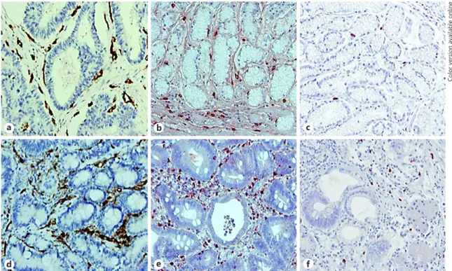
a b c
d e f
Fig. 3. Immunohistochemical staining for CD31, tryptase, and chymase in stage II (a–c) and stage IV (d–f) hu-man gastric cancer. a, d In endothelial cells immunoreactive for CD31. b, e In tryptase-positive mast cells. c, f In chymase-positive mast cells. Blood vessels and mast cells are distributed around the gastric glands. The number of blood vessels and mast cells is higher in stage IV than in stage II biopsy specimens, and the number of chymase-positive cells is lower than the number of tryptase-chymase-positive cells (reproduced from [47]).
By 1960, two proteases, with chymotrypsin- and tryp-sin-like activity were identified in mast cells [30–32], and enzyme activity was recognized to localize within intra-cellular granules. The enzymes were purified in the 1980s and renamed tryptase and chymase [33, 34]. Mast cells from different anatomical sites contain different profiles of these enzymes as well as of other proteases (Fig. 3). Hu-man mast cells were divided into two subtypes, depend-ing on the expression of different proteases in their gran-ules [35]. The first, containing tryptase only, was
desig-nated MCT or “immune cell-associated” mast cells,
predominantly located in the respiratory and intestinal mucosa, where they colocalize with T lymphocytes. A type of mast cell that contained both tryptase and chy-mase, along with other proteases such as
carboxypepti-dase A and cathepsin G (Fig. 4), was referred to as MCTC,
and these cells are predominantly found in connective tissue areas, such as the skin, submucosa of the stomach and intestine, breast parenchyma, myocardium, lymph nodes, conjunctiva, and synovium. A third type of mast
cell, called MCC, expresses chymase without tryptase and
is to be found mainly in the submucosa and mucosa of the stomach, small intestinal submucosa, and colonic
muco-sa [36]. Interestingly, the human MCT type was seen to
correspond most closely to rodent MMCs, whereas the
MCTC type resembled rodent CTMCs.
Other Stains
Chloroacetate-esterase is found in mast cells. Speci-mens are incubated with naphthol AS-D chloroacetate in the presence of freshly formed diazonium salt. Enzymat-ic hydrolysis of ester linkages liberates free naphthol
com-pounds which couple with diazonium salt, forming high-ly colored deposits at the site of enzyme activity [37]. Un-like most enzyme stains, chloroacetate-esterase can be used on fixed, paraffin-embedded tissue. The slides are incubated in a solution containing the substrate naphthol AS-D chloroacetate, and the esterase contained in the neutrophils and mast cells then binds with the chloroac-etate. This releases the naphthol group, which binds to the diazonium dye pararosanalin (basic fuchsin), another component of the incubating solution. Pararosanilin or basic fuchsin gives a deep pink-red color to the granules, while hematoxylin counterstains the nuclei blue.
Berberine forms a strongly fluorescent complex with heparin, proportional to the heparin content in the mast cell granules [38], enabling the quantitation of heparin by both microscope fluorometry [38] and flow cytometry [39]. Berberine staining may also be used for the visual-ization of mast cells in tissue sections [40].
Mast cells stain intensely at a pH of 8.00–10.5 with the anionic bis-azo dye, Biebrich scarlet [41], and stain strong-ly with naphtol AS-D chloroacetate as a substrate and di-azo-coupling fixation in acetone or neutral formalin [42]. Histamine can be detected histochemically by means of a fluorescent reaction with o-phthalaldeyde [43, 44].
Acknowledgements
This study was supported by a grant from the “Associazione Italiana Mastocitosi.”
Disclosure Statement
There are no conflicts of interest.
a b c
Fig. 4. Dual immunofluorescence for tryptase (red, a), cathepsin-G (green, b), and both proteins (orange, c) in a biopsy specimen of human cutaneous mastocytosis (reproduced from [48]).
References
1 Baccari GC, Pinelli C, Santillo A, Minucci S, Rastogi RK: Mast cells in nonmammalian ver-tebrates. Int Rev Cytol 2011;290:1–53. 2 von Recklinghausen F: Über Eiter- und
Bin-degewebskörperchen. Virchows Arch Patho-log Anat Physiol Klin Med 1863;83:247–287. 3 Bradding P, Holgate ST: Immunopathology
and human mast cell cytokines. Crit Rev On-col Hematol 1999;31:119–133.
4 Rest JR, Lee RL: Staining of mast cell granules by the Ziehl-Neelsen method and differential diagnosis of malignant dermal tumours in the dog. Vet Rec 1979;104:79.
5 Simoes JP, Schoning P: Canine mast cell tu-mors: a comparison of staining techniques. J Vet Diagn Invest 1994;6:458-465.
6 Enerbäck L: Mast cells in rat gastrointestinal mucosa. I. Effects of fixation. Acta Pathol Mi-crobiol Scand 1966;66:289–302.
7 Enerbäck L: Mast cells in rat gastrointestinal mucosa. 2. Dye-binding and metachromatic properties. Acta Pathol Microbiol Scand 1966;66:303–312.
8 Enerbäck L: The evolution of the concept of a specific mucosal mast cell; in Befus AD, Bie-nenstock J, Denburg JA (eds): Mast Cell Dif-ferentiation and Heterogeneity. New York, Raven Press, 1986, pp 1–26.
9 Ehrlich P: Beiträge zur Theorie und Praxis der histologischen Färbung (thesis, 1878); in: The Collected Papers of Paul Ehrlich. Elsevier, 2013, pp 29–64.
10 Ackroyd W: Metachromism or colour change. Chem News 1876;33:60.
11 Ehrlich P: Beiträge zur Kenntnis der granuli-erten Bindegewebszellen und der eosino-philen Leukozyten. Arch Anat Physiol 1879; 36:166–169.
12 Ehrlich P: Farbenanalytische Untersuchun-gen zur Histologie und Klinik des Blutes. Dtsch Med Wochenschr 1894;20:135–136. 13 Ehrlich P: Die Anemie. 1. Normale und
Pa-thologische Histologie des Blutes. Wien, Holder, 1898.
14 Holmgren H, Wilander O: Beitrag zur Kenn t- nis der Chemie und Funktion der Ehrlich-schen Mastzellen. Z Mikr Anat Forsch 1937; 42:242–278.
15 Jorpes E HH, Wilander O: Über das Vorkom-men von Heparin in den Gefässwänden und in den Augen. Ein Beitrag zur Physiologie der Ehrlichschen Mastzellen. Z Mikr Anat Forsch 1937;42:279–301.
16 Kramer H, Windrum GM: The metachromat-ic staining reaction. J Histochem Cytochem 1955;3:227–237.
17 Lennert K: Zur pathologischen Anatomie von Urticaria pigmentosa und Mastzellenreticu-lose. Klin Wochenschr 1962;40:61–67. 18 Klatt EC, Lujes RJ, Meyer PR: Benign and
ma-lignant mast cell proliferations. Diagnosis and separation using a pH-dependent toluidine blue staining in tissue section. Cancer 1983; 51:1119–1124.
19 D’mello AXP, Sylvester TV, Ramya V, Britto FP, Shetty PK, Jasphin S: Metachromasia and metachromatic dyes: a review. Int J Adv Health Sci 2016;2:12–17.
20 Spicer SS HR, Leppy TJ (eds): Histochemistry of connective tissue mucopolysaccharides; in: The Connective Tissue. Baltimore, Williams and Wilkins, 1967.
21 Scott JE, Quintarelli G, Dellovo MC: The chemical and histochemical properties of Al-cian blue. Histochemie 1964;4:73–85. 22 Spicer SS: Histochemical properties of
muco-polysaccharixe and basic protein in mast cells. Ann NY Acad Sci 1963;103:322–333. 23 Irani AM, Schwartz LB: Mast cell
heterogene-ity. Clin Exp Allergy 1989;19:143–155. 24 Spicer SS: A correlative study of the
histo-chemical properties of rodent acid mucopoly-saccharides. J Histochem Cytochem 1960;8: 18–35.
25 Combs JW: Differentiation and proliferation of embryonic mast cells of the rat. J Cell Biol 1965;25:577–592.
26 Kitamura Y, Go S, Hatanaka K: Decrease of mast cells in W/Wv mice and their increase by bone marrow transplantation. Blood 1978;52: 447–452.
27 Lammie A, Dobnojak M, Gerald W, Saad A, Cote R, Cordon-Cardo C: Expression of c-Kit and Kit ligand proteins in normal human tis-sues. J Histochem Cytochem 1994;42:1417– 1425.
28 Qi JC, Li L, Li Y, Moore K, Madigan MC, Kat-soulotos G, Krilis SA: An antibody raised against in vitro-derived human mast cells identifies mature mast cells and a population of cells that are FcεRI(+), tryptase (–), and chymase (–) in a variety of human tissues. J Histochem Cytochem 2003;51:643–653. 29 Escribano L, Díaz-Agustín B, Bellas C,
Na-valón R, Nuñez R, Sperr WR, Schernthaner G-H, Valent P, Orfao A: Utility of flow cyto-metric analysis of mast cells in the diagnosis and classification of adult mastocytosis. Leuk Res 2001;25:563–570.
30 Benditt EP, Arase M: An enzyme in mast cells with properties like chymotrypsin. J Exp Med 1959;110:451–460.
31 Benditt EP: An enzyme in mast cells with some properties resembling chymotrypsin. Fed Proc 1956;15:507.
32 Glenner GG, Cohen LA: Histochemical dem-onstration of a species-specific trypsin-like enzyme in mast cells. Nature 1960;185:846– 847.
33 Schechter NM, Choi JK, Slavin DA, Deresien-ski DT, Sayama S, Dong G, Lavker RM, Proud D, Lazarus GS: Identification of a chymotryp-sin-like proteinase in human mast cells. J Im-munol 1986;137:962–970.
34 Schwartz LB, Lewis RA, Austen KF: Tryptase from human pulmonary mast cells. Purifica-tion and characterizaPurifica-tion. J Biol Chem 1981; 256:11939–11943.
35 Irani AA, Schechter NM, Craig SS, DeBlois G, Schwartz LB: Two types of human mast cells that have distinct neutral protease composi-tions. Proc Natl Acad Sci USA 1986;83:4464– 4468.
36 Irani A-MA, Schwartz LB: Human mast cell heterogeneity. Allergy Asthma Proc 1994;15: 303–308.
37 Moloney WC, McPherson K, Fliegelman L: Esterase activity in leukocytes demonstrated by the use of naphthol AS-D chloroacetate substrate. J Histochem Cytochem 1960;8: 200–207.
38 Enerbäck L: Berberine sulphate binding to mast cell polyanions: a cytofluorometric method for the quantitation of heparin. His-tochemistry 1974;42:301–313.
39 Enerbäck L, Berlin G, Svensson I, Rundquist I: Quantitation of mast cell heparin by flow cytofluorometry. J Histochem Cytochem 1976;24:1231–1238.
40 Dimlich RVW, Meineke HA, Reilly FD, Mc-Cuskey RS: The fluorescent staining of hep-arin in mast cells using berberine sulfate: compatibility with paraformaldehyde oro-phthalaldehyde induced fluorescence and metachromasia. Stain Technol 1980;55:217– 223.
41 Spicer SS: Histochemical properties of muco-polysaccharide and basic protein in mast cell. Ann NY Acad Sci 1963;103:322–333. 42 Gomori G: Chloroacyl esters as histochemical
substrates. J Histochem Cytochem 1953;1: 469–470.
43 Shelley WB, Öhman S, Parnes HM: Mast cell stain for histamine in freeze-dried embedded tissue. J Histochem Cytochem 1968;16:433– 439.
44 Juhlin L, Shelley WB: Detection of histamine by a new fluorescent o-phthalaldehyde stain. J Histochem Cytochem 1966;14:525–528. 45 Ribatti D, Vacca A, Nico B, Crivellato E,
Ron-cali L, Dammacco F: The role of mast cells in tumour angiogenesis. Br J Haematol 2001; 115:514–521.
46 Patruno R, Marech I, Zizzo N, Ammendola M, Nardulli P, Gadaleta C, Introna M, Capri-uolo G, Rubini RA, Ribatti D, Gadaleta CD, Ranieri G: c-Kit expression, angiogenesis, and grading in canine mast cell tumour: a unique model to study c-Kit driven human malig-nancies. BioMed Res Int 2014;2014:1–8. 47 Ribatti D, Guidolin D, Marzullo A, Nico B,
Annese T, Benagiano V, Crivellato E: Mast cells and angiogenesis in gastric carcinoma. Int J Exp Pathol 2010;91:350–356.
48 Ribatti D, Nico B, Finato N, Crivellato E, Bel-trami CA: Co-localization of tryptase and ca-thepsin-G in mast cells in cutaneous mastocy-tosis. Cancer Lett 2009;279:209–212.
![Fig. 1. Semi-thin section of rat peritoneal mast cells stained with toluidine blue. Nu-merous cytoplasmic metachromatic gran-ules are recognizable (reproduced from [45]).](https://thumb-eu.123doks.com/thumbv2/123dokorg/5418494.59122/2.892.331.839.95.289/section-peritoneal-stained-toluidine-cytoplasmic-metachromatic-recognizable-reproduced.webp)

![Fig. 4. Dual immunofluorescence for tryptase (red, a ), cathepsin-G (green, b ), and both proteins (orange, c ) in a biopsy specimen of human cutaneous mastocytosis (reproduced from [48]).](https://thumb-eu.123doks.com/thumbv2/123dokorg/5418494.59122/5.892.193.838.94.292/immunofluorescence-tryptase-cathepsin-proteins-specimen-cutaneous-mastocytosis-reproduced.webp)
