UNIVERSITY OF MESSINA
Department of Chemical, Biological, Pharmaceutical
and Environmental Sciences
Doctorate School in Chemical Sciences (
XXXII
cycle)
S.S.D. CHIM/08
Design, synthesis and biological
evaluation of novel inhibitors of human
and protozoan proteases
PhD Thesis of:
Santina Maiorana
Supervisor:
Prof. Maria ZAPPALÀ
Co-Supervisor:
Prof. Roberta ETTARI
PhD Coordinator:
Prof. Paola DUGO
CONTENTS
1.AIM OF THE RESEARCH
1.1THE IMPORTANCE OF PROTEASES AS DRUG TARGETS 1
1.2DEVELOPMENT OF IMMUNOPROTEASOME INHIBITORS 2
1.3DEVELOPMENT OF RHODESAIN INHIBITORS 6
2.DESIGN AND SYNTHESIS OF NOVEL AMIDES AS NONCOVALENT IMMUNOPROTEASOME INHIBITORS
2.1INTRODUCTION 9
2.2CONSTITUTIVE PROTEASOME 10
2.3IMMUNOPROTEASOME 12
2.4THE ROLE OF IMMUNOPROTEASOME IN HUMAN DISEASES 14
2.5PROTEASOME AND IMMUNOPROTEASOME INHIBITORS 18
3.RESULTS AND DISCUSSION
3.1SYNTHESIS OF AMIDES 2-33 26
3.2KINETIC PARAMETERS CALCULATED ACCORDING TO THE BINDING MODE OF
EACH INHIBITOR 28
3.3BIOLOGICAL ACTIVITY AND DOCKING STUDIES 29
4.EXPERIMENTAL SECTION
4.1CHEMISTRY 37
4.2BIOLOGICAL ACTIVITY 51
4.3 DOCKING STUDIES 52
5. DESIGN, SYNTHESIS AND BIOLOGICAL EVALUATION OF NOVEL RHODESAIN INHIBITORS FOR THE TREATMENT OF HUMAN AFRICAN TRYPANOSOMIASIS
5.1INTRODUCTION 54
5.2THE PROTOZOAN 54
5.3HAT STAGES AND SYMPTOMS 57
5.4DIAGNOSIS 58
5.5PHARMACOLOGICAL APPROACHES 59
5.6NEW TARGETS FOR THE TREATMENT OF HAT 64
5.7CYSTEINE PROTEASES AS PROMISING TARGETS 65
5.8RHODESAIN: STRUCTURE AND FUNCTIONS 66
6.RESULTS AND DISCUSSION
6.1SYNTHESIS OF COMPOUNDS 35-40 73
6.2BIOLOGICAL ACTIVITY AND DOCKING STUDIES 75
6.3SYNTHESIS AND BIOLOGICAL ACTIVITY OF COMPOUNDS 41-51 83
7.EXPERIMENTAL SECTION 7.1CHEMISTRY 88 7.2BIOLOGICAL ACTIVITY 115 7.3 DOCKING STUDIES 116 8.SUPPLEMENT 8.1AIM OF THE RESEARCH 118
8.2RESULTS AND DISCUSSIONS 123
8.3EXPERIMENTAL SECTION 130
9.ABBREVIATIONS 136
10.REFERENCES 139
1 1. AIMOFTHERESEARCH
1.1 The importance of proteases as drug targets
Proteases represent one of the most relevant group of enzymes. They catalyze the hydrolysis of peptide bonds and they are classified into endopeptidases, which cleave internal peptide bonds, and exopeptidases, which break terminal peptide bonds. Exopeptidases are further subclassified into aminopeptidases and carboxypeptidases. Based on the nature of their active sites, proteases are also divided in five main classes: aspartic, cysteine, serine and threonine proteases and metalloproteases.1
Proteases play many important roles in several key pathways, thus representing potential drug targets for the treatment of various diseases.
Many protease inhibitors for the treatment of several diseases are already commercially available. As a few examples, captopril (Capoten ®) targets the angiotensin-converting enzyme for the treatment of hypertension; saquanivir (Invirase ®) has been developed to act on the HIV proteases for the treatment of HIV; Bortezomib (Velcade ®) has been proven to be effective on multiple myeloma by inhibiting a proteolytic complex named proteasome; Denosumab (Prolia ®) targets the cathepsin K as therapeutic approach for the treatment of osteoporosis. Many other new protease inhibitors are in currently in the early stage of the clinical trials for the treatment of several diseases.
The key aspect of a protease is the ability to identify and to cleave its substrate in a specific way, thus also a protease inhibitor must possess specific structural features to accommodate into the protease binding pockets.
The challenge to develop novel drugs for new protease targets has revealed particular issues concerning the target selectivity, which is essential for the design of new inhibitors. The general approach to develop a specific inhibitor is to create a special motif to block the active site; generally a small molecule. Mostly, novel inhibitors have been discovered on the basis of the structure of known protease substrates. Many non-peptide or peptide-based inhibitors have been developed to date in literature, however a promising challenge is the development of peptidomimetics, which with respect to peptide-based inhibitors can lead an improvement of pharmacokinetic (i.e. metabolic stability) and pharmacodynamic properties (i.e. possibility to lock a defined conformation of the peptide, able to interact with the target enzyme in a specific way).
2
The design of novel protease inhibitors has made progress over the last years and currently, there are two major strategies to design new promising inhibitors. The first involves a fast screening (HTS, high-throughput screening) of libraries of small molecules already on hand or newly synthesized. Several interesting inhibitor structures have been discovered with this approach, but such screenings frequently reveals a number of uninteresting compounds that demonstrate to be nonspecific, thus being alkylating or oxidizing agents.
The second major approach to design novel inhibitors is the structure-based drug design made on the basis of X-ray crystallography.2,3 Structural information are frequently used by medicinal chemists in order to improve their lead compounds.
A significant difference between inhibitors is also based on the mechanism of action. An irreversible inhibition is normally desirable in the case of parasitic proteases, on the contrary in the case of human proteases it is more convenient to develop a reversible inhibitor to avoid undesidered or off-targets effects.
Selectively targeting critical protease of infectious organisms (e.g., viruses, bacteria and protozoa) is fundamental for the development of chemotherapeutic drugs as well as the selective inhibition of dysregulated human proteases can be fundamental in other disease.4
1.2 Development of immunoproteasome inhibitors
The development of immunoproteasome-selective inhibitors represents a promising strategy for the treatment of various diseases such as hematologic malignancies, autoimmune or inflammatory diseases; within this context, the main research goal of my PhD work was focused on the design, synthesis, and biological evaluation of a series of amide derivatives acting as immunoproteasome inhibitors. The compounds were designed to act as noncovalent inhibitors, thus being a promising therapeutic strategy because of the lack of all the drawbacks associated to the irreversible inhibition.
The 20S proteasome is the major non-lysosomal proteolytic system in eukaryotic cells that is involved in the degradation of many proteins. The 20S catalytic core of 26S proteasome shows a barrel-like structure, with the two outer and the two inner rings composed of seven different α and β subunits, respectively. The caspase-like (C-L),
3
trypsin-like (T-L) and chymotrypsin-like (ChT-L) activities of the proteasome are located into the catalytic subunits β1c, β2c and β5c, respectively.5 In addition to the constitutive proteasome (cCP), vertebrates express a specialized form of proteasome, named immunoproteasome (iCP) which is mainly present in lymphocytes and monocytes. An increase of IFN-γ and TNF-α is reflected into the replacement of the β1c, β2c, and β5c subunits with the immuno-subunits β1i, β2i, and β5i, respectively (Fig.1).6
Figure 1. Structure of constitutive and immuno-proteasomes.
While the substrate preference of β5i and β2i subunits are comparable with their cCP analogues, the β1i subunit shows a remarkable chymotrypsin-like activity, while the caspase-like activity is reduced to background levels.7 The expression of immunoproteasome is associated with a wide range of inflammatory diseases, such as Crohn’s disease, inflammatory bowel and ulcerative diseases or autoimmune diseases like rheumatoid arthritis or systemic lupus erythematosus.6
The activity of immunoproteasome is also overexpressed in neoplastic diseases, including multiple myeloma (MM), hence targeting iCPs could be a very promising strategy for the treatment of these hematologic malignancies.8-10
Within this context, several efforts have been spent over the last decades to generate noncovalent proteasome/immunoproteasome inhibitors of β5i and/or β1i subunits. In this scenario, the research group with whom I worked during my PhD has been actively involved in the development of novel 20S proteasome inhibitors; in particular, a series of amides11-13 were identified, some of which inhibited the ChT-L activity of 20S
4
proteasome with Ki values in the submicromolar range. Docking studies allowed us to show the noncovalent binding mode of the most active inhibitors by simulations into the yeast 20S proteasome crystal structure.
In order to design novel immunoproteasome inhibitors, we decided to firstly screen several amide derivatives, already synthesized in our laboratories, against the three immuno-subunits, to identify active compounds. Among the tested compounds, N-benzyl-2-(2-oxopyridin-1(2H)-yl)acetamide (1) showed a relevant result, selectively inhibiting the β1i subunit with a Ki of 2.23 μM. Therefore, compound 1 was selected as hit compound to design a panel of derivatives characterized by structural variations at the N-substituent and at the methylene linker between the pyridone scaffold and the amide function (Fig. 2).14
Pyridones are often introduced as peptidomimetic scaffolds in the backbone of protease inhibitors, including cysteine or serine proteases, being a suitable surrogate for a portion of the peptide or a convenient strategy to lock a defined conformation of the peptide. 15-17
The amide group was functionalized with hydrophobic aliphatic or aromatic substituents (compounds 2-5), in agreement to the structural features required for the accommodation intothe S1 pockets of β1i and β5i subunits that are large and hydrophobic, whereas the 2-pyridone at the P3 site was kept unchanged due to the nature of S3 subsite that is small and polar.9
The glycine at P2 was replaced with a β-alanine homologue (compounds 6-11), to evaluate if a longer distance between the amide portion and the pyridone scaffold could allow a better accommodation of these moieties into S1 and S3 pocket, respectively. The glycine residue at the P2 site was also replaced with a phenylalanine (Phe) (compounds
12-18), or with the homologous homophenylalanine (HPhe) (compounds 19-25), in
order to explore the size of the S2 pocket of the catalytic site of the immuno-subunits. With regard to the stereochemistry, precursors 12-25 have been developed in homochiral form with S absolute configuration, which appears to be the most suitable one. Noteworthy, the designed compounds lack of the electrophilic warhead and act as non-covalent proteasome inhibitors that, with respect to non-covalent inhibitors, might be a promising alternative to use in therapy, because of the lack of all drawbacks and side effects related to irreversible inhibition.18
5 Figure 2. Design of novel immunoproteasome inhibitors.
Another compound (i.e. compounds 26) was selected as lead compound to design a second series of amide derivatives as selective-immunoproteasome inhibitors The optimization of the lead compound 26, inhibitor of the ChT-L (β5c, Ki=0.56 µM) proteasome activity, consisted in the replacement of the naphtyridinone scaffold at the P3 site with a 6,7-dimethoxyisoquinolin-1(2H)-one scaffold, chosen considering the strong preference of immunoproteasome core-particles for polar groups at the P3 site. Further modifications have been examined on the N-terminal group in terms of size with the aim to improve the binding affinity to the immune-core particles of the new inhibitors. On this regard, the amide group was N-alkyl or N-aryl substituted, in agreement to the strong preference of the immunoproteasome for bulky hydrophobic groups at the P1 site (compounds 27-33).
6 Figure 3. Design of novel immunoproteasome inhibitors.
An additional rigidification of the structure has been obtained by the introduction of an insaturated spacer that, could be responsible of an irreversible inhibition of the target, thus acting as Michael acceptor.
1.3 Development of rhodesain inhibitors
Human African Trypanosomiasis (HAT, also known as sleeping sickness) is an endemic parasitic disease, which affects more than 30 countries in sub-Saharan Africa, where it represents a significant cause of morbidity and mortality, mainly in the rural areas, despite the significant decrease in the number of new reported cases over the last years.19
HAT is caused by two species of Trypanosoma: T. brucei gambiense, widespread in central and western Africa, responsible for a chronic form of the disease, and T. brucei rhodesiense, the most common subspecies in southern and eastern Africa, which causes a rapid-onset acute form of HAT, with a higher mortality rate.20
The disease is spread by the bite of the tsetse flies of the Glossina genus and clinically developes in two main stages. The first stage, or hemolymphatic stage, is characterized by fever, shaking chills, muscle aches and fatigue and it persists for weeks or months; if untreated, can evolve into the second stage, during which the protozoan invades the central nervous system, inducing neurological symptoms, sleep disorders, coma and eventually death.21
Due to the turnover of the variant surface glycoproteins (VSGs) of the Trypanosoma coat,22 which induces a high degree of antigenic variations, the preparation of an appropriate vaccine has not been possible yet. Therefore, chemotherapy is the only way
7
to control the infection; at the moment, there are five drugs available for HAT treatment: suramin and pentamidine, effective during the stage 1 since they are not able to cross the blood-brain barrier (BBB); melarsoprol, eflornithine and nifurtimox, which are active during the stage 2. However, melarsoprol causes encephalopathy in 5-10% of treated patients while eflornithine, although less toxic is effective only against T. b. gambiense.23 The combination of eflornithine and nifurtimox, represents the first-line treatment of the second-stage of the gambiense trypanosomiasis.24 More recently, in 2018, also fexinadazole, has been approved for the treatment of gambiense trypanosomiasis. As a consequence, there is an urgent need to identify new targets for HAT treatment. Within this context, rhodesain, a Clan CA, family C1 (papain-family) cathepsin L-like cysteine protease of T. brucei rhodesiense, represents one of the most valuable targets for the development novel anti-HAT drugs.25-26 The importance of rhodesain is due to its several functions: it is required to cross the BBB, thus inducing the neurological stage of HAT,27 it is involved in the turnover of the trypanosome VSGs,28 which form a densely packed coat surrounding the parasite. In addition, rhodesain causes the degradation of host anti-VSG immunoglobulins, which contributes to reducing the host immune system reaction.29 Lastly, rhodesain also takes part in the degradation of protozoan proteins and intracellularly transported host proteins within the lysosomes.30 All these functions clearly highlight the relevance of rhodesain both for Trypanosoma survival and at the same time for the progression of the disease in the patients.
During the last period of my PhD, I focused my research activity on the optimization of the high reactive vinyl ketone 3 which was previously identified as a potent rhodesain inhibitor of T. b. rhodesiense, with a k2nd value of 67000·x 103 M-1 min-1 coupled with a potent binding affinity (Ki = 38 pM).31
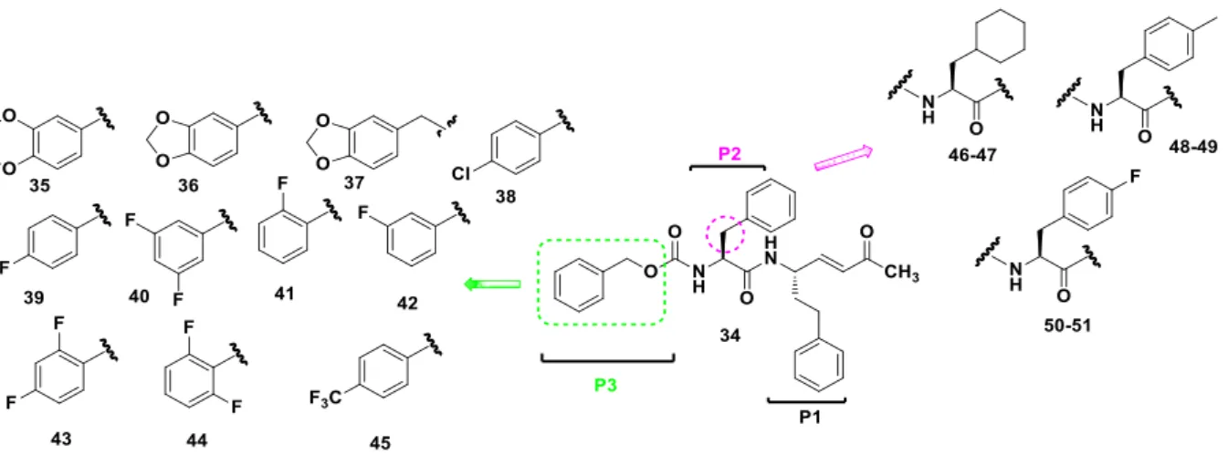
Based on the structure of the lead compound 3, we designed a new series of Michael acceptors (Fig. 4). Herein, the highly reactive vinyl ketone warhead and the HPhe at the P1 site were kept unchanged. The HPhe moiety at the P1 position assures resistance towards endopeptidases, leading to a greater stability in vivo with respect to the corresponding analogs bearing a natural amino acid side chain at P1.32 We then decided to replace the Cbz group with some benzo-fused rings (i.e. 35-37), one of which is the 2,3-dihydrobenzo[b][1,4]dioxine moiety, present in potent rhodesain inhibitors. A panel of aromatic moieties, variously decorated with halogen atoms (i.e. 38-45), were also
8
introduced at the P3 site, in agreement to the structure of peptidic inhibitors bearing at the P3 site 3,5-difluoro- or 4-CF3-phenyl moieties or taking into consideration non peptide rhodesain inhibitors based on a triazine nucleus variously decorated with halogen–substituted aromatic nuclei. This approach could also allow us to investigate the size of S3 pocket and to concurrently allow for the formation of halogen bonds and/or hydrophobic interactions. We also investigated the relevance of the L-Phe residue at the P2 site, by synthesizing the L-cyclohexylalanine, 4-F-L-phenylalanine and 4-methyl-L-phenylalanine derivatives. Cyclohexyl residues at the P2 position have been described in inhibitors of human protease that show some improvment of the binding or of metabolic stability;33 4-Me-L-Phe P2 substituent should afford highly rhodesain selective inhibitors according to a previous study 32 that suggest, moreover, that the introduction of small substituents on the phenyl ring of the P2 side chain could represent a good strategy for the design of novel rhodesain-selective inhibitors.
9 2. DESIGN AND SYNTHESIS OF NOVEL AMIDES AS NONCOVALENT IMMUNOPROTEASOME INHIBITORS
2.1 Introduction
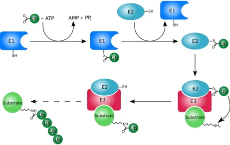
The ubiquitin-proteasome system (UPS) is the major non-lysosomal proteolytic pathway, which plays a fundamental role in the turnover of intracellular protein of eukaryotic cells, thus being responsible for cells’ homeostasis. This role is absolved by the degradation of misfolded or aberrant proteins, which are no longer useful to the cells.34-35 Proteolysis, mediated by the proteasome, is a complex process which includes several steps: in the first step, ATP-dependent, proteins are first targeted to be recognizable for the degradation by the addition of polyubiquitin chains to a residue of lysine of the substrate; in the second step, the polyubiquitinated proteins are processed by 26S proteasome into ubiquitin and short peptides (Fig. 5).35-36 In particular, the ubiquitin-activating enzyme E1 catalyzes the activation of the ubiquitin through the formation of a thioester bond. This process needs the presence of ATP and occurs through the formation of ubiquitin-AMP intermediate and ubiquitin-E1 thiol ester is the final product.37 After this, ubiquitin is transferred, via thioester bond, to the ubiquitin-conjugating enzyme E2, which adds ubiquitin to the lysine residue of either the target protein or the elongating polyubiquitin chain, with the help of the ubiquitin-ligase enzyme E3.
Figure 5. Schematic representation of the ubiquitination process.
In normal cells, the main function of the proteasome is to regulate different cellular functions, such as cell cycle regulation, cell differentiation and apoptosis, immune
10
surveillance.38-39 In addition to the constitutive proteasome, eukaryotic cells express other two variants of proteasome: immunoproteasome, mainly expressed in monocytes and lymphocytes, and thymoproteasome, expressed by thymic cortical epithelial cells.36,40
2.2 Constitutive proteasome
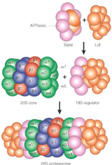
The constitutive 26S proteasome (Fig. 6) is a multi-protease complex which includes a barrel-like 20S core particle (CP), covered by two 19S regulatory caps.5 Each 19S cap is composed by a lid and a base: the lid is responsible for the recognition and the binding of the polyubiquitin chains, while the base includes ATPases that allow the entrance of substrates into the proteolytic 20S core.41
Figure 6. Proteasome structure
The central catalytic 20S CP is characterized by four stacked rings: two outer α-rings and two inner β-rings, which contain different α subunits (α1-α7) and different β subunits (β1-β7), respectively.42 The β-rings contain the three proteolytic activity located in three subunits, endowed with different substrate specificities: the β1 subunit, also known as “post-glutamate peptide hydrolase” (PGPH) or “caspase-like” (C-L), which cleaves after acidic residues; the β2 subunit, which holds a “trypsin-like” (T-L) activity and cleaves after basic residues; lastly, the β5 subunit is responsible for the “chymotrypsin-like” (ChT-L) activity and cleaves after hydrophobic residues.43-44
11
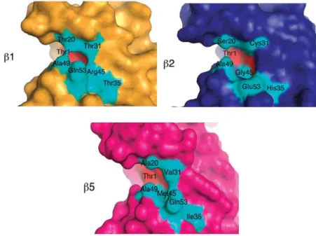
S1 pockets, which are the closest ones to the active site, have the major influence upon the different substrate specificity of the proteasome catalytic subunits (Fig. 7).5,45,46 The β1 subunit exhibits an Arg45 residue which preferably interacts with a Glu residue at P1 position of the protein substrate, thus conferring the post-glutamyl peptidyl hydrolase activity to this subunit. Similarly, the low steric hindrance of β2-Gly45 allows the formation of a large binding pocket with a Glu residue (Glu53) at the bottom, which accepts bulky basic P1 residues. Finally, the β5-Met45 generates a wide pocket which allows the accomodation of bulky hydrophobic P1 residues.
Figure 7. S1 pockets of the catalytic subunits binding sites.5
Each active site of the proteasome shows a γ-OH of a N-terminal threonine residue acting as nucleophile, crucial for the hydrolytic activity and peptide bond cleavage.5,47 In particular, the β5 subunit is mainly involved in protein degradation and is thus considered a main target for the development of anticancer agents.
The immunoproteasome and the thymoproteasome originate by the replacement of the three constitutive catalytic subunits (β1c, β2c, β5c) with the three inducible low-molecular-mass protein (LMP) subunits (β1i; β2i; β5i/β5t) (Fig. 8).40,48
12 Figure 8. Different forms of proteasome.
2.3 Immunoproteasome
The immunoproteasome, mainly expressed by monocytes and lymphocytes, is responsible for cell-mediated immunity and for the generation of MHC (Major Histocompatibility Complex) class I ligands.40,49 The thymoproteasome, expressed by thymic cortical epithelial cells, is instead involved in CD8+ T cells positive selection.48,50 The expression of immunoproteasome is inducible in many tissues; in fact, it has been also detected in various cell types, not only in immune cells, under the stimuli of cytokines, such as INF-γ or TNF-α. The replacement of the catalytic subunits with the correspondent immune subunits leads to the formation of the 20S iCP. 51 The 20S iCP contains the LMP2/β1i and LMP7/β5i subunits, which are upregulated as a consequence to the IFN-γ,52 and the MECL-1/β2i subunit, which is a multicatalytic endopeptidase complex.53 While MECL-1 and LMP7 shows the same type of activities of their counterparts, the LMP2 subunit express a remarkable ChT-L activity, cleaving peptides after hydrophobic residues.
This change of activity is considered an essential requirement for the specific function of epitope generation for antigen presentation. In fact, peptides generated by the hydrolase activity of LMP2 are endowed with non-polar amino acids, which represents a perfect motif for binding to the MHC class I molecules.
A comparison between the S1 pockets of β5c and β5i subunits shows that the residues responsible for the ChT-L activity (Ala20, Met45, and Cys52) are maintained, while the amino acid 31 of β5i changes in terms of size (Met31 which replaces Val31), retaining the hydrophobic feature. However, a different conformation of Met45 allow the
13
formation of a larger S1 pocket, which is also stabilized by van der Waals interactions with the aliphatic side chains of the amino acid Gln53.6
In the end, the S1 pocket of β5c mainly accommodates peptides with small amino acids (Ala or Val), whereas the S1 pocket of β5i well accommodates large non-polar residues (Tyr, Trp, Phe). Therefore, β5c and β5i cleave different fluorogenic substrates Ac-WLA-AMC and Ac-ANW-Ac-WLA-AMC respectively.
Other differences were also observed between β1c and β1i: the substitutions T20V, T31F, R45L and T52A cause the enhancement of the hydrophobicity of β1i S1 pocket, but results in a reduction of dimension. Therefore, peptide hydrolysis occurs after small hydrophobic residues. On the contrary, in the S3 site of β1i subunit, the substitutions T22A and A27V generally downsize and polarize the S3 pocket. To summarize, a β5i-selective inhibitor requires bulky hydrophobic amino acids, like Trp or Phe, at the P1 site and small polar groups at the P3, while a β1i-selective inhibitor should be endowed with branched non-polar amino acids in P1 and small polar amino acids at the P3.6,7 Lastly subunit β2i is the only one not encoded on MHC cluster and it was found to be identical to β2c with the sole replacement of Asp53 (β2c) with Glu (β2i).
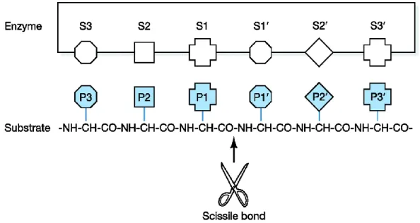
In spite of the presence of the N-terminal nucleophilic threonine residue in all the active sites, the preference cleavage for the various proteolytic subunits is due to the structure of substrate binding pockets. Based on the proximity to the active sites, the binding pockets are termed: nonprimed sites (S1, S2, S3,... Sn), located on the left of the scissile peptide bond; primed sites (S1’, S2’ S3’,... Sn’), located on the right. As a consequence, substrate residues which interact with these binding pockets are named as P1, P2, P3,... Pn sites and P1’, P2’, P3’,... Pn’ sites, respectively (Figure 9).34
14
Figure 9. Peptide orientation (from N-terminus, to the C-terminus), in the active centre
of β-subunits.
2.4 The role of immunoproteasome in human diseases
As stated before, under the stimuli of interferon-γ (IFN-γ), β1i/LMP2, β2i/MECL-1, and β5i/LMP7 are incorporated into the iCPs. Among the subunits, the β1i and β5i play a key role in antigen processing.54 The immunoproteasome is also normally expressed in several non-immune tissues, thus his role is not only limited to MHC class I presentation.55
It was elucidated that the immunoproteasome is involved in protein homeostasis regulation, cell proliferation and differentiation, cytokine expression, being involved in many biological and pathological processes.55
The development of iCPs selective inhibitors is considered a valuable approach to investigate the biological role of each subunit and, at the same time, to treat the same pathologies in which the subunit is involved.
Immunoproteasome is mainly involved in inflammatory, autoimmune diseases and hematologic malignancies.
Since the inflammatory cytokines play a key role in the expression of the immunoproteasome, the role of the multicatalytic complex in inflammatory and autoimmune diseases has been widely explored.
15
High levels of immunoproteasomes were detected in a large number of autoimmune and inflammatory diseases such as Crohn’s disease, 56 ulcerative colitis,57 inflammatory bowel disease58-59 and hepatitis. In Crohn’s disease, an increased expression of the immunosubunits was observed; in particular, the replacement of the constitutive proteasome subunit β1 by the inducible immunosubunit β1i was noticed.56 Also in the inflammatory bowel disease, an increased level of LMP2/β1i in the inflamed colon tissues, with respect to normal tissues, was detected.59
Many studies were also carried out on experimental models of colitis, thus assessing the involvement of both β1i and β5i subunits.
A murine model of dextran-sulfate sodium (DSS)-induced colitis showed an up-regulation of β1i subunit: as a result, transgenic mice lacking the β1i subunit did not suffer of this kind of inflammatory disease.58 In another case of DSS-induced colitis, the inhibition of LMP7/β5i by ONX 0914, a β5i-selective inhibitor, strongly reduced the symptoms of experimental colitis and suppressed a number of cytokines in treated mice, resulting in the reduction of inflammation.57 ONX-0914 proved to block, both in vitro and in vivo, the presentation of LMP7-specific MHC-I-restricted antigens, the production of interleukin-23 (IL-23), by activated monocytes, and of IFN-γ and IL-2 by T cells.
Regarding autoimmune diseases, proteasome inhibitors represents a promising strategy for the treatment of these kind of disorders. Rheumatoid arthritis (RA), one of the most important autoimmune disease, is characterized by synovial inflammation, auto-antibody production and cartilage and bone destruction. Muchamuel et al.49 showed that the selective inhibition of immunoproteasome by ONX-0914 reduced the levels of auto-antibodies in mouse model of RA.
Always in mouse model was found that the inhibition of LMP7/β5i reduced Th1 differentiation, without affecting Th2 differentiation; the suppression of T cell activation seems to be beneficial for the treatment of RA.60
Other relevant studies suggested that the inhibition of the immunoproteasome is efficacious in preventing systemic lupus erythematosus (SLE), a complex autoimmune disease characterized by the production of auto-antibodies and IFN-α. The proteasome inhibitors Bortezomib and Carfilzomib, targeting both the constitutive and immuno-subunits, and the immunoproteasome-selective inhibitor ONX-0914 are effective in the
16
treatment of murine lupus through a dual inhibition of pathogenic IFN-α production and reactive plasma cells. It was also demonstrated that the levels of auto-antibodies, produced by plasma cells, decreased with the reduction in plasma cell numbers in spleen and bone marrow.61
Cascio et al. 62 showed that protein synthesis moderates the responsiveness of differentiating and malignant plasma cells to proteasome inhibitors. It was suggested that protein synthesis could play a role in the proteasomal proteolytic burden and PIs sensitivity. The average proteolytic work accomplished by the proteasome can change by several orders of magnitude, both in MM cells and in differentiating plasma cells; it was suggested that the increased workload is associated with an enhanced sensitivity of PIs.
Another study concerning Multiple Sclerosis (MS), a chronic demyelinating immune-mediated disease of the central nervous system, investigated the potential function of ONX 0914. The β5i-selective inhibitor reduced the disease progression, by reducing levels of cytokine-producing CD4 (+) cells in treated mice, thus validating its potential applicability for the treatment of MS.63 These findings highly clarified the importance of targeting LMP7 as a rational approach to treat inflammatory and autoimmune disorders. A relevant implication of the immunoproteasome was confirmed also in rejection antibody-mediated. MECL-1 (β2i) increased in the graft and blood samples during chronic antibody-mediated rejection. In rat cardiac allografts, Bortezomib delayed acute rejection, attenuated the humoral response and strongly reduced the level of circulating donor-specific antibodies, in a dose-dependent manner.64
Immunoproteasome-inhibitors represent a promising strategy also in preventing transplant rejection.
Proteasome inhibition is an important strategy to treat hematologic malignancies; the nonspecific inhibitors cause side effects and toxicities that may let the therapy fail. Bortezomib (Velcade®) and Carfilzomib (Kyprolis®),also called conventional proteasome inhibitors, showed their effects on MM and other hematologic malignancies but they unfortunately are not selective, targeting both cCPs and iCPs indiscriminately. Targeting the immunoproteasome is a valuable strategy against models of MM that does not respond to conventional drugs.65
17
The collateral effects of conventional proteasome inhibitors limit their applications; consequently, selectively targeting proteasome elements responsible for inflammatory pathways would increase the therapeutic index.66-67
The main collateral effect of Bortezomib is the peripheral neuropathy; however, it was also suggested that bortezomib-induced peripheral neuropathy (BIPN) occurs through a proteasome-independent mechanism.68 The accumulation of Bortezomib in the dorsal root ganglia cells, mitochondria-mediated disregulation of Ca2+ homeostasis and disregulation of neurotrophins were suggested to be responsible of the pathogenesis of BIPN.69 On the contrary, Carfilzomib showed a reduced peripheral neuropathy. In cellular lysates, Bortezomib, but not Carfilzomib, inhibited the serine proteases cathepsins G, A chymase, dipeptidyl peptidase-II and HtrA2/Omi, the latter being involved in neuronal survival.68
High levels of immunoproteasome were detected in MM cells, suggesting that immunospecific proteasome inhibitors could be a strategy to develop a potential treatment for hematological malignancies.65
Carlfilzomib, an α̍,β̍-epoxyketone, showed a strong antitumor activity in MM cells but also an equal inhibition in normal cells was observed (80%), thus cells death was not selective.70
The immunoproteasome-specific inhibitor IPSI-001, a peptidyl aldehyde able to selectively target the β1i subunit of the immunoproteasome,65 induces apoptosis in MM cells and other hematological malignancies, being able to overcome conventional drug resistance, also to Bortezomib; in addition also dexamethasone-resistant cells (MM1.R cells) showed comparable responses as dexamethasone-sensitive cells (MM1.S) to the inhibitor.
As a matter of fact, a non-covalent inhibition should be preferred in order to avoid the inhibition of other human proteases and reduce collateral effects.
Proteasome inhibition represents a valuable strategy for the treatment of Waldenström macroglobulinemia (WM), a kind of non-Hodgkin lymphoma (NHL).
It was proved that WM cells express higher level of iCPs in comparison with cCPs,71 Oprozomib (ONX-0912), an orally active proteasome inhibitor with potent activity on both β5i and β5c subunits, demonstrated is responsible for the toxicity in WM cells and for the reduction of bone marrow (BM)–derived interleukin-6 and insulin-like growth
18
factor 1 (IGF-1) secretion, thus inhibiting BM-induced p-Akt and phosphorylated extracellular signal related kinase (p-ERK) activation in WM cells.71
2.5 Proteasome and immunoproteasome inhibitors
Many proteasome inhibitors targeting both the immuno- and constitutive proteasomes indiscriminately have been approved over the years.
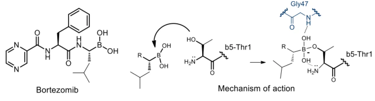
Bortezomib (Velcade®, Fig. 2) represents the first proteasome inhibitor approved by the FDA in 2003 for the treatment of MM and later in 2006 for mantle cell lymphoma. 72-75
Figure 10. Bortezomib structure and mechanism of inhibition of the β5 subunit
Bortezomib is a dipeptidyl boronic acid able to inhibit all three catalytic activities of both the constitutive and immuno-proteasomes through the formation of a reversible tetrahedral adduct (Fig. 10), by reaction with the Thr1Oγ residue, as deduced from the crystal structure of 20S constitutive proteasome in complex with Bortezomib, reported by Groll and coworkers.76
Bortezomib showed similar potencies when evaluated against β1i, β5c and β5i subunits, with IC50 values of 5.5 nM, 7.0 nM, and 3.3 nM, respectively, all ChT-L activities.77 The remaining catalytic sites are also inhibited by Bortezomib, but in particular the inhibition of the T-L activity of both isoforms was very poor. The most important side effect of Bortezomib is the peripheral neuropathy.68 BIPN seemed, anyway, to be reversible in the most of of patients after dose reduction; although this side effect was more frequent in the presence of baseline neuropathy, the overall occurrence was independent of prior neurological disorders.78
19
Carfilzomib (Kyprolis®, Fig. 11) is a second-generation proteasome inhibitor approved by the FDA in 2012 for the treatment of refractory MM.
Figue 11. Carfilzomib structure
Carfilzomib is a tetrapeptide α’,β’-epoxyketone with a N-acyl morpholine cap, obtained by optimization79 of epoxomicin,80 a natural and potent irreversible proteasome inhibitor.81-82 The mechanism of action of epoxomicin, together with that of the related derivatives bearing an α’,β’-epoxyketone warhead (Fig. 12), including Carfilzomib, was investigated by Groll and coworkers.83
R O O HN O O H H Thr1 R O O O N O H H Thr1 R O HO NH OH O Thr1
Figure 12. Mechanism of action of α’,β’-epoxyketone inhibitors.
Both the hydroxyl and the amino groups of Thr1 are involved in a sequential double nucleophilic attack on the α’,β’-epoxyketone warhead, leading to the formation of a stable morpholino ring. Given the key role in the mechanism of action of the free N-terminal amino group, it was suggested that the α’,β’-epoxyketone derivatives are able to inhibit only the N-teminal nucleophile proteases, like the proteasome.
Studies carried out on both the constitutive and immuno-isoforms showed that Carfilzomib is a very potent proteasome inhibitor, able to inhibit both β5c and β5i
20
subunits at low nanomolar concentration, with a slight preference for the constitutive isoform (IC50 values of 6 nM and 33 nM, respectively).84
With regard to the other proteolytic subunits, Carfilzomib showed a lesser amount of activity, thus suggesting, as previously mentioned, that the selective inhibition of the ChT-L proteasome activities is sufficient to exert an antitumor response in hematological malignancies.70
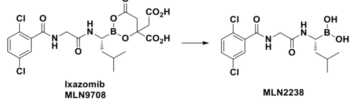
Ixazomib or MLN9708, (Ninlaro®, Fig. 13) was approved by FDA in November 2015, in combination with lenalidomide and dexamethasone, for the treatment of patients with MM that received at least one prior therapy. It is an orally bioavailable prodrug of the boronic acid MLN2238, that overcomes the intravenous or subcutaneous routes of administration of Bortezomib and Carfilzomib. 85
Figure 13. In vivo activation of Ixazomib to MLN2238
Ixazomib preferentially targets the β5c subunit with an IC50 value of 3.4 nM; however, it also shows a lower activity towards β1c and β2c with IC50 values of 31 nM and 3500 nM, respectively.85 MLN9708 showed an improved pharmacokinetic, pharmacodynamic and antitumor activity with respect to Bortezomib. Like Bortezomib, Ixazomib targets both the immuno- and constitutive proteasomes,8 however, no IC50 values have been reported in the literature.
More recently, considering the key role played by the immunoproteasome in various diseases, medicinal chemists focused their research on the discovery of potent selective inhibitors of this isoform. Many interesting inhibitors which show a high degree of selectivity towards the immunoproteasome have been developed.
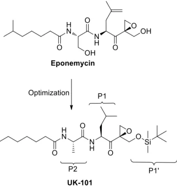
UK-101 (Fig. 14) is a potent peptide inhibitor with a reactive epoxyketone warhead, which selectively targets β1i subunit.86 A previous SAR study carried out on epoxomicin and eponemycin, two natural product able to inhibit the proteasome,80,87 provided crucial
21
structural data. In detail, when tested in vitro, eponemycin showed remarkable inhibitory properties against the immunoproteasome compared to the constitutive isoform, in particular towards the β1i subunit.88
Figure 14. Development of UK-101 from eponemycin.
Based on these data, further SAR studies were carried out, starting from the idea to maintain the N-terminal hydrocarbon tail and the C-terminal hydroxyl group of eponemycin. This work resulted into a series of epoxyketone derivatives, including UK-101, which was identified as the most promising β1i-selective inhibitor.86
UK-101 contains a hydrophobic heptanoic tail at the N-terminal, an alanine residue at the P2 position and the hydroxyl group at the P1’site protected with a tert-butyldimethylsilyl (TBDMS) group at the C-terminal moiety, whereas the P1 leucine plays a key role for the inhibition of the ChT-L activity. In in vitro assays, UK-101 selectively inhibited β1i subunit with IC50 values in the range 100-500 nM. On the contrary, a lack of activity was noticed against the other proteolytic activities, with the only exception of the β5c subunit.86
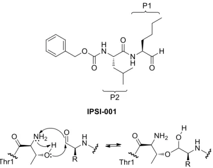
Another interesting inhibitor with a good selectivity towards immmunoproteasome core particles β1i and β5i is represented by the dipeptidyl aldehyde IPSI-001 (Fig. 15).65 As a matter of fact, when tested against the proteasome, IPSI-001 showed a great selectivity towards iCPs. More in detail, comparing the inhibitions of β1i vs β1c and β5i vs β5c, a
22
165- and 100-fold preference respectively for the iCPs β1i and β5i was observed, with an increased selectivity for LMP2/β1i subunit.
Figure 15. IPSI-001 structure and mechanism of action
The mechanism of action of IPSI-001 (Fig. 11) proceeds through the nucleophilic attack of the hydroxyl group of Thr1 on the carbonyl carbon, thus leading to the formation of a tetrahedral hemiacetal. Furthermore, this class of compounds binds to proteasome catalytic sites with a time-independent inhibition.34 IPSI-001 is a dipeptide containing at the P1 site a norLeucine residue, at the P2 site a Leu residue, while at the N-terminal cap it bears a carbobenzyloxy (Cbz) protecting group.
Orlowski et al.65 demonstrated that increasing the hydrophobic group of the P1 aminoacid determines a great selectivity towards the immunoproteasome, while the activity towards constitutive proteasome decreased.
ONX-0914 is a tripeptide epoxyketone (Fig. 16), reported as the first β5i-selective inhibitor49,89 and actually in preclinical studies to evaluate the potential clinical application on autoimmune disorders such as RA, and SLE and for the inflammatory bowel disease.
X-ray structure of ONX-0914 covalently bound to the murine immuno- and constitutive proteasomes, suggested that the presence of a Phe residue at the P1 site induces a major conformational change in the S1 pocket of β5c, but only minor alterations in β5i.7
23
These results suggested also that the N-terminal morpholinyl acetyl cap does not interact with β5i subunit but, probably, is very important to enhance the solubility and to improve the physicochemical properties of ONX-0914.
Figure 16. ONX 0914 structure
In Raji cell lysates, this inhibitor showed a great selectivity towards β5i subunit, with a 10-fold increased activity compared to β5c subunit. With regard to the remaining proteolytic sites, they were inhibited by ONX-0914 only at higher concentrations. Further investigations to verify the selectivity of this inhibitor for β5i were carried out: in β5i knockout mice, the inhibitory effect of ONX-0914 was not observed.49 Furthermore, various studies have reported the promising potential of this compound in pathologies where immunoproteasome plays a key role, as inflammatory and autoimmune diseases (i. e. RA). ONX 0914 is able to inhibit the release of pro-inflammatory cytokines, IL-23 and TNF-α in activated monocytes and to block the disease progression, as well as to alleviate the severity of disease symptoms 49.
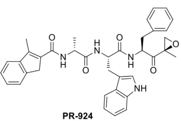
PR-924 (named also IPSI), currently in preclinical trial for the treatment of MM, is another β5i-selective inhibitor with a α’,β’-epoxyketone warhead (Fig. 17) [88]. PR-924, when tested against Raji cell lysates 90 exhibited an IC50 value of 2.5 nM towards β5i, with a 100-fold lower activity against β5c. The remaining catalytic subunits were inhibited only at higher concentrations (i.e. β1i: IC50 value of 1.84 µM), or not inhibited at all.
PR-924 showed antileukemic activity, but only at the concentrations that inhibit both the constitutive (β5c) and immunoproteasome (β5i) subunits: the sole β5i inhibition is not sufficient to induce an anti-leukemia effect.
24
A recent study reported that PR-924 induces apoptotic cell death when tested in several MM cell lines.91 No significant toxicities were observed in control cell lines when they were exposed at 20 µM concentration of PR-924.
Figure 17. PR 924 structure
KZR-616 is a potent, selective dual immunoproteasome inhibitor of the subunits LMP7/LMP2 that shows IC50 of 39 nM/139 nM. KZR-616 was developed by Kezar Life Sciences and is presently in clinical trials for treatment of rheumatic disease.
KZR-616 (Fig. 18) acts as selective covalent inhibitor of the immunoproteasome that potently blocks inflammatory cytokine production in vitro and disease progression in mouse models of SLE. Durable disease remission in animals was achieved at well tolerated doses without affecting normal T-cell dependent immune responses.
Figure 18. KZR-616 structure
Compared to ONX 0914, KZR-616 maintained LMP7 and LMP2 selective inhibition, a comparable cytokine inhibition profile and similar rapid pharmacokinetics. Moreover, efficacy of KZR-616 was comparable to ONX 0914 in the anticollagen antibody induced arthritis (CAIA) model at half the dosage while subcutaneous administration gave comparable results at similar dosages. KZR-616 showed no inhibition at 10 μM against a broad selectivity panel of 20 serine, metallo-, cysteine, and aspartyl proteases and 11
25
hydrolases. Based in part on these results, KZR-616 was selected as a clinical candidate for treatment of autoimmune disease.92
26 3. Results and discussion
3.1 Synthesis of amides 2-33
The synthesis of amides 2-11 was achieved according to a previously reported procedure,11 starting from 2(1H)-pyridone 52 that was N-alkylated with ethyl bromoacetate or methyl 3-bromopropionate in the presence of NaH, to give esters
53-54. These latter intermediates were converted into the corresponding carboxylic acids 55-56 by alkaline hydrolysis with LiOH. Coupling reactions between the carboxylic acids 55-56 and the suitable amines, in the presence of EDC∙HCl, HOBt, as coupling reagents,
and DIPEA as a base, gave the desired amides 2-5 and 6-11 in good yields (Scheme 1).
Scheme 1. Reagents and conditions: (a) NaH, DMF, 0°C, 1h, N2; ethyl bromoacetate or methyl 3-bromopropionate, r.t. 12h; (b) LiOH, MeOH/H2O/dioxane (1:1:1), 0°C-r.t., 6 h ; (c) DCM/DMF, 0°C, HOBt, EDC∙HCl, 10 min, then DIPEA and a suitable amine, r.t., 12h.
The synthesis of the P2 fragments of compounds 12-25 was achieved starting from the commercially available 2-hydroxy-3-phenyl propanoic acid methyl ester 57 and (R)-2-hydroxy-3-phenyl butanoic acid ethyl ester 58, which were converted into the more reactive methanesulfonates 59-60, by reaction with mesyl chloride in the presence of triethylamine (Scheme 2). Intermediates 59-60 were then condensed to the pyridone scaffold 52 to give the esters 61-62, which were then converted into the corresponding carboxylic acids 63-64 by alkaline hydrolysis. Coupling reactions, carried out as reported in Scheme 1, between the carboxylic acids 63-64 and the suitable amines gave compounds 12-18 and 19-25 in high yields.
27 Scheme 2. Reagents and conditions: (a) MsCl, Et3N, dry CH2Cl2, r.t., 2h, N2; (b) compound 52, NaH, dry DMF, 0°C-r.t., then 59 or 60, 12h, N2; (c) LiOH, MeOH/H2O/dioxane (1:1:1), 0°C-r.t., 12h; (d) DMF, 0°C, HOBt, EDC∙HCl, 10 min, then DIPEA and a suitable amine, 0°C-r.t., 12h
The synthesis of amides 27-33 (Scheme 3) was performed starting from commercial available 6,7-dimethoxyisoquinolin-1(2H)-one 65 that was N-alkylated with methyl 4-bromocrotonate in the presence of NaH, using dry DMF as solvent, under N2 atmosphere, to give ester 66. The latter intermediate was converted into the corresponding carboxylic acid 67 by alkaline hydrolysis with LiOH. Coupling reactions between the carboxylic acids 67 and the suitable amines, in the presence of HATU, an efficient coupling reagent, and DIPEA as a base, gave the peptides 27-33 in good yields.
Scheme 3. Reagents and conditions: (a) NaH, DMF, 0°C, 1h, N2; then methyl 4-bromocrotonate, rt, 12h ; (b) LiOH, MeOH, 0°C-rt, 12h; (c) DMF, 0°C, HATU, 10 min, then DIPEA, amines, rt, 12h.
28 3.2 Kinetic parameters calculated according to the binding mode of each inhibitor
Reversible inhibitors usually bind to the enzyme via non-covalent interactions, although exceptions are known in which covalently bound inhibitors result in reversible inhibition due to hydrolytically labile bonding (e.g., peptidyl aldehydes). Reversible inhibitors can act in a competitive or non-competitive manner. Competitive inhibitors compete with the substrate for access to the active site of the enzyme and can be displaced by increasing substrate concentrations. In contrast, in the case of non-competitive inhibitors, the substrate and inhibitor bind independently to the enzyme. The inhibitor promotes a conformational change within the catalytic site that does not prevent the binding of the substrate but that promotes the formation of a non-productive complex [E·S·I].
Irreversible inhibitors bind covalently to the enzyme, resulting in the permanent inactivation of the target protease. Consequently, these inhibitors cannot be displaced by increasing the substrate concentration.
For reversible inhibitors, IC50 values are typically used to describe inhibitory potency. However, for irreversible inhibitors, IC50 values are dependent on both the substrate concentration and the duration of the incubation of enzyme with inhibitor prior to the addition of substrate. Thus, irreversible inhibitors should be described by the following kinetic parameters: i) the dissociation constant (Ki) of the non-covalent enzyme-inhibitor complex [E·I], which is a measure of the binding affinity; ii) the inactivation rate constant, also called the first-order rate constant of inhibition (kinac min-1), which describes the rate of formation of the covalent enzyme-inhibitor complex E-I; and iii), the second-order rate constant of inhibition (k2nd M-1min-1), which is calculated from the
29 3.3 Biological activity and docking studies
All the synthesized compounds were tested for their ability to inhibit each one of the catalytic subunits of c20S and i20S, by measuring the rate of hydrolysis of the appropriate fluorogenic substrate (Suc-Leu-Leu-Val-Tyr-AMC for β5i and β5c; Boc-Leu-Arg-Arg-AMC for β2i and β2c; Ac-Pro-Ala-Leu-Boc-Leu-Arg-Arg-AMC for β1i and Z-Leu-Leu-Glu-Boc-Leu-Arg-Arg-AMC for β1c). MG-132 (Z-Leu-Leu-Leu-al), a reversible inhibitor of both proteasome and immunoproteasome, was used as positive control.
First, compounds underwent a preliminary screening on each proteolytic subunit at 50 µM. Compounds able to inhibit the enzymatic activity by more than 60% were characterized in detail: continuous assays were thus performed (progress curve method, at seven different concentrations, ranging from those that minimally inhibited to those that fully inhibited the immunoproteasome or the proteasome subunit) to determine the Ki values reported in Table 1.
Among all the tested compounds, some of them (i.e. 1, 7, 10-13, 19 and 21) turned to be active on the iCPs. Interestingly, compounds 1 and 7, which were N-benzyl substituted amides selectively inhibited the β1i subunit, with a consistent improvement of activity observed for the β-alanine derivative with respect to the glycine derivative (Ki = 0.021 µM vs 2.23 µM).
The β-alanine derivatives 10 and 11, bearing a cyclohexyl and an n-butyl substituent, on the contrary, targeted both the β1i and β5i subunits. When a Phe residue was introduced at P2 site (e.g. compounds 12 and 13), the activity was switched on both the constitutive and immuno-core particles (β5i and β5c), with a strong preference for the constitutive core-particle (see e.g. compound 12, Ki = 8.81 µM and 45.5 µM for β5c and β5i, respectively). It is worth noting that two the compounds bearing a HPhe residue at the P2 site turned to be active on both the ChT-L activities of i20S (i.e. 19) or against the sole β5i (i.e. 21).
30 Table 1. Activity on cCPs and iCPs of compounds 1-25.
1-5 6-11 12-18 19-25
% of inhibition at 50 µM or Ki (µM)
Comp. R β1c β2c β5c β1i β2i β5i
1 benzyl 32% 21% n.i. 2.23±0.26 17% n.i.
2 phenylethyl 24% n.i. n.i. 44% n.i. n.i.
3 phenylpropyl 32% n.i. n.i. n.i. 11% n.i.
4 cyclohexyl 36% n.i. n.i. 22% 10% 3%
5 n-butyl 36% n.i. n.i. n.i. 5% n.i.
6 phenyl 23% 1% 25% n.i. 11% 25%
7 benzyl 37% n.i. n.i. 0.021±0.002 25% 17%
8 phenylethyl 24% n.i. 3% 27% n.i. 46%
9 phenylpropyl 22% n.i. 18% 37% n.i. 15.17±0.63
10 cyclohexyl 23% n.i. 12% 2.92±0.87 n.i. 5.74±0.63
11 n-butyl 19% n.i. 4% 3.09±1.06 n.i. 14.29±3.0
12 phenyl 15% 6% 8.81±1.11 23% n.i. 45.5± 2.6
13 benzyl 14% 8% 3.02±0.29 19% n.i. 7.77±1.51
14 phenylethyl 10% 13% 20.4±2.2 10% n.i. 43%
15 phenylpropyl 15% 12% 50% 25% n.i. 45%
16 cyclohexyl 5% 6% 45% 16% n.i. 39%
17 n-butyl n.i. n.i. 48% 30% 9% 33%
18 i-pentyl 8% n.i. 55% 23% n.i. 30%
19 phenyl n.i. 23% n.i. 5.9± 0.16 n.i. 5.81±0.37
20 benzyl n.i. 26% n.i. 21% n.i. 38%
21 phenylethyl n.i. 31% n.i. 48% 14% 3.85± 0.46
22 phenylpropyl n.i. 33% n.i. 36% 4% 34%
23 cyclohexyl n.i 30% n.i. 40% 6% 42%
24 n-butyl n.i. 31% n.i. 29% n.i. 37%
31
To help interpret the structure–activity relationship (SAR) data and elucidate the molecular mechanism of i20S inhibition of the most active compounds 1, 7, 13 and 21, docking studies were carried out in collaboration with Prof. Lavecchia (Federico II University of Napoli), using the GOLD Suite docking package 93 with the X-ray crystal structure of the murine i20S in complex with the epoxyketone inhibitor PR-957 (PDB: 3UNF).7 This structure was selected due to the high degree of amino acid sequence identity (>90%) between mouse and human i20S subunits around the respective catalytic sites. Moreover, the few non-identical residues are located at the interface with the concomitant adjacent subunits and are thus absent from the catalytic sites. Furthermore, the crystal structure of PR-957 bound to β1i reveals two well-defined water molecules within the S3 pocket, which coordinate a tight H-bond network between β1i-A50N, β2i-S118Oγ and the backbone amide nitrogen and carbonyl oxygen of the P3 alanine residue of PR-957. Accordingly, the intervening water molecules were included in the docking experiments.
Docking of the amide derivative 1 to the β1i subunit revealed a binding mode similar to that of PR-957 (Fig. 19a), with an almost identical positioning of the backbone amides, the P1 phenyl moieties adopting the same spatial arrangement and the P3 alanine residue of PR-957 nicely overlapped with the pyridone ring of 1 (Fig. 19b). The P1 benzyl substituent extends deeply into the S1 pocket, forming hydrophobic contacts with V20, F31, L45, A49 and A52. The 1 backbone amide is engaged in H-bonds with the NH group of S21 backbone and the carbonyl oxygen of G47 backbone, whereas the carbonyl oxygen of the pyridone ring accepts further H-bonds from A49N, S48Oγ and A50N, these last two through one of the two intervening water molecules located crystallographically in the PR-957 structure and included in our model. Moreover, the second water molecule forms a H-bond with the first one, while simultaneously engaging the β2i-S11833Oγ, which is at the bottom of the S3 pocket and contributes to a further stabilization of the ligand into the active site. Additional hydrophobic interactions occur between the pyridone ring and the V20 and V27 side chains located in proximity of S3 pocket of β1i subunit.
32 Figure 19. Binding modes of compounds 1 (A, yellow orange sticks) and 7 (C, lime green
sticks) into the β1i (slate)/β2i (light pink) active site of mouse i20S, represented as a ribbon model. Only amino acids located within 4 Å of the bound ligand are displayed and labelled. H-bonds discussed in the text are depicted as dashed grey lines. The defined water molecules forming tight H-bonds to the protein are displayed as red spheres. S1-S3 specificity pockets are labeled. An overlay of 1 (B, docked pose) and 7 (D, docked pose) with PR-957 (white sticks, X-ray crystal pose) is shown in the β1i/β2i active site of mouse i20S.14
The β1i and β1c subunits strongly differ in their amino acid lining in the unprimed substrate binding channel. The hydrophobic active site surrounding of subunit β1i is replaced by a more polar one in subunit β1c. In particular, the amino acid substitutions V20T, F31T, L45R and A52T increase the polarity and the size of the β1c S1 pocket. Therefore, the more hydrophilic S1 pocket of subunit β1c opposes binding of the hydrophobic P1 substituents. These findings are in agreement with the poor inhibitory activity displayed by this series of derivatives towards β1c.
Elongation of the linker between the pyridone ring and the amide function from one to two methylene units yielded compound 7, which showed the highest inhibitory potency and selectivity towards β1i (Ki = 0.021 μM). As a result of the docking of 7 into the β1i subunit, we obtained a complex showing an overall binding mode similar to that of 1 (Fig. 19c,d), with the ligand forming H-bonds with G47O, S21N and S21Oγ of β1i
33
through the amide group as well as the same pattern of hydrophobic interactions within the S1 pocket. These results are in consonance with the SAR data showing that compounds bearing a benzyl (7), cyclohexyl (10) or n-butyl (11) residue at P1 inhibit β1i much stronger than β1c, which confirms that the S1 pocket of β1i is more hydrophobic than that of β1c.7 In addition, neither β1i nor β1c is inhibited by compounds featuring a bulky phenylethyl (8) or phenylpropyl (9) substituent at P1, an outcome that supports structural data displaying that β1i/c have a smaller S1 pocket than β5i/c.7
The pyridone moiety of 7 fits into the S3 pocket formed by S48, S118 and H114, which is beneficial for improving potency and selectivity for β1i subunit (Fig. 19c). In particular, the pyridone carbonyl oxygen forms a tight H-bond network with A49N, S48Oγ and A50N of β1i through one water molecule and with β2i-S118Oγ via the second water molecule. Additionally, the aromatic pyridone core makes an edge-to-face π-stacking interaction with the H114 imidazole ring in the subunit β2i. Compared to β1c, the S3 pocket of β1i is characterized by the amino acid replacements T22A, A27V as well as Y114H in the neighbouring subunit β2i. These differences lead to a more size-restricted and more hydrophilic S3 pocket in β1i compared to its counterpart β1c.
A second series of derivatives was designed, in which a residue of phenylalanine was used to replace the glycine P2 group. Among them, compound 13 was moderately active against β5i (Ki = 7.77 μM) and displayed a fairly good potency against β5c (Ki = 3.02 μM). As depicted in Fig. 20a,b, 13 exhibited a binding mode in β5i similar to that of derivatives 1, 7 and PR-957. The P1 benzyl substituent fits into the S1 pocket, the backbone amide is stabilized by H-bonds with G47O, T21N and T21Oγ of β5i and the pyridone group protrudes into the S3 site with the carbonyl engaged in H-bonds with A49N and C48S. The P2 phenylalanine moiety projects into the central core of the 20S cavity, making little or no contacts with the β5i subunit. Docking of 13 to the human c20S crystal structure 94 elucidates that the P1 benzyl and the P3 pyridone ring fit into subunit β5c by interacting with T21, A27, A49, S129 and D144 of the neighbouring subunit β6 via an intervening water molecule. This latter interaction stabilizes the ligand into the S3 pocket and accounts for the enhanced potency of 13 for β5c.
34 Figure20. Binding modes of compounds 13 (A, pink sticks) and 21 (C, violet) into the
β5i (aquamarine)/β6 (wheat) active site of mouse i20S, represented as a ribbon model. Only amino acids located within 4 Å of the bound ligand are displayed and labelled. H-bonds discussed in the text are depicted as dashed grey lines. S1-S3 specificity pockets are labeled. An overlay of 13 (B, docked pose) and 21 (D, docked pose) with PR-957 (white sticks, X-ray crystal pose) is shown in the β5i/β6 active site of mouse immunoproteasome. Compound 21 overlaps with the X-ray crystal pose of PR-924 (slate sticks) bound to the humanized yeast i20S (PDB: 5L5H).14
Noteworthy, substitution of the glycine residue at P2 with a homophenylalanine residue yielded a series of derivatives in which the activity against c20S subunits β1 and β5 was completely lost. Among them, compound 21 showed selective inhibitory activity against β5i (Ki = 3.85 μM). Docking of 21 into the subunit β5i revealed a different binding mode in comparison to 1 and 7 derivatives, with the P2 substituent protruding into the S3 subsite instead of pointing toward the initially assumed S2 binding pocket (Fig. 20c). As a consequence, the pyridone scaffold assumes a folded conformation, engaging a very weak H-bond with β5i-G23N (distance of 4 Å).95 The backbone amide establishes a H-bonding network involving T21O and A49N. The homophenylalanine residue deeply extends into the S3 subsite making O-H/π interactions with the hydroxyl groups of S27 from β5i and S129 from β6. The phenylethyl at P1 perfectly fits into the spacious S1
35
pocket of β5i and is stabilized by a cation- π contact with K33, a sulfur–arene interaction with M4596-97 and by C-H/ π interactions with the side chains of M31, K33, and A49. Interestingly, the overlay of the docked pose of 21 with the β5i-specific inhibitors PR-957 (PDB ID: 3UNF) and PR-924 bound to the chimeric hβ5/hβ6 substrate binding channel (PDB ID: 5L5H)98 reveals that 21 and PR-924 adopt a similar kinked binding mode, with an identical positioning of the amide scaffolds and a similar orientation of the P1 and P3 functions, whereas PR-957 adopts a linear orientation (Fig. 20d). The superior β5i-selectivity of 21 compared to ligands that target the strictly conserved peptide binding sites seems to result from its ability to exploit subpockets other than the substrate-binding channels. Moreover, insights into the selectivity of 21 towards i20S over c20S can be gained when considering amino acid compositions of β5c and β5i S1 pockets. Although the S1 specificity pockets of both β5c and β5i are formed by the same residues, the conformation of M45 in β5c is different from that in β5i, thus resulting in peculiarly sized S1 specificity pockets. 7 The observed selectivity of 21 can be rationalized by the impaired accommodation of the phenylethyl moiety into the smaller β5c S1 pocket because of the closed conformation of M45.
In conclusions, with this investigation we identified a series of amides with Ki values in the low micromolar or submicromalar range towards one or two chymotrypsin-like activities of immunoproteasome (β5i and β1i subunits). Amide 7 was identified as lead compound, due to the selective inhibition of β1i subunit in the submicromolar range (Ki = 21 nM). Docking studies allowed us to clarify the binding mode of the amides in the catalytic site of immunoproteasome proteolytic subunits, thus explaining the preferential inhibition of immunoproteasome with respect to proteasome.
Worthy of note, the noncovalent inhibition, characteristic of our amides, is strongly desirable, because free of drawbacks and side-effects related to covalent inhibition. Our future efforts will be devoted to optimize the identified lead compound 7 in terms of potency and selectivity and to check activity against a panel of hematological malignancies or against autoimmnune diseases in which immunoproteasome is overexpressed.
Also the second series of compounds was also evaluated by means of fluorimetric assays as described above. Interestingly, compound 33 revealed a high binding affinity for the
36
β1i subunit (0.27 µM) coupled with good inhibitory properties towards β5i and β5c subunits (Table 2).
Table 2. Activity of compounds 27-33 on cCPs and iCPs
% inhibition at 100 µM or Ki (µM)
27-33
Comp. R 𝜷1c 𝜷2c 𝜷5c 𝜷1i 𝜷2i 𝜷5i
27 phenyl 17 15 49 10 12 50
28 benzyl 23 9 n.i. 3 n.i. 65
29 phenylethyl n.i. 10 60 14 4 45
30 phenylpropyl n.i. 5 10.61 ±0.25 50 3 64
31 cychlohexyl n.i. n.i. 23 n.i. 42 55
32 n-butyl n.i. 10 19 n.i. 42 49
33 i-pentyl n.i. 4 1.18±0.03 0.27±0.31 20 1.91±0.02
Preliminary docking studies of the most active inhibitor, 33, into the β1i active site of mouse i20S show that the dimethoxy-isoquinolinonic portion is interposed between S2 and S3 pockets, orienting one of the methoxyl group towards Ser118 to form an H-bond, while the hydrocarbon chain is located into the S1’ subpocket. Furthermore, the ligand forms H-bonds with Gly47 and Thr21 (Figure 21).
37 4. Experimental Section
4.1 Chemistry.
All reagents and solvents were purchased from commercial suppliers and used without any further purification. Elemental analyses were performed on a C. Erba Model 1106 Elemental Analyzer and the results are within ±0.4% of the theoretical values. Merck silica gel 60 F254 plates were used for analytical TLC; column chromatography was carried out on Merck silica gel (200–400 mesh). 1H-NMR and 13C-NMR spectra were recorded on a Varian 300 MHz NMR spectrometer operating at frequencies of 300.13 and 75.47 MHz, or on a Varian 500 MHz spectrometer operating at 499.74 and 125.73 MHz for 1H-NMR and 13C-NMR spectra, respectively. The residual signal of the deuterated solvent was used as internal standard. Chemical shifts are given in δ (ppm) and coupling constants (J) in Hz. Splitting patterns are described as singlet (s), broad singlet (bs), doublet (d), triplet (t), quartet (q), multiplet (m), doublet of doublet (dd), or triplet of doublets (td).
Ethyl (2-oxo-2H-pyridin-1-yl)-acetate (53).
A suspension of NaH (91 mg, 3.78 mmol) in dry DMF (10 mL) at 0 °C under N2, was treated with a solution of pyridone 52 (300 mg, 3.15 mmol) in dry DMF (20 mL). After 1 h, ethyl bromoacetate (490 µL, 4.42 mmol) was added to the mixture and the reaction was stirred for a further 12 h at room temperature. The reaction was quenched with saturated NH4Cl (5 mL) and the product was extracted with CH2Cl2 (3 × 70 mL). The combined organic phases were washed with water (3 × 100 mL), dried over Na2SO4, filtered and concentrated in vacuo. The crude was purified by column chromatography (CHCl3/MeOH 95:5) to give compound 53 as a yellow oil (354 mg, 62%); Rf = 0.70 (CHCl3/MeOH 9:1). 1H NMR (300 MHz, CDCl3) δ: 1.21 (t, J = 7.3 Hz 3H), 4.16-4.20 (m, 2H), 4.60 (s, 2H), 6.15-6.19 (m, 1H), 6.53-6.56 (m, 1H), 7.19-7.33 (m, 2H).
Methyl 3-(2-oxopyridin-1(2H)-yl)propanoate (54).
According to the same procedure described for compound 53, compound 54 (300 mg, 3.15 mmol) was reacted with methyl 3-bromo-proprionate (481 µL, 4.41 mmol). Compound 54 was obtained as a yellow oil (474 mg, 83%), after purification by column chromatography (CHCl3/MeOH 95:5); Rf = 0.70 (CHCl3/MeOH 95:5). 1H NMR (300