Contents lists available atScienceDirect
Neurobiology of Disease
journal homepage:www.elsevier.com/locate/ynbdiToxic properties of microsome-associated alpha-synuclein species in mouse
primary neurons
Emanuela Colla
a,⁎, Giulia Panattoni
a, Alessio Ricci
a, Caterina Rizzi
a, Lucia Rota
a, Nicola Carucci
a,
Verdiana Valvano
a, Francesco Gobbo
a, Simona Capsoni
a, Michael K. Lee
b,c, Antonino Cattaneo
a,daBio@SNS Laboratory, Scuola Normale Superiore, Pisa, Italy bDepartment of Neuroscience, University of Minnesota, United States cInstitute for Translational Neuroscience, University of Minnesota, United States
dNeurotrophins and Neurodegenerative Diseases Laboratory, Rita Levi-Montalcini European Brain Research Institute, Rome, Italy
A R T I C L E I N F O
Keywords: α-Synuclein Microsomes Endoplasmic reticulum Oligomers Aggregates Neurodegeneration Parkinson's diseaseA B S T R A C T
α-synuclein (αS) is a small protein that self-aggregates into α-helical oligomer species and subsequently into larger insoluble amyloid fibrils that accumulate in intraneuronal inclusions during the development of Parkinson's disease. Toxicity ofαS oligomers and fibrils has been long debated and more recent data are sug-gesting that both species can induce neurodegeneration. However while most of these data are based on dif-ferences in structure between oligomer and aggregates, often preassembled in vitro, the in vivo situation might be more complex and subcellular locations whereαS species accumulate, rather than their conformation, might contribute to enhanced toxicity. In line with this observation, we have shown thatαS oligomers and aggregates are associated with the endoplasmic reticulum/microsomes (ER/M) membrane in vivo and how accumulation of solubleαS oligomers at the ER/M level precedes neuronal degeneration in a mouse model of α-synucleino-pathies. In this paper we took a further step, investigating the biochemical and functional features ofαS species associated with the ER/M membrane. We found that by comparison with non-microsomal associatedαS (P10), the ER/M-associatedαS pool is a unique population of oligomers and aggregates with specific biochemical traits such as increased aggregation, N- and C-terminal truncations and phosphorylation at serine 129. Moreover, when administered to murine primary neurons, ER/M-associatedαS species isolated from diseased A53T human αS transgenic mice induced neuronal changes in a time- and dose-dependent manner. In fact the addition of small amounts of ER/M-associatedαS species from diseased mice to primary cultures induced the formation of beads-like structures or strings offibrous αS aggregates along the neurites, occasionally covering the entire process or localizing at the soma level. By comparison treatment with P10 fractions from the same diseased mice resulted in the formation of scarce and small puncta only when administered at high amount. Moreover, in-creasing the amount of P100/M fractions obtained from diseased and, more surprisingly, from presymptomatic mice induced a significant level of neuronal death that was prevented when neurons were treated with ER/M fractions immunodepleted ofαS high molecular weight (HMW) species. These data provide the first evidence of the existence of two different populations of αS HMW species in vivo, putting the spotlight on the association to ER/M membrane as a necessary step for the acquisition ofαS toxic features.
1. Introduction
Accumulation ofα-synuclein (αS) aggregates in intracellular pro-teinacious inclusions called Lewy Bodies (LB) or Lewy neurites, ac-cording to their subcellular location, is a classical hallmark of Parkinson's disease (PD) and α-synucleinopathies (Goedert et al., 2012).αS is a small, soluble acidic protein highly expressed throughout
the nervous system and with a well-described presynaptic localization (Iwai et al., 1995; Maroteaux et al., 1988). Point mutations in theαS gene (Appel-Cresswell et al., 2013; Krüger et al., 1998; Lesage et al., 2013; Polymeropoulos et al., 1997; Zarranz et al., 2004) and gene amplifications (Chartier-Harlin et al., 2004; Singleton, 2003) have been found in family pedigrees affected by autosomal dominant, early onset PD, althoughαS neurotoxicity contributes to both genetic and sporadic
https://doi.org/10.1016/j.nbd.2017.12.004
Received 19 July 2017; Received in revised form 7 December 2017; Accepted 11 December 2017
⁎Corresponding author at: BIO@SNS Laboratory, Scuola Normale Superiore, Piazza dei Cavalieri 7, Pisa 56126, Italy.
E-mail address:[email protected](E. Colla).
Abbreviations:αS, alpha-synuclein; PD, Parkinson's disease; ER/M, endoplasmic reticulum/microsomes; HMW, high molecular weight; SpC, spinal cord; Ctx, cortex; PreS, pre-symptomatic; Tg, transgenic; nTg, non-Tg
Available online 12 December 2017
0969-9961/ © 2017 The Authors. Published by Elsevier Inc. This is an open access article under the CC BY license (http://creativecommons.org/licenses/BY/4.0/).
forms of PD (Shulman et al., 2011).
Aggregation of αS in insoluble inclusion is a complex nucleation reaction that includes at least two key steps: the transition from an unfolded monomer which has a naturally intrinsic unfolded con-formation [(Burré et al., 2013; Chandra et al., 2003; Fauvet et al., 2012; Theillet et al., 2016; Weinreb et al., 1996), although lately these data have been questioned (Bartels et al., 2011; Dettmer et al., 2015; Wang et al., 2011)] to an oligomer-type of structure and the transition to an insolubleβ-sheet rich protofibril (Cremades et al., 2012; Deleersnijder et al., 2013). Many variables are thought to influence these transitions: point mutations, such as those associated with genetic PD (Conway et al., 1998); C-terminal truncation (Li et al., 2005); protein level and stability (Li, 2004); environmental factors (Uversky et al., 2002); post-translational modifications, such as ubiquitination, phosphorylation, nitration/oxidation (Oueslati et al., 2010).
Membrane interaction appears to be another condition that can influence formation of αS fibrils (H.-J. Lee et al., 2002). In fact,αS is believed to shift between a free and a membrane-bound state in a dy-namic equilibrium with the membrane-bound state accounting for 10–15% of the total protein amount. αS can bind synaptic vesicles and its binding is believed to mediate its synaptic function (Burré, 2015).αS has been implicated in a broad range of presynaptic functions that in-cludes binding and promoting SNARE complex assembly to favor docking of synaptic vesicles to the cell membrane (Burré et al., 2010, 2014), lipid transport and metabolism (Golovko et al., 2005, 2009), neurotransmitters release and brain plasticity (Bendor et al., 2013). Association with membranes is mediated by αS N-terminal seven 11-amino-acids repeats that are predicted to form an amphipathicα-helix. αS has been found to bind high-curvature membranes such as in pre-synaptic vesicles, through the acquisition of a multimeric structure with a defined orientation (Burré et al., 2014). Thus, under normal condi-tionsαS can shuffle between a native unfolded structure to a multimeric vesicle-bound conformation at the presynaptic terminals. However it is not clear how the transition from these native conformations to a toxic type of aggregates occurs.
We have recently described the presence of toxicαS high molecular weight (HMW) species (oligomers and aggregates) associated with the endoplasmic reticulum/microsomal vesicles (ER/M) in vivo, in diseased Prp-A53T transgenic (Tg) mice (Colla et al., 2012a).αS HMW species were sensitive to protease degradation suggesting that theseαS species were associated with the microsomal membrane on the cytosolic side. Importantly, the appearance ofαS oligomers at the ER/M level tem-porally preceded the onset of neurodegeneration and ER stress-induced cell death in a Tg mice, suggesting the microsomal membranes might be a unique place to foster the accumulation of toxic species ofαS (Colla et al., 2012b).
In this paper we took a deeper look into the ER/M-associatedαS HMW species, comparing them to the rest ofαS aggregates purified through low speed centrifugation. Ourfindings provide evidence of the existence of a toxic species ofαS in vivo, associated with the ER/M vesicles that has unique biochemical traits and is more aggressive in spreading and inducing cell death in neuronal primary cultures. 2. Materials and methods
2.1. Animal models
Transgenic mice expressing human A53TαS under the control of the mouse prion protein (PrP) promoter [line G2-3(A53T)] have been de-scribed previously (Colla et al., 2012a, 2012b; M. K. Lee et al., 2002; Li et al., 2005; Martin et al., 2006). This model develops fatal neurological disease at ~ 12 months of age which rapidly progresses to end state within 14–21 days of onset. Diseased mice show a drastic reduction in motor function, accumulation of intracellular αS inclusions and neu-ronal death. For this study, sick mice at 12–14 months of age, pre-symptomatic mice at 9 months and age-matched nTg littermates were
used. Presymptomatic animals did not show any motor dysfunction or αS pathology in the central nervous system. All animal studies were approved and complied in full by the national and international laws for laboratory animal welfare and experimentation (EEC council di-rective 86/609, 12 December 1987 and Didi-rective 2010/63/EU, 22 September 2010).
2.2. Membrane fractions preparation
Membrane pellet fractions were obtained as previously described (Colla et al., 2012a). Briefly, fresh tissues were homogenized in a 1:10 (wt/vol) volume of lysis buffer (250 mM sucrose, 20 mM HEPES, 10 mM KCl, 1.5 mM MgCl2, 2 mM EDTA, protease and
phosphate-in-hibitors cocktails) using a Teflon pestle homogenizer. Initial homo-genates were centrifuged at 1000 × g to remove nuclei and unbroken cells. The resulting supernatant was centrifuged at 10,000 ×g for 20 min at 4 °C to obtain thefirst membrane pellet (called P10), while the supernatant was centrifuged at 100,000 × g for 1 hr at 4 °C to ob-tain the microsomal pellet (called P100). Both pellet fractions were washed with homogenization buffer once and centrifuged again at the same speed as previously mentioned. P10 and P100 were then re-suspended in 200 or 100μl of lysis buffer, respectively, and their pro-tein content was determined.
2.3. Western blot
Immunoblot and dot blot analyses were performed as previously described (Colla et al., 2012a, b). The following antibodies were used: syn1 (1:5000; BD Transduction Laboratories, Franklin Lakes, NJ); LB509 (1:5000; Abcam, Cambridge, MA); mouse pser129-αS (1:1000; DAKO, Glostrup, Denmark), syn303 (1:1000; Biolegend, San Diego, CA); A11 (ThermoFisher Scientific, Eugene, OR). Quantitative analysis of immune detected bands was done using ImageLab Software (Bio-rad, Hercules, CA).
2.4. Primary hippocampal and cortical neurons preparation and P10 and P100 fractions treatment
Primary neuronal cultures were prepared from wild-type (WT) newborn (P0) hippocampus and cortex of mouse strain B6.129, ac-cording to a procedure byBeaudoin et al., (2012). Hank's Balanced Salt Solution was used as mechanical dissection medium. Tissues me-chanically dissected were treated with 0.1% trypsin for 7 min at 37 °C and then collected with regular DMEM medium containing fetal bovine serum (FBS) and DNase. Preparations were centrifuged at 1000 rpm for 5 min and pellet was resuspended in Neurobasal medium containing 2% B27, 1X Glutamax, 6 mg/ml glucose, 10% FBS, 12.5μM glutamate and 1X Gentamicin. Dissociated neurons were plated on poly-D-lysine
coated coverslips placed in 24 well dishes at a concentration of about 100,000 cells/cm2. At day in vitro (DIV) 1 the medium was replaced with new fresh Neurobasal medium containing 2% B27, 1X gentamicin and 1X Glutamax. At DIV2, 1/3 of the medium was removed and re-placed with fresh medium containing 2.5μM cytosine arabinoside for 48 hrs to reduce glial contamination.
Neurons were treated at DIV7 with 0.5, 1 or 2μg of various pellet fractions according to the experiment. To avoid sample variability, 4–5 pellet fractions isolated from different mice with the same phenotype were pooled. For internalization experiments, neurons werefixed after 2 days, 1 week or 2 weeks of treatment.
2.5. Cell lines
Human neuroblastoma SH-SY5Y cells were transfected with a pcDNA3.1 plasmid carrying human WT αS cDNA tagged at the C-terminal with Myc. Cells stably carrying the plasmid were kept poly-clonal and routinely cultured in DMEM medium containing 10% FBS,
1% penicillin/streptomycin and 500μg/ml of G418 as selection anti-biotic.
For pellet fractions treatment, 50,000 cells/well were plated on poly-D-lysine coated coverslips placed in 24 well dishes. The day after,
pellet treatment was done following the same protocol described for neurons except that, in case of SH-αS-Myc WT, 5 μg of various mem-brane fractions were added. Cells werefixed after 2 days of treatment.
2.6. Confocal immunofluorescence
Cells werefixed with 2% paraformaldehyde and 5% sucrose solu-tions for 15 min at room temperature (RT) and permeabilized with 0.3% Triton x-100 (Tx-100). Standard immunofluorescence was per-formed as previously described (Colla et al., 2012a). The following antibodies were used: syn1 (1:2500), rabbit pser129-αS (EP1536Y, 1:1000, Abcam, Cambridge, MA), Map2 (1:10000, Abcam) and Lamp1 (1:1000, Abcam), NeuN (1:1000 Millipore, Merck, Darmstadt, Ger-many); syn303 (1:1000), Tau (1:10000 Synaptic system, Goettingen, Germany), mouseαS (1:200 Cell Signal, Leiden, The Netherlands).
Quantitative analysis of totalfluorescence, after background sub-traction or particles count perfield was done using Image J software.
For the “two-stages” immunofluorescence, live cultures were in-cubated before fixation with 3% FBS for 30 min at 4 °C with gentle rocking and then incubated with primary antibody dissolved in 3% FBS for 30 min at 4 °C. Cells were then washed,fixed and stained following the standard procedure. Stacked images were acquired with a laser scanning confocal microscope SP2 system (Leica Microsystems) using a 63 × objective.
2.7. Tunel staining
Afterfixation, neuronal cultures were incubated with 1X Terminal Deoxynucleotidyl Transferase (TdT) Buffer (3 mM trizma base, 14 mM cacodylate sodium, 0.1 mM cobalt chloride, pH 7.2) for 15 min at 37 °C and subsequently with TdT reaction mixture containing TdT buffer, 1 mM Biotin-16-dUTP, 400 U/μl terminal deoxynucleotidyl transferase [Roche, Indianapolis, IN] for 1 hr at 37 °C. After incubation, the enzyme activity was blocked by washing with 2X Saline‑sodium citrate (SSC). Unspecific binding sites were blocked by incubating for 30 min with a blocking solution containing 1% BSA and 0.1% Tx-100 at RT. Neurons were incubated with 1:3000 Streptavidin Alexa Fluor® 568 conjugate (ThermoFisher Scientific), for 1 hr at RT in the dark. After incubation, neurons were washed repeatedly with PBS and standard immuno-fluorescence protocol for NeuN antibody and DAPI staining was carried out.
Pictures were taken using Nikon Eclipse E600 epi-fluorescence mi-croscope (Nikon Corp, Tokyo, Japan) with 20 × objective. Cell counting was done using ImageJ software (National Institute of Health).
2.8. Immunodepletion
Immunodepletion ofαS species was performed by incubating 20 μg of P100 fractions (pooled from 4 mice) obtained from SpC of diseased and presymptomatic animals, with 500 ng of A11, Syn1 or Syn303 antibodies previously conjugated with protein A/G agarose (ThermoFisher Scientific). After 1 hr incubation at 4 °C, samples were centrifuged at low speed (2500 rpm for 5 min) to remove im-munoprecipitated complex conjugated with the agarose. The super-natant that represented the immunodepleted sample was carefully re-moved, avoiding any agarose contamination. 2μg of each sample was then loaded on a denaturing SDS Page and immunoblotted with Syn1 antibody to check forαS levels. According to the experiment 1 or 2 μg was then administered to neuronal cultures.
2.9. Statistical analysis
All values are expressed as the mean ± SD. Differences between means were analyzed using Student's t-test and one-way ANOVA fol-lowed by Tukey multiple comparisons test (Prism, Graph Pad Software, LaJolla CA).
3. Results
3.1. Biochemical characterization ofαS aggregates
Because we recently identified αS oligomers and aggregates asso-ciated with the ER/M membrane in presymptomatic and diseased transgenic mice (Colla et al., 2012a, b), we decided to focus on the biochemical and functional characterization of these aggregates to understand whether distinct populations ofαS HMW species exist at the same time in neurons. To do this we isolated all theαS HMW species from a disease-affected area such as the spinal cord (SpC) of sick A53T transgenic (Tg) mice and separated them into two different populations according to their sedimentation rate. These populations were named P10 and P100. P10 represents a crude membrane pellet fraction that sediments at 10,000 × g, containing the majority of αS aggregated species (M. K. Lee et al., 2002; Li et al., 2005) and is rich in mi-tochondria and synaptosomes. P100 is the microsomal pellet, a fraction that sediments at 100,000 × g and is rich in ER, Golgi and synaptic vesicles. According to this protocol, there are virtually no aggregates that remain in the cytosol after centrifugation (Colla et al., 2012a). In order to correlate changes inαS compositions in different fractions to the onset ofαS-driven pathology, P10 and P100 fractions were also obtained from SpC of presymptomatic (aged 9 months) mice and from pathology-free regions like cortex (Ctx) of diseased age-matched lit-termates.
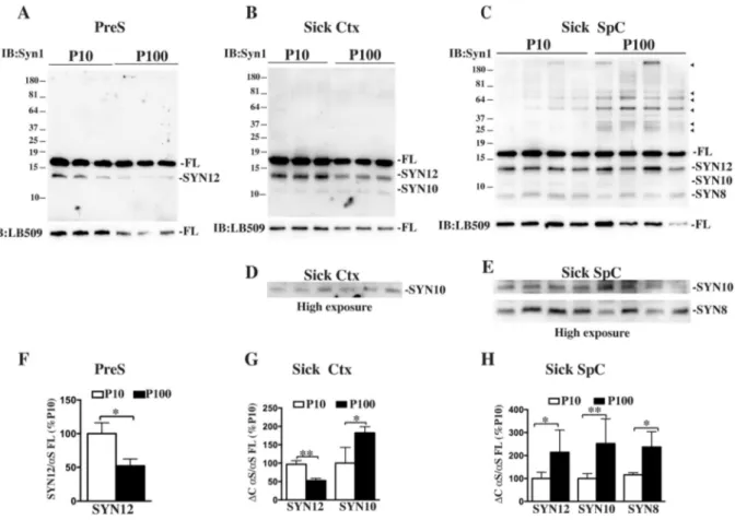
Since C- and N-terminal truncations of theαS protein are thought to be involved with aggregation and neuronal toxicity (Li et al., 2005), we mapped the accumulation of truncated species in P10 and P100 using an antibody raised against the central portion of the protein, the NAC domain (Syn1 clone 42). Analysis of truncation showed how accumu-lation of the most abundant truncated fragments Syn12, Syn10 and Syn8 changed with disease onset within the two aggregates populations (Fig. 1). In presymptomatic mice, increased accumulation of Syn12, the most abundant and normally occurring C-terminal truncated fragment (Li et al., 2005), was only found in P10. With disease onset, however, the whole truncation pattern dramatically changed and P100 showed increased protein truncation compared to P10, with an elevated accu-mulation of all the truncated fragments analyzed, including Syn8, the aggregation-specific N- and C-terminal truncated specie. Analysis of Ctx from diseased mice, a region not affected by αS pathology in this mouse model, showed a puzzling truncation pattern with a decreased accu-mulation of Syn12 in P100 and concomitant increased of Syn10. No Syn8 accumulation was found in presymptomatic or Ctx from diseased mice confirming lack of aggregation in these two areas.
Thus theαS species associated with P100 show an increase in αS truncation compared to P10 that is temporally and spatially linked to the disease onset and concurrent withαS aggregation.
Since western blot analysis showed differences in the content of αS HMW bands between P10 and P100 from sick SpC, we determined the amount ofαS oligomers and aggregates accumulated within the pellet fractions using specific markers of aggregation such as syn303 anti-body, specific for oxidized/aggregated αS and A11, specific for oli-gomer species. Dotblot analysis confirmed previous data that only af-fected tissues in diseased mice accumulated aggregated αS (Supplemental Fig. 1), although P100 immunoreactivity to syn303 was significantly higher than in P10. Moreover P100 from sick SpC con-tained moreαS oligomers compared with P10 as detected by the A11 that were also present in some of the P100 fractions of presymptomatic mice and more phosphorylatedαS at serine 129, compared to P10, with
specific accumulation of phosphorylated HMW bands. Analysis of Ctx of diseased mice showed a slight increase in oligomers/aggregates and phosphorylatedαS in P100, with respect to P10, although much less abundant than in affected SpC, suggesting that early signs of αS ag-gregation appear initially in the P100/M fraction.
Taken together these results suggest that specific features such as increased aggregation, truncation and phosphorylation can define a distinctive specie ofαS associated with the P100/M vesicles.
3.2. Spreading ofαS aggregate fractions in mouse primary hippocampal and cortical neurons
In order to understand whetherαS species present in P10 and P100 fractions induce toxicity to neurons, we added P10 and P100 fractions isolated from presymptomatic (SpC) and diseased mice (SpC or Ctx) in the conditioned medium of hippocampal or cortical primary neurons obtained from WT mice. To avoid sample variability, P10 or P100 were obtained by pooling fractions purified from 3 to 4 age-matched mice of the same condition.
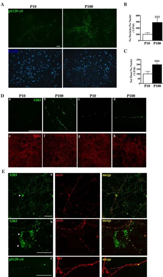
Application of 1μg of P100 fraction from sick mice to neuronal cultures resulted in the appearance of long strings of beads-like struc-tures after two weeks of treatment, that were positive forαS aggregates specific antibodies such as pSer129-αS [Fig. 2 A, E(c)] and syn303 [Fig. 2D, E (a,b)] and organized in a neurite-like pattern. Partial co-localization with neuronal synaptic and axonal markers such as mouse αS and Tau confirmed that these structures made of αS aggregates were indeed associated with the neurites network (Fig. 2C). Occasionally these strings appeared to co-localize and stain the cell soma or cover the
entire process, resembling necrotic neurites. In this latter case when cultures where stained with the neuronal marker syn1, the neurites network appeared to be less dense and more scattered when treated with P100 from sick mice, especially for cortical cultures, suggesting that the formation of these strings of αS aggregates might be detri-mental for cells. While for P100 those structures were more abundant and covered the neuronal network extensively, administration of the same amount of P10 produced very few small and scattered strings that were more immature in size and number (Fig. 2). No major differences were detected in the formation of these strings of aggregates between hippocampal and cortical neurons (Fig. 2D). Further administration of P10 and P100 from presymptomatic mice or P100 from Ctx of sick mice or nTg littermates failed to produce any string-like structure positive for syn303 (Supplemental Fig. 2) or pSer129-αS (data not shown).
Formation ofαS positive strings were also dose- and time-dependent since reducing by half (0.5μg) the original dose of P100 fraction from SpC of sick mice orfixing neuronal cultures at earlier time points re-sulted in few scattered fibrillary structures or numerous premature puncta that while their amount increased over time, failed to form an elongated and complete string (Supplemental Fig. 3A,C) when analyzed at early time points. In both cases administration of P10 from sick mice failed to produce comparable string-like structures positive for syn303 or pSer129-αS.
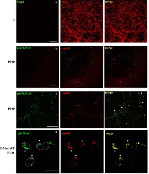
To understand whether these strings ofαS aggregates accumulated endogenously or were externally attached to the plasma membrane we used a protocol of“two-stage” immunofluorescence where labeling of P100-associated aggregates with pSer129-αS antibody was done in living neurons, beforefixation and permeabilization. In this case the
Fig. 1. Accumulation ofαS truncated fragments in P10 and P100 pellet fractions.
A, B, C, Immunoblotting showing how neurodegeneration affects the levels of αS truncated fragments in P10 and P100 fractions obtained from SpC of presymptomatic (PreS) (A) or sick Ctx (B) and SpC (C) of A53T Tg mice. 2μg of each fractions was run on a denaturating SDS-Page, transferred and blotted with syn1 antibody that recognizes the most abundant truncated fragments, Syn12, Syn10, Syn8 and HMW species (arrows heads) ofαS. Human αS monomer was visualized with human specific antibody LB509. FL, αS full length monomer. D, E, High exposure images of immunoblots in B and C, respectively, to better visualize accumulation of truncated fragments Syn10 and Syn8 in P100.F, G, H, Quantitative analysis of relative density ofαS truncated fragments in P10 and P100 respectively in PreS, sick Ctx or SpC, obtained by normalization with human αS full length monomer detected with LB509. Values are expressed as % of P10 and are given as the mean ± SD (n = 3 or 4). *p < 0.05, **p < 0.001, Student's t-test.
plasma membrane remains intact so onlyαS species that are not in-ternalized by the neurons can be labeled. After incubation with the pSer129-αS antibody, cultures were extensively washed and then fixed, permeabilized and stained with the syn303 antibody in order to label all the aggregates, inside and outside the cell membrane. Surprisingly, all the P100 aggregates were labeled only with syn303 but not with pSer129-αS, indicating that P100-associated αS species are contained
within the neurons and not merely externally attached to the cell membrane (Fig. 3b). The lack of merged signal between syn303 and pSer129-αS also suggested that an active transport in living cells of membrane-bound antibodies was not significant in our conditions. Switching the antibody used in live neurons, i.e. incubating live primary cultures with syn303 and then staining with pSer129-αS antibody, did not affect the outcome of the two-stage immunofluorescence
Fig. 2. Treatment with P100 fractions induced de-position of beads-like structures made of αS ag-gregates in primary murine neurons.
Formation of beads-like structures made of αS ag-gregates after administration of 1μg of P10 or P100 fractions pooled from 3 to 4 SpC of sick mice to hip-pocampal [A, B (a,b)], or cortical primary neuronal cultures [B (c,d), C]. Cultures werefixed after 2 weeks of treatment and stained with pSer129-αS or Syn303 antibodies. A, Representative immunofluorescence images showingαS-positive strings after P10 and P100 treatment in hippocampal cultures stained with the pSer129-αS antibody. P100 administration induced deposition of beads-like structures organized in a string and following a neurite-like pattern. These strings were positive forαS aggregates specific anti-bodies such as pSer129-αS. Compared to P100, P10 treatment induced the formation of small puncta that resembled those obtained for P100 but were less abundant and more immature in size and number. Images were taken with Nikon Eclipse E600 epi-fluorescence microscope using a 20× objective. Scale bar = 100μm. B, C, Quantitative analysis of images in A showing increased accumulation of pSer129-αS po-sitive structures in P100 treated cultures. Fluorescence signal was measured as totalfluorescence after back-ground subtraction (B) or as particles count (C) using Image J software. Both parameters were normalized with the total nuclei count (labeled with DAPI) per field. Values are expressed as % of P10 and are given as the mean ± SD (n = 6). ***p < 0.0001, Student's t-test.D, Confocal images showing that no major dif-ference between hippocampal (a,b) or cortical (c,d) cultures were detected in the formation ofαS-positive strings after administration of P10 or P100 obtained from SpC of sick mice. OccasionallyαS strings after P100 treatment appear to cover the entire process, resembling necrotic neurites (b, d). To check the neuronal viability, cultures were also stained with syn1 antibody (red). Stacked images were acquired with a Leica confocal microscope SP2 system using a 63 × objective. Scale bar = 50μm. E, Confocal images showing partial co-localization with neuronal markers such as mouseαS (mαS) (a,b) and Tau (c) in cortical primary cultures treated with P100 fractions from SpC of diseased mice. Occasionally these strings positive forαS aggregates co-localize and stain the cell soma (a and b arrow heads). Stacked images were acquired using a 63 × objective. Scale bar = 50μm (a), 20μm (b,c).
experiment, since no syn303 staining was detected without permeabi-lization (Supplemental Fig. 4A). However, when neurons were fixed before the incubation with pSer129-αS and syn303, αS strings were mostly co-labeled by both antibodies, especially in the case of larger puncta, ruling out the possibility that competition for the same target could interfere with the aggregates staining by both antibodies at the same time. To confirm that αS aggregates that are externally associated with the cell membrane can be efficiently labeled following the “two-stage” immunofluorescence protocol we repeated this assay using neuroblastoma cell lines (SH-SY5Y) stably expressing WT αS tagged with Myc. When the P100 fraction was given to these cells and“two stage” immunofluorescence was performed, most of the aggregates were double labeled with pSer129-αS and syn303 antibodies, showing that most of the aggregates were found associated outside the cell membrane. Additionally, incubation of live neuronal cultures treated with buffer (B) with Map2 (Fig. 3a) or grp94 antibodies and SH-αS-Myc WT cells treated with grp94, according to the “two-stage” immuno-fluorescence protocol (Supplemental Fig. 4B) did not result in any staining unless cells were previously fixed, confirming that no anti-bodies can label cellular endogenous targets without previousfixation and permeabilization of the cellular membrane as done in our experi-mental settings. Moreover since the endosomal-lysosomal pathway has been implicated in the internalization of exogenous in vitro preformed αS fibrils (Karpowicz et al., 2017), double staining with syn303 and Lamp1, a lysosomal marker, was done in cortical neuronal culture after 2 days or 1 week of treatment with P100 fractions from SpC of sick Tg
mice. We found little co-localization between syn303 and Lamp1 at both times, suggesting that the endosomal pathway might not be the preferential route of entry of P100-associated aggregates in primary neurons (Supplemental Fig. 5).
In summary, treatment with P100 fractions from diseased mice that contain microsomal-associated αS species induced the formation of string-like structures, positive forαS aggregates specific antibodies, that followed a neurite pattern and co-localized with synaptic and axonal markers.
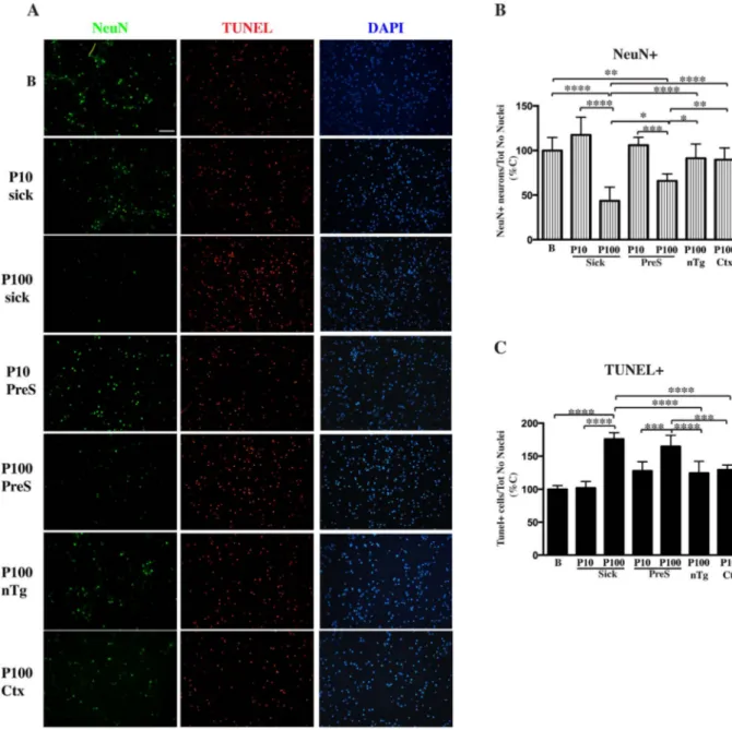
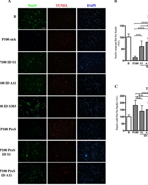
3.3. Induction of apoptosis in mouse hippocampal and cortical primary culture treated with P100 from SpC of presymptomatic and diseased mice
Increasing the concentration of the above fractions to 2μg resulted in induction of apoptosis only in primary cultures treated with P100 fractions obtained from SpC of diseased mice and more surprisingly from presymptomatic littermates, as shown by the increase in TUNEL staining and the concomitant decrease of neuronal survival measured by NeuN labeling (Fig. 4). No neuronal degeneration in cultures treated with P10 from sick and presymptomatic mice, P100 Ctx or P100 nTg was observed. Although TUNEL levels were comparable between pre-symptomatic and sick P100 fractions, the NeuN-positive count was actually higher in presymptomatic P100-treated samples, suggesting that αS species associated with microsomal fractions obtained from healthy aged animals were less aggressive in inducing cellular death. In thefirst experiments, although early signs of cellular death were visible
Fig. 3. Internalization and spreading of P100 fractions into neuronal cultures.
Two-stage immunofluorescence showed how αS ag-gregates spread into within the neurons to formαS positive strings. Live hippocampal neurons treated with buffer (B) (a), P100 fractions from sick SpC (b) or SH-SY5Y neuroblastoma cells stable forαS expression (SH-αS-Myc WT) treated with 5 μg of P100 sick SpC (d) were incubated at 4 °C with Map2 or pSer129-αS antibody, for 30 min before beingfixed, permeabilized and stained with mαS or syn303 antibody, respec-tively. Confocal stacked images were acquired with a 63 × objective. Absence of merged signals showed that no antibodies can label endogenous target without previousfixing and permeabilization of the cell membrane indicating that most of the αS ag-gregates are internalized by neurons. When neurons treated with P100 sick SpC were insteadfixed (c) be-fore pSer129-αS and syn303 incubation, both anti-bodies labelαS strings (arrow heads), although not with the same efficiency. On the contrary, merged signals between pSer129-αS and syn303 in SH-αS-Myc WT treated with P100 pellet from Sick SpC (d, arrow heads) demonstrated thatαS aggregates are not in-ternalized in this cell model. Thus only in the case of αS aggregates externally attached to the neurite membrane or the cell membrane being permeabilized, both antibodies can co-labelαS aggregates. In d) a white line showing the cell borders was drawn by overlapping fluorescence with regular white light images taken at the same time. Scale bar = 50μm (a,b,c), 20μm (d).
as soon as 24–48 hrs after addition, neurons were fixed after 3 days of treatment when neuronal degeneration in P100 sick treated cultures became widespread. When we let the treatment go further and fixed cultures after a week, cell death was so exhausted in P100 sick-treated neurons that very few cells (DAPI-positive nuclei) were left in the well. In this case also neurons treated with P100 from presymptomatic mice showed remarkable levels of cell death and neuronal loss, comparable to what has been observed previously in cultures treated with P100 sick and fixed after 3 days (Supplemental Fig. 6). At the same time also cultures treated with P10 from sick animals started to show a slight decrease in NeuN-positive neuronal count and an increased in TUNEL staining, showing that P10-associatedαS aggregates can also be toxic but not as aggressive as the P100 counterpart in inducing neuronal death. Although TUNEL levels in sick P10-treated neurons were
comparable with the amount of cell death in cultures treated with presymptomatic P100, the decrease in neuronal loss was not as re-markable, suggesting that neuronal degeneration was delayed com-pared to PreS-P100 treated cultures. Interestingly P10 from pre-symptomatic mice was never able to induce apoptosis in primary cultures in our experimental setting, while a mild decrease in neurons viability was observed for P100 Ctx-treated cultures, although was not significant, for prolonged treatment. Thus, only microsomal fractions obtained from diseased or presymptomatic affected areas have an en-hanced ability to induce cellular death in primary neuronal cultures therefore are more toxic than P10. Interestingly, the only feature that the P100 from presymptomatic and diseased areas have in common in terms ofαS structural or posttranslational modifications is the presence of αS oligomers, suggesting that other modifications such as
Fig. 4. Induction of neuronal death in primary neurons after treatment with P10 and P100.
Primary hippocampal neurons were treated with 2μg of various P10 and P100 pellet obtained from presymptomatic or diseased SpC and Ctx of Tg mice and age-matched Ntg littermates or buffer (B). Cultures were fixed after 3 days of treatment and co-labeled with NeuN and Tunel staining. Cultures were also counterstained with Dapi. A, Representative fluorescent images showing induction of apoptosis and concomitant decrease in neurons viability only in primary cultures treated with P100 fractions obtained from SpC of diseased mice and more surprisingly from presymptomatic littermates. Fluorescent images were acquired with a Nikon epi-fluorescence microscope using a 20× objective and cell counting was done using Image J software. Scale bar = 100μm. B, C, Quantitative analysis of images in the panel A for live NeuN-positive neurons (B) or dead Tunel-positive neurons (C). Live or dead cell counts were normalized for the number of Dapi-positive nuclei. Values are expressed as % of control neurons treated with buffer and are given as the mean ± SD (n = 6). *p < 0.05, **p < 0.001, ***p < 0.0001, ****p < 0.00001, one-way ANOVA, followed by Tukey post-hoc test.
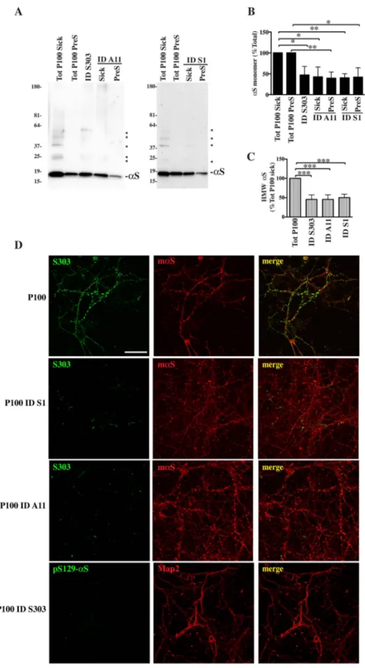
Fig. 5. Immunodepletion of αS toxic species from P100 prevents formation ofαS strings.
To check ifαS species associated with microsomes were indeed responsible for cellular modifications described above we perform immunodepletion ofαS species from the P100 fractions by incubating P100 with protein agarose conjugated with Syn1 (S1), Syn303 (S303) or A11.A, Shows immunoblotting of P100 immune-depleted (ID) fractions run along total P100 pellets from presymptomatic (Tot P100 PreS) or diseased mice (Tot P100 Sick). Samples were trans-ferred to a nitrocellulose membrane and blotted with Syn1 antibody.B, C, Quantitative analysis ofαS full length monomer (B) or HMW species (C) as shown in A indicates a reduction inαS content for all species after immunodepletion in P100 ID fractions. Densitometric analysis of HMW bands was done by measuring the relative intensity for the whole sample lane above the monomer. Values are expressed as % of corresponding total P100 pools, sick or presympto-matic, and given as the mean ± SD (n = 3). *p < 0.05, **p < 0.001 One-way ANOVA, followed by Tukey post-hoc test.C, 1μg of ID and original P100 samples from diseased were given to hippocampal neurons. Cells werefixed and immunostained with syn303/mαS or pSer129-αS/Map2 after 2 weeks. Stackedfluorescent images taken at the confocal mi-croscope showed a drastic decrease in the formation of αS aggregates-positive strings. Scale bar = 50 μm.
phosphorylation and truncation are not necessary for αS acquired toxicity.
3.4. Partial depletion of theαS species from P100 fractions from diseased and presymptomatic mice prevents spreading and induced toxicity in primary cultures
To establish whetherαS species contained in P100 fractions were indeed responsible for the induction of apoptosis, immunodepletion with various αS antibodies that recognize specific αS conformations (A11 forαS oligomers, syn303 for αS aggregates and Syn1 for total αS) was performed from pooled P100 fractions from SpC of diseased and presymptomatic mice. Briefly, P100 fractions were incubated with the above antibodies, previously conjugated with protein-binding agarose, for 1 hr at 4 °C. After spinning at low speed the supernatant was care-fully recovered and checked for the content ofαS species. αS monomer and HMW species levels in immunodepleted (ID) samples were dra-matically reduced to about 40–45% or 45–50%, respectively, of the original total pools amount (Fig. 5A, B, C). ID samples were then dosed to cortical neuronal cultures and formation ofαS strings and neuronal death was evaluated. While neurons treated with P100 obtained from SpC of sick mice developed αS aggregates-positive strings, cultures treated with ID sick samples did not. More specifically, neurons treated with ID Syn1 sick or ID A11 sick fractions showed scattered dot-likeαS structures not yet organized in a string pattern, more abundant in ID A11 than ID Syn1, while ID syn303 sick-treated cultures showed only very few small puncta positive forαS (Fig. 5C).
When we checked for toxicity, in the case of neurons treated with P100 pooled from SpC of sick mice, cell death was already apparent after 24–48 hrs of treatment. Nevertheless, to see if αS depletion could effectively rescue neurons, cultures were not fixed immediately but the treatment was continued to make sure that any possible rescuing effect that we would see was not due to a time frame difference. When cul-tures were then analyzed for cell survival, neuronal degeneration in P100 sick-treated cultures was exhaustive. In fact, the number of NeuN-positive neurons (Fig. 6) had dramatically dropped only in samples treated with P100 sick fractions but not in cultures treated with ID sick fractions, suggesting that depletion ofαS helped keep neurons healthy. Interestingly immunodepletion of matureαS aggregates with syn303 was more efficient in preventing αS-induced toxicity than the abroga-tion of totalαS in ID Syn1 samples, suggesting that toxicity is due to the presence of αS HMW conformers rather than αS itself after disease onset. Moreover while treatment with ID Syn1 and A11 seemed to only delay cell death as shown by increased Tunel positivity compared to P100 and control samples (Fig. 6C), immunodepletion with syn303 constitutively blocked apoptosis reducing Tunel level lower than un-treated samples.
In the case of presymptomatic fractions, the number of NeuN-posi-tive neurons was significantly reduced only in cultures treated with P100 PreS, but not in P100 PreS ID treated samples. Here im-munodepletion with Syn 1 or A11 fully rescued neuronal cultures and blocked cellular death as shown by reduced Tunel levels.
Thus reduced spreading ofαS aggregates due to immunodepletion prevents neuronal degeneration in murine primary neurons, indicating thatαS species contained in P100 pools are responsible for neurode-generation after administration to primary neuronal cultures. 4. Discussion
The results presented here indicate that within the heterogeneity of αS HMW species there is a small, unique fraction that is more ag-gressive in terms of propagation and toxicity. This population of ex-tremely toxicαS species is associated with the microsomal vesicles and presents specific biochemical traits such as increased aggregation, truncation and phosphorylation at serine 129 that makes them unique. The hypothesis of an extremely detrimental factor or species within
the αS HMW population has been long postulated (Caughey and Lansbury, 2003) and not only in PD but in other neurodegenerative diseases as well (Ugalde et al., 2016). Initially questions arose regarding the potential toxicity ofαS oligomers as opposed to mature fibrils to explain, for example, early observations on why dopaminergic neurons bearing LBs appeared to be healthier than neighboring neurons (Tompkins and Hill, 1997). Since then a significant body of evidence, describingαS different oligomer conformations in vitro and their asso-ciated mechanism of toxicity has been generated (Roberts and Brown, 2015), despite that the in vivo presence ofαS oligomers has been con-troversial and particularly difficult to characterize (Colla et al., 2012b). On the opposite, more recent data (Luk et al., 2009, 2012; Volpicelli-Daley et al., 2011) including an investigation led by V. Baekelandt showed that both in vitro pre-formed oligomers andfibrils are indeed neurotoxic but display different seeding capacity and induce different histopathological and behavioral abnormalities when injected in rats (Peelaerts et al., 2015). Thus it appears that structure does not ne-cessarily dictate toxicity, or at least in part, since bothαS oligomers and fibrils seems to contribute to neuronal degeneration.
While most of the evidence accumulated until now to prove the enhanced capacity of oligomers or aggregates to induce toxicity focused on structural differences, in this paper, we took a different approach and we focused our attention on the different subcellular location of αS HMW species rather than their conformation.
By isolatingαS HMW species from a Tg mouse model for αS ex-pression, and separating them according to their molecular weight and/ or membrane-association, we showed how the ER/M-associatedαS pool is remarkably neurotoxic and able to seed formation of αS-positive strings in primary neurons when compared to the rest of αS HMW species that precipitates at low speed (P10).αS association and locali-zation within the ER/M system in normal and diseased conditions has been demonstrated in cultured cells, in neurons of Tg mice and PD brain by biochemical isolation, cell imaging and electron microscopy (Colla et al., 2012a, 2012b; H.-J. Lee et al., 2002; Lee et al., 2005). Given the presynaptic localization ofαS, its ability to bind to synaptic vesicles through the SNARE complex and to inhibit the ER-Golgi transport (Burre et al., 2010; Burré et al., 2014; Cooper et al., 2006; Thayanidhi et al., 2010), it is not surprising tofind a weak association of αS with microsomal vesicles, which is a raw pellet fraction containing ER, Golgi e synaptic vesicles, in normal conditions (Colla et al., 2012a). Instead, with αS aggregation and disease onset, the partitioning between membranous-bound and freeαS changes and there is an increased re-distribution ofαS to P100/M membranes (Colla et al., 2012a). This could be due to an increase ofαS protein stability and reduced turnover correlated to change in conformation and protein solubility or to an increased sequestration of freeαS by aggregates. Interestingly, at the presynaptic level, others have shown how free presynapticαS can be recruited into insoluble aggregates (Volpicelli-Daley et al., 2011) and how presynapticαS small aggregates might be more detrimental than LB and correlate better with synapses dysfunction in vivo (Kramer and Schulz-Schaeffer, 2007).
Association with the microsomal membrane seems to be very spe-cific since although P10 contains membranous components that include the mitochondria and MAM, we never foundαS HMW species co-pre-cipitating with this organelle when we purified mitochondria from af-fected areas in the same mouse model (Colla et al., 2012b). In contrast, we found a strong association between αS aggregates and the ER membrane as shown by the ability ofαS aggregates to float and par-tition with the membranous component on a lipidflotation assay when isolated as a P100/Microsomal pellet. Very interestingly when the membranous component of P100 was removed by the addition of non-ionic detergent and the lipidflotation analysis was repeated, ER-asso-ciated αS aggregates did not partition with the membrane fraction, demonstrating that αS aggregates actively bind the ER/Microsomal membrane (Colla et al., 2012a). In addition accumulation ofαS pa-thological changes such as formation ofαS aggregates, truncation and
phosphorylation are more likely to occur close to the ER/M membranes. Similarly because of this association it is also plausible that in patho-physiological conditions the first changes implicated in αS acquired toxicity occur at the ER/M membranes level. Thus association with the microsomal membranes seems to be a key step forαS acquired toxicity. Although it remains unclear what is the pathological role of all the changes associated with neurotoxicity thatαS undergoes, the cortical region in Prp A53T mice may be a good model to study early changes in αS toxic modifications. In fact, although the cortical regions in Prp A53T mice have no overt accumulation ofαS aggregates and LB-like inclusions, nor neuronal degeneration or gliosis (M. K. Lee et al., 2002), the P100 fractions obtained from Ctx of sick mice did show mild early changes in protein truncation, phosphorylation or accumulation ofαS oligomers/aggregates that may resemble an intermediate condition between presymptomatic and disease. At the same time P100 Ctx fraction did not significantly induce cell death when given to primary
cultures, indicating these initial changes inαS are not sufficient to in-duce an overt neuronal degeneration.
Once administered to primary neurons, P100/M-associatedαS toxic species from diseased mice quickly spread into neurons in a similar manner to what has been described by V. Lee and coworkers ( Volpicelli-Daley et al., 2011, 2014). In their work, in vitro assembledαS fibrils initially induced the deposition of strings of small puncta within neurites, resembling what we obtained when low amount of P100 Sick SpC was used or when neuronal cultures werefixed at early time points (Supplemental Fig. 3), which subsequently developed into fibrous structures covering processes and soma. Although we only occasionally saw accumulation offibrous strings in the neuronal cell body, probably because of the difficulty in finely adjusting the titration of P10 and P100 fractions in our cultures, the fibrous structures described by Volpicelli-Daley's paper are comparable to what we obtained by treating neurons with increasing concentration of P100/M fractions
Fig. 6. Immunodepletion ofαS toxic species from P100 prevents neurodegeneration in murine primary cultures.
2μg of ID and total P100 samples from diseased and presymptomatic mice or buffer (B) were administered to hippocampal neurons in culture. A, Representative images of im-munostaining with NeuN antibody or Tunel shows an increase in cell survival and concomitant reduction in neuronal death in cultures treated with ID samples obtained from pre-symptomatic and sick mice. Fluorescent images were acquired with a Nikon epi-fluorescence microscope using a 20× objective. Scale bar = 100 μm. B, C, Cell survival graph of NeuN-positive neurons (B) or Tunel staining (C) after administration of ID and total fractions showed how reduction in P100-associatedαS toxic species prevents cell death. Values were normalized for the number Dapi-positive nuclei and expressed as % of neurons treated with buffer. In the graph each column represents the mean ± SD (n = 6). *p < 0.05, **p < 0.001, ***p < 0.0001, ****p < 0.00001, One-way ANOVA, followed by Tukey post-hoc test.
from Sick SpC. In addition the dose- and time-dependent formation of αS fibrous strings and their co-localization with mouse endogenous αS may suggest that mouseαS could become entangled and recruited into these bead-like structures, similarly to what have been previously de-scribed after administration of in vitro preformedαS fibrils ( Volpicelli-Daley et al., 2011). However, since the co-localization with mouseαS was only partial, it is unlikely that the formation ofαS strings in cul-tured neurons is only due to aggregated mouseαS. In the same way the limited co-localization between Tau andαS strings suggested a lower chance for Tau to cross-seedsαS aggregates deposition after P100 ad-ministration.
In terms of amount of cell toxicity of P100/M fractions from sick SpC was far more detrimental than what V. Lee's paper suggested (Volpicelli-Daley et al., 2011), considering that the dosage we used was lower and far less concentrated. Thus, a small amount of P100/M-as-sociated αS was sufficient to induce a massive apoptotic response in primary neurons (Figs. 4, 6). Levels of cell death in neurons treated with P100 PreS were remarkably higher than with P10 Sick, indicating that before accumulation of αS oligomers/aggregates and aggregation-re-lated modifications, some toxic αS species associated with the micro-somal membrane were already present. Despite that, we were never able to observe deposition of any αS puncta or string in presympto-matic-treated cultures with aggregates-specific antibodies (syn303 or pSer129-αS) or with a human αS antibody, confirming that soluble toxic forms of αS have different seeding properties from fibrils (Peelaerts et al., 2015) and thatαS oligomers do not need to become mature aggregates to be toxic. Also because of the toxicity ofαS asso-ciated with P100 PreS, it appears that accumulation of truncated and/ or phosphorylated species is only secondary and does not account for the acquired ability to induce neuronal death. Moreover, im-munodepletion experiments showed how the toxic function ofαS de-pends on its internalization and propagation rather than a mere acti-vation of a cell death cascade from the cell membrane. In fact treatment with P100 ID samples prevented induction of neuronal death and a correspondent striking reduction in the formation of αS-positive neuritic strings (Figs. 5, 6). Thus our data point to a change in structure and a specific interaction with the ER/M, shown by the abundant presence of solubleαS oligomers in the P100/M fractions of diseased and presymptomatic mice (Supplemental Fig. 1 and (Colla et al., 2012b) as the main culprit ofαS propagation and neuronal degenera-tion.
5. Conclusions
Our results have demonstrated the in vivo existence of a toxic pool of αS HMW species associated with the ER/M vesicles. Our data suggests that thefirst pathological changes in αS behavior appear in the small portion ofαS associated with the microsomal vesicles, (probably at a presynaptic level). It is not yet clear whether otherαS aggregates (that we concentrated in P10) derived from the microsomes-associated pool or form independently. While more studies will be necessary, our findings provide the first insights in the pathological behavior of αS in vivo, shining a critical spotlight on the association with the P100/M membranes as a necessary step toward the acquisition ofαS toxicity.
Supplementary data to this article can be found online athttps:// doi.org/10.1016/j.nbd.2017.12.004.
Acknowledgements
We thanked Dr. Laura Marchetti from SNS for confocal microscopy technical assistance, Prof. Mohamed Farah from Johns Hopkins School of Medicine and Prof. Darren Moore from Van Andel Research Institute for reviewing the manuscript.
Funding
This work has been supported by the Italian Ministry of University and Research (MIUR) (PROGGR09EC) through the Career Reintegration grant scheme (RLM Program for Young Researcher) and from Scuola Normale Superiore. The authors declare no competing interests. Author contributions
EC, GP, AR, VV performed the experiments; EC designed the re-search, wrote the paper and analyzed the data; CR, LR and FG isolated murine primary neurons and helped with cell cultures; NC, SC provided technical assistance with mice husbandry; MKL provided the murine transgenic model and reviewed the manuscript; AC reviewed the manuscript. All authors read and approved thefinal manuscript. Competing interests
The authors declare that they have no competing interests. Ethics approval
All animal studies were approved and complied in full by the na-tional and internana-tional laws for laboratory animal welfare and ex-perimentation (EEC council directive 86/609, 12 December 1987 and Directive 2010/63/EU, 22 September 2010).
References
Appel-Cresswell, S., Vilarino-Guell, C., Encarnacion, M., Sherman, H., Yu, I., Shah, B., Weir, D., Thompson, C., Szu-Tu, C., Trinh, J., Aasly, J.O., Rajput, A., Rajput, A.H., Jon Stoessl, A., Farrer, M.J., 2013. Alpha-synuclein p.H50Q, a novel pathogenic mutation for Parkinson's disease. Mov. Dis. Off. J. Mov. Dis. Soc. 28, 811–813.http:// dx.doi.org/10.1002/mds.25421.
Bartels, T., Choi, J.G., Selkoe, D.J., 2011.α-Synuclein occurs physiologically as a helically folded tetramer that resists aggregation. Nature 477, 107–110.http://dx.doi.org/10. 1038/nature10324.
Beaudoin, G.M.J., Lee, S.-H., Singh, D., Yuan, Y., Ng, Y.-G., Reichardt, L.F., Arikkath, J., 2012. Culturing pyramidal neurons from the early postnatal mouse hippocampus and cortex. Nat. Protoc. 7, 1741–1754.http://dx.doi.org/10.1038/nprot.2012.099. Bendor, J.T., Logan, T.P., Edwards, R.H., 2013. The function ofα-synuclein. Neuron 79,
1044–1066.http://dx.doi.org/10.1016/j.neuron.2013.09.004.
Burré, J., 2015. The synaptic function ofα-synuclein. J. Parasit. Dis. 5, 699–713.http:// dx.doi.org/10.3233/JPD-150642.
Burré, J., Sharma, M., Tsetsenis, T., Buchman, V., Etherton, M.R., Sudhof, T.C., 2010. α-Synuclein promotes SNARE-complex assembly in vivo and in vitro. Science 329, 1663–1667.http://dx.doi.org/10.1126/science.1195227.
Burré, J., Vivona, S., Diao, J., Sharma, M., Brunger, A.T., Südhof, T.C., 2013. Properties of native brainα-synuclein. Nature 498, E4-6-7.http://dx.doi.org/10.1038/ nature12125.
Burré, J., Sharma, M., Südhof, T.C., 2014.α-Synuclein assembles into higher-order multimers upon membrane binding to promote SNARE complex formation. Proc. Natl. Acad. Sci. 111, E4274–E4283.http://dx.doi.org/10.1073/pnas.1416598111. Caughey, B., Lansbury, P.T., 2003. Protofibrils, pores, fibrils, and neurodegeneration:
separating the responsible protein aggregates from the innocent bystanders. Annu. Rev. Neurosci. 26, 267–298.http://dx.doi.org/10.1146/annurev.neuro.26.010302. 081142.
Chandra, S., Chen, X., Rizo, J., Jahn, R., Südhof, T.C., 2003. A broken alpha-helix in folded alpha-synuclein. J. Biol. Chem. 278, 15313–15318.http://dx.doi.org/10. 1074/jbc.M213128200.
Chartier-Harlin, M.-C., Kachergus, J., Roumier, C., Mouroux, V., Douay, X., Lincoln, S., Levecque, C., Larvor, L., Andrieux, J., Hulihan, M., Waucquier, N., Defebvre, L., Amouyel, P., Farrer, M., Destée, A., 2004.α-Synuclein locus duplication as a cause of familial Parkinson's disease. Lancet 364, 1167–1169.http://dx.doi.org/10.1016/ S0140-6736(04)17103-1.
Colla, E., Coune, P., Liu, Y., Pletnikova, O., Troncoso, J.C., Iwatsubo, T., Schneider, B.L., Lee, M.K., 2012a. Endoplasmic reticulum stress is important for the manifestations of α-synucleinopathy in vivo. J. Neurosci. 32, 3306–3320.http://dx.doi.org/10.1523/ JNEUROSCI.5367-11.2012.
Colla, E., Jensen, P.H., Pletnikova, O., Troncoso, J.C., Glabe, C., Lee, M.K., 2012b. Accumulation of toxic-synuclein oligomer within endoplasmic reticulum occurs in α-synucleinopathy in vivo. J. Neurosci. 32, 3301–3305.http://dx.doi.org/10.1523/ JNEUROSCI.5368-11.2012.
Conway, K.A., Harper, J.D., Lansbury, P.T., 1998. Accelerated in vitrofibril formation by a mutant alpha-synuclein linked to early-onset Parkinson disease. Nat. Med. 4, 1318–1320.http://dx.doi.org/10.1038/3311.
K., Strathearn, K.E., Liu, F., Cao, S., Caldwell, K.A., Caldwell, G.A., Marsischky, G., Kolodner, R.D., Labaer, J., Rochet, J.-C., Bonini, N.M., Lindquist, S., 2006. Alpha-synuclein blocks ER-Golgi traffic and Rab1 rescues neuron loss in Parkinson's models. Science 313, 324–328.http://dx.doi.org/10.1126/science.1129462.
Cremades, N., Cohen, S.I.A., Deas, E., Abramov, A.Y., Chen, A.Y., Orte, A., Sandal, M., Clarke, R.W., Dunne, P., Aprile, F.A., Bertoncini, C.W., Wood, N.W., Knowles, T.P.J., Dobson, C.M., Klenerman, D., 2012. Direct observation of the interconversion of normal and toxic forms ofα-synuclein. Cell 149, 1048–1059.http://dx.doi.org/10. 1016/j.cell.2012.03.037.
Deleersnijder, A., Gerard, M., Debyser, Z., Baekelandt, V., 2013. The remarkable con-formational plasticity of alpha-synuclein: blessing or curse? Trends Mol. Med. 19, 368–377.http://dx.doi.org/10.1016/j.molmed.2013.04.002.
Dettmer, U., Newman, A.J., Soldner, F., Luth, E.S., Kim, N.C., von Saucken, V.E., Sanderson, J.B., Jaenisch, R., Bartels, T., Selkoe, D., 2015. Parkinson-causing α-sy-nuclein missense mutations shift native tetramers to monomers as a mechanism for disease initiation. Nat. Commun. 6, 7314.http://dx.doi.org/10.1038/ncomms8314. Fauvet, B., Mbefo, M.K., Fares, M.-B., Desobry, C., Michael, S., Ardah, M.T., Tsika, E.,
Coune, P., Prudent, M., Lion, N., Eliezer, D., Moore, D.J., Schneider, B., Aebischer, P., El-Agnaf, O.M., Masliah, E., Lashuel, H.A., 2012.α-Synuclein in central nervous system and from erythrocytes, mammalian cells, and Escherichia coli exists pre-dominantly as disordered monomer. J. Biol. Chem. 287, 15345–15364.http://dx.doi. org/10.1074/jbc.M111.318949.
Goedert, M., Spillantini, M.G., Del Tredici, K., Braak, H., 2012. 100 years of Lewy pa-thology. Nat. Rev. Neurol. 9, 13–24.http://dx.doi.org/10.1038/nrneurol.2012.242. Golovko, M.Y., Faergeman, N.J., Cole, N.B., Castagnet, P.I., Nussbaum, R.L., Murphy, E.J., 2005. Alpha-synuclein gene deletion decreases brain palmitate uptake and alters the palmitate metabolism in the absence of alpha-synuclein palmitate binding. Biochemistry (Mosc) 44, 8251–8259.http://dx.doi.org/10.1021/bi0502137. Golovko, M.Y., Barceló-Coblijn, G., Castagnet, P.I., Austin, S., Combs, C.K., Murphy, E.J.,
2009. The role ofα-synuclein in brain lipid metabolism: a downstream impact on brain inflammatory response. Mol. Cell. Biochem. 326, 55–66.http://dx.doi.org/10. 1007/s11010-008-0008-y.
Iwai, A., Masliah, E., Yoshimoto, M., Ge, N., Flanagan, L., Rohan de Silva, H., Kittel, A., Saitoh, T., 1995. The precursor protein of non-Aβ component of Alzheimer's disease amyloid is a presynaptic protein of the central nervous system. Neuron 14, 467–475.
http://dx.doi.org/10.1016/0896-6273(95)90302-X.
Karpowicz, R.J., Haney, C.M., Mihaila, T.S., Sandler, R.M., Petersson, E.J., Lee, V.M.-Y., 2017. Selective imaging of internalized proteopathicα-synuclein seeds in primary neurons reveals mechanistic insight into transmission of synucleinopathies. J. Biol. Chem. 292, 13482–13497.http://dx.doi.org/10.1074/jbc.M117.780296. Kramer, M.L., Schulz-Schaeffer, W.J., 2007. Presynaptic-synuclein aggregates, not Lewy
bodies, cause neurodegeneration in dementia with Lewy bodies. J. Neurosci. 27, 1405–1410.http://dx.doi.org/10.1523/JNEUROSCI.4564-06.2007.
Krüger, R., Kuhn, W., Müller, T., Woitalla, D., Graeber, M., Kösel, S., Przuntek, H., Epplen, J.T., Schöls, L., Riess, O., 1998. Ala30Pro mutation in the gene encoding alpha-sy-nuclein in Parkinson's disease. Nat. Genet. 18, 106–108.http://dx.doi.org/10.1038/ ng0298-106.
Lee, H.-J., Choi, C., Lee, S.-J., 2002. Membrane-bound-synuclein has a high aggregation propensity and the ability to seed the aggregation of the cytosolic form. J. Biol. Chem. 277, 671–678.http://dx.doi.org/10.1074/jbc.M107045200.
Lee, M.K., Stirling, W., Xu, Y., Xu, X., Qui, D., Mandir, A.S., Dawson, T.M., Copeland, N.G., Jenkins, N.A., Price, D.L., 2002. Human alpha-synuclein-harboring familial Parkinson's disease-linked Ala-53→ Thr mutation causes neurodegenerative disease with alpha-synuclein aggregation in transgenic mice. Proc. Natl. Acad. Sci. U. S. A. 99, 8968–8973.http://dx.doi.org/10.1073/pnas.132197599.
Lee, H.-J., Patel, S., Lee, S.-J., 2005. Intravesicular localization and exocytosis of alpha-synuclein and its aggregates. J. Neurosci. 25, 6016–6024.http://dx.doi.org/10. 1523/JNEUROSCI.0692-05.2005.
Lesage, S., Anheim, M., Letournel, F., Bousset, L., Honoré, A., Rozas, N., Pieri, L., Madiona, K., Dürr, A., Melki, R., Verny, C., Brice, A., for the French Parkinson's Disease Genetics Study Group, 2013. G51Dα-synuclein mutation causes a novel Parkinsonian-pyramidal syndrome: SNCA G51D in Parkinsonism. Ann. Neurol. 73, 459–471.http://dx.doi.org/10.1002/ana.23894.
Li, W., 2004. Stabilization of -synuclein protein with aging and familial Parkinson's dis-ease-linked A53T mutation. J. Neurosci. 24, 7400–7409.http://dx.doi.org/10.1523/ JNEUROSCI.1370-04.2004.
Li, W., West, N., Colla, E., Pletnikova, O., Troncoso, J.C., Marsh, L., Dawson, T.M., Jäkälä, P., Hartmann, T., Price, D.L., Lee, M.K., 2005. Aggregation promoting C-terminal truncation of alpha-synuclein is a normal cellular process and is enhanced by the familial Parkinson's disease-linked mutations. Proc. Natl. Acad. Sci. U. S. A. 102, 2162–2167.http://dx.doi.org/10.1073/pnas.0406976102.
Luk, K.C., Song, C., O'Brien, P., Stieber, A., Branch, J.R., Brunden, K.R., Trojanowski, J.Q., Lee, V.M.-Y., 2009. Exogenous alpha-synucleinfibrils seed the formation of Lewy body-like intracellular inclusions in cultured cells. Proc. Natl. Acad. Sci. U. S. A. 106, 20051–20056.http://dx.doi.org/10.1073/pnas.0908005106.
Luk, K.C., Kehm, V., Carroll, J., Zhang, B., O'Brien, P., Trojanowski, J.Q., Lee, V.M.-Y., 2012. Pathological-synuclein transmission initiates Parkinson-like neurodegeneration in nontransgenic mice. Science 338, 949–953.http://dx.doi.org/10.1126/science. 1227157.
Maroteaux, L., Campanelli, J.T., Scheller, R.H., 1988. Synuclein: a neuron-specific protein localized to the nucleus and presynaptic nerve terminal. J. Neurosci. 8, 2804–2815. Martin, L.J., Pan, Y., Price, A.C., Sterling, W., Copeland, N.G., Jenkins, N.A., Price, D.L., Lee, M.K., 2006. Parkinson's disease alpha-synuclein transgenic mice develop neu-ronal mitochondrial degeneration and cell death. J. Neurosci. 26, 41–50.http://dx. doi.org/10.1523/JNEUROSCI.4308-05.2006.
Oueslati, A., Fournier, M., Lashuel, H.A., 2010. Role of post-translational modifications in modulating the structure, function and toxicity ofα-synuclein. In: Progress in Brain Research. Elsevier, pp. 115–145.
Peelaerts, W., Bousset, L., Van der Perren, A., Moskalyuk, A., Pulizzi, R., Giugliano, M., Van den Haute, C., Melki, R., Baekelandt, V., 2015.α-Synuclein strains cause distinct synucleinopathies after local and systemic administration. Nature 522, 340–344.
http://dx.doi.org/10.1038/nature14547.
Polymeropoulos, M.H., Lavedan, C., Leroy, E., Ide, S.E., Dehejia, A., Dutra, A., Pike, B., Root, H., Rubenstein, J., Boyer, R., Stenroos, E.S., Chandrasekharappa, S., Athanassiadou, A., Papapetropoulos, T., Johnson, W.G., Lazzarini, A.M., Duvoisin, R.C., Di Iorio, G., Golbe, L.I., Nussbaum, R.L., 1997. Mutation in the alpha-synuclein gene identified in families with Parkinson's disease. Science 276, 2045–2047. Roberts, H., Brown, D., 2015. Seeking a mechanism for the toxicity of oligomeric
α-sy-nuclein. Biomol. Ther. 5, 282–305.http://dx.doi.org/10.3390/biom5020282. Shulman, J.M., De Jager, P.L., Feany, M.B., 2011. Parkinson's disease: genetics and
pa-thogenesis. Annu. Rev. Pathol.: Mech. Dis. 6, 193–222.http://dx.doi.org/10.1146/ annurev-pathol-011110-130242.
Singleton, A.B., 2003. Synuclein locus triplication causes Parkinson's disease. Science 302http://dx.doi.org/10.1126/science.1090278.(841–841).
Thayanidhi, N., Helm, J.R., Nycz, D.C., Bentley, M., Liang, Y., Hay, J.C., 2010. Synuclein delays endoplasmic reticulum (ER)-to-Golgi transport in mammalian cells by antag-onizing ER/Golgi SNAREs. Mol. Biol. Cell 21, 1850–1863.http://dx.doi.org/10. 1091/mbc.E09-09-0801.
Theillet, F.-X., Binolfi, A., Bekei, B., Martorana, A., Rose, H.M., Stuiver, M., Verzini, S., Lorenz, D., van Rossum, M., Goldfarb, D., Selenko, P., 2016. Structural disorder of monomericα-synuclein persists in mammalian cells. Nature 530, 45–50.http://dx. doi.org/10.1038/nature16531.
Tompkins, M.M., Hill, W.D., 1997. Contribution of somal Lewy bodies to neuronal death. Brain Res. 775, 24–29.http://dx.doi.org/10.1016/S0006-8993(97)00874-3. Ugalde, C.L., Finkelstein, D.I., Lawson, V.A., Hill, A.F., 2016. Pathogenic mechanisms of
prion protein, amyloid-β and α-synuclein misfolding: the prion concept and neuro-toxicity of protein oligomers. J. Neurochem.http://dx.doi.org/10.1111/jnc.13772. Uversky, V.N., Li, J., Bower, K., Fink, A.L., 2002. Synergistic effects of pesticides and
metals on thefibrillation of α-synuclein: implications for Parkinson's disease. Neurotoxicology 23, 527–536.http://dx.doi.org/10.1016/S0161-813X(02)00067-0. Volpicelli-Daley, L.A., Luk, K.C., Patel, T.P., Tanik, S.A., Riddle, D.M., Stieber, A.,
Meaney, D.F., Trojanowski, J.Q., Lee, V.M.-Y., 2011. Exogenousα-synuclein fibrils induce Lewy body pathology leading to synaptic dysfunction and neuron death. Neuron 72, 57–71.http://dx.doi.org/10.1016/j.neuron.2011.08.033.
Volpicelli-Daley, L.A., Luk, K.C., Lee, V.M.-Y., 2014. Addition of exogenousα-synuclein preformedfibrils to primary neuronal cultures to seed recruitment of endogenous α-synuclein to Lewy body and Lewy neurite–like aggregates. Nat. Protoc. 9, 2135–2146.http://dx.doi.org/10.1038/nprot.2014.143.
Wang, W., Perovic, I., Chittuluru, J., Kaganovich, A., Nguyen, L.T.T., Liao, J., Auclair, J.R., Johnson, D., Landeru, A., Simorellis, A.K., Ju, S., Cookson, M.R., Asturias, F.J., Agar, J.N., Webb, B.N., Kang, C., Ringe, D., Petsko, G.A., Pochapsky, T.C., Hoang, Q.Q., 2011. A soluble-synuclein construct forms a dynamic tetramer. Proc. Natl. Acad. Sci. 108, 17797–17802.http://dx.doi.org/10.1073/pnas.1113260108. Weinreb, P.H., Zhen, W., Poon, A.W., Conway, K.A., Lansbury, P.T., 1996. NACP, a
protein implicated in Alzheimer's disease and learning, is natively unfolded. Biochemistry (Mosc) 35, 13709–13715.http://dx.doi.org/10.1021/bi961799n. Zarranz, J.J., Alegre, J., Gómez-Esteban, J.C., Lezcano, E., Ros, R., Ampuero, I., Vidal, L.,
Hoenicka, J., Rodriguez, O., Atarés, B., Llorens, V., Tortosa, E.G., del Ser, T., Muñoz, D.G., de Yebenes, J.G., 2004. The new mutation, E46K, ofα-synuclein causes Parkinson and Lewy body dementia: newα-synuclein gene mutation. Ann. Neurol. 55, 164–173.http://dx.doi.org/10.1002/ana.10795.