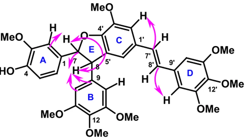
I
UNIVERSITY OF CATANIA
INTERNATIONAL PhD IN CHEMICAL SCIENCES - XXIV CYCLE
CURRICULUM: ORGANIC CHEMISTRY
Bhusainahalli Vedamurthy Maheswarappa
NATURAL POLYPHENOLS AS LEAD COMPOUNDS IN THE
SYNTHESIS OF ANTITUMOR AGENTS AND OTHER USEFUL
PRODUCTS
Tutor: Prof. CORRADO TRINGALI
_______________________________________________
II INDEX II ABBREVATIONS VI ACKNOWLEDGEMENTS VII 1. INTRODUCTION 1 1.1 Stilbenolignans 12 1.2 Benzo[k,l]xanthene lignans 17 1.3 Hydantoins 22 1.4 Main Objectives 27
1.4.1 Biomimetic synthesis of stilbenolignans 28
1.4.2 Biomimetic synthesis of benzo[k,l]xanthene lignamides 28
1.4.3 Enzymatic synthesis of hydantoin-related compounds 29
1.4.4 Structural characterization, biological evaluation and SAR studies 29
2. RESULTS AND DISCUSSION 30
2.1 Biomimetic synthesis of stilbenolignans 30
2.1.1 Synthesis of monomers 32 2.1.1.1 Synthesis of monomer 83 32 2.1.1.2 Synthesis of monomer 86 35 2.1.1.3 Synthesis of monomers 90-92 39 2.1.1.4 Synthesis of monomers 95-96 40 2.1.1.5 Synthesis of monomer 98 40 2.1.2 Synthesis of Dimers 41
2.1.2.1 Biomimetic synthesis of stilbenolignan (±)-99 41
2.1.2.2 Biomimetic synthesis of stilbenolignan (±)-100 48
2.1.2.3 Biomimetic synthesis of stilbenolignan (±)-101 56
2.1.2.4 Biomimetic synthesis of stilbenolignan (±)-102 59
2.1.2.5 Biomimetic synthesis of stilbenolignan (±)-103 62
2.1.2.6 Biomimetic synthesis of stilbenolignan (±)-104 64
2.1.2.7 Biomimetic synthesis of stilbenolignan (±)-105 67
2.1.2.8 Biomimetic synthesis of stilbenolignan (±)-106 73
III 2.1.4. Antiproliferative activity and absolute configuration of stilbenolignans 78
2.1.4.1 Antiproliferative activity of the racemic stilbenolignans 78 2.1.4.2 Chiral resolution of the active racemates and absolute configuration of the enantiomers 79 2.1.4.3 Antiproliferative activity of pure stilbenolignans and SAR 84
2.2 Biomimetic synthesis of benzo[k,l]xanthene lignans 87
2.2.1 Synthesis of benzo[k,l]xanthene 112 87
2.2.1.1 Step1. Preparation of amide 111 87
2.2.1.2 Step2. Preparation of compound 112 89
2.2.2 Synthesis of benzo[k,l]xanthene 115 95
2.2.2.1 Step1. Preparation of amide 114 95
2.2.2.2 Step2. Preparation of compound 115 97
2.2.3 Synthesis of benzo[k,l]xanthene 118 103
2.2.3.1 Step1. Preparation of amide 117 103
2.2.3.2 Step2. Preparation of compound 118 105
2.2.4 Synthesis of benzo[k,l]xanthene 121 109
2.2.4.1 Step1. Preparation of amide 120 110
2.2.4.2 Step2. Preparation of compound 121 112
2.2.5 Synthesis of benzo[k,l]xanthene 124 115
2.2.5.1 Step1. Preparation of amide 123 115
2.2.5.2 Step2. Preparation of compound 124 117
2.2.5 Reaction mechanism of benzo[k,l]xanthene 117
2.3 Synthesis of caffeic hydantoins 118
2.3.1 Enzymatic synthesis of caffeic hydantoin 128 121 2.3.1.1 Step 1. Synthesis of caffeic N N'-dicyclohexylurea 125 121 2.3.1.2 Step 2. Enzymatic conversion of 125 to caffeic hydantoin 128 124
2.3.2 Enzymatic synthesis of caffeic hydantoin 130 133 2.3.2.1 Step1. Synthesis of caffeic N,N’-diisopropylurea 129 133 2.3.2.2 Step 2. Enzymatic conversion of 129 to caffeic hydantoin 130 136
2.3.3 Study of the mechanism of formation of the hydantoins 140
IV
2.3.4.1 Step1. Synthesis of Ferulic acid N,N'-dicyclohexylurea 132 141
2.3.4.2 Step2. Attempted Synthesis of Ferulic Hydantoin 143
2.3.4.3 Step1. Synthesis of Coumaric acid N-acylurea 134 144
2.3.4.4 Step2. Attempted Synthesis of Coumaric Hydantoin 147
2.4 Theoretical Study of the mechanism of formation of the hydantoins 128 147
2.4.1 Mechanism of the Nucleophilic 1,2- and 1,4-Addition 151
2.4.2 Transition-State Structures 152
2.4.3 Energetics of the 1,2- and 1,4-Nucleophilic Addition 155
2.4.4 Formation of hydantoin 128 156
3. CONCLUSIONS 158
4. EXPERIMENTAL SECTION 160
4.1 General Section 160
4.2 Materials and method 161
4.3 Biomimetic synthesis of stilbenolignans 161
4.3.1 Synthesis of monomers 161
4.3.1.1 Synthesis of monomer 83 161
4.3.1.2 Synthesis of monomer 86 163
4.3.1.3 Synthesis of monomers 90-92, 95-96, and 98 164
4.3.2 Synthesis of Dimers 164
4.3.2.1 Biomimetic synthesis of stilbenolignan (±)-99 164
4.3.2.2 Biomimetic synthesis of stilbenolignan (±)-100 165
4.3.2.3 Biomimetic synthesis of stilbenolignan (±)-101 166
4.3.2.4 Biomimetic synthesis of stilbenolignan (±)-102 167
4.3.2.5 Biomimetic synthesis of stilbenolignan (±)-103 168 4.3.2.6 Biomimetic synthesis of stilbenolignan (±)-104 168
4.3.2.7 Biomimetic synthesis of stilbenolignan (±)-105 169
4.3.2.8 Biomimetic synthesis of stilbenolignan (±)-106 169
4.4 Biomimetic synthesis of benzo[k,l]xanthene lignans 170
4.4.1 Synthesis of benzo[k,l]xanthene 112 170
V
4.4.1.2 Step2. Preparation of compound 112 171
4.4.2 Synthesis of benzo[k,l]xanthene 115 171
4.4.2.1 Step1. Preparation of amide 114 171
4.4.2.2 Step2. Preparation of compound 115 172
4.4.3 Synthesis of benzo[k,l]xanthene 118 172
4.4.3.1 Step1. Preparation of amide 117 172
4.4.3.2 Step2. Preparation of compound 118 173
4.4.4 Synthesis of benzo[k,l]xanthene 121 173
4.4.4.1 Step1. Preparation of amide 120 173
4.4.4.2 Step2. preparation of compound 121 174
4.4.5 Synthesis of benzo[k,l]xanthene 124 174
4.4.5.1 Step1. Preparation of amide 123 123
4.4.5.2 Step2. Preparation of compound 124 175
4.5 Synthesis of caffeic hydantoins 175
4.5.1 Enzymatic synthesis of caffeic hydantoin 128 175 4.5.1.1 Step1. Synthesis of caffeic N,N'-dicyclohexylurea 125 175 4.5.1.2 Step2. Enzymatic conversion of 125 to caffeic hydantoin 128 176
4.5.2 Enzymatic synthesis of caffeic hydantoin 130 176 4.5.2.1 Step1. Synthesis of caffeic N,N’-diisopropylurea 129 176 4.5.2.2.Step2. Enzymatic conversion of 129 to caffeic hydantoin 130 177
4.5.3 Attempted Synthesis of Ferulic and Coumaric Hydantoin 177 4.5.3.1 Step1. Synthesis of Ferulic acid N,N'-dicyclohexylurea 132 177
4.5.3.2 Step2. Attempted Synthesis of Ferulic Hydantoin 178
4.5.3.3 Step1. Synthesis of Coumaric acid N,N'-dicyclohexylurea 134 178
4.5.3.4 Step2. Attempted Synthesis of Coumaric Hydantoin 179
4.6 Theoretical Approach 179
VI
ABBREVIATIONS
BOP Benzotriazole-1-yl-oxy-tris-(dimethylamino)-phosphonium hexafluorophosphate BuLi n-Butyl lithium
13C NMR Carbon Nuclear Magnetic Resonance
CD Circular Dichroism
COSY COrrelation SpectroscopY DNA Deoxyribonucleic acid DCM Dichloromethane DCC N,N-Dicyclohexylcarbodiimide DMAP Dimethylaminopyridine DIC N,N-Diisopropylcarbodiimide DMF N,N-dimethylformamide DPPH 2,2-diphenyl-1-picrylhydrazyl EWG Electron Withdrawing Group
EtOH Ethanol
EtOAc Ethyl acetate
HSQC Heteronuclear Single Quantum Coherence HMBC Heteronuclear Multiple-Bond Correlation HPLC High Performance Liquid Chromatography HRP Horseradish Peroxidase
HOBt N-hydroxybenzotriazole
LPO Laccase from Pleurotus ostreatus LTV Laccase from Trametes versicolor
MeOH Methanol
MW Microwave
NOESY Nuclear Overhauser Enhancement Spectroscopy
RT Room Temperature
1H NMR Proton Nuclear Magnetic Resonance
THF Tetrahydrofuran
TLC Thin Layer Chromatography TMP Trimethylpyridine
VII
ACKNOWLEDGEMENTS
This work was carried out at the Department of Chemistry, Faculty of Mathematical, Physical and Natural Sciences, University of Catania, (Italy) and Institut de Chimie Moléculaire de l'Université de Bourgogne (ICMUB), Dijon, France (during a short period visit).
I want to express my immense depth acknowledgement to:
Supervisor of this thesis Prof. Corrado Tringali, who accepted me as doctorate student for his project and was the main source of the great ideas, techniques and backbone of this thesis. I experienced with him great warmth of care, and we experienced together all the ups and downs of routine work, the shared happiness of success and the depression of failure when (sometimes) everything went wrong in laboratory. He managed to teach me how to write scientific reports and reproduce them in scientific journals. It is a real pleasure to work and communicate with such a polite, honest, helping natured and open-minded person.
Co-supervisor - Dr. Carmela Spatafora, who guided me organic chemistry in laboratory activities. She took lot of care towards me and she has helped me greatly during all three years, special appreciation for the proofreading (her patience is valuable) and language translation from Italian to English for all administrative work and has always been extremely supportive and nice to me. It was her great help in science and beyond science life that I got the chance to initiate and complete this thesis. It is great pleasure to work with such an optimistic genteel lady. I felt great love and affection from both guide and co-guide in my three years research work.
I would like to acknowledge Prof. Dominique Fasseur-Vervandier and Dr. Malik Chalal for their love and affection in my short period of stay in ICMUB, Dijon.
I would like to express my immense depth acknowledgement to Prof. Vincenzo Amico to providing laboratory space to complete my research work. I want to say thanks to Prof. Sebastiano Sciuto, Prof.ssa Rosa Chillemi, and all Professors from Chemistry Department, who gave positive critics and suggestions in my yearly viva to improve quality of this thesis. I would like to thank Prof. Antonio Rescifina for theoretical calculations.
VIII Also I want to thanks, an external examiner Prof. Philippe Cotelle from University of Lille, who gave me suggestions in my annual presentations.
I want to express my acknowledgement to Prof. B.S. Sherigara, Prof.Tapas Kumar Kundu, Prof. S. Sampath, Prof. J. Keshavayya, Prof. K.S. Rangappa, Dr. Lokesh, Dr. K.M. Lingu, Dr. A. Hari Kishore, who gave me dimension and shape in research.
I would like to thank present and former colleagues of “Bioactive natural product group”, for their assistance and providing an excellent working atmosphere: Dr. Carmelo Daquino, Nino Marino, Ionella Ranno, Lella Susinna, Maria Rita Nobile, Rita Tosto, Carmelo Girmenia, Danilo Aleo and all others.
I want to thanks all 24th cycle International Ph.D program students: Valentina La Paglia Fragola, Martina Pannuzzo, Zelica Minniti, Valentina Greco, Pietro Gemmellaro and Salvatore Battiato and Noufal kandoth for enjoyable outings in Catania.
I want to express my gratitude towards JNCASR. IISc Professors and friends who made my stay in campus fruitful, and M.Sc friends, Bhasker H. G, Pradeep P.C, Harsha V, Ravindra, Siddesh, Maruthi rao, Manthesh, Niranjan, etc for their constant encouragement and cooperation.
I would like to thank my maternal uncles, who feed me immediately after my birth to till my life. Umapathy B.C, Shivalingappa B.C, Shadaksharappa B.C, Onkarappa B.C, Shivappa B.C, Parameshwarappa B.C, Shekrappa B.C, and Basappa B.C. and also I want to acknowledge my all aunts’ who took care towards me from childhood to till know. I want to express my gratitude towards, my sisters, my sibling brothers and sisters who gave me company to make my childhood joy able.
I would like to thank my father Maheshwarappa B.M, who was dreaming of my bright future in his life time and I want to dedicate this thesis for his 4th year death anniversary. Last but not least my mother Sakamma B.C, without she, I am an imagination. I had unforgettable love from her, she is praying god each and every movement for my success. How can I express thanks to her? No, it is not possible. Simply I want to say love you Amma....
1
1. INTRODUCTION
Polyphenols or, more properly, the plant metabolites originated by the shikimic acid pathway show a noticeable structural variety, ranging from simple phenolic acids, for instance gallic acid or caffeic acid, to oligomers (tannins) and polymers (lignins or melanins). This large group of chemical substances, mainly found in higher plants, include more than 8.000 compounds, many of whom display important biological properties. In recent years, an increasing attention towards many dietary polyphenols has been also due to their recognised antioxidative and cancer chemopreventive properties.1 Indeed, a considerable
number of studies is concentrated on a limited number of low molecular weight dietary polyphenols, such as the stilbenoid resveratrol (1) found in grape and
red wine, the green tea catechins, and in particular epigallocatechin-3-gallate (EGCG, 2), the hop chalcone xanthohumol (3), the soya isoflavone genistein
(4), the yellow pigment curcumin (5) found in turmeric (curry), ellagic acid (6), a
component of pomegranate, the widespread caffeic acid (7) and its natural
derivatives chlorogenic acid and (8) and CAPE (9), this latter found in
honeybee propolis. In particular, resveratrol (1) is considered one of the most
important polyphenols with cancer chemopreventive activity and displays an impressive variety of biological activities.2
Some dimeric polyphenols, biosynthetically related to phenolic acids or stilbenoids, collectively named ‘lignans’ are of high biomedical importance. A well-known example is podophyllotoxin (10), a natural aryldihydrolignan with
cytotoxic properties, which was submitted to chemical modifications to obtain the anticancer drug etoposide (11). Many other dimeric lignans have been
reported in the literature, some of them with unusual structures or displaying promising biological properties like antibacterial, anticancer, antiestrogenic,
2 antiviral, fungistatic, estrogenic, antioxidant, and insecticidal activities.3 Other
lignans are reported as anti-mitotic, anti-angiogenic and anti-radical agents.4
OH OCH3 OCH3 HO O O 5 OH 1 HO OH OH O OH HO OH O O HO OH O O HO OH OH HO O OH O OH HO OH O 3 2 7 8 9 O HO O 4 O OH OH OH O O O O OH HO OH OH 6 OMe O OH HO OH
3 MeO OMe OMe O OH O O O 10 MeO OH OMe O O O O O O OH OO HO 11
From a biosynthetic point of view, lignans are dimers generated by β-β’ (8-8’) oxidative coupling of two cinnamic acid residues. Many lignans and neolignans include a heterocycle in their structure. This is normally generated through intramolecular cyclization of a reactive intermediate formed in the oxidative coupling step, as exemplified by the biosynthesis of the lignan (+)-pinoresinol (14) (Scheme1 O OH MeO O O H OMe O O OH O H OMe MeO H+ H+ OH OMe O O OH MeO OH OH MeO O OH MeO O OH MeO O OH MeO O OH MeO . . . . . 12 13 14 Oxidase 8 8' 5 1 8 8' 8 ). Scheme 1
4 This biosynthetic pathway has been widely studied and it is known that pinoresinol originates from the monolignol coniferyl alcohol (12): in the first step
an oxidase enzyme gives rise to the coniferyl alcohol radical, reported here as mesomeric forms. Subsequently, a coupling reaction affords a reactive quinone-methide intermediate (13) which by cyclization gives the final product 14.2
According to the IUPAC recommendations 2000,5,6 ‘neolignans’ are
distinguished in referring to dimers with a carbon linkage between two C6C3
units different from 8-8’, as for example (+)-dehydrodiconiferyl alcohol (16),
whose biosynthesis, outlined in Scheme 2, proceeds via the quinone-methide intermediate 15 formed through an 8-5’ coupling of coniferyl radicals.7
The term ‘oxyneolignans,’ should be reserved to dimers linked by an ether oxygen atom where there are no direct carbon–carbon bonds between the C6C3 units. An important point on lignan and lignin biosynthesis is that the
regio- and stereospecificity of bimolecular phenoxy radical coupling is not controlled by peroxidases, laccases or other oxidases, as demonstrated by the formation of racemic mixtures as product of in vitro oxidative coupling experiments
5 O OMe OH H+ OH OH OMe O OMe OH OH O OMe O OMe OH O OMe OH O O H OH O H OMe OMe . . 15 8 5' 16 Scheme 2
As shown by Graph 1, the number of references (articles) retrieved through Scifinder Scholar using the term ‘lignans’ is in constant growth since 1997 to 2010 and a similar trend is observed if the search is restricted to the patents. These data confirm the biomedical importance of this family of natural products: in fact, the most interesting bioactive lignas are an attractive target for chemical synthesis and pharmacological optimization.
6 Among the various synthetic methodologies employed in the synthesis of lignans, biomimetic coupling reactions are frequently carried out on natural precursors, and may afford ‘unnatural’ products through a radical phenolic oxidative coupling mechanism mimicking the ‘natural’ biosynthetic process. Thus, it is possible in principle to obtain compounds unprecedented in literature although maintaining a basic ‘natural’ skeleton and possibly offering a bioactivity profile similar to, or better than, that of a natural analogue. Due to the lack of stereocontrol both in metal- or enzyme mediated phenolic radical coupling, racemic mixtures are frequently obtained.10 Nevertheless, a number
of interesting products has been obtained in such a way, and in some cases the bioactive racemate has been resolved to obtain the most active enantiomer. In this regard, one interesting example is the reported dimerization of methyl caffeate (17) with Ag2O4 which afforded a neolignan racemate related to the
natural dimer 3’,4-di-O-methylcedrusin (18). This latter was isolated as one of
the active principles of ‘dragon’s blood’, the blood-red latex produced by some
Croton spp. growing in South America and employed in traditional medicine for
its wound-healing and anti-cancer properties. The racemate was then resolved to give the (2R,3R)-enantiomer (19) showing promising antitumor properties
against breast cancer cell lines and resulting most potent than its (2S,3S)-enantiomer not only as antiproliferative but also as antiangiogenic agent.9 In
addition, the related neolignan racemate (±)-20 (only the 2R,3R-enantiomer is
reported) resulted highly active against chloroquine-resistant Plasmodium
7 HO OH O OMe 17 HO O OH OMe OMe OMe 18 O MeO O O OH OH OMe 19 OH OH O O O O O OH OH 20
Ralph et al10 employed ethyl ferulate 21 as substrate and Ag
2O (in dry acetone)
as the oxidative agent; an intermediate quinone-methide 22 is formed by 8-5’
coupling of the electron-delocalized phenoxy radical and this gives rise to the cyclization product, the racemic trans 2,3-dihydrobenzofuran neolignan 23 (only
one enantiomer is reported), obtained in ca 30% yield, according to Scheme 3.
OH O MeO EtO O O MeO EtO O O MeO EtO O O MeO EtO O O MeO EtO O OMe OEt O OEt O OMe O OMe O OH OMe O OEt O OEt O OMe O OEt O OMe O OEt Ag2O . . . . 4 5 1 8 . . 8 5' 21 22 23 Scheme 3
8 In the last decade, metal-mediated biomimetic syntheses based on phenolic oxidative coupling have been frequently paralleled by coupling methods mediated by enzymes, mainly peroxidases or laccases.
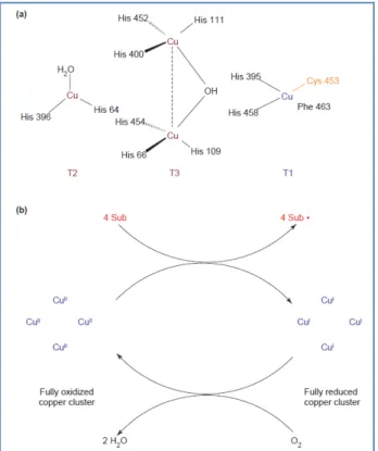
Laccases (Figure 1) are oxidoreductases belonging to the multinuclear
copper-containing oxidases; these enzymes are widely distributed in fungi and in some bacteria and higher plants and are able to catalyse the monoelectronic oxidation of substrates at the expense of molecular oxygen.11 The active site of
these enzymes consists of a metallic cluster containing four copper atoms, all of them being involved in the redox process via a radical cyclic mechanism12
(Figure 2a). The oxidation of a specific phenolic derivative depends on the redox potential difference between the target compound and the so called ‘T1’-copper. The overall outcome of the catalytic cycle is the reduction of one molecule of oxygen to two molecules of water and the concomitant oxidation of four substrate molecules to produce four radicals13 (Figure 2b). These reactive
intermediates can then produce dimers, oligomers and polymers.
9
Figure 2: Laccases: active-site structure and catalytic cycle. (a) Model of the
catalytic cluster of the laccase from Trametes versicolor made of four copper atoms. Type I (T1) copper confers the typical blue colour to the protein and is the site where substrate oxidation takes place. Type 2 (T2) and Type 3 (T3) copper form a trinuclear cluster, where reduction of molecular oxygen and release of water takes place.14 (b) Schematic representation of a laccase
catalytic cycle producing two molecules of water from the reduction of one molecule of molecular oxygen and the concomitant oxidation (at the T1 copper site) of four substrate molecules to the corresponding radicals.22 Sub: substrate
molecule; Sub: oxidized substrate radicals.
Laccases show exceptional substrate versatility and therefore are potentially suitable biocatalysts for the mild oxidation of organic compounds. Interest in these essentially ‘eco-friendly’ enzymes, working with air and producing water as the only by-product, has grown significantly in recent years. So far, the main limitation to their use has been their scarce availability. However, mainly to satisfy the demand for new ‘green’ processes by the textile and pulp and paper industries, some of these enzymes have recently been cloned and overexpressed and are becoming commercially available.15 Following the
10 increased interest in these essentially ‘green’ catalysts, significant number of reports has been published in the past decade, focusing on the biochemical properties of these proteins and/or on their applications in technological and bioremediation processes in addition to their use in chemical reactions.
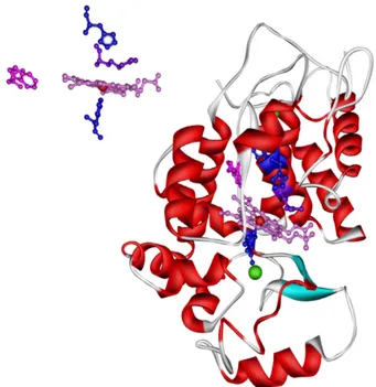
Peroxidases act on phenols by H2O2-dependent one-electron oxidation.
Horseradish Peroxidase (HRP) (Figure 3) in the presence of H2O2 is one of the
most frequently used enzymes for phenolic oxidative coupling (Figure 4).
11
Figure 4: Mechanism of Horseradish Peroxidase (HRP)
Various HRP-mediated dimerizations of phenolic precursors have been carried out; for instance, the 4-allylphenol 25 was used by Liu et al.16 as a putative
biogenetic precursor of the oligomeric neolignans isolated from Illicium spp., linked through the aromatic rings and exemplified by magnolol (25), a dimeric o,o-coupled product displaying neuroprotective activity and other promising
biological properties.17 The biomimetic synthesis of 25 was carried out by
HRP-catalysed dimerization of 24 with 40% yield, significantly higher than that
obtained by the conventional chemical methods.
OH OH OH
25 24
12 Nitrogenated lignanes are quite rare in nature; two antifeedant lignanamides isolated from Xylopia aethiopica18
OH NH O OH MeO OMe O N H O OH OH O N H OH OMe O H OH MeO N H O O N H MeO OH OH 26 27 28
were obtained by chemical and enzymatic synthesis. This latter was carried out treating the phenolic amide 26 with
HRP/H2O2 and afforded the same products of the chemical synthesis in better
yields, namely (±)-27 (18%) and (-)-28 (10%).
1.1 Stilbenolignans
Dimeric products have been also obtained by oxidative coupling of polyphenols different from phenylpropanoids. In particular, many chemists have studied the oxidative coupling of resveratrol (or resveratrol analogues) under a variety of conditions because resveratrol oligomers have unusual structures and a wide range of biological activities,19,20,21 but low natural abundance; ε-viniferin (29)
firstly isolated in 1977,22 is a typical example of stilbenoid dimers. Viniferins
exhibit a variety of biological activities, among them anti-HIV, antimicrobial, anti-inflammatory and anticarcinogenic.19, 23 Their biosynthetic origin is
reasonably due to oxidative coupling of a stilbenoid precursor, so we include these dimers within the class of stilbenolignans, although this term has been mainly used for mixed dimers with a stilbenoid portion.
13 In synthetic radical coupling of resveratrol (1), the main product most frequently
obtained was the 5–8-coupling product, that is, resveratrol trans-dehydrodimer (30) or δ-viniferin, regardless of whether enzymes (peroxidase24 or laccase25)
or inorganic oxidants were used.
O O H OH OH O H O H OH O OH OH O H O H 30 29
By treating 1 with 2,2-diphenyl-1-picrylhydrazyl free radical (DPPH) a major
product was isolated in 18% yield and identified as 30.26 On the other hand, the
treatment of 1 with FeCl3 failed to give 30, affording the isomeric dimer
ε-viniferin (29) as the sole product.27 Riva et al28 reported that the laccase
enzyme-mediated dimerization of resveratrol afforded 30. In a more recent
work the same authors reported the laccase enzyme-mediated dimerization of a series of resveratrol analogues.29 An exemplificative product of this synthesis
is again the stilbenoid dimer 30 (type A structure, Scheme 4) but two other kinds of structure, type B and C, are reported.28
14 OH R R R R O O H R R R R R R R R O OH R R R O H R R R R R O O O H R R R R R R R R = H, OH, OCH3 laccase from Trametes pubescens type A structure type B structure type C structure Scheme 4
A recent paper by Snyder et al.30 describes elegant and versatile routes to a
few dimeric species by adding and/or constructing additional rings onto brominated stilbenes. Some key steps include intramolecular Friedel–Crafts alkylation. Sako described the non-enzymatic synthesis of 30 involving the
treatment of 1 with AgOAc and other metallic oxidants.31 Similarly, Takaya and
co-workers described the treatment of 1 with several oxidizing reagents and
found that Tl(NO3)3 and K3[Fe(CN)6] were the best oxidants to transform 1 into 29, whereas FeCl3 in acetone and MnO2 in dichloromethane were the best
catalysts to produce the dimer 30.32 Fang et al33 investigated the radical
scavenging activity and detailed mechanism of resveratrol 1 and its analogues
2,2-diphenyl-1-15 picrylhydrazyl (DPPH•) radicals in ethanol and ethyl acetate at 25 °C, using UV-Vis spectroscopy: they found that the reaction rates increase with increasing the electron-rich environment in the molecules, and the compound bearing o-dihydroxyl groups (3,4-dihydroxy-trans-stilbene) is the most reactive one among the examined resveratrol analogues. Lin’s group described the oxidative coupling of isorhapontigenin (31) by means of formic acid and one electron
oxidants (Ag2O, FeCl3·6H2O).34 These researchers managed to obtain dimeric
species of various natural skeletons, mainly of the δ-viniferin or ε-viniferin type. Hou and co-workers dimerized a cleverly designed resveratrol derivative with help of the horseradish peroxidase.35 The bulky substituents did not allow the enzyme to convert the substrate into a δ-viniferin analogue and subsequently quadrangularin A (32) was obtained.
OH OH O H OMe HO OH OH OH OH O H 31 32
S. Velu et al36 achieved biomimetic synthesis of unnatural derivatives by subjecting three resveratrol analogues to oligomerization by means of one-electron oxidants. Upon varying the metal oxidant, the solvent, and the oxygenated substitution pattern of the starting material, these researchers
16 managed to obtain a series of unusual oligomeric stilbenes, among them compounds 33 and 34. OH O H MeO OMe OH OH OMe MeO 33 34
The dimeric compound with an 1,4-benzodioxane ring, (-)-aiphanol (37) is an
unprecedented natural dimer where the dioxane bridge connects a phenylpropane unit with a stilbene unit. This mixed stilbenolignan was recently obtained through bioassay-guided isolation from the seeds of Aiphanes
aculeata37and resulted a potent inhibitor of both COX-1 (IC
50 = 1.9 µM) and
COX-2 (IC50 = 9.9 µM). A synthesis of 37 as a racemic mixture has been
carried out by Banwell et al.38 through a cross-coupling between stilbenoid and phenylpropanoid substrates. In this synthesis (Scheme 5), the natural stilbenoid piceatannol (35), prepared by condensation of a suitable ylide and an aromatic
aldehyde (followed by deprotection of the primary product) undergoes an Ag2CO3-promoted oxidative coupling with sinapyl alcohol 36 affording
(±)-aiphanol (only the levorotatory 2R,3R-enantiomer is reported here) and a mixture of related dimers (33 - 40). All these compounds inhibited both COX-1
and COX-2 (IC50 in the range 0.17–9.5 µM). Interestingly, (±)-aiphanol proved
to be a potent anti-angiogenic agent, completely inhibiting blood vessels growth at 100 µg/mL. Also compounds 39 and 38 were comparable angiogenesis
inhibitors, whereas 40 resulted less active. Compounds 38 - 40 proved more
active than PI-88, an anti-angiogenic polysulphated oligosaccharide now in clinical development as anti-cancer agent.39 In the frame of this work,
17 piceatannol (35) was tested as anti-angiogenic inhibitor and resulted almost
active as (±)-aiphanol. OH OMe OH MeO OH O H OH OH OH O H O O OH OH OMe OMe OH O H O O OH OH OMe OMe OH O H O O OH OH OMe OMe OH O H O O OH OH OMe OMe Ag2CO3 35 36 37 39 38 40 + Scheme 5 1.2 Benzo[k,l]xanthene lignans
Benzo[kl]xanthene lignans are very rare polyphenolic chemical compounds both among natural products and synthetic analogues. Recently reported representatives are yunnaneic acid H (41), rufescidride (42) and mongolicumin
A (43) isolated respectively from Salvia yunnanensis,40 Cordia rufescens,41 and
Taraxacum mongolicum.42 Interestingly, the aryldihydronaphtalene lignan
rabdosiin (44) was also isolated from S. yunnanensis, It is also worth noting
that, due to their rarity in nature and the low yield of previous synthetic reactions to obtain analogues,43 and these compounds are almost unexplored
with regard to their biological properties and possible pharmacological applications. Even for the previously known rufescidride (42), the only available
reference about its biological properties is, to the best of our knowledge, a patent referring to its antimicrobial activity.44
18 O O H OH OH O O O O OH OH OH OH CO2H CO2H O O H OH OH O O O O O H OH OH O O OH OH O H OH OH O O O O OH OH OH OH CO2H CO2H O H 41 42 43 44
Among the few synthetic reactions affording benzoxanthene lignans there is a study of Maeda et al; they prepared 45 from the natural coumarin esculetin; the
reaction of 45 with Ag2O in benzene-acetone (Scheme 6), followed by
acetylation of the reaction mixture, afforded the aryldihydronaphtalene (46)
(28% yield) accompanied by the unusual benzo[kl]xanthene (47) (10%) as
major products. Without acetylation, 46 was obtained in 22% yield. Minor
amounts of the benzo[kl]xanthene (48) and the 1,4-benzodioxane (49) were
also obtained. The formation of dihydrobenzofuran neolignans (8-5’ coupling) is blocked by the presence of the methoxy group at C-2 position. A hypothetical mechanism for the formation of the benzoxanthenes, through biradical
intermediates, was also proposed. Compounds 46, 47 and 49 resulted much
more effective inhibitors of lipid peroxidation in rat liver microsomes than α-tocopherol. Differently from benzoxanthene lignans, many natural analogs of
19 the aryldihydronaphtalene lignan 46 are known, among them magnoshinin,45
thomasic acid,46 thomasidioic acid47 and rabdosiin.48
O H O H OMe O OMe O O AcO OAc OAc OMe OMe OAc OMe R O AcO OAc OAc O OMe OMe O O MeO O MeO OAc OAc O OMe MeO Ac2O Ag2O 45 46 47 R = COOMe 48 R = H 49 Scheme 6
Recently, C. Tringali et al,49 employed MnO
2 as oxidative agent in a biomimetic
coupling reaction of caffeic acid phenethyl ester (CAPE, 9). This natural product
is a component of propolis and it is reported as anti-inflammatory, antioxidant and antitumoral agent.50
According to
Scheme 7, the manganese-mediated reaction afforded with good yield the benzo[kl]xanthene lignan (50) as major product, accompanied by
minor amounts of the aryldihydronaphtalene lignan (51). Interestingly, when
Ag2O was employed as oxidative agent, the neolignan 52 was obtained as
20 O H O H O O O O OH OH O H O O O O OH OH O O H O H O O O O O OH OH OH O O MnO2 + 52 Ag2O 50 51 9 Scheme 7
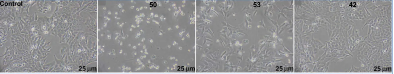
Very recently, G. Bifulco et al52 investigated interaction of six synthetic
benzo[kl]xanthene lignans (50, 53, 54, 55, 56, 57) and the natural metabolite
rufescidride 42 with DNA through a combined STD-NMR and molecular
docking approach, paralleled by in vitro biological assays on their antiproliferative activity towards two different cancer cell lines: SW 480 (Figure 5 O OR OR O O OMe OMe RO O OR OR O O O O RO 53. H 54. CH3 55. COCH3 50. H 56. CH3 57. COCH3 ) and HepG2.
The most active compound was the benzo[kl]xanthene lignan 50, potently
active both on colon (IC50 = 2.57 mM) and hepatic cancer cells (IC50 = 4.76
21 proliferation after a 48 h treatment, selectively on colon cells (IC50 = 3.21 mM),
and to a lesser extent, on hepatic cells (IC50 = 26.66 mM). It is worth noting that
compounds 50 and 57, bearing the phenylethyl pendants at positions 1 and 2,
are significantly more active than compounds 53 and 55, which have methyl
ester groups in the same positions.
Figure 5: Phase contrast microscopy observation of SW480 colon cancer cells treated or not
for 48 h with 50, 53 and 42.
The study of intercalation between base pairs was performed taking into account two binding cavities (Figure 6
Within both models A and B, the common polycyclic aromatic moiety intercalates between base pairs forming π-π interactions with the macromolecular counterparts and the two phenylethyl groups are accommodated along the minor groove establishing van derWaals contacts(see Ref. 54 for more details) (
), in order to evaluate if this type of interaction to the DNA, and the consequent hydrogen bond formations, were base-dependent.
Figure 7). Finally, Bifulco et al data suggest that the benzo[kl]xanthene lignans are suitable lead compounds for the design of DNA selective ligands with potential antitumour properties.
Figure 6: Schematic representation of DNA models (A and B) used in docking calculations
22 Figure 7: 3D interactions of 50-model A (a and b) and 50-model B (c and d) complexes
1.3 Hydantoins
As will be detailed in the following, the above reported analysis of the literature was the basis to a research work carried out during three years with the aim to obtain new bioactive stilbenolignans and benzoxanthene lignans. A step of this work was the synthesis of caffeoyl amides, and in this occasion we observed the formation of caffeoyl N-acylurea an unexpected product in specific reaction conditions. We tried to exploit this unexpected product as starting material for the preparation of previously unreported dimers through an enzyme-mediated oxidative coupling; promising results were obtained with Laccase from
Trametes versicolor (LTV) enzyme. This reaction afforded again an unexpected
product, which was submitted to spectral analysis and established as a hydantoin-related compound. Thus, we report here a summary of literature data on hydantoins.
Hydantoin (57) which is also known as glycolilurea, was discovered in 1861 by
23 product of purine metabolic degradation, during the course of his now classic
study of uric acid.53 The new substance was named “hydantoin”, since it had
been obtained through the reduction, or hydrogenation, of allantoin. The hydantoin moiety is found in several natural products, including some unusual compounds of marine origin, such as midpacamide (59),54 isolated from a
marine sponge of the genus Agelas, or the hydantoin-related compounds 60 - 62, 55, 56,57 obtained from the red sea sponge Hemimycale arabica. Among
these, 60 showed potent in vitro anti-growth and anti-invasive properties
against PC-3M prostate cancer cells in MTT and spheroid disaggregation assays.58 Hydantoin 60 also showed anti-invasive activity in orthotopic
xenograft of PC-3M cells in nude mice model. Some 1-phenethyl and 5-(E)-benzylidene hydantoins, as 63, inhibited EGFR autophosphorylation and
polyGAT phosphorylation and were found to inhibit the growth and proliferation of human A-431 cells.59,60 Therefore, these hydantoins were considered good
scaffolds for future design of tyrosine kinase inhibitors.
N H NH O O O H NH NH O O O H N H NH O O N H Br N H NH O O N H NH O N H N H2 O O N Br Br C H3 O N H N N H O O CH3 60 61 62 59 57 58
24 Hydanthoin-related compounds are also known as useful drugs, such as phenytoin 64, employed for the treatment of epilectic seizures.61
Phenylmethylenehydantoins substituted with alkyl groups, for instance 65, were
found to exhibit good anticonvulsant activity62
N H NH O O Me Me Me N H NH O O N N H O O OH 65 64 63
Hydantoins have been investigated for other promising pharmacological activities. In fact, hydantoin derivatives have been identified as antimuscarinics,63 antiulcers, and antiarrythmics,64 antivirals, antidiabetics,65
serotonin and fibrinogen receptor antagonists,66 inhibitors of the glycine binding
site of the NMDA receptor67 and antagonists of leukocyte cell adhesion acting
as allosteric inhibitors of the protein–protein interaction.68 Peptide hydantoin
analogs were patented for their ability to inhibit thrombocyte aggregation, metastasis, and osteoclast binding to bone surfaces.69
Moreover, substituted hydantoins are important building blocks for the synthesis of unnatural amino acids both in racemic form by alkaline degradation
70 and in an enantioselective way by enzymatic resolution.71 In fact,
hydantoins can serve as useful intermediates in the synthesis of optically pure amino acids employing hydantoinases and other related biocatalysts.72
For this reason, there is high interest in developing new strategies for a straightforward synthesis of selectively substituted hydantoins both in solution
25 and in the solid phase. A classical synthesis of hydantoins is that of Bucherer– Bergs from ketones or cyanohydrins, treated with ammonium carbonate and potassium cyanide respectively to give hydantoins.73
O R R' N H N H O O R R' R' O H NC R KCN (NH4)2CO3 (NH4)2CO3 Scheme 8
S. Paul et al74 synthesized 1,5-disubstituted hydantoins/thiohydantoins 68 in good yield by a microwave-promoted solvent-free condensation of arylaldehydes 66 and phenylurea/thiourea 67 using polyphosphoric ester (PPE)
as a reaction mediator. R O CHO N H2 NHPh Y R N N H O Y Ph Y: O, S + polyphosphoric esterMW 66 67 68 Scheme 9
A novel intermolecular α-amination process of esters using CuCl as catalyst and di-tert-butyldiaziridinone as nitrogen source forms hydantoins effectively under mild reaction conditions. Yian Shi et al75 found that ester 69 can be aminated at the R position using di-tert-butyldiaziridinone (70)76 as a nitrogen
source and CuCl as a catalyst, leading to a direct formation of hydantoin (71).
R O OMe N N O N N R O O + 69 70 71 CuCl, PBu3 CHCl3 65 oC R = Ar, vinyl Scheme 10
26 Enantiomerically pure hydantoins 73 are prepared from optically pure α-amino
amides 72 utilizing triphosgene. A mechanism for the racemization observed
with 1,1'-carbonyldiimidazole (CDI) for this type of reaction is proposed.77
R N H O R' NH2 NH N R O O R' triphosgene DCM 0 oC, 1.5 h 40 oC, 12 h 72 73 Scheme 11
Carbodiimides78 such as dicyclohexylcarbodiimide (DCC) and
diisopropylcarbodiimide (DIC) are very popular reagents often used to activate carboxylic acid groups to nucleophilic substitution.79 The mechanism and
kinetics of reaction of carbodiimides with carboxylic acids have been extensively investigated.80 A. Volonterio et al81
R1 R2 HOOC N N R2 R1 O O R3 R4 N N R3 R4 + one-pot R1 = EWG, R2 = H, COOEt, CF3
R3 = alkyl or aryl, R4 = alkyl or aryl
74
75 76
recently demonstrated that carbodiimides 75 when treated with suitable carboxylic acids 74, namely,
activated α,β-unsaturated acids, in the absence of a nucleophile are useful reagents for the straightforward synthesis of 1,3,5-trisubstituted hydantoins 76
through a regiospecific domino process consisting of a condensation step between the two reactants, leading to the formation of O-acyl isourea intermediates that undergo nucleophilic aza-Michael reaction or halogen displacement, respectively, followed by a final N-O acyl migration step.
27 Recently, the same group82
In another work A. Volonterio et al
developed a general, straightforward method for the preparation of 1,5-disubstituted hydantoins in good to excellent yields through a two-step strategy relying on a highly regioselective domino reaction between N-tert-butyl- or N-trityl-substituted carbodiimides and activated α,β-unsaturated carboxylic acids or α-haloarylacetic acids followed by selective deprotection of the tertiary alkyl substituent at the 3-position of the ring.
83 N N R R' Br Ph CO2H N N R R' O O Ph + 2,4,6-trimethylpyridine DCM, r.t 75 76 77
carried out reaction of carbodiimide 75 with
α-Br(Cl)-aryl acetic acids 76 produces N,N′-substituted 5-arylhydantoins 77
under very mild conditions and high yields. When the carbodiimides are generated in situ by Staudinger reaction, the process becomes a one-pot, three-component sequential synthesis of libraries of differently substituted 5-arylhydantoins.
Scheme 13
1.4 Main Objectives
The above reported data show that some polyphenol dimers, such as lignans, neolignans and stilbenoid compounds, because of their biological properties as well as their structural variety, are an attractive target for chemical synthesis or modification. Thus, these polyphenols may be considered ‘lead compounds’ to obtain products with useful properties, in particular as possible antitumor agents. The observation that lignans, neolignans and related compounds can be obtained by a biomimetic oxidative coupling starting from a phenolic precursor prompted us to carry out a research project aimed to obtain new bioactive products by using biomimetic synthetic methodologies.
28 In particular, the main objectives pursued during this three year research work have been oriented to the synthesis of new dimers of simple derivatives of natural polyphenols such as resveratrol or caffeic acid and their analogues. To this purpose, we employed both chemical and enzymatic methods, as detailed in the Discussion.
The biological activity of natural stilbenolignans prompted us to carry out a to biomimetic synthesis of resveratrol synthetic analogues, to obtain new ‘unnatural’ derivatives; dimerization was obtained by means of one-electron oxidants and employing horseradish peroxidase and Laccase from Trametes
versicolor. Radical dimerization, both metal- or enzyme-mediated, normally
affords racemic mixtures, but enantiopure neolignans could be obtained by resolution of the racemic mixture through chiral HPLC.
1.4.1 Biomimetic synthesis of stilbenolignans
Nitrogenated lignans (Lignanamides) are quite rare respect to oxygenated lignans and their biological activity has not been extensively evaluated. As reported above from C.Tringali et al, benzo[k,l]xanthene related compounds can be obtained by oxidative coupling of phenolic precursors, employing metal-mediated processes. The above biological results inspired the synthesis of new nitrogenated DNA ligands designed for establishing additional minor groove contacts, improving the affinity for the biological target and contributing to sequence selectivity of the DNA-interacting compounds. Thus, part of this doctorate project has been devoted to carry out some coupling reactions finalized to optimize the biomimetic synthesis of new benzo[k,l]xanthene lignanamides, starting from natural polyphenols and in particular from caffeic acid derivatives. To this purpose, we planned to employ the metal-mediated 1.4.2 Biomimetic synthesis of benzo[k,l]xanthene lignanamides
29 oxidative coupling reaction of caffeoyl amides employing Mn(OAc)3 as oxidative
agent.
As mentioned above hydantoin-related compounds are interesting small molecules exhibiting promising biological activities. Thus we tried to exploit some unexpected results of this research work to synthesize hydantoins by a simple biocatalyzed method starting from caffeic N-acylureas.
1.4.3. Enzymatic synthesis of hydantoin-related compounds
The final goal of the above reported project was to obtain a library of bioactive compounds to be addressed to biological evaluation with the aim to establish structure-activity relationships for a future optimization of the most promising products. This work was completed only for a group of stilbenolignans and is in progress for the other families of compounds. Of course, this required a careful spectroscopic characterization of all the intermediates and final products obtained, in order to establish unambiguously their structures.
1.4.4. Structural characterization, biological evaluation and SAR studies
Biological evaluation has been carried out (or is in progress) in cooperation with other laboratories: antiproliferative and pro-apoptotic activity will be examined primarily, in parallel with DNA-interacting properties for benzoxanthene lignans. Other biological evaluations such as antiangiogenic, antioxidative activity (radical scavenging, DNA-protective, etc.), inhibition of tubulin polimerization and others, will be carried out as a prosecution of this work.
30
2. RESULTS AND DISCUSSION
The research activity carried out in these three years has been addressed to the main objectives of my doctorate project, as outlined in the Introduction. This was focused on the biomimetic synthesis of new lignans and stilbenolignans in both enzymatic- and metal-mediated ways. Some of these dimeric polyphenols may be considered as ‘lead compounds’ to obtain optimized products, in particular as possible antitumor agents. More generally, our goal was to obtain useful products employing natural polyphenols as starting material. As detailed below, the main part of my doctorate research activity has been devoted to the synthesis of stilbenolignans, dimeric compounds related to viniferins, natural dimers of the stilbenoid polyphenol resveratrol (Section 2.1). The compounds obtained were evaluated for antiproliferative activity toward tumor cells and the results are reported here. A second project was focused on synthetic approaches to benzo[k,l]xanthene lignanamides, obtained as oxidative coupling products of caffeic acid amides (Section 2.2). Benzo[k,l]xanthene lignans are rare natural products with antiproliferative and DNA-interacting properties, and the lignanamides have never been previously synthesized and submitted to biological evaluation. During the preparation of these dimeric products, some unexpected reactions occurred in the presence of Laccase enzyme, leading to hydantoin-related products (Section 2.3); because of the biological properties of hydantoins, also this synthetic methodology, of possible interest in ‘green chemistry’, is discussed.
2.1 Biomimetic synthesis of stilbenolignans
The synthesis of stilbenolignans dimers and their evaluation as antiproliferative agents has been carried out in collaboration with Prof. Dominique
Fasseur-31 Vervandier, at the Institut de Chimie Moléculaire de l'Université de Bourgogne, Dijon (France). I have worked for a period in this laboratory, in particular for the synthesis of two new stilbenoid monomers, discussed in detail in the following (see below structures 83 and 86). Further six stilbenoid monomers were gained
from French laboratory. All eight monomers were subjected to metal- and/or enzyme–mediated oxidative coupling in our laboratory, which afforded eight dimers as racemic mixtures. All the synthesized dimers were subjected in France to antiproliferative activity bioassays against colon cancer cells SW480, and apart from (±)-104 and (±)-105, all the other racemic mixtures resulted
significantly active; therefore these mixtures were subjected to separation of the pure enantiomers. Among these dimers three stilbenolignans, namely, (±)-99,
(±)-100 and (±)-105 are new compounds, which have been carefully
characterized through mass spectrometry, mono and bidimensional NMR spectroscopy to assign all proton and carbon signals. The remaining dimers have been previously reported in literature, although their biological activity has not been investigated before this work. Unfortunately, one of the monomers synthesized in France (namely 98) was not available for a new synthesis of the
active dimer (±)-106; so this mixture could not be separated into enantiomers
and also not characterized thoroughly. Thus, five racemic mixtures were separated into five couples of pure enantiomers through preparative chiral HPLC and their Circular Dichroism (CD) spectra were run to establish their absolute configuration.
A detailed discussion of all this work is reported below In Sections from 2.1.1 to 2.1.4
32
2.1.1 Synthesis of monomers 2.1.1.1
During my stay in France, I have synthesized the stilbenoid monomer 83
(4-(4-vinylstyryl)phenol) through Wittig reaction; in this reaction the protected aldehyde 80 reacts with a triphenylphosphonium ylide 82 to give the alkene 83
and triphenylphosphine oxide. Thus, the first steps were the preparation of both the protected aldehyde and the Wittig reagent.
Synthesis of monomer 83
Step1:
The commercially available p-hydroxy benzaldehyde (78) was employed as
starting material and chlorotrimethylsilane (CTMS, 79) as protecting agent
(Scheme14). The reaction mixture was refluxed in pyridine for 10 hours under argon. After removal of the solvent under reduced pressure, the protected benzaldehyde 80 was obtained. 1H NMR spectrum is reported in
Preparation of hydroxy protected benzaldehyde 80
Figure 9 and data are reported in experimental part.
OH CHO Si Cl O Si CHO Pyridine, 10hr reflux, Ar + + HCl 78 79 80 Scheme 14
33 1 2 3 4 5 6 7 8 9 10 ppm 0.252 6.866 7.721 9.823 8. 90 2. 01 2. 07 1. 00 Figure 9: 1H NMR spectrum of 80 Step2:
1-Iodomethyl-4-vinylbenzene (81) and triphenylphosphine were refluxed in dry
toluene solvent for 4 hours (Scheme 15). After observing the formation of the insoluble triphenylphosphonium ylide 82, the product was filtered off from the
reaction mixture and dried under reduced pressure. 1H and 31P NMR spectra
are reported in
Preparation of triphenyl phosphonium salt 82
Figure 10 and 11 P(Ph)3I
I
+ -P(Ph)3, 4hr Toluene, Reflux 81 82respectively and data are reported in experimental part.
34 4.5 5.0 5.5 6.0 6.5 7.0 7.5 8.0 8.5 ppm 2. 07 1. 10 1. 03 1. 00 2. 11 2. 09 Figure 10: 1H NMR spectrum of 82 -100 -50 100 50 0 ppm 22.67 Figure 11: 31P NMR spectrum of 82 Step3:
The reaction between 80 and 82 was carried out in dry THF at − 80 oC by
adding the butyl lithium basic reagent in ether solution (Scheme 16). The Preparation of compound 83
35 reaction is sensitive to atmospheric oxygen, thus it was carried out under argon atmosphere. It furnished a mixture of the E and Z-isomers of the compound 83
(4-hydroxy-4’-ethenylstilbene). The trans-isomer was separated from the cis-isomer through preparative silica-gel column chromatography. 1H NMR
spectrum is reported in Figure 12
Si OH O Si CHO P(Ph)3I OH 80 82 83 + -+ Ph3PO + 1. BuLi, dry THF - 80 oC 45 min, RT 24h 2. H2O, 2hrs +
and data are reported in experimental part.
Scheme 16 5.0 5.5 6.0 6.5 7.0 7.5 ppm 1. 04 0. 95 1. 00 1. 03 1. 97 2. 14 6. 08 Figure 12: 1H NMR spectrum of 83 2.1.1.2
During my stay in France, I have synthesized the stilbenoid monomer 86
(4-hydroxy-4’-bromostilbene) through Wittig reaction; in this reaction the protected aldehyde 80 reacts with a triphenylphosphonium ylide 85 to give the alkene 86
36 and triphenylphosphine oxide. Thus, the first steps were the preparation of both the protected aldehyde and the Wittig reagent.
Step1:
1-bromo-4-(bromomethyl)benzene (84) and triphenylphosphine were refluxed
in dry toluene solvent for 4 hours (Scheme 17). After observing the formation of the insoluble triphenylphosphonium ylide 85, the product was filtered off from
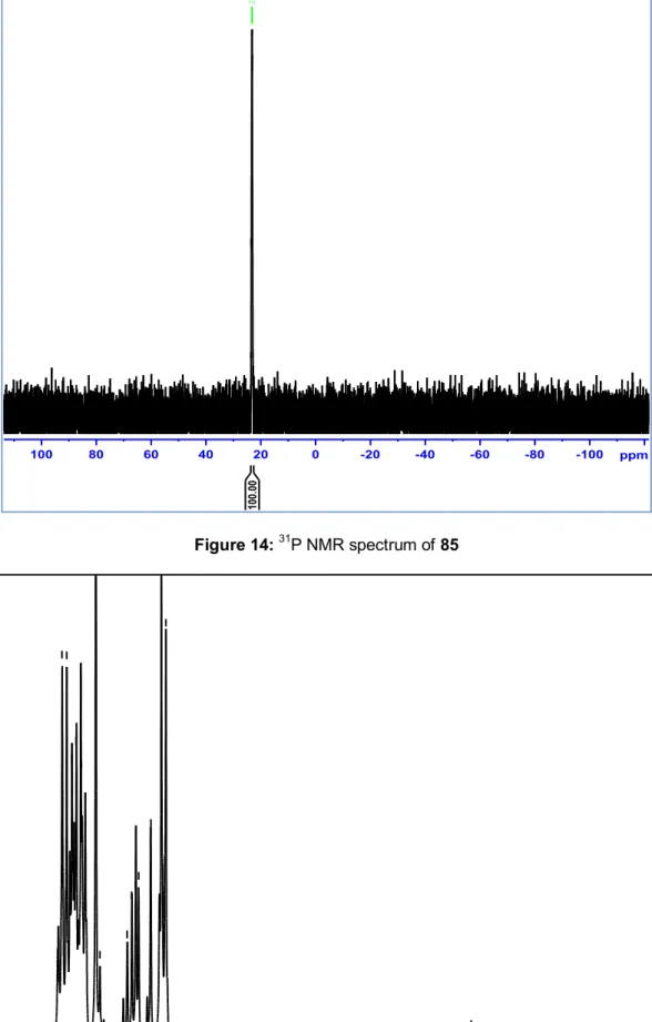
the reaction mixture and dried under reduced pressure. 1H and 31P NMR
spectra are reported in
Preparation of compound 85 Figure 13 and 14 Br P(Ph)3Br Br Br + -P(Ph)3, 4 hr Toluene, Reflux 84 85
respectively and data are reported in experimental part.
Scheme 17
Step2:
The reaction between 80 and 85 was carried out in dry THF at − 80 oC by
adding the butyl lithium basic reagent in ether solution (Scheme 18). The reaction is sensitive to atmospheric oxygen, thus it was carried out under argon atmosphere. It furnished a mixture of the E and Z-isomers of the compound 86
(4-[2-(4-bromophenyl)ethenyl]phenol). The trans-isomer was separated from the cis-isomer through preparative silica-gel column chromatography. The characterization of the stilbene 86 was made by 1H and 13C NMR spectra
(
Preparation of compound 86
37 Br P(Ph)3Br O Si CHO Br OH Si OH + 1. BuLi, dry THF - 80 oC, 45 min, RT, 24 h 2. H2O, 2hrs 80 85 86 + Ph3PO + + -Scheme 18 Figure 13: 1H NMR spectrum of 85
38 -100 -80 -60 -40 -20 100 80 60 40 20 0 ppm 23.20 100. 00 Figure 14: 31P NMR spectrum of 85 4.5 5.0 5.5 6.0 6.5 7.0 7.5 ppm
39 Figure 16: 13CNMR spectrum of compound 86
2.1.1.3
These monomers were gained from French laboratory by collaboration with Prof. Dominique Fasseur-Vervandier, at the Institut de Chimie Moléculaire de l'Université de Bourgogne, Dijon (France)
Synthesis of monomers 90-92 P(Ph)3Br R1 R2 R3 O Si CHO OH R3 R2 R1 Si OH + 1. BuLi, dry THF - 80 oC, 45 min, RT, 24 h 2. H2O, 2hrs 80 87 - 89 90 - 92 + Ph3PO + + 87. R1 = R2 = R3 = H 88. R1 = R3 = H, R2 = OMe 89. R1 = R3 = OMe, R2 = H 90. R1 = R2 = R3 = H 91. R1 = R3 = H, R2 = OMe 92. R1 = R3 = OMe, R2 = H Scheme 19
40
2.1.1.4
These monomers were gained from French laboratory by collaboration with Prof. Dominique Fasseur-Vervandier, at the Institut de Chimie Moléculaire de l'Université de Bourgogne, Dijon (France). The synthesis of these monomers is reported in the Scheme 20
Synthesis of monomers 95-96 P(Ph)3Br R1 R2 R3 O Si CHO MeO OH R3 R2 R1 OMe Si OH + 1. BuLi, dry THF - 80 oC, 45 min, RT, 24 h 2. H2O, 2hrs 93 88, 94 95-96 + Ph3PO + + -88. R1 = R3 = H, R2 = OMe 94. R1 = R2 = R3 = OMe 95. R1 = R3 = H, R2 = OMe 96. R1 = R2 = R3 = OMe Scheme 20 2.1.1.5
This monomer was gained from French laboratory by collaboration with Prof. Dominique Fasseur-Vervandier, at the Institut de Chimie Moléculaire de l'Université de Bourgogne, Dijon (France). The synthesis of this monomers is reported in the Scheme 21
Synthesis of monomer 98 Br O H OH MeO OH OH MeO + Pd(2), Ligand DMF, Cs2CO3 97a 97b 98 Scheme 21
41
2.1.2 Synthesis of Dimers 2.1.2.1
In order to obtain previously unreported stilbenolignans, new stilbenoid monomers were synthesized in cooperation with the laboratory of Prof. Dominique Vervandier-Fasseur. One of these substrates is compound 83
(E)-4-(4-vinylstyryl)phenol which was used as substrate, in the presence of Laccase from Trametes versicolor (LTV), acetate buffer (pH = 4.7) and EtOAc as co-solvent (
Biomimetic synthesis of stilbenolignan (±)-99
Scheme 22 O H O O H a or b 83 99
a) LTV, acetate buffer pH 4.7, EtOAc, 24hr. b) Mn(III)Ac, CH2Cl2, 24 hrs.
2
).
Sc Scheme 22
The dimerization afforded the product (±)-99 as racemic mixture in 24% yield.
This reaction was carried out also with Mn(OAc)3 in dichloromethane but this
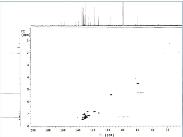
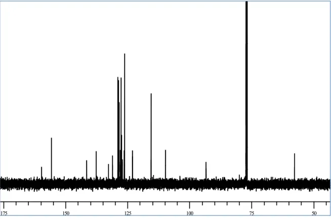
method gives very poor yield (ca 10%). After purification on DIOL silica gel column with petroleum ether: ethyl acetate 9.5:0.5 as the eluent. A spectroscopic characterization of the product was carried out. The structure of this product has not been previously reported, so we employed both mono and two-dimensional NMR methods (COSY and HSQC) to assign all the signals and to establish the structure apart from previously reported data. The 1H and 13C NMR spectra are reported in Figure 17 and 18, respectively, and Table 1;
assignments were aided by the careful analysis of COSY, HSQC, and HMBC spectra, reported in Figure 19 ,20 and 21, respectively. The mass spectrum
42 shows ESI-MS peak at m/z 465.1 [M+ Na]+ confirming the formation of a dimer
with molecular formula C32H26O2 and spectrum is reported in Figure 22.
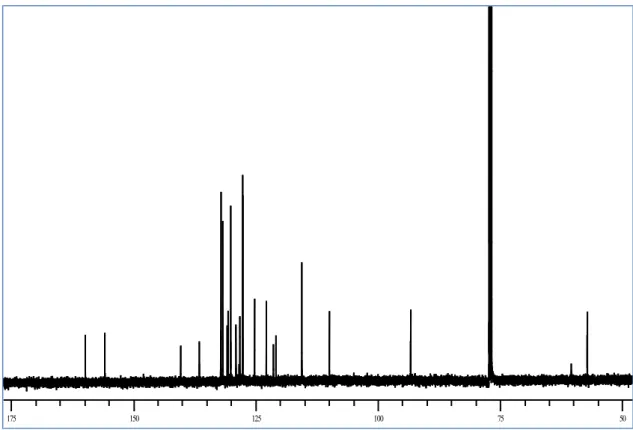
Figure 17: 1H NMR spectrum of (±)-99 25 25 50 50 75 75 100 100 125 125 150 150 175 175 Figure 18:.13C NMR spectrum of (±)-99
43 Figure 19: COSY spectrum of (±)-99
44 Figure 21: HMBC spectrum of (±)-99
45 Table1: 1H and 13C NMR data of (±)-99
The 13C NMR spectrum showed 25 signals: the majority of the signals were in
the sp2 region (from 159.6 to 109.7 ppm), but two methine signals in the sp3
region (57.4 and 93.2 ppm) were also observed, suggesting the formation of a dihydrobenzofuran ring. In the sp2 region there are 9 quaternary carbon
H δH (mult, J Hz) in CDCl3 C δC in CDCl3 C-1 132.5 H-2,6 7.22 (d,8.5, 2H) C-2,6 127.9 H-3,5 6.82 (d,8.5, 2H) C-3,5 115.5 C-4 155.6 H-7 5.49 (d,8.75, 1H) C-7 93.2 H-8 4.56 (d,8.75, 1H) C-8 57.4 C-9 140.9 H-10,14 7.17 (d,8.5, 2H) C-10,14 128.6 H-11,13 7.26 (m,2H) C-11,13 126.3 C-12 137.0 H-15a 6.74 (dd,17.5, 10.5, 1H.) C-15 136.5 H-16b 5.24 (d,10.5, 1H) C-16 113.9 H-16c 5.75 (d, 17.5, 1H) C-1' 128.4 H-2' 7.35 (bs,1H) C-2' 125.9 H-3’ 6.94 (d,8.5, 1H) C-3' 109.7 C-4' 159.6 C-5' 128.6 H-6' 7.19 (bs,1H) C-6' 122.8 H-7' 7.02 (d,16.25, 1H) C-7' 131.0 H-8' 6.88 (d,16.25, 1H) C-8' 126.7 C-9' 136.0 H-10',14' 7.36 (d,7.5, 2H) C-10',14' 127.6 H-11',13' 7.41 (d, 8.5, 2H) C-11',13' 126.4 C-12' 137.2 H-15’a 6.70 (dd,17.5, 10.5, 1H) C-15’ 136.4 H-16’b 5.28 (d,10.5, 1H) C-16’ 113.4 H-16’c 5.78 (d, 17.5, 1H) OH 4.9 (bs, 1H)
46 signals, 12 CH signals, and 2 CH2 signals. The 1H NMR spectrum of (±)-99 in
the region from 7.42 and 6.82 ppm shows clear similarities with the spectra of the above discussed dihydrobenzofuran neolignans. In particular, a COSY correlation was observed between the two doublet signals at δ = 7.37 and 6.94 ppm assigned to a four-proton AA’BB’ system, and between the two doublets at δ = 7.41 and 7.18 ppm, integrating for eight protons (two AA’BB’ systems). These signals indicates that there are three para substituted aromatic rings (see below); the 13Cchemical shift analysis and HSQC spectrum indicated that
one of these rings bear a phenol group whereas the other two, bearing an ethenyl group, give isochronous signals (Figure 23
OH Ha' Hb' Ha Hb Hb Hc Ha Ha' Hb' Ha Hb ). Figure 23
The signals of the two ethenyl groups are partly overlapped and also in this case the COSY/HSQC analysis proved useful, showing the spin-spin couplings in these two ABC systems, as reported below: both ‘a’ signals, closer to the
aromatic ring, resonate at lower fields (6.74 and 6.70 ppm), whereas the other proton signals (b and c) are appear at higher fields (5.24, 5.28, 5.76 and 5.78
ppm).
Two further doublets at δ = 7.04 and 6.90 ppm (1H each) show a cross-peaks correlation in the COSY spectrum, and may be assigned to AB system with a large coupling constant (16.3 Hz), clearly due to two trans olefinic protons (Figure 24)