A randomized placebo-controlled phase 3 trial of an antisense
oligonucleotide, drisapersen, in Duchenne muscular dystrophy
Nathalie Goemans
a,*, Eugenio Mercuri
b, Elena Belousova
c, Hirofumi Komaki
d,
Alberto Dubrovsky
e, Craig M. McDonald
f, John E. Kraus
g,1, Afrodite Lourbakos
h,
Zhengning Lin
h, Giles Campion
h, Susanne X. Wang
h, Craig Campbell
ifor the DEMAND III
study group
aDepartment of Pediatrics and Child Neurology, University Hospitals Leuven, Leuven, Belgium bPediatric Neurology, Catholic University, Rome, Italy
cResearch and Clinical Institute of Pediatrics, Pirogov Russian National Research Medical University, Moscow, Russia dDepartment of Child Neurology, National Center of Neurology and Psychiatry, Tokyo, Japan
eFundacion Cenit, Instituto de Neurociencias, Fundación Favaloro, Buenos Aires, Argentina fSchool of Medicine, University of California, Davis, Sacramento, CA, USA
gGlaxoSmithKline, Research Triangle Park, NC, USA hBioMarin Pharmaceutical Inc., Novato, CA, USA
iPaediatric Neurology, Schulich School of Medicine, Western University, London, Canada Received 21 March 2017; received in revised form 23 September 2017; accepted 17 October 2017
Abstract
This 48-week, randomized, placebo-controlled phase 3 study (DMD114044; NCT01254019) evaluated efficacy and safety of subcutaneous drisapersen 6 mg/kg/week in 186 ambulant boys aged≥5 years, with Duchenne muscular dystrophy (DMD) resulting from an exon 51 skipping amenable mutation. Drisapersen was generally well tolerated, with injection-site reactions and renal events as most commonly reported adverse events. A nonsignificant treatment difference (P= 0.415) in the change from baseline in six-minute walk distance (6MWD; primary efficacy endpoint) of 10.3 meters in favor of drisapersen was observed at week 48. Key secondary efficacy endpoints (North Star Ambulatory Assessment, 4-stair climb ascent velocity, and 10-meter walk/run velocity) gave consistent findings. Lack of statistical significance was thought to be largely due to greater data variability and subgroup heterogeneity. The increased standard deviation alone, due to less stringent inclusion/exclusion criteria, reduced the statistical power from pre-specified 90% to actual 53%. Therefore, a post-hoc analysis was performed in 80 subjects with a baseline 6MWD 300–400 meters and ability to rise from floor. A statistically significant improvement in 6MWD of 35.4 meters (P= 0.039) in favor of drisapersen was observed in this subpopulation. Results suggest that drisapersen could have benefit in a less impaired population of DMD subjects.
© 2017 The Authors. Published by Elsevier B.V. This is an open access article under the CC BY-NC-ND license (http://creativecommons.org/ licenses/by-nc-nd/4.0/).
Keywords: Duchenne muscular dystrophy; Drisapersen; Dystrophin; Antisense oligonucleotide; Exon skipping; six-minute walking distance
1. Introduction
Duchenne muscular dystrophy (DMD, OMIM 310200) is a rare neuromuscular disease, affecting one in 3500–5000 newborn boys[1–3] as reported from a number of screening programs worldwide[4,5]. It is an X-linked recessive condition
caused by mutations in the DMD gene, with deletions flanking exon 51 most commonly observed [6]. This results in disruption of the transcriptional open-reading frame and leads to prematurely aborted dystrophin synthesis [7]. The lack of functional dystrophin results in progressive damage and degeneration of muscle fibers, followed by a predictable clinical trajectory [8]. Initial development of motor skills is followed by a plateau phase, after which progressive muscle function deterioration with age is observed (decline to be expected from 7 years of age) [9–15]. As a result, most subjects become wheelchair-bound by their mid-teens [8]. * Corresponding author. University Hospitals Leuven, Herestraat 49, B3000
Leuven, Belgium.
E-mail address:[email protected](N. Goemans). 1 Member of the DEMAND III study team 2.
https://doi.org/10.1016/j.nmd.2017.10.004
0960-8966/© 2017 The Authors. Published by Elsevier B.V. This is an open access article under the CC BY-NC-ND license (http://creativecommons.org/ licenses/by-nc-nd/4.0/).
Available online atwww.sciencedirect.com
Neuromuscular Disorders 28 (2018) 4–15
www.elsevier.com/locate/nmd
Subsequently, respiratory failure and cardiomyopathy emerge, with most subjects developing nocturnal hypoventilation or respiratory failure requiring non-invasive mechanical ventilation or death before the age of 30 years[16]. DMD is currently mainly managed by standard of care treatments such as glucocorticoids [16,17], physiotherapy, management of spine deformity, and cardiorespiratory dysfunction[8,18]. This better clinical management has improved prognosis and life expectancy over the last few decades[19,20]. Ataluren received conditional approval in the European Union but can only be used for treating DMD caused by a nonsense mutation (nmDMD) in ambulatory boys aged≥5 years[21,22].
Drisapersen is a 2′-O-methyl-phosphorothioate antisense oligonucleotide (AON) that induces exon 51 skipping during pre-messenger RNA splicing[23], generating a shorter in-frame largely functional dystrophin protein[24]. This approach could be used to treat approximately 13% of DMD subjects[6]. In a comprehensive clinical development program, 2 randomized, placebo-controlled, phase 2 studies (DMD114117 [25], and DMD114876 [McDonald et al. Unpublished results]) examined the effect of 6 mg/kg/week drisapersen in ambulant boys with a six-minute walk distance (6MWD) of≥75 meters and a rise from floor (RFF) time of≤7 seconds (except for 2 boys). Both studies have provided evidence that continuous subcutaneous (sc) drisapersen 6 mg/kg/week improved mean 6MWD versus placebo after 24 weeks by 35 meters (P= 0.014) and 27 meters (P= 0.069), respectively. This was maintained after 48 weeks in DMD114117 (mean difference 36 meters, P= 0.051)[25]. The treatment benefit in the randomized, placebo-controlled, phase 2 studies was supported by long-term phase 1/2 extension data (~3.4 years)[26]compared with matched natural history (NH) controls with a comparable observational time frame
[27].
The current phase 3 study (DEMAND III; DMD114044; NCT01254019) evaluated the efficacy and safety of sc drisapersen 6 mg/kg/week over 48 weeks in a larger cohort (N= 186) of ambulant DMD subjects.
2. Subjects and methods
The CONSORT checklist is available as supporting information.
2.1. Study population
The DMD114044 study was performed in accordance with the International Conference on Harmonisation Good Clinical Practice guidelines, the Declaration of Helsinki (2008), and applicable country-specific requirements. Written informed consents from parents/caregivers and assent (from appropriately aged subjects) were obtained for all subjects prior to any study procedure.
Ambulant boys aged≥5 years, with DMD resulting from an exon 51 skipping amenable mutation, were eligible for inclusion. Inclusion criteria included: a life expectancy >1 year; 3 pre-treatment 6MWD tests ≥75 meters within 20% of each other; glucocorticosteroid therapy for ≥6 months and stable
dose and regimen for≥3 months prior to screening, with the expectation of remaining on similar dose/regimen for the study duration. Key exclusion criteria included: any additional missing exon that could not be treated with exon 51 skipping; use of concomitant medication (anticoagulants, antithrombotics, antiplatelet agents, idebenone and other forms of coenzyme Q10); any liver and renal impairment; and symptomatic cardiomyopathy.
2.2. Study design and treatment
This randomized, double-blind, placebo-controlled, phase 3 study was conducted between December 30, 2010 and June 28, 2013 at 44 centers in 19 countries: Argentina, Belgium, Brazil, Canada, Chile, Czech Republic, Denmark, France, Germany, Italy, Japan, Korea, the Netherlands, Norway, Poland, Russian Federation, Spain, Taiwan, and Turkey.
Following screening, eligible subjects were centrally randomized by an interactive voice response system and a random allocation sequence (without using any stratification factors) in a 2:1 ratio to receive either sc drisapersen 6 mg/kg/week or volume-matched placebo for 48 weeks.
The study was fully blinded with respect to treatment allocation. All treatments were prepared and administered by trained and qualified unblinded personnel who were not involved in the efficacy assessments. To minimize the risk of injection-site reactions, rotation of the injection site on a weekly basis was recommended.
2.3. Endpoints
The primary efficacy endpoint was change from baseline 6MWD at week 48[9,28]. Key secondary efficacy endpoints were the change from baseline in: North Star Ambulatory Assessment (NSAA)[16,29], 4-stair climb ascent velocity, and 10-meter walk/run velocity. Other secondary efficacy endpoints included: other timed-function tests (RFF time and 4-stair climb descent velocity), muscle strength, pulmonary function, molecular efficacy (exon 51 skipping at mRNA level and dystrophin expression) [30,31], serum creatine kinase (CK), and functional and health outcome assessments (Clinical Global Impression of Improvement [CGI-I] [32], pediatric quality of life [PedsQL] neuromuscular module [33], health utility index [HUI], and activities of daily living).
Pharmacokinetic evaluations included the drisapersen concentration in plasma and muscle tissue. Safety and tolerability endpoints included adverse events (AEs), serious AEs, local tolerability, laboratory parameters (including lactate dehydrogenase [LDH], which was evaluated post-hoc), vital signs, electrocardiograms, echocardiography, and physical examination.
2.4. Assessments
The primary efficacy endpoint and most secondary efficacy assessments were conducted at baseline, and subsequently every 12 weeks or at early withdrawal. Site staff members were trained on functional efficacy endpoints by highly experienced 5 N. Goemans et al. / Neuromuscular Disorders 28 (2018) 4–15
and well-trained physiotherapists, who provided refresher training as required throughout the study.
The 6MWD was assessed as reported previously[9,28]. An age- and height-based equation fitted to normative data by Geiger et al. was applied to the 6MWD data and a percent-predicted 6MWD was calculated for each subject[34,35].
Timed-function test velocities for RFF, 4-stair ascent and descent, and 10-meter walk/run were calculated as described by McDonald et al.[36].
Muscle strength was evaluated by handheld myometry using a microFET dynamometer (Biometrics BV, Almere, The Netherlands), whereas pulmonary function was assessed using a handheld Koko spirometer (PDS Instrumentation, Louisville, KY, USA) and a magnehelic manometer (Dwyer Instrument, Michigan City, IN, USA). Pulmonary function testing included the following parameters: forced vital capacity (FVC) [37], forced expiratory volume in 1 second (FEV1), peak expiratory flow (PF), and peak cough flow (PCF)[38].
The CGI-I was a single-item question designed to provide a brief, stand-alone assessment of the clinician’s view of the subject’s global functioning (with 7 potential responses ranging from “much improved” to “very much worse”) after initiating study drug, compared to their global functioning just prior to initiating treatment [32]. Health-related quality of life was measured via the PedsQL, with total score ranging from 0–100 and higher scores indicating a better health-related quality of life. The appropriate age-specific version of the PedsQL was completed, with children aged≤7 years completing a different questionnaire than children aged >7 years [33]. The HUI is comprised of a family of generic health profiles and preference-based systems for the purposes of measuring health status, reporting health-related quality of life, and producing utility scores.
Blood samples for pharmacokinetic assessments were collected pre-dose and between 1 to 4 hours post-dose, at week 8, 12, 24, or 36 (depending on which visit the muscle biopsy was taken) and at week 47. At week 0, samples were collected pre-dose and at 0.5, 1, and 3 hours post-dose.
AEs were coded using MedDRA System Organ Class and Preferred Term.
2.5. Statistical methods
It was planned to randomize 180 subjects in order to recruit at least 162 evaluable subjects. It was calculated that 162 evaluable subjects would provide 90% power to detect 30 meters difference of change from baseline in 6MWD between drisapersen and placebo, assuming a common standard deviation of 55 meters (using a two-group t-test with a 0.05 two-sided significance level). It is noteworthy that most subjects included in DMD114117 study had a baseline 6MWD>300 meters and a smaller SD for the change in 6MWD after drisapersen treatment[25].
2.5.1. Pre-specified analyses
The primary efficacy analysis of the change from baseline in the 6MWD used the intent-to-treat (ITT) population to which a
mixed model for repeated measures (MMRM) was applied, that included fixed terms for treatment, visit, treatment-by-visit interaction, country grouping, baseline value, and baseline value-by-visit interaction. The MMRM was used to assess longitudinal data and allows to correct missing data.
If a statistically significant treatment difference (5% level) is observed for the primary efficacy endpoint, the 3 aforementioned key secondary endpoints are tested in a sequential manner, using the same methodology. For other secondary efficacy endpoints, statistical comparisons were made and the results were considered supportive.
Furthermore, pre-specified subgroup summary statistics of the primary efficacy endpoint based on age (≤7 years; >7 years) were performed.
2.5.2. Post-hoc analyses
In more recent years of DMD research, NH study outcomes highlighted the importance of baseline characteristics, such as age, 6MWD, and the ability to RFF, as prognostic factors for loss of ambulation[9–15,39]. Subjects with a baseline 6MWD of less than 300 meters and/or who are unable to perform the RFF test at baseline are at greater risk of imminent loss of ambulation than subjects with a baseline 6MWD of above 300 meters and/or who are able to perform the RFF test[10,13,14]. Subjects with a baseline 6MWD of above 400 meters are too stable to detect a treatment difference in a study of one year duration. In addition, a 6MWD<300 meters is associated with a significant reduction in lower extremity fat fraction on magnetic resonance imaging hence less muscle substrate is present to effect a change in lower extremity ambulatory endpoints [40]. Therefore, the same MMRM analysis was performed in a subgroup of subjects with a baseline 6MWD result between 300 and 400 meters and who were able to RFF.
The pre-specified analysis for the CGI-I was only for the dichotomized variable of subjects who improved much or very much versus those who did not improve category. This analysis was not carried out due to the small sample size in the very much improved and much improved category for the placebo group with the non-convergence of the statistical model. Instead, an analysis of all 7 CGI-I categories using the Cochrane-Mantel-Haenszel test with row mean score was conducted. The Cochrane-Mantel-Haenszel test was selected as controls were stratified and matched at baseline.
For all post-hoc analyses in this manuscript, nominal
P-values are reported without adjustment of multiplicity.
Statistical significance is defined as nominal P< 0.05, which does not take into account the post-hoc nature of the analyses.
3. Results
3.1. Study population
A total of 186 subjects were randomized by 44 centers in 19 countries to drisapersen 6 mg/kg/week (N= 125) or placebo
(N= 61; Fig. 1). All subjects were included in the ITT population. At baseline in both treatment arms (N= 186), the age ranged from 5 to 16 years and the 6MWD from 107 to 566 meters. However, an imbalance in baseline disease severity characteristics was observed between the 2 treatment arms
(Table 1), with a lower mean 6MWD and greater mean age
observed in the drisapersen than in the placebo group (337.5 versus 348.0 meters and 8.3 versus 8.0 years old, respectively). Furthermore, 15.2% of subjects included in the drisapersen group were unable to perform the RFF test at baseline
compared with 9.8% of subjects included in the placebo group. This imbalance reflected the inclusion of an older and more impaired population in the drisapersen group at baseline.
3.2. Efficacy
3.2.1. Primary endpoint
Over 48 weeks, 10% of the placebo group compared with 12% of the drisapersen group lost ambulation. Of those who lost ambulation, 5 of 6 placebo-treated subjects and 14 Fig. 1. DMD114044 subject flow diagram.
aSubjects could only have one primary reason for withdrawal.
bThe ITT population was defined as all subjects randomized to the study who received at least one dose of study medication and had at least one post-baseline efficacy assessment.
cAll subjects who received at least one dose of study medication were included in the safety population.
dThe PK population was defined as all subjects who were randomized to the study and from whom at least one blood sample was obtained for assessment of drisapersen levels.
eThe PP population was defined as all ITT subjects who had no major protocol deviations. fA subject could have more than one major protocol deviation.
6MWD= six-minute walk distance; ITT = intent-to-treat; PK = pharmacokinetic; PP = per-protocol.
7 N. Goemans et al. / Neuromuscular Disorders 28 (2018) 4–15
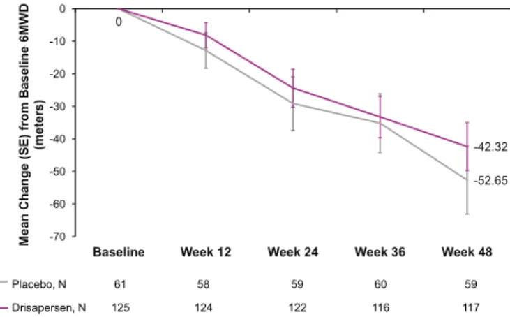
of 15 drisapersen-treated subjects had a baseline 6MWD <300meters; 3 of 6 placebo and 9 of 15 drisapersen-treated subjects were unable to RFF at baseline. A decrease in mean change (standard error [SE]) from baseline 6MWD to week 48 was observed in the placebo (−52.7 [10.4] meters) as well as in the drisapersen group (−42.3 [7.4] meters), with a mean (95% confidence interval [95% CI]) treatment benefit of 10.3 (−14.7; 35.3) meters in favor of drisapersen (P= 0.415;Fig. 2).
A pre-specified MMRM subgroup analysis by age (≤7 years; >7 years), showed a greater mean (95% CI) treatment difference in change from baseline 6MWD over placebo for the drisapersen treatment group in younger subjects (21.5 [−6.6, 49.6] meters; P= 0.131) than in older subjects (6.9 [-29.0, 42.9] meters; P= 0.703) at week 48.
A post-hoc MMRM analysis was performed in a subgroup of 83 subjects (52 drisapersen; 31 placebo) with a baseline 6MWD between 300 to 400 meters and a mean difference in 6MWD at week 48 of 27.8 meters (95% CI: −7.5, 63.1). Three subjects in this subgroup were unable to perform the RFF test at baseline; all belonged to the drisapersen group of whom 1 lost ambulation at week 60 visit (12 week visit in the extension study). Upon removal of these 3 subjects, a significant mean
treatment benefit in 6MWD of 35.4 meters (95% CI: 1.8, 69.0) in favor of drisapersen was observed (P= 0.039).
3.2.2. Secondary efficacy endpoints
3.2.2.1. Ambulatory function endpoints. Using the
pre-specified statistical analysis model, no statistically significant differences were observed for the secondary ambulatory function efficacy endpoints: NSAA (P= 0.757), 4-stair climb ascent velocity (P= 0.718), 4-stair climb descent velocity (P= 0.513), 10-meter walk/run velocity (P = 0.881), percent-predicted 6MWD (P= 0.320), and time to RFF (P = 0.658;
Fig. 3A).
Similarly as for the primary efficacy endpoint, a post-hoc analysis was performed in which the same MMRM analysis was applied to the subgroup of 80 subjects (49 drisapersen; 31 placebo) with a baseline 6MWD result between 300 to 400 meters and who were able to RFF (Fig. 3B). As was the case for the 6MWD, the mean difference in percent-predicted 6MWD was in favor of drisapersen in this subpopulation. For the other secondary efficacy endpoints, larger mean improvements observed in NSAA, RFF velocity, 10-meter walk/run velocity, and 4-stair descent velocity in the drisapersen versus the placebo group were observed.
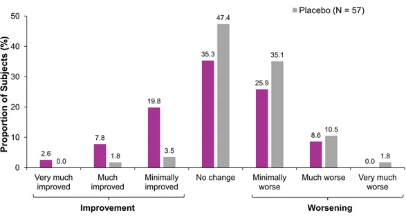
3.2.2.2. Other secondary endpoints. At week 48, 10% of
drisapersen-treated subjects compared to 2% of placebo-treated subjects were considered responders (“much improved” or “very much improved”) on the CGI-I scale. The pre-specified inferential analysis of “(very) much improved” in CGI-I could not be carried out due to the fact that only 1 placebo subject was in the “much improved” category. Hence, a post-hoc analysis of all CGI-I categories was performed, showing a statistically significant difference in favor of drisapersen in the entire study population (P= 0.002;Fig. 4), and in the subgroup of subjects with a baseline 6MWD of 300–400 meters and able to RFF (P= 0.001).
There were no clinically meaningful treatment differences between drisapersen and placebo for the PedsQL neuromuscular module, HUI assessments, and activities of daily living (data not shown). In addition, no clinically meaningful treatment difference was observed for any of the pulmonary function tests (including FVC, FEV1, PF, and PCF).
3.2.2.3. Laboratory and biomarker assessments. At week 48,
the adjusted mean [SE] CK serum concentration declined significantly in the drisapersen (−5273.5 [359.1] IU/L) versus the placebo (−1228.5 [500.6] IU/L) group (P< 0.001). The difference was statistically significant from week 12 onwards (Fig. 5A). Similarly, the mean [SE] LDH serum concentration also declined more in the drisapersen (−375.3 [21.40)] than in the placebo (−119.2 [36.58] IU/L) group at week 48 (P< 0.001). Also this difference was statistically significant from week 12 onwards (Fig. 5B).
3.2.2.4. Pharmacokinetic analysis. Blood samples for
pharmacokinetic analysis were taken using sparse sampling, therefore no pharmacokinetic parameters were calculated using non-compartmental analysis. The median trough drisapersen Table 1
Baseline demographics and clinical characteristics (safety population).
Mean (SD) Drisapersen 6 mg/kg/week (N= 125) Placebo (N= 61) Age, years 8.3 (2.4) 8.0 (2.4) Height, cm 124.0 (10.8) 122.0 (9.6) Weight, kg 30.1 (10.3) 26.9 (7.6)
Time since first symptoms, months* 71.8 (31.6) 66.77 (31.3) Time since diagnosis, months 58.0 (35.2) 54.2 (32.8) Time since first corticosteroid taken, months 35.6 (29.0) 29.1 (25.8)
6MWD, meters 337.5 (95.6) 348.0 (92.2)
RFF time, seconds† 12.34 (14.98) 13.41 (15.88)
% subjects unable to RFF 15.2% 9.8%
* N= 122 for the drisapersen group and N = 58 for the placebo group. † N= 106 for the drisapersen group and N = 55 for the placebo group. 6MWD= six-minute walk distance; RFF = rise from floor; SD = standard deviation. -52.65 0 -42.32 -70 -60 -50 -40 -30 -20 -10 0
Baseline Week 12 Week 24 Week 36 Week 48
Mean
Change (SE) from
Baseline 6MWD (m eters) Placebo, N 61 58 59 60 59 Drisapersen, N 125 124 122 116 117 *
Fig. 2. Pre-specified analysis of the mean change (SE) from baseline to week 48 in 6MWD (intent-to-treat population).
6MWD= six-minute walk distance: SE = standard error. *P= 0.415.
plasma concentration increased over time up to 48 weeks of dosing (Figure S2A), whereas the mean drisapersen muscle tissue homogenate concentration slowly increased over time reaching steady state at approximately 36 weeks post-treatment (Figure S2B).
3.3. Safety
The majority of subjects reported at least one on-treatment AE (98% drisapersen; 95% placebo), most of which were mild to moderate in intensity (7% of drisapersen and 3% of placebo subjects reported severe AEs; Table 2). Serious AEs were reported in 10% of drisapersen-treated and 8% of placebo-treated subjects, of which none were reported in more than 1 subject in either treatment group. Two (1.6%) subjects in the drisapersen group had a drug-related serious AE that led to study discontinuation (glomerulonephritis [N= 1] and intracranial venous sinus thrombosis and spinal pain [N= 1]). No deaths occurred during the study and no subjects withdrew due to meeting pre-defined criteria for dose interruption.
AEs of special interest were reported in 91% of the drisapersen-treated and 61% of the placebo-treated group. The
most commonly reported on-treatment AEs of special interest were local injection-site and renal AEs.
Injection-site AEs occurred more frequently in the drisapersen-treated (78%) than in the placebo-treated (16%) group; with erythema (50%), discoloration (33%), reaction (19%), pain (18%), induration (14%), and pruritus (14%) most commonly reported in the drisapersen group. None were reported as serious AEs and the majority of events (65% of all events) was resolved or was being resolved over the observational time frame or was resolved with sequelae (18% of all events). AEs with an outcome of not resolved injection-site reactions (16% of all events; followed-up from December 30, 2010 to March 17, 2014) were primarily injection-site extravasation (2/2 events; 100%), lipodystrophy acquired (8/8 events; 100%), lipoatrophy (3/3 events; 100%) pigmentation disorder (6/7 events; 86%), injection-site atrophy (9/13 events; 69%), site reaction (40/106 events; 38%), injection-site discoloration (64/230 events; 28%), injection-related reaction (1/4 events; 25%), injection-site erythema (72/494 events; 15%), injection-site induration (5/35 events; 14%), injection-site pain (3/41 events; 7%), injection-site swelling (1/20 events; 5%), and injection-site pruritus (1/34 events; 3%). Fig. 3. Forest plot of secondary ambulatory endpoints.
A) Pre-specified analysis in the intent-to-treat population.
B) Post-hoc analysis in a subpopulation with baseline 6MWD results between 300 and 400 meters and who were able to perform the RFF test.
6MWD= six-minute walk distance; RFF = rise from floor; NSAA = North Star Ambulatory Assessment; SD = standard deviation; CI = confidence interval. 9 N. Goemans et al. / Neuromuscular Disorders 28 (2018) 4–15
Photographs of the injection-site reactions are not shown in this publication as no permission was obtained from the patients.
Renal AEs were reported more commonly in the drisapersen (64%) than in the placebo (33%) group, with proteinuria (34%), hematuria (17%), protein in urine (14%), red blood cells positivity (11%), and increased cystatin C (11%) most frequently reported in the drisapersen group. Increases in α1-microglobulin, serum cystatin C, and KIM-1 in the drisapersen treatment group were consistent with possible mild proximal tubule dysfunction and subclinical proteinuria. One drisapersen-treated subject had a serious AE of moderate glomerulonephritis.
Hepatic AEs were observed in 6% of drisapersen-treated subjects, none were considered clinically significant. There were no clinically relevant differences between treatment groups in clinical chemistry, hematology, or coagulation values of potential clinical concern. Platelet count was slightly reduced in the drisapersen group. However, no AEs related to decreased thrombocyte count were reported.
4. Discussion
This phase 3 study is one of the largest placebo-controlled trials in DMD to date. The primary efficacy endpoint was change from baseline 6MWD over 48 weeks. The 6MWD test has been adapted to evaluate muscle function and endurance in neuromuscular disorders, and has been used as the primary outcome measure in several other international multicenter clinical trials for DMD [21,25,41]. The pre-planned analysis of the present phase 3 study demonstrated a small and insignificant mean treatment benefit in 6MWD of 10.3 meters over 48 weeks (P= 0.415). This is in contrast with the outcomes of the 2 randomized, placebo-controlled, phase 2 studies
showing an improvement in mean 6MWD at 24 weeks of 35 meters (P= 0.014)[25]and 27 meters (P= 0.069), respectively. One possible explanation for this discrepancy is the increased data variation (pre-specified SD of 55 meters increased to an actual SD of 87 meters due to less stringent inclusion/exclusion criteria) in the phase 3 study reducing the statistical power to detect a treatment difference of 30 meters in mean change from baseline 6MWD from 90% to 53%. The 87 meters SD for change in 6MWD over 48 weeks is more consistent with the literature in DMD subjects with wide variation in baseline 6MWD found in NH studies and clinical trials[15,21].
A second explanation for the discrepancy is the lack of NH knowledge at the start of the drisapersen clinical program, with recent NH studies highlighting the importance of baseline characteristics, such as age, 6MWD, and the ability to RFF
[9–15]. Subjects with a baseline 6MWD<300 meters are at greater risk of imminent loss of ambulation with large changes in 6MWD over 48 weeks being driven by those subjects who lose ambulation. In contrast subjects with a baseline 6MWD>400 meters change very little over 48 weeks as they are often in the stage of natural maturation and can present with a ceiling effect that limits the measurable improvement with a dystrophin restoration treatment [9–15,39]. In addition, the inability to RFF is a separate prognostic factor for imminent loss of ambulation, with 43% of the DMD subjects unable to perform RFF test losing ambulation within 1 year[39]. Taking into account these learnings from NH [9–15,39], subjects included in the phase 3 study had a more severe disease status at baseline (mean age= 8.2 years, mean 6MWD = 341 meters, and mean RFF time= 13 seconds) compared with subjects included in the phase 2 studies (mean age= 7.3 and 7.8 years, mean 6MWD= 409 meters [in both studies], and mean 2.6 7.8 19.8 35.3 25.9 8.6 0.0 0.0 1.8 3.5 47.4 35.1 10.5 1.8 0 10 20 30 40 50 Very much improved Much improved Minimally improved No change Minimally worse
Much worse Very much
worse Proportion of Subjects (% ) CGI-I at Week 48 Drisapersen (N = 116) Placebo (N = 57) Improvement Worsening
Fig. 4. Distribution of the Clinical Global Impression of Improvement scale at week 48. CGI-I= Clinical Global Impression of Improvement.
RFF time= 5 seconds [in both studies], respectively) [25]
[McDonald et al. Unpublished results]. Furthermore, all subjects included in the phase 2 studies (except 2 subjects in DMD114876) were able to perform the RFF test at baseline in ≤7 seconds [25] [McDonald et al. Unpublished results]. In contrast, due to the lack of a RFF criterion during phase 3 study enrollment, 13.4% of subjects were unable to perform the RFF test, predicting a high risk of losing ambulation in the following year [39]. The 6MWD is not an appropriate measure of
treatment efficacy for subjects at high risk of losing ambulation prior to the completion of a 48-week clinical trial.
A pre-specified subgroup analysis, taking into account baseline age (≤7; >7 years), partially addressed this disease status imbalance issue and showed a greater mean treatment benefit in favor of drisapersen for the younger (21.5 meters) than the older population (6.9 meters). A post-hoc MMRM analysis addressing the imbalance in baseline 6MWD and RFF time was performed in a subgroup of subjects with a baseline Fig. 5. Mean (±standard error) serum concentration over 48 weeks.
A) Creatine kinase. B) Lactate dehydrogenase.
* P< 0.05, ** P < 0.01, *** P < 0.001.
11 N. Goemans et al. / Neuromuscular Disorders 28 (2018) 4–15
6MWD result between 300 and 400 meters and able to RFF, and showed a statistically significant treatment benefit of 35.4 meters in favor of drisapersen. This is in line with the fact that in order for drisapersen to provide benefit, sufficient muscle tissue needs to be preserved[42]. Less target lower extremity tissue will become available as ambulation declines due to progressive replacement of healthy muscle with fat and fibrous tissue in subjects with increasing age[43,44]. Other endpoints such as upper limb function may be more appropriate to measure therapeutic effect in these subjects [45–47]. These results are also in line with those from the phase 3 clinical trial with ataluren in 228 nmDMD subjects showing a non-significant improvement over placebo of 15 meters in the total population with a greater statistically significant improvement over placebo of 47 meters in the subgroup of subjects with a baseline 6MWD of 300–400 meters (N=99)[48].
A significantly greater percentage of the drisapersen-treated subjects (30%) showed improvement on the CGI-I scale compared with placebo-treated subjects (5%). This may suggest that the CGI-I does not have the same limitations as other endpoints as it captures the physicians’ global impression of improvement. In contrast, there were no clinically meaningful treatment differences between drisapersen and placebo for the PedsQL neuromuscular module, HUI assessments, and activities of daily living. This is not surprising as a weak correlation of the PedsQL physical function scale to variation in disease was previously reported in DMD [12,33] and the PedsQL neuromuscular module uses a similar frequency of difficulty construct. No statistically significant differences were observed for the pulmonary function tests but data from NH demonstrate it can be difficult to observe significant treatment effects after a period of only 48 weeks in ambulatory subjects and respiratory decline mainly occurs in DMD subjects older than the population included in the current study[49–51].
Elevated serum CK is used as a diagnostic biomarker for
DMD [52,53] because dystrophin deficiency and associated
muscle fiber damage in DMD result in the release of CK from the intracellular compartment of muscle fibers into the circulation. Serum CK concentration, a pre-specified secondary efficacy endpoint, was statistically significantly reduced in subjects treated with drisapersen versus placebo as early as 12 weeks and up to 48 weeks (Figure S3). Although in humans CK is known to decrease with age and stage of the disease, the observed reduction was irrespective of baseline age and 6MWD. This is in line with placebo-controlled studies in mdx mice and mdx/utrn (+/-) mice treated with mouse surrogate exon 23 skipping by 2′-O-methyl-phosphorothioate AONs
[54,55] or by phosphorodiamidate morpholino AONs [56]
demonstrating exon skipping in various muscles and a related decrease in serum CK. Elevated serum LDH is also considered a biomarker of muscle disease in DMD subjects[57–60] and LDH isoform expression is dysregulated in DMD muscles
[58,59]. Post-hoc analysis showed that the reduction in serum LDH in the drisapersen group reached statistical significance compared to placebo as early as 12 weeks and up to 48 weeks. Thus it seems that CK and LDH serve as plausible pharmacodymanic biomarkers for drisapersen.
Table 2
Incidence of on-treatment adverse events (safety population). Drisapersen 6 mg/kg/week (N= 125) Placebo (N= 61) On-treatment AEs AEs 123 (98) 58 (95)
Drug-related AEs (as determined by investigator)
111 (89) 31 (51)
Severe AEs 9 (7) 2 (3)
Serious AEs 13 (10) 5 (8)
AEs leading to permanent discontinuation of study treatment
2 (2) 0
Any AE of special interest* 114 (91) 37 (61)
Injection-site reaction 97 (78) 10 (16)
Renal effects 80 (64) 20 (33)
Inflammation 33 (26) 16 (26)
Coagulation 9 (7) 9 (15)
Hepatic effects 7 (6) 0
Low thrombocyte counts 0 0
Most common on-treatment AEs (Reported in≥5% of subjects in either treatment group)† Injection-site erythema 62 (50) 4 (7) Proteinuria 42 (34) 11 (18) Injection-site discoloration 41 (33) 2 (3) Nasopharyngitis 38 (30) 25 (41) Headache 33 (26) 11 (18) Vomiting 28 (22) 13 (21) Pyrexia 27 (22) 15 (25) Fall 27 (22) 12 (20) Cough 24 (19) 12 (20) Injection-site reaction 24 (19) 1 (2) Diarrhea 23 (18) 9 (15) Injection-site pain 23 (18) 2 (3) Hematuria 21 (17) 5 (8)
Protein urine present 17 (14) 4 (7)
Injection-site induration 17 (14) 0
Injection-site pruritus 17 (14) 0
Upper respiratory tract infection 15 (12) 8 (13)
Gastroenteritis 15 (12) 6 (10)
Abdominal pain 14 (11) 7 (11)
Red blood cells urine positive 14 (11) 4 (7)
Cystatin C increased 14 (11) 2 (3)
Pain in extremity 11 (9) 12 (20)
Injection-site bruising 11 (9) 6 (10)
Epistaxis 11 (9) 5 (8)
Red blood cells urine 11 (9) 4 (7)
Urine protein/creatinine ratio increased 11 (9) 2 (3)
Rhinitis 10 (8) 3 (5)
Arthralgia 10 (8) 1 (2)
Oropharyngeal pain 10 (8) 1 (2)
Upper abdominal pain 9 (7) 3 (5)
Injection-site hematoma 9 (7) 2 (3) Injection-site atrophy 9 (7) 0 Injection-site urticaria 9 (7) 0 Contusion 8 (6) 7 (11) Back pain 8 (6) 5 (8) Influenza 7 (6) 4 (7) Excoriation 7 (6) 3 (5) Ligament sprain 7 (6) 3 (5) Ear pain 6 (5) 4 (7) Ear infection 4 (3) 5 (8)
Blood fibrinogen decreased 3 (2) 6 (10)
All values are N (%).
* Defined as AEs resulting from any of the Laboratory Safety Parameter Stopping criteria for hepatic or renal effects, thrombocyte counts, inflammation and coagulation abnormalities, and any AEs resulting from injection-site reactions.
† Reported in order of descending frequency of total group. AE= adverse event.
Pharmacokinetic analyses suggest that a steady state drisapersen plasma concentration was not reached until 36 weeks of treatment whereas optimal drisapersen muscle tissue and plasma concentration were not reached until 36 or 48 weeks of treatment, respectively. This suggests that, given the long half-life of drisapersen, maximal response would not have been reached until the end of the phase 3 study. Furthermore in a more impaired DMD population, accumulation of adipose and fibrous tissue limits the amount of muscle that drisapersen can target. In contrast, the DMD114117 study, which showed a statistically significant treatment effect on the pre-specified primary endpoint, used a 3-week loading dose of drisapersen and the study enrolled a younger population of DMD subjects with a higher baseline 6MWD (mean 409 meters) and relatively preserved RFF (mean 5 seconds).
Sc administration of drisapersen was generally tolerated and the safety profile was consistent with previous studies[25,61]. The most common AEs were injection-site reactions and renal events (mainly subclinical proteinuria due to competitive binding of drisapersen, therefore leading to leakage of proteins). One drisapersen-treated subject had a serious AE of glomerulonephritis and discontinued from the study. No deaths occurred during the study and the incidence of serious AEs leading to withdrawal was low (1.6%) in the drisapersen group. It is known from literature that sc administration of AONs results in the occurrence of local skin reactions originating around the injection-site and manifests itself as erythema, induration, itching, discomfort, pain, or more severely as ulceration or necrosis[62]. Although none of the injection-site reactions were reported as a serious AE in the current study, 16% remained unresolved upon the end of the study. It has been reported in literature that these persistent injection-site reactions could potentially evolve further to sclerosis, calcification, and ulcerations over time[62]. Therefore, ongoing monitoring and management are required. Any new events occurring after 2015 were to be reported to the sponsor. As described for many other AONs[62], the evolving injection-site reactions were one of the reasons that drisapersen did not reach approval.
In summary, compared with the phase 2 studies [25]
[McDonald et al. Unpublished results], the current phase 3 study demonstrated a smaller but positive trend toward improvement in 6MWD versus placebo for the pre-specified analysis. Post-hoc analysis based on the evolving understanding of the NH of DMD in a subgroup of subjects with a baseline 6MWD between 300 and 400 meters and who were able to RFF showed a greater mean treatment benefit of drisapersen over placebo after 48 weeks of follow-up, with a comparable 6MWD treatment difference observed as in the phase 2 studies. Furthermore, pre-specified subgroup analysis showed a greater treatment benefit of drisapersen in the younger than in the older population, reinforcing the need to treat early to maintain functional ambulatory capacity. Even though these results were promising and subjects treated with drisapersen have maintained stable ambulatory function in an extension study for 3.4 years[26], further clinical development of drisapersen has been terminated by the study sponsor.
Selecting a well-defined, less variable DMD population randomized for prognostic factors (6MWD, age, and RFF time) and longer trial duration may aid in showing a clearer statistically significant treatment effect in future trials with AONs.
Funding
This study (NCT01254019; DEMAND III; DMD114044) was initially sponsored by GlaxoSmithKline (Research Triangle Park, NC, USA) and sponsorship was then transferred to BioMarin Pharmaceutical Inc. (Novato, CA, USA). In collaboration with their academic colleagues, the funders were actively involved in the study design, data collection and analysis, decision to publish, and preparation of the manuscript. Conflict of interest
The authors have read the journal’s policy and have the following conflicts: Nathalie M. Goemans has received funding for trials from Prosensa Therapeutics BV limited to the study costs. She has also served on clinical steering committees and/or as a consultant and received compensation from BioMarin Pharmaceutical Inc., Eli Lilly, Italfarmaco, PTC Therapeutics, and Summit. C. Campbell has been a site investigator for DMD clinical trials sponsored by Acceleron, BioMarin, GSK, PTC Therapeutics, Eli Lilly, and Sarepta, is a member of the DSMB for Catabasis, and received consulting fees from GlaxoSmithKline and Shire. E. Mercuri has served on advisory boards for Sarepta Therapeutics. E. Belousova was a principal investigator in one of the trial centers and received a honorarium as the principal investigator. H. Komaki has received funding from Pfizer, Eli Lilly, Daiichi-Sankyo, Taiho Pharma, Nippon Shinyaku, and Sanofi. He has also served as a consultant and received compensation from PTC Therapeutics. A. Dubrovsky received honoraria and travel expenses for advisory board participation. C.M. McDonald acts as a consultant for BioMarin, Sarepta Therapeutics, PTC Therapeutics, Eli Lilly, Pfizer, Akashi Therapeutics, Catabasis Pharmaceuticals, MarathonPharmaceuticals, Italfarmaco, and Santhera Pharmaceuticals. J.E. Kraus is an employee and shareholder of GlaxoSmithKline, the original sponsor for the study. A. Lourbakos, Z. Lin, G. Campion, and S.X. Wang are, or were, employees of BioMarin Pharmaceutical Inc.
Acknowledgements
The DEMAND III study group: A. Araujo, E. Bertini, P. Born, C. Cances, B. Chabrol, J.-H. Chae, J. Colomer Oferil, G.P. Comi, J.-M. Cuisset, G. D’Anjou, I. Desguerre, R. Erazo Torricelli, R. Escobar, D. Feder, A. Ferlini, R. Giugliani, E. Henricson, A. Herczegfalvi, Y.-J. Jong, S. Kimura, J.-B. Kirschner, K. Kleinsteuber, A. Kostera-Pruszczyk, M. Kudr, W. Mueller-Felber, E.H. Niks, K. Ogata, C. Palermo, M. Pane, I. Pascual, Y. Pereon, S. Raskin, M. Rasmussen, U. Reed, U. Schara, K. Selby, C. Sobreira, Y. Takeshima, J.J. Vilchez Padilla, G. Vita, P. Vondracek, G. Wiegand, and E. Wilichowski.
The authors would also like to thank all non-author collaborators for their assistance in the protocol development, 13 N. Goemans et al. / Neuromuscular Disorders 28 (2018) 4–15
study execution, data analysis, and development of this manuscript, including Sjef de Kimpe, Katie A. Rolfe, Claire Wardell, Carolyn Watson, Jenny Scott, Joanna Nakielny, Naashika Nyako, Yuqing Yang, Shawn Jones, and Barbara Jeter. The authors would also like to thank Ismar Healthcare, Aji Nair (BioMarin), and Elaina Jurecki (BioMarin) for their support with the writing of the manuscript, which was funded by BioMarin Pharmaceutical Inc.
Appendix: Supplementary material
Supplementary data to this article can be found online at
doi:10.1016/j.nmd.2017.10.004. References
[1] Mendell JR, Lloyd-Puryear M. Report of MDA muscle disease symposium on newborn screening for Duchenne muscular dystrophy. Muscle Nerve 2013;48(1):21–6.
[2] Emery AE. Population frequencies of inherited neuromuscular diseases-a world survey. Neuromuscul Disord 1991;1(1):19–29.
[3] Mah JK, Korngut L, Dykeman J, Day L, Pringsheim T, Jette N. A systematic review and meta-analysis on the epidemiology of Duchenne and Becker muscular dystrophy. Neuromuscul Disord 2014;24(6):482–91.
[4] Ellis JA, Vroom E, Muntoni F. 195th ENMC international workshop: newborn screening for Duchenne muscular dystrophy 14-16th December, 2012, Naarden, The Netherlands. Neuromuscul Disord 2013;23(8):682–9. [5] Moat SJ, Bradley DM, Salmon R, Clarke A, Hartley L. Newborn bloodspot screening for Duchenne muscular dystrophy: 21 years experience in Wales (UK). Eur J Hum Genet 2013;21(10):1049–53. [6] Aartsma-Rus A, Fokkema I, Verschuuren J, Ginjaar I, van Deutekom J,
Van Ommen GJ, et al. Theoretic applicability of antisense-mediated exon skipping for Duchenne muscular dystrophy mutations. Hum Mutat 2009;30(3):293–9.
[7] Monaco AP, Bertelson CJ, Liechti-Gallati S, Moser H, Kunkel LM. An explanation for the phenotypic differences between patients bearing partial deletions of the DMD locus. Genomics 1988;2(1):90–5. [8] Bushby K, Finkel R, Birnkrant DJ, Case LE, Clemens PR, Cripe L, et al.
Diagnosis and management of Duchenne muscular dystrophy, part 1: diagnosis, and pharmacological and psychosocial management. Lancet Neurol 2010;9(1):77–93.
[9] McDonald CM, Henricson EK, Han JJ, Abresch RT, Nicorici A, Atkinson L, et al. The 6-minute walk test in Duchenne/Becker muscular dystrophy: longitudinal observations. Muscle Nerve 2010;42(6):966–74.
[10] McDonald CM, Henricson EK, Abresch RT, Florence JM, Eagle M, Gappmaier E, et al. The 6-minute walk test and other endpoints in Duchenne muscular dystrophy: longitudinal natural history observations over 48 weeks from a multicenter study. Muscle Nerve 2013;48(3): 343–56.
[11] Mazzone E, Vasco G, Sormani MP, Torrente Y, Berardinelli A, Messina S, et al. Functional changes in Duchenne muscular dystrophy: a 12-month longitudinal cohort study. Neurology 2011;77(3):250–6.
[12] Henricson E, Abresch R, Han JJ, Nicorici A, Goude Keller E, de Bie E, et al. The 6-minute walk test and person-reported outcomes in boys with Duchenne muscular dystrophy and typically developing controls: longitudinal comparisons and clinically-meaningful changes over one year. PLoS Curr 2013;5.
[13] Pane M, Mazzone ES, Sivo S, Sormani MP, Messina S, D’Amico A, et al. Long term natural history data in ambulant boys with Duchenne muscular dystrophy: 36-month changes. PLoS ONE 2014;9(10):e108205. [14] Mazzone ES, Pane M, Sormani MP, Scalise R, Berardinelli A, Messina S,
et al. 24 month longitudinal data in ambulant boys with Duchenne muscular dystrophy. PLoS ONE 2013;8(1):e52512.
[15] Goemans N, van den Hauwe M, Wilson R, van Impe A, Klingels K, Buyse G. Ambulatory capacity and disease progression as measured by the 6-minute-walk-distance in Duchenne muscular dystrophy subjects on daily corticosteroids. Neuromuscul Disord 2013;23(8):618–23. [16] Henricson EK, Abresch RT, Cnaan A, Hu F, Duong T, Arrieta A, et al.
The cooperative international neuromuscular research group Duchenne natural history study: glucocorticoid treatment preserves clinically meaningful functional milestones and reduces rate of disease progression as measured by manual muscle testing and other commonly used clinical trial outcome measures. Muscle Nerve 2013;48(1):55–67.
[17] Matthews E, Brassington R, Kuntzer T, Jichi F, Manzur AY. Corticosteroids for the treatment of Duchenne muscular dystrophy. Cochrane Database Syst Rev 2016;(5):CD003725.
[18] Bushby K, Finkel R, Birnkrant DJ, Case LE, Clemens PR, Cripe L, et al. Diagnosis and management of Duchenne muscular dystrophy, part 2: implementation of multidisciplinary care. Lancet Neurol 2010;9(2): 177–89.
[19] Ishikawa Y, Miura T, Ishikawa Y, Aoyagi T, Ogata H, Hamada S, et al. Duchenne muscular dystrophy: survival by cardio-respiratory interventions. Neuromuscul Disord 2011;21(1):47–51.
[20] Eagle M, Baudouin SV, Chandler C, Giddings DR, Bullock R, Bushby K. Survival in Duchenne muscular dystrophy: improvements in life expectancy since 1967 and the impact of home nocturnal ventilation. Neuromuscul Disord 2002;12(10):926–9.
[21] Bushby K, Finkel R, Wong B, Barohn R, Campbell C, Comi GP, et al. Ataluren treatment of patients with nonsense mutation dystrophinopathy. Muscle Nerve 2014;50(4):477–87.
[22] Haas M, Vlcek V, Balabanov P, Salmonson T, Bakchine S, Markey G, et al. European Medicines Agency review of ataluren for the treatment of ambulant patients aged 5 years and older with Duchenne muscular dystrophy resulting from a nonsense mutation in the dystrophin gene. Neuromuscul Disord 2015;25(1):5–13.
[23] Hammond SM, Wood MJ. PRO-051, an antisense oligonucleotide for the potential treatment of Duchenne muscular dystrophy. Curr Opin Mol Ther 2010;12(4):478–86.
[24] Hoffman EP, Bronson A, Levin AA, Takeda S, Yokota T, Baudy AR, et al. Restoring dystrophin expression in Duchenne muscular dystrophy muscle. Progress in exon skipping and stop codon read through. Am J Pathol 2011;179(1):12–22.
[25] Voit T, Topaloglu H, Straub V, Muntoni F, Deconinck N, Campion G, et al. Safety and efficacy of drisapersen for the treatment of Duchenne muscular dystrophy (DEMAND II): an exploratory, randomised, placebo-controlled phase 2 study. Lancet Neurol 2014;13(10):987– 96.
[26] Goemans NM, Tulinius M, van den Hauwe M, Kroksmark AK, Buyse G, Wilson RJ, et al. Long-term efficacy, safety, and pharmacokinetics of drisapersen in Duchenne muscular dystrophy: results from an open-label extension study. PLoS ONE 2016;11(9):e0161955.
[27] Goemans N, Tulinius M, Kroksmark AK, Wilson R, van den Hauwe M, Campion G. Comparison of ambulatory capacity and disease progression of Duchenne muscular dystrophy subjects enrolled in the drisapersen DMD114673 study with a matched natural history cohort of subjects on daily corticosteroids. Neuromuscul Disord 2017;27:203–13.
[28] McDonald CM, Henricson EK, Han JJ, Abresch RT, Nicorici A, Elfring GL, et al. The 6-minute walk test as a new outcome measure in Duchenne muscular dystrophy. Muscle Nerve 2010;41(4):500–10.
[29] Mazzone ES, Messina S, Vasco G, Main M, Eagle M, D’Amico A, et al. Reliability of the North Star Ambulatory Assessment in a multicentric setting. Neuromuscul Disord 2009;19:458–61.
[30] van Deutekom JC, Janson AA, Ginjaar IB, Frankhuizen WS, Aartsma-Rus A, Bremmer-Bout M, et al. Local dystrophin restoration with antisense oligonucleotide PRO051. N Engl J Med 2007;357(26): 2677–86.
[31] Aartsma-Rus A, Janson AAM, Kaman WE, Bremmer-Bout M, den Dunnen JT, Baas F, et al. Therapeutic antisense-induced exon skipping in
cultured muscle cells from six different DMD patients. Hum Mol Genet 2003;12(8):907–14.
[32] Busner J, Targum SD. The clinical global impressions scale: applying a research tool in clinical practice. Psychiatry (Edgmont) 2007;4(7):28–37. [33] McDonald CM, Henricson EK, Abresch RT, Florence J, Eagle M, Gappmaier E, et al. The 6-minute walk test and other clinical endpoints in Duchenne muscular dystrophy: reliability, concurrent validity, and minimal clinically important differences from a multicenter study. Muscle Nerve 2013;48(3):357–68.
[34] Henricson E, Abresch R, Han JJ, Nicorici A, Goude Keller E, Elfring G, et al. Percent-predicted 6-minute walk distance in Duchenne muscular dystrophy to account for maturational influences. PLoS Curr 2012; 4:RRN1297.
[35] Geiger R, Strasak A, Treml B, Gasser K, Kleinsasser A, Fischer V, et al. Six-minute walk test in children and adolescents. J Pediatr 2007;150(4): 395–9.
[36] McDonald CM, Henricson EK, Abresch RT, Han JJ, Escolar DM, Florence JM, et al. The cooperative international neuromuscular research group Duchenne natural history study-a longitudinal investigation in the era of glucocorticoid therapy: design of protocol and the methods used. Muscle Nerve 2013;48(1):32–54.
[37] Hankinson JL, Odencrantz JR, Fedan KB. Spirometric reference values from a sample of the general U.S. population. Am J Respir Crit Care Med 1999;159(1):179–87.
[38] Quanjer PH, Stocks J, Polgar G, Wise M, Karlberg J, Borsboom G. Compilation of reference values for lung function measurements in children. Eur Respir J Suppl 1989;4:184S–261S.
[39] Mazzone ES, Coratti G, Sormani MP, Messina S, Pane M, D’Amico A, et al. Timed rise from floor as a predictor of disease progression in Duchenne muscular dystrophy: an observational study. PLoS ONE 2016;11(3):e0151445.
[40] Willcocks RJ, Rooney WD, Triplett WT, Forbes SC, Lott DJ, Senesac CR, et al. Multicenter prospective longitudinal study of magnetic resonance biomarkers in a large Duchenne muscular dystrophy cohort. Ann Neurol 2016;79:535–47.
[41] Mendell JR, Rodino-Klapac LR, Sahenk Z, Roush K, Bird L, Lowes LP, et al. Eteplirsen for the treatment of Duchenne muscular dystrophy. Ann Neurol 2013;74(5):637–47.
[42] Wu B, Cloer C, Lu P, Milazi S, Shaban M, Shah SN, et al. Exon skipping restores dystrophin expression, but fails to prevent disease progression in later stage dystrophic dko mice. Gene Ther 2014;21(9):785–93. [43] Bonati U, Hafner P, Schädelin S, Schmid M, Naduvilekoot Devasia A,
Schroeder J, et al. Quantitative muscle MRI: a powerful surrogate outcome measure in Duchenne muscular dystrophy. Neuromuscul Disord 2015;25(9):679–85.
[44] Wenzhu L, Zheng Y, Zhang W, Wang Z, Xiao J, Yuan Y. Progression and variation of fatty infiltration of the thigh muscles in Duchenne muscular dystrophy, a muscle magnetic resonance imaging study. Neuromuscul Disord 2015;25(5):375–80.
[45] Seferian AM, Moraux A, Annoussamy M, Canal A, Decostre V, Diebate O, et al. Upper limb strength and function changes during a one-year follow-up in non-ambulant patients with Duchenne muscular dystrophy: an observational multicenter trial. PLoS ONE 2015;10(2):e0113999. [46] Pane M, Mazzone ES, Fanelli L, De Sanctis R, Bianco F, Sivo S, et al.
Reliability of the performance of upper limb assessment in Duchenne muscular dystrophy. Neuromuscul Disord 2014;24(3):201–6.
[47] Servais L, Deconinck N, Moraux A, Benali M, Canal A, Van Parys F, et al. Innovative methods to assess upper limb strength and function in non-ambulant Duchenne patients. Neuromuscul Disord 2013;23(2): 139–48.
[48] McDonald CM, Sweeney HL, Luo X, Elfring G, Kroger H, Riebling P, et al. Use of the six-minute walk distance (6MWD) across Duchenne muscular dystrophy DMD studies. 5th International Congress of Myology 2016; P8–148.
[49] Mayer OH, Finkel RS, Rummey C, Benton MJ, Glanzman AM, Flickinger J, et al. Characterization of pulmonary function in Duchenne muscular dystrophy. Pediatr Pulmonol 2015;50(5):487–94.
[50] Phillips MF, Quinlivan RCM, Edwards RHT, Calverley PMA. Changes in spirometry over time as a prognostic marker in patients with Duchenne muscular dystrophy. Am J Respir Crit Care Med 2001;164(12):2191– 4.
[51] Khirani S, Ramirez A, Aubertin G, Boulé M, Chemouny C, Forin V, et al. Respiratory muscle decline in Duchenne muscular dystrophy. Pediatr Pulmonol 2014;49(5):473–81.
[52] Emery AE, Muntoni F. Duchenne muscular dystrophy. 3rd ed. Oxford: Oxford University Press; 2003. p. 46–75.
[53] Somer H, Dubowitz V, Donner M. Creatine kinase isoenzymes in neuromuscular diseases. J Neurol Sci 1976;29:129–36.
[54] Tanganyika-de Winter CL, Heemskerk H, Karnaoukh TG, van Putten M, de Kimpe SJ, van Deutekom J, et al. Long-term exon skipping studies with 2’-O-methyl phosphorothioate antisense oligonucleotides in dystrophic mouse models. Mol Ther Nucleic Acids 2012;1:e44. [55] Heemskerk H, de Winter C, van Kuik P, Heuvelmans N,
Sabatelli P, Rimessi P, et al. Preclinical PK and PD studies on 2’-O-methyl-phosphorothioate RNA antisense oligonucleotides in the mdx mouse model. Mol Ther 2010;18:1210–17.
[56] Wu B, Xiao B, Cloer C, Shaban M, Sali A, Lu P, et al. One-year treatment of morpholino antisense oligomer improves skeletal and cardiac muscle functions in dystrophic mdx mice. Mol Ther 2011;19:576– 83.
[57] Somer H, Donner M, Murros J, Konttinen A. A serum isozyme study in muscular dystrophy. Particular reference to creatine kinase, aspartate aminotransferase, and lactic acid dehydrogenase isozymes. Arch Neurol 1973;29:343–5.
[58] Yasmineh WG, Ibrahim GA, Abbasnezhad M, Awad EA. Isoenzyme distribution of creatine kinase and lactate dehydrogenase in serum and skeletal muscle in Duchenne muscular dystrophy, collagen disease, and other muscular disorders. Clin Chem 1978;24:1985–9.
[59] Pearson CM, Kar NC, Peter JB, Munsat TL. Muscle lactate dehydrogenase patterns in two types of X-linked muscular dystrophy. Am J Med 1965;39:91–7.
[60] Hathout Y, Marathi RL, Rayavarapu S, Zhang A, Brown KJ, Seol H, et al. Discovery of serum protein biomarkers in the mdx mouse model and cross-species comparison to Duchenne muscular dystrophy patients. Hum Mol Genet 2014;23(24):6458–69.
[61] Goemans NM, Tulinius M, van den Akker JT, Burm BE, Ekhart PF, Heuvelmans N, et al. Systemic administration of PRO051 in Duchenne’s muscular dystrophy. N Engl J Med 2011;364(16):1513–22.
[62] van Meer L, Moerland M, Gallagher J, van Doorn MB, Prens EP, Cohen AF, et al. Injection site reactions after subcutaneous oligonucleotide therapy. Br J Clin Pharmacol 2016;82:340–51.
15 N. Goemans et al. / Neuromuscular Disorders 28 (2018) 4–15