Universit`a degli Studi di Ferrara
DIPARTIMENTO DI BIOCHIMICA E BIOLOGIA MOLECOLARE Corso di Dottorato di Ricerca in Biochimia Biologia Molecolare e Biotecnologie
Ciclo XXI
Coordinatore Prof. Francesco Bernardi
Molecular bases of the modulation of coagulation factor levels
Settore Scientifico Disciplinare BIO/11
Candidato:
Contents
1 General Introduction 1
1.1 Hemostasis . . . 2
1.2 Blood coagualtion . . . 3
1.3 Clotting factors . . . 9
1.4 Clotting factors levels . . . 11
1.5 Level’s analysis . . . 18
2 Combined effect of hemostatic gene polymorphisms and the risk of myocardial infarction in patients with advanced coro-nary atherosclerosis. 26 2.1 Abstract . . . 27
2.2 Intoduction . . . 27
2.3 Materials & Methods . . . 28
2.4 Results . . . 33
2.5 Discussion . . . 36
2.6 Supporting Information . . . 42
2.7 References . . . 42
3 Vitamin K-induced modification of coagulation phenotype in VKORC1 homozygous deficiency. 47 3.1 Abstract . . . 48
3.2 Introduction . . . 48
3.3 Patients, materials and methods . . . 49
3.4 Results . . . 51
3.5 Discussion . . . 58
3.6 References . . . 60
4 Reduced FVII and FVIII levels and shortened thrombin-generation times during a healthy diet in middle-aged women with mild to moderate CVD risk. 65 4.1 Abstract . . . 66
4.2 Introduction . . . 66
4.3 Methods . . . 67
4.4 Results . . . 70
4.5 Discussion . . . 72
4.6 References . . . 77
5 Stimulation of P2 (P2X7) receptors in human dendritic cells induces the release of tissue factor-bearing microparticles 82 5.1 Abstract . . . 83
5.2 Introduction . . . 83
5.3 Material and Methods . . . 85
5.4 Results . . . 88 5.5 Discussion . . . 94 5.6 References . . . 97 6 General discussion 103 6.1 General discussion . . . 104 6.2 Conclusions . . . 112 7 Summary 116
Abbreviation
vWF von Willebrand Factor
GPIb-V-IX Glicoprotein Ib-V-IX
II Prothrombin
IIa Thrombin
TF Tissue Factor
FV Factor V
FVa Activated FV
FVII Factor VII
FVIIa Activated Factor VII
FVIII Factor VIII
FVIIIa Activated Factor VIII
FIX Factor IX
FIXa Activated Factor IX
FX Factor X
FXa Activated Factor X
FXI Factor XI
FXIa Activate Factor XI
MPs Microparticles
PAR Protease-Activated Receptor
TAFI Thrombin Activatable Fibrinolysis Inhibitor
TFPI Tissue Factor Pathway Inhibitor
AT Antithrombin
PC Protein C
APC Activated Protein C
TM Thrombomodulin
Chapter 1
1.1 Hemostasis
Hemostasis is a dynamic process whereby blood is maintained fluid under normal conditions, but is allowed to clot in case of trauma. It is a tightly regulated mechanism involving various cellular and molecular components. Blood coagulation is part of this important host defense mechanism.
In the resting state endothelial cells, that tile the walls of the blood vessels, inhibit the platelet adherence and thus the activation of the blood coagu-lation. Moreover, the synthesis of prostacyclin and heparin-like substances, and the presence of protein complexes (thrombin-thrombomodulin), leading to generation of anticoagulant proteins (activated protein C), prevent clot formation in normal blood vessels [1].
After vessel injury, damaged endothelial cells expose negatively charged phos-pholipids and release procoagulant proteins [2], platelets adhere to macro-molecules in subendothelial tissues and aggregate to form the primary hemo-static plug that temporary blocks blood loss.
The interaction between platelets and the damaged endothelium requires von Willebrand Factor (vWF), a large multimeric plasma protein. vWF acts as a bridge by binding to exposed collagen in the sub-endothelium and Glicopro-tein Ib-V-IX (GPIb-V-IX), a specific receptor on platelet surface [1]. Platelets activation triggers specific morphologic and biochemical alterations on their membrane surface; phospholipid composition changes, resulting in negatively charged phopshatidylserine exposure on the outer leaflet [3, 4]. Platelet α-granules release fibrinogen, Factor V (FV), vWF and other proteins involved in hemostasis; δ-granules secrete Calcium ions and ADP. Glycoprotein IIb-IIIa (G-IIb-IIb-IIIa) expression on platelet membrane surface mediates interac-tion with fibrinogen, vWF, fibronectin and platelet aggregainterac-tion [1, 9]. Later, as wound healing occurs, fibrin clot are broken down and removed [5].
1.2 Blood coagualtion
In a classical view coagulation is represented as a “cascade”or “waterfall”mo-del, divided into two pathways: an “intrinsic pathway”, so named because all the components are present in blood and an “extrinsic pathway”, in which the subendothelial cell membrane protein tissue factor (TF) is required in ad-dition to circulating components. The initiation of both pathways resulted in activation of Factor X (FX) and the eventual generation of a fibrin clot through a common pathway [6]. Although these concepts represented a sig-nificant advance in the understanding of coagulation and served for many years as a useful model, more recent clinical and experimental observations [7] explain how the cascade/waterfall hypothesis does not fully and com-pletely reflect the events of hemostasis in vivo [8].
A cell-based model of coagulation explain, in a more physiological way, how coagulation cascade evolves in consequence of a vascular injury, underlaying the roles of cellular elements. Several cells play different roles in the coagula-tion process, due to their procoagulant and anticoagulant properties. Blood platelets and TF-bearing Microparticles (MPs) play a major role in sup-porting procoagulant reactions, supplying negatively charged phospholipid essential for the correct assembly of molecular complexes. Vascular endothe-lial cells play a key role in maintaining the anticoagulant properties of the vasculature.
Formation of an impermeable platelet and fibrin plug at the site of vessel injury is essential, but it is also required that procoagulant substances acti-vated in this process remain localized on damaged site.
According to this model, coagulation pathway proceeds as a sequence of events localized on the site of vessel injury.
1.2.1 Initiation phase
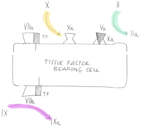
The process of blood coagulation starts by the exposure of TF-expressing cells to flowing blood (Fig.1.1). TF is expressed constitutively on cells such as smooth muscle cells and fibroblasts but not on resting endothelium; it is also expressed in several other districts and constitutes an hemostatic envelope normally not in contact with blood [9]. Disruption of the endothelium or activation of endothelial cells or monocytes results in the exposure of TF on blood flow [10]. Stronger evidence suggests that TF circulates in blood exposed on the surface of MPs; that derive from various cell types : white blood cells, endothelial cells, and platelets, and might play important roles in development of pathological hemostasis (thrombosis) opposing to normal clotting [11].
Figure 1.1: Factor VIIa bound to TF activates both factor X and factor IX. Factor Xa formed by factor VIIa/TF binds to factor Va on that cell and converts a small amount of prothrombin to thrombin
Exposed TF interacts with Factor VII/activated Factor VII (FVII/FVIIa) and the new-formed complex TF/FVIIa activates small amounts of FX and Factor IX (FIX). Activated FX (FXa) associates with its cofactor, activated Factor V (FVa), and forms the so called “prothrombinase-complex”on the surface of the TF-bearing cells [12], and leads the activation of small quantity of circulating Prothrombin (II) into Thrombin (IIa).
The adhesion process partially activates the platelets and promotes se-cretion of partially activated FV from their α-granules [13].
Zymogen FV can also be activated by FXa [14] or by noncoagulant pro-teases [15].
Low level activity of the TF pathway probably occurs at all times in the extravascular space. The coagulation proteins leave the vasculature, perco-late through the tissues, and are found in the lymph roughly in proportion to their molecular size [16]. Thus, FVII is probably bound to extravascular TF
even in the absence of an injury[12], and the extravascular FX and FIX can be activated as they pass through the tissues. This idea is consistent with the finding that low levels of the activation peptides from coagulation factors are present in the blood of normal individuals [17]. This process does not lead to clot formation under normal circumstances, because the really large com-ponents of the coagulation process, platelets and Factor VIII (FVIII)/vWF complex, are kept sequestered in the vascular space. Coagulation only pro-ceeds when damage to the vasculature allows platelets and Factor VIII/vWF exposure into the extravascular tissues.
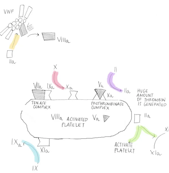
1.2.2 Amplification phase
The small amount of thrombin generated on the TF-bearing cell in the ini-tiation phase, has several important functions; one of that is activation of platelets. Although platelets have already adhered at the site of injury and become partially activated, the addition of thrombin can induce a higher level of procoagulant activity than adhesive interactions alone [18]. As a result platelets release partially activated forms of FV onto their surfaces.
Another function of thrombin formed during the initiation phase is the activation of the cofactors FV and FVIII on the activated platelet surface. In this process, the FVIII/vWF complex is dissociated, permitting vWF to mediate additional platelet adhesion and aggregation at the site of injury (Fig. 1.2).
Thrombin also activates Factor XI (FXI), activated by the prekallekrein/ki-ninogen/Factor XII cascade in the classic “intrinsic parhway”, which acts as a “booster”of thrombin generation on the platelet surface [19, 20]. This finding also strengthen the hypothesis that the intrinsic mechanism gives no contribute to in vivo coagulation process.
By the end of the amplification phase, the “stage”is set for large-scale throm-bin generation in the propagation phase.
1.2.3 Propagation phase
The propagation phase of clot formation occurs on the surface of activated platelets. First, activated-Factor IX (FIXa) generated during the initiation phase can now bind to its cofactor, activated-Factor VIII (FVIIIa), on the platelet surface, assembling in the so called “tenase-complex”.
Second, additional FIXa can be supplied by platelet-bound activated-FactorXI (XIa).
Third, because FXa cannot move effectively from the TF-bearing cell to the activated platelet, FXa must be provided directly on the platelet surface by
the FIXa/FVIIIa complex.
Fourth, the FXa rapidly associates with FVa bound to the platelet during the amplification phase, producing a burst of thrombin generation of suf-ficient magnitude to clot fibrinogen [9, 21, 22]. Hence more than 95% of the total amount of thrombin production takes place after initial clot
forma-Figure 1.2: On the surface of an activated platelet, factor IXa formed on the TF-bearing cell can incorporate into a Xase complex. Addition factor IXa is formed by plateletbound factor XIa. Factor Xa formed on the platelet surface is channeled into IIase complexes, leading to a burst of thrombin generation. Because factor XI is activated on the platelet surface by thrombin, PK, HK, and factor XII are not required for thrombin generation in this model.
tion, during propagation phase [23]. This excess of thrombin has been pro-posed to play an important role in stabilizing the clot by: activating Factor XIII (FXIII), the fibrin stabilizing factor [24]; cleaving the platelet protease-activated receptor-4 (PAR-4), that contribute to full activation of human platelets [25]; activating thrombin activatable fibrinolysis inhibitor (TAFI) [26]. TAFI is a carboxypeptidase that removes terminal lysine residues from fibrin, thereby removing potential binding sites for fibrinolytic enzymes and enhancing clot resistance to fibrinolysis [27]. Greater levels of thrombin ac-tivity are needed to activate TAFI than to form a fibrin clot. Failure in TAFI activation is thought to contribute significantly to the bleeding tendency in hemophilia [28].
1.2.4 Termination phase
Once a fibrin platelet clot is formed over a damaged area, the clotting process must be limited to avoid thrombotic occlusion in other normal areas of the vasculature [12].
The TF/FVIIa activity is inhibited by the Kunitz type inhibitor, Tissue Fac-tor Pathway InhibiFac-tor (TFPI) [29–31], secreted by endothelium. TFPI binds to FXa forming a quaternary complex with TF/FVIIa that quickly limits coagulation [35].
The serine protease inhibitor Antithrombin (AT)[32] neutralize enzymes of the coagulation system; its physiological role is to protect the circulation from free enzymes and limit the coagulation process to sites of vascular in-jury. Circulating AT is a relatively inefficient serpin, but its activity is stim-ulated by heparin and presumably by heparin-like molecules such as sulfated glycosaminoglycans that are synthesized and expressed by endothelial cells [34]. The increasing efficiency of AT by heparin is the molecular basis for the use of heparin as a therapeutic anticoagulant. Antithrombin is the major thrombin-inactivating protein [33].
TFPI and AT inhibit coagulation acting directly against proteins and macro-molecular complexes, instead another mechanism act in an indirect way switch off coagulation.
The protein C (PC) anticoagulant system inhibits the procoagulant func-tions of FVIIIa and FVa, the cofactors in the tenase and prothrombinase complexes respectively [36, 37].
The key component in the system is protein C, a vitamin K-dependent zy-mogen (proenzyme), activated by thrombin bound to the membrane protein thrombomodulin (TM) on the surface of intact endothelial cells. Activated PC (APC) cleaves a few peptide bonds in each of the phospholipid membrane bound cofactors FVa and FVIIIa. APC can also cleave the intact form of
FV, converting FV to an anticoagulant cofactor of APC in the inactivation of FVIIIa in the tenase complex [9].
APC activity is enhanced by another vitamin Kdependent inhibitory cofac-tor, protein S (PS). In human plasma, about 30% of protein S circulates as free protein; the remaining is bound to the complement regulatory protein C4b-binding protein. Only the free form of PS functions as a cofactor to APC.
1.3 Clotting factors
Procoagulant and anticoagulant proteins are composed of multiple structural domains. Among the different proteins, these domains have both a high de-gree of structural and functional homology.
1.3.1 Signal peptide
Both procoagulant and anticoagulant proteins found in plasma are initially synthesised with a signal peptide. This usually highly hydrophobic short peptide, needed for translocation of the growing polypeptide chain into the endoplasmatic reticulum, is cleaved off prior to secretion.
1.3.2 Propeptide/γ-Carboxyglutamic Acid-Rich Domain
All vitamin K-dependent proteins, prothrombin, FVII, FIX, FX, PC PS, contain a γ-Carboxylation recognition site located on the propeptide domain between the signal peptide and the γ-Carboxyglutamic Acid-Rich domain (Gla domain). This region directs, in a Vitamin-K dependent mechanism, γ-Carboxylation of the γ-Carboxyglutamic acid residues located in the adja-cent, approximately 40 AA residues long Gla domain. Gla domain is crucial for the Ca2+ mediated binding of these proteins with negatively chargedmembranes. After carboxylation the propeptide is cleaved off.
1.3.3 Epidermal Growth Factor Domain
Several procoagulant and anticoagulant proteins contain two or more epi-dermal growth factor (EGF)-like domains. These domains are about 43 to 50 amino acid residues in length and their structure is determined by three typical disulfide-bonds. The function of EGF-like domains on many clotting proteins, although not fully understood, appears to be involved in the for-mation of protein complexes. The EGF-like domains in FVII are important for the binding to TF. The second EGF-like domain of Factor IX contains a binding site for activated FVIII. The second EGF-like domain of protein C has been shown to be involved in the binding of protein S. The TM’s binding sites for thrombin and PC are located on the fifth and the fourth EGF-like domains, respectively.
1.3.4 Kringle Domain
Kringle domains are about 100 amino acid residues in length and charac-terised by a pattern of three disulfide bonds. These domains are likely to be involved in protein complex formation. Only two procoagulant proteins, prothrombin and FXII contain kringle domains. The second kringle of pro-thrombin probably contains the main binding site for activated factor V.
1.3.5 Catalytic Domain: Serine Protease Module
The catalytic domain of all procoagulant enzymes have an active site and an internal core that is nearly identical to that of trypsin. Conversion of an inactive proenzyme to an active enzyme depends on limited proteolysis and for some proteins the cleavage of so-called activation peptides.The enzyme activity of all clotting proteases depends on a serine, an aspartic acid and a histidine within the active site, which is characteristic for all serine proteases. The molecular surfaces surrounding the active site are responsible to specifically define the enzyme substrate [38].
1.4 Clotting factors levels
Within the vast majority of the healthy population the range of pro and anticoagulant components varies significantly. Counting the only thrombin generation, the responsiveness for a given TF initiating dose in the healthy population is quite extraordinary different [39].
The basis for the heterogeneity of the procoagulant response in human pop-ulation is probably a consequence of both genetic and environmental factors, which influence the status of the proteomic phenotype when evaluated in either a healthy or pathological state [40].
Recently has been estimate that there are 24500 human protein coding genes [45]; considering all molecular mechanisms that fold a protein from a primary mRNA transcript to the final physiologic function, e.g. alternative splicing and post translational modification, we might estimate a huger amount of pri-mary transcript protein products. The initial peptide product and most of its biosynthetic modifications are dictated primarily by genetics, with each mod-ification system under the direction of multiple genes. A variety of instruc-tions are included in the genetic package for the primary product which tar-get the protein through multiple, chemically driven assembly and transport processes which also incorporate additional post-translational modifications. The completed, modified, intracellular product must be further processed proteolytically at multiple sites prior to export. In addition, extracellular modifications, by both biologically directed processes and environmental ac-cidents, may further alter the circulating product. As a consequence of all these modification events, the number of members of the phenotypic pro-teome will probably extend well over the million(s) range [40].
In addition other elements could influence the variation of circulating clotting proteins; it has been reported that even with equivalent genetic deficiencies, there is a wide range of expression of risk events, either thrombotic or hemor-rhagic, suggesting that multiple genetic, acquired and environmental factors influence the possibility of an individual to undergo a clinically significant hemorrhagic or thrombotic event.
Concomitant presence of genetic defects in an individual, so called “gene-gene interaction”, also influence the normal or predicted levels of clotting factor.
It is well established that plasma concentrations of several clotting factors, i.e. fibrinogen, FVII, FVIII, vWF, FIX, FXII, high molecular-weight kinino-gen, and prekallikrein, increase with progressing age in healthy humans [46]. Moreover, in elderly people there are seasonal variation in fibrinogen concen-tration and the highest plasma levels have been observed during the coldest months [47, 48]. However, high plasma levels of the coagulation activation
markers in older populations do not necessarily reflect a high risk of arterial or venous thrombosis [49].
Disease states, drug consumption, and also human behavior or attitude i.e. smoking, diet, sedentariness, could in such aspect influence clotting factor levels, conditioning the fragile hemostatic balance.
Hence, only a few information dealing with the meaning of the heterogeneity of the plasma vitamin K-dependent proteins or the influence of this hetero-geneity on their biological performance with respect to activation, catalytic function or turnover is available.
1.4.1 “Normal”proteins levels
Various coagulation proteins circulate in blood at very different concentra-tions related to their specific roles in the blood coagulation system [56–59].
Table 1.1 Coagulation Factor Levels
Factor Plasma concentration µg/mL
Fibrinogen 3000 Prothrombin 9 Factor V 10 Factor VII 0.5 Factor VIII 0.1 Factor IX 5 Factor X 8 Factor XI 5 Factor XII 30 Factor XIII 10 Protein C 4 Protein S (free) 10 Prekallikrein 50
High molecular weight kininogen 70
AntithrombinIII 290
Fibrinogen is the predominant clotting factor having a concentration that is approximately 30 000-fold higher than that of FVIII. The high level of fib-rinogen is required for the formation of the fibrin clot, whereas the low con-centration of FVIII is more than sufficient to support FIXa in the activation of FX. Taking into account other vitamin K-dependent proteins, FVII is the least abundant, FIX and FX have intermediate levels and prothrombin cir-culating at the highest concentration. Several studies conducted in knockout
mice may help in understanding relative importance of various coagulation factors in vivo [60].
The embryonic lethal phenotype, associated with TF deficiency, demon-strates the crucial importance of the TF pathway [61–63], also confirmed by the absence of natural TF deficiency in humans.
FVII deficient mice develop normally in utero but die shortly after birth due to severe bleeding [64]. The difference in severity between TF and FVII knockout animals suggests a role for TF during embryogenesis in fibrin for-mation.
Prothrombin and FV deficiency are associated with partial embryonic lethal-ity and fatal haemorrhage [65–67]. In contrast, FIX and FVIII-deficient mice develop normally in utero but acquire haemophilia-like disease after birth [68, 69].
Fibrinogen-deficient mice have normal fetal development and suffer a moderate-to-severe bleeding phenotype similar to human fibrinogen deficiency [70]. This shows that thrombin generation is more important than fibrin deposi-tion.
Mice deficient in TAFI have no pathologic phenotype, demonstrating that TAFI deficiency is fully compatible with life [71].
Hemostasis is based on anticoagulant protein too. No TFPI deficiency are described in humans, which may indicate that lack of TFPI is not compat-ible with life; TFPI knockout mice have a lethal phenotype suffering from uncontrolled activation of coagulation with consumption of coagulation fac-tors [72].
Homozygous AT knockout mice have a lethal phenotype, demonstrating the importance of the protein for control of coagulation [73].
The protein C system is physiologically very important, which is clearly demonstrated by the severe thromboembolic disease associated with homozy-gous deficiency of protein C in both man and mice [74]. Mice lacking the protein C or TM genes are affected by a lethal phenotype, actually TM deficiency is particularly severe affecting embryogenesis even before the de-velopment of a functional cardiovascular system [75, 76].
Within healthy population, there is a wide range of hemostatic pheno-type; as a result it is difficult to define the “edge”of normal/abnormal, or pathologic, clotting factors levels.
Since thrombin plays a central role in formation of the primary hemostatic plug, the total amount of thrombin formed and the rate of its generation could provide a good reflection of the potential coagulation activity; but an effective clot cannot be formed without adequate levels of procoagulant fac-tors.
(generally 50% to 150% of the level in normal pooled plasma), as evaluated in several papers [39–44]. This suggests that a wide range of factor levels is compatible with normal hemostatic function. However, even within the normal range, variations can affect the thrombin generation [41].
The effect of coagulation factors levels on thrombin generation’s pattern is complex. For most of the coagulation factors, changing the level between 50% and 150% has little effect on the rate and pattern of thrombin genera-tion [41, 50].
Decreasing level of factors VIII, IX, or XI to < of 50% results in a mod-est decline in thrombin generation, with a dramatic decline only after levels fall below 10% to 20% of normal. Thus, the wide range of levels found in the normal population probably has little effect on thrombin generation and, therefore, little effect on hemostatic function. The pattern for FX is little different than the other factors. Again, changes between 50% and 150% have essentially no effect on thrombin generation. However, thrombin generation is maintained down to FX levels as low as 1% to 5% in in vitro experiments before falling off sharply [50, 51]. Thus, variation in FX levels probably con-tributes little to the pattern of thrombin generation unless combined with a deficiency of other factors.
The relationship between prothrombin levels and thrombin generation is dif-ferent from the other coagulation factors, actually total amount of thrombin produced are proportional to the prothrombin level [41]. The rate of throm-bin generation activity achieved during clot formation significantly affect the structure and stability of the resulting fibrin clot [52]. This means that any variation in prothrombin level is reflected in the pattern of thrombin genera-tion and could have an effect on the hemostatic effectiveness of the resulting clot.
The relationship between prothrombin level and thrombin generation is achiev-able also for supernormal prothrombin concentrations: elevated plasma pro-thrombin results in an increase in the rate and amount of pro-thrombin gen-eration. This may be the reason that elevated levels of prothrombin are correlated with a risk of arterial and venous thrombosis [53].
FVIII and FXI levels above the normal range result in a modest increase in the rate of thrombin generation, and elevated levels of these factors have been reported to be associated with the risk of thrombosis [54, 55].
1.4.2 Inherited and acquired coagulation disorder
As already mentioned, the hemostatic Vitamin K-dependent factors of coagu-lation are encoded by genes with virtually identical exon/intron distributions [77], suggesting that they have evolved relatively recently from a commonan-cestor by a process of gene duplication and divergence [78]; in this context it is believable that both the genes and the proteins have common regulatory mechanisms.
It has been provided the direct quantification of the genetic and environ-mental correlations between the plasma levels of a group of closely related vitamin K-dependent hemostasis factors, i.e. prothrombin, factor VII, IX, X, PC and functional PS. This investigation indicates how, actually, a set of common genes affects for a large proportion the phenotypic variation in vita-min K-dependent proteins, and similarly how a set of common unmeasured environmental factors also appears to influence plasma levels of vitamin K-dependent proteins [79].
A major contributor to our understanding of the complex hemostatic process, however, has occurred because of individuals with hereditary deficiencies of molecular elements fundamental to the hemostatic pathway.
Bleeding disorders
Genetic defects causing bleeding disorders can affect different proteins in the coagulation process.
The most common forms of haemophilia are due to different types of ge-netic defects (deletions, insertions, point mutation) resulting in inherited deficiency of FVIII (haemophilia A) or FIX (haemophilia B). Other coagula-tion factor deficiencies are very rare [82, 83]. Haemophilias occur in severe, moderate and mild forms, corresponding plasma levels being <1% of the normal plasma concentration, 1-5% and 5-30% respectively [83]. In the most common human hemophilias, the linkages between the degree of pathology and the molecular severity of the mutations are also not always correlated [86]. These observations imply that the genetic/environmental background in which a gene deficiency is observed may produce very different phenotypic responses.
von Willebrands disease (vWD) is a relatively common bleeding disorder af-fecting both males and females [85]. vWF is important for platelet adhesion and for maintaining the normal level of FVIII [87, 88], thus patients with vWD could have a primary haemostasis defect, caused by deficient adhesion of platelets to exposed subendothelial collagen, or, associated to severe forms of vWD, a secondary deficiency of FVIII, as the vWF is a carrier of FVIII in blood.
Faulty platelet function or thrombocytopenia can cause bleeding defects but clinically important bleeding only occurs when the abnormalities are severe [80, 81]. The symptoms associated with the inherited platelet disorders rarely are severe haemorrhage. Inherited deficiency of GPIb-V-IX (Bernard-Soulier
syndrome) is characterized by thrombocytopenia, giant platelets and lack of vWF binding and affected individuals have bleeding tendency. Acquired platelet function defects can originate by different medication (e.g. aspirin) and by different chronic diseases such as renal failure or autoimmune disease. Combined deficiency of the vitamin K-dependent coagulation proteins is an-other very rare inherited cause of bleeding. This may be caused by ge-netic defects in one of the two known enzymes that are involved in the γ-carboxylation of the vitamin K-dependent coagulation proteins: the γ-carboxylase and the vitamin K epoxide reductase [89].
Acquired bleeding problems can also be related to deficiency of vitamin K, which is required in the biosynthesis of many of the coagulation pro-teins. Malabsorption of the lipidsoluble vitamin K results in deficient γ-carboxylation of the vitamin K-dependent coagulation proteins, which in severe cases may result in increased bleeding. Even more common is vitamin K deficiency due to excessive intake of vitamin K antagonists, e.g. warfarin, used as anticoagulant therapy.
Bleeding disease can be due to the development of autoantibodies against a coagulation factor, the most common is directed against FVIII [90–93]. These conditions mainly affect elderly people. The molecular mechanisms respon-sible for the development of the autoantibodies are poorly understood. In disseminated intravascular coagulation (DIC), acquired bleeding is due to the consumption of platelets and coagulation factors due to widespread pathological proteolysis [93, 95]. In this condition, multiple proteolytic en-zyme systems including the coagulation and fibrinolytic systems are activated causing microvascular thrombosis and major disturbances of the capillary circulation. DIC may complicate malignancy, traumatic injury, surgery or pregnancies, and is often caused by severe infections with septicaemia. Thrombotic disorders
Epidemiological studies illustrate that significant thrombotic risk is asso-ciated with the inheritance of common mutations (polymorphisms) which influence functions or concentrations of coagulation proteins and cells [96]. But determining the global risk for thrombotic events (venous and arterial) is difficult because thrombosis is a multicausal disorder. The pathogenesis of thrombosis involves inherited and acquired risk factors. Most acquired risk factors are of short duration, like pregnancy, surgery, and immobilization; other are environmental factors, such as obesity [97, 98], oral contraception [99, 100], hormone replacement therapy [100], age [101], alcohol use [102] and potentially smoking [103]; in addition there are lifelong inherited fac-tors. Thus, a thrombotic episode may appear to be induced by an acquired
risk factor when in fact the disease is due to a combination of genetic and acquired risk factors. The natural balance between pro- and anticoagulant forces is affected by most of the inherited risk factors for thrombosis.
The majority of the genetic risk factors impair the function of the protein C anticoagulant system. The most common, found in 20-40% of patients with thrombosis, is a single point mutation in the FV gene, which keeps full pro-coagulant capacity but causes the APC resistance phenotype (FV Leiden). This mutation impairs degradation of mutant FVa by APC, causing the loss of one APC cleavage sites in FVa. Furthermore impairs the degradation of FVIIIa, because mutant FV shows poor anticoagulant-cofactor activity for APC in the degradation of FVIIIa.
A single point mutation (G20210A) in the 3’ untranslated region of the pro-thrombin gene is the second most common genetic risk factor for thrombosis (three- to five-fold increased risk) found in 68% of patients with thrombosis and in around 2% of healthy controls [104–106]. The prothrombin func-tion is unaffected by the mutafunc-tion but the levels of prothrombin in plasma are slightly increased as a result of the mutation, which may be the basis for the increased risk. Also other impairment in providing normal levels of FVIII [107], FIX [108], FXI [109] and fibrinogen [110] have been reported to be independent risk factors of venous thromboembolism (VTE). Mech-anisms through which these modifications could influence thrombotic risk, however, are not completely understood. Most individuals with a single ge-netic risk factor, although they have a lifelong increased risk, will not suffer from thrombosis during their lives because the associated risk is relatively low. People affected by more than one risk factor, either genetic or acquired are at higher risk and it is now considered that venous thromboembolism is a typical multigenetic/multifactorial disease.
The antiphospholipid syndrome (lupus anticoagulant) is an acquired risk fac-tor for both arterial and venous thrombosis [111–113]. Pregnant women with the antiphospholipid syndrome have an increased risk of spontaneous abor-tions.
1.5 Level’s analysis
It is a well known problem in clinical hematology that traditional coagulation tests, such as the prothrombin time (PT) and activated partial thromboplas-tin time (APTT), do not assess the whole coagulation system. These tests use clot formation as their endpoint, which occurs when only around 5% of all physiologically relevant thrombin is formed [114, 115] and also are insensi-tive to prothrombotic states. Coagulation factor assays can identify specific deficiencies but these do not always closely correlate with the clinical pheno-type. Hence neither the prothrombin time, nor the activated partial throm-boplastin time, nor the level of the individual clotting factors can accurately predict the presence and severity of a bleeding tendency. The observation that thrombin generation varies up to 40-fold when measurements are done with individual coagulation factors at the extremes of the normal ranges in a synthetic plasma system [39], also demonstrate the limitations of the tra-ditional tests.
Thrombin is the central enzyme in the coagulation cascade; estimation of an individuals potential to generate thrombin may correlate more closely with a hyper- or hypo-coagulable phenotype, compared to traditional coagulation tests. Measurement of an individuals capacity to generate thrombin, how-ever, captures the end result of the interaction between proteases and their inhibitors and is therefore potentially more useful as a reflection of a throm-botic (high thrombin generation) or haemorrhagic (low thrombin generation) phenotype compared to conventional coagulation tests [116].
Controlled TF concentration used to trigger the assay may evidence differ-ent role of procoagulant proteins among coagulation cascade. The positive feedback activation of thrombin by the intrinsic pathway can only be demon-strated at low (<1 pmol/L) TF concentrations [117]. Furthermore the assay is sensitive to FVIII and FIX at TF concentrations up to 5 pmol/L [118]. Small differences in TF concentration can therefore cause significant dif-ferences in the thrombin generating potential of plasma. Low TF trigger concentration may exacerbate differences in thrombograms, the thrombin generation curve, of patients with the same pathology, e.g. hæmophilia; in-stead higher quantity could masks this result due to the decreased sensitivity of the system [119]. Conversely, thrombin generation measurements at low TF concentrations in hæmophilia are difficult because of the low signal ob-tained and it may not always be possible to obtain full curves [119], limiting the information gained from the measurements. Low TF concentrations are also needed to demonstrate the protein C independent effect of protein S on anticoagulant process [120].
on thrombin generation. FIXa was found to be sensitive to differences in thrombin generation at very low FVIII concentrations [121]. Even if throm-bin generation assay in common laboratory usage is not sensitive to the actions of the protein C pathway, in vitro sensitivity can be increased by adding truncated human recombinant thrombomodulin (sTM) [122], APC [123], or the snake venom Protac [124, 125]. The use of these agents makes the assay more sensitive to deficiencies in protein C (not detected if APC is used), protein S, FV R506Q (factor V Leiden) and conditions associated with acquired protein C resistance.
Thus thrombin generation measurements can be performed in several ways that have different sensitivities for various haemostatic or thrombotic defects. As small variations in pre-analytical variables can cause significant changes in the sensitivity for these defects, results cannot be interpreted without detailed information on how the assay was performed. Since throm-bin is the central enzyme in the coagulation cascade, evaluating hypothetical thrombin generation based upon the individuals blood composition may cor-relate more closely with a hyper- or hypo-coagulable phenotype, compared to traditional coagulation tests. Finally, even when a single well standardized test is available it is unlikely that it will be suitable in all situations and separate modified tests designed for specific clinical situations may need to be developed.
Bibliography
[1] Furie B, et al. Cell, 1988, 53: 505-518
[2] Pearson JD. Baillieres Best Practice & Research: Clinical Haematology, 1999, 12: 329-341.
[3] Bevers EM, et al. European Journal of Biochemistry, 1982, 122: 429-436. [4] Bevers EM, et al. Biochimica et Biophysica Acta, 1983, 736: 57-66.
[5] Riddel J P, et al. Journal of Pediatric Oncology Nursing, 2007, 24(3): 123-131. [6] Luchtman-Jones L, et al. Annals of Medicine, 1995, 27(1): 47-52.
[7] Kleinschnitz C, et al. Journal of Experimental Medicine, 2006, 203(3): 513-518. [8] Hoffman M, et al. Thrombosis and Haemostasis, 2001, 85: 958-965.
[9] Dahlback B. Journal of Internal Medicine, 2005, 257: 209-223.
[10] O’Shaughnessy D, et al. Pratical hemostasis and thrombosis. Malden, MA: Blackwell Publishing ltd, 2005.
[11] Osterud B, et al. Seminars in Thrombosis and Hemostasis, 2006, 32: 11-23. [12] Hoffman M. Journal of Thrombosis and Thrombolysis, 2004, 16(1/2): 17-20. [13] Briede JJ, et al. Thrombosis and Haemostasis, 2001, 85: 509-513.
[14] Monkovic D, et al. Biochemistry, 1990, 29: 1118-1128.
[15] Allen DH, et al. Journal of Biological Chemistry, 1995, 270: 1408-1415. [16] Miller GJ, et al. Thrombosis and Haemostasis, 2000, 83: 427-432. [17] Bauer KA, et al. Blood, 1990, 76: 731-736.
[18] Alberio L, et al. European Journal of Clinical Investigation, 1999, 29: 1066-1076.
[19] Baglia FA, et al. Biochemistry, 1998, 37: 2271-2281. [20] Oliver JA, et al. Blood, 2002, 100: 539-546.
[21] Hoffman M. Journal of Thrombosis and Thrombolysis, 2004, 16(1/2): 17-20. [22] Loscalzo J. S. A. I. Thrombosis and hemorrhage (3rd ed.), 2003, Philadelphia:
Lippincott Williams & Wilkins.
[23] Mann KG, et al. Thrombosis and Haemostasis, 2003, 1: 1504-1514.
[24] Lorand L. Annals of the New York Academy of Sciences, 2001, 936: 291-311. [25] Ofosu FA. Transfusion and Apheresis Science, 2003, 28(3): 265-8.
[26] Bajzar L, et al. Journal of Biological Chemistry, 1995, 270: 14477-84. [27] Nasheim M. Current Opinion in Hemathology, 1998, 5: 309-313. [28] Mosnier LO, et al. Thrombosis and Haemostasis, 2001, 86: 1035-39. [29] Rapaport S.I. Blood, 1989, 73: 359.
[30] Broze GJ, et al. Biochemistry, 1990, 29: 7539.
[31] Broze GJ Jr. Journal of Thrombosis and Haemostasis, 1995, 74: 90.
[32] Van ’t Veer C, et al. Journal of Biological Chemistry, 1997, 272(7): 4367-77. [33] Beresford CH, et al. International Journal of Biochemistry, 1990, 22(2), 121-8. [34] Weitz JI. Journal of Clinical Investigation, 2003, 111: 952-4.
[35] Kumar V, et al. Robbins and Cotran: Pathologic basis of disease (7th ed.),
2005, Philadelphia: Elsevier.
[36] Dahlback B, et al. The molecular basis of blood disease, 2001, 3 ed., Philadel-phia: WB Saunders Company: 614-56.
[37] Esmon CT. Chest, 2003, 124(s3): 26-32.
[38] Stassen JM, et al. Current medicinal Chemistry, 2004, 11: 2245-2260. [39] Butenas S, et al. Blood, 1999, 94(7), 2169-2178.
[40] Mann GK, et al. Journal of Thrombosis and Haemostasis, 2004, 2: 1727-34. [41] Allen GA, et al. Journal of Thrombosis and Haemostasis, 2004, 2: 402-413. [42] Dielis AWJH, et al. Journal of Thrombosis and Hæmostasis, 2008, 6: 125-31.
[43] Hockin MF, et al. Journal of Biological Chemistry, 2002, 277: 18322-33. [44] Brummel-Ziedins KE, et al. Journal of Thrombosis and Hæmostasis, 2004, 2:
281-8.
[45] Venter JC, et al. Science, 200, 291(5507): 1304-51.
[46] Kannel WB, et al. Journal of the American Medical Association, 1987, 258: 1183-86.
[47] Frohlic M,et al. Atherosclerosis Thrombosis and Vascular Biology, 1997, 17: 2692-97
[48] Stout RW, et al. Age Ageing, 1996, 17: 3321-25.
[49] Mari D, et al. Experimental Gerontology, 2008, 43: 66-73.
[50] Al Dieri R, et al. Thrombosis and Haemostasis, 2002, 88: 576-582.
[51] Allen GA, et al. Blood Coagulation and Fibrinolysis, 2000, 11(s1): S3-S7. [52] Wolberg AS, et al. Blood, 2003, 101: 3008-3013.
[53] Poort S, et al. Blood, 1996, 88: 3698-3703.
[54] Meijers JC, et al. New England Journal of Medicine, 2000, 342: 696-701. [55] Maede TW, et al. Lancet, 1980, 1: 1050-1054.
[56] Mann KG, et al. Methods in Enzymology, 1993, 222: 1-10.
[57] Furie B, et al. New England Journal of Medicine, 1992, 326: 800-6. [58] Davie EW. Thrombosis and Haemostasis, 1995, 74: 1-6
[59] Stenflo J. et al. The molecular basis of blood disease, 2001, 3 ed., Philadelphia: WB Saunders Company, 579-613.
[60] Carmeliet P, et al. Cardiovascular Research, 1998, 39: 8-33. [61] Carmeliet P, et al. Nature, 1996, 383, 73-5.
[62] Toomey JR, et al. Blood, 1996, 88, 1583-7.
[63] Bugge TH, et al. Proceedings of the National Academy of Sciences of the USA, 1996, 93: 6258-63.
[65] Sun WY, et al. Proceedings of the National Academy of Sciences of the USA, 1998, 95: 7597-602.
[66] Xue J, et al. Proceedings of the National Academy of Sciences of the USA, 1998, 95: 7603-7.
[67] Cui J, et al. Nature, 1996, 384: 66-8.
[68] Bi L, et al. Nature Genetics, 1995, 10: 119-21. [69] Kundu RK, et al. Blood, 1998, 92: 168-74.
[70] Suth TT, et al. Genes and development, 1995, 9: 2020-33.
[71] Nagashima M, et al. Journal of Clinical Investigation, 2002, 109: 101-10. [72] Huang ZF, et al. Blood, 1997, 90: 944-51.
[73] Ishiguro K, et al. Journal of Clinical Investigation, 2000, 106: 873-8. [74] Jalbert LR, et al. Journal of Clinical Investigation, 1998, 102: 1481,8. [75] Rosenberg RD. Thrombosis and Haemostasis, 1997, 78:705-9.
[76] Weiler H, et al. Journal of Thrombosis and Haemostasis, 2003, 1: 1515-24. [77] Tuddenham EGD, et al. Oxford University Press, 1994.
[78] Patthy L. Seminars in Thrombosis and Haemostasis, 1990, 16: 245-59. [79] Souto JC, et al. Thrombosis and Haemostasis, 2001, 85: 88-92.
[80] George JN. Lancet, 2000, 355: 1531-9.
[81] Majerus PW. The molecular basis of blood disease, 2001, 3 ed., Philadelphia: WB Saunders Company: 764-90.
[82] Mannucci PM, et al. Blood, 2004, 104: 1243-52.
[83] Mannucci PM, et al. New England Journal of Medicine, 2001, 344: 1773-9. [84] Bolton-Maggs PH, et al. Lancet, 2003, 361(9371): 1801-9.
[85] Mannucci PM. New England Journal of Medicine, 2004, 351: 683-94. [86] Aledort L. Pthophysiology of Haemostasis and Thrombosis, 2003, 33: 2-3. [87] Sadler JE. Annual Review in Biochemistry, 1998, 67: 395-424.
[89] Zhang B, et al. Journal of Thrombosis and Haemostasis, 2004, 2: 1564-72. [90] Collins PW. Blood Coagulation and Fibrinolysis, 2003, 14(s1): s23-7. [91] Kraemer DM, et al. Journal of Internal Medicine, 2003, 254: 301-3. [92] Delgado J, et al. British journal of Haematology, 2003, 121: 21-35. [93] Hay CR. Baillieres Clinical Haematology, 1998, 11: 287-303. [94] Levi M. British Journal of Haematology, 2004, 124: 567-76. [95] Dempfle CE. Thrombosis and Haemostasis, 2004, 91: 213-24. [96] Rosendaal FR. Haemostasis, 1999, 29(s1): 1-9.
[97] Romano M, et al. Journal of Clinical Endocrinology & Metabolism, 2003, 88(11): 5321-5326.
[98] Rauramaa R, at al. Medicine & Science in Sports & Exercise, 1999, 31(S11): S631-4.
[99] Rosendaal FR, et al. Journal of Thrombosis and Hæmostasis, 2003, 1(7): 1371-80.
[100] Rosing J, et al. American Journal of Obstetric & Gynecology, 1999, 180(6 Pt 2): S375-82.
[101] Haapanen-Niemi N, et al. Preventive Medicine, 1999, 28(4): 343-348. [102] Catena C, et al. Journal of Hypertension, 2003, 21(2): 281-8.
[103] Thorneycroft IH, et al. Journal of Reproductive Medicine 2003, 48(S11): 911-920.
[104] Rosendaal FR. Lancet, 1999, 353: 1167-73.
[105] Nicolaes GA, et al. Hematology/oncology clinics of North America, 2003, 17: 37-61.
[106] Bertina RM. Clinical Chemistry, 1997, 43: 1678-83. [107] Koster T, et al. Lancet, 1995, 345: 152-5.
[108] Van Hylckama Vlieg A, et al. Blood, 2000, 95: 3678-82.
[109] Maijers JCM, et al. New England Journal of Medicine, 2000, 342: 696-701. [110] Koster T, et al. Thrombosis and Hæmostasis, 1994, 71: 719-22.
[111] Levine JS, et al. New England Journal of Medicine, 2002, 346: 752-63. [112] Arnout J. Thrombosis and Haemostasis, 2001, 86: 83-91.
[113] Triplett DA. Haemostasis, 1996, 4: 358-67.
[114] Hemker et al. Thrombosis and Hæmostasis, 1995, 74: 134-8. [115] Rand MD, et al. Blood, 1996, 88: 3432-45.
[116] van Veen JJ, et al. British Journal of Hæmatology, 2008, 142: 889-903. [117] Keularts IM, et al. Thrombosis and Hæmostasis, 2001, 85: 1060-5. [118] van Veen JJ, et al. Blood Coagulation and Fibrinolysis, 2008, 19: 183-9. [119] Lewis SJ, et al. British Journal of Hæmatology, 138: 775-82.
[120] Sere KM, et al. Blood, 104: 3624-30.
[121] McIntosh JH, et al. Journal of Thrombosis and Hæmostasis, 2003, 1: 1005-11.
[122] Van Hylckama Vlieg A, et al. British Journal of Hæmatology, 2007, 138: 769-74.
[123] Regnault V, et al. Pathophysiology of Hæmostasis and Thrombosis, 2003, 33: 23-9.
[124] Gatt A, et al. Thrombosis and Hæmostasis, 2003, 98: 691-2. [125] Hezard N, et al. Thrombosis and Hæmostasis, 2007, 97: 165-6.
Chapter 2
Combined effect of hemostatic gene
polymorphisms and the risk of myocardial
infarction in patients with advanced coronary
atherosclerosis.
Martinelli N, Trabetti E, Pinotti M, Olivieri O, Sandri M, Friso S, Pizzolo F, Bozzini C, Caruso P, Cavallari U, Cheng S, Pignatti PF, Bernardi F, Cor-rocher R, Girelli D.
2.1 Abstract
Relative little attention has been devoted until now to the combined effects of gene polymorphisms of the hemostatic pathway as risk factors for Myocar-dial Infarction (MI), the main thrombotic complication of Coronary Artery Disease (CAD). The aim of this study was to evaluate the combined ef-fect of ten common prothrombotic polymorphisms as a determinant of MI. Methodology/Principal Findings We studied a total of 804 subjects, 489 of whom with angiographically proven severe CAD, with or without MI (n= 307; n= 182; respectively). An additive model considering ten common poly-morphisms [Prothrombin 20210G>A, PAI-1 4G/5G, Fibrinogen β-455G>A, FV Leiden and R2, FVII -402G>A and -323 del/ ins, Platelet ADP Receptor P2Y12 -744T>C, Platelet Glycoproteins Ia (873G>A), and IIIa (1565T>C)] was tested. The prevalence of MI increased linearly with an increasing num-ber of unfavorable alleles (χ2 for trend = 10.68; P= 0.001). In a multiple
logistic regression model, the number of unfavorable alleles remained signif-icantly associated with MI after adjustment for classical risk factors. As compared to subjects with 3-7 alleles, those with few (≤2) alleles had a de-creased MI risk (OR 0.34, 95%CIs 0.13-0.93), while those with more (≥8) alleles had an increased MI risk (OR 2.49, 95%CIs 1.03-6.01). The number of procoagulant alleles correlated directly (r = 0.49, P= 0.006) with endoge-nous thrombin potential. Conclusions The combination of prothrombotic polymorphisms may help to predict MI in patients with advanced CAD.
2.2 Intoduction
Myocardial Infarction (MI), the leading complication of coronary atheroscle-rotic disease (CAD), generally occurs in the late stages of disease because of coronary thrombosis superimposed on a ruptured/unstable plaque [1]. In clinical practice it is well-known that, in spite of the documented presence of advanced CAD, only a subset of patients develops acute MI during their life-course [2]. The reasons for individual differences in susceptibility to MI are poorly understood. In principle, subjects with an increased tendency to form blood clots (i.e. with hypercoagulability) may be at increased risk, as observed for venous thrombosis. Lessons from animal models suggest that excessive thrombin generation may be particularly harmful during the later stages of atherosclerosis, when thrombotic complications often occur [3,4]. However, this is difficult to assess in clinical practice, since we lack a unique and reliable laboratory marker of hypercoagulability [5]. Moreover, functional tests evaluating concentration and/or function of blood
coagu-lation proteins are often subjected to multiple transient interferences, e.g. due to the use of antithrombotic and anticoagulant agents or the presence of concomitant inflammation. Genetic polymorphisms with a documented functional effect on blood coagulation proteins may represent a useful tool, by reflecting the individuals lifelong exposure to even a mild prothrombotic state. During the last decade, extensive studies on various individual poly-morphisms as risk factors for CAD and MI have yielded largely inconclusive results [6-11]. These results reflect at least two critical issues: 1) the multifac-torial and multistep pathogenesis of CAD, involving many different biochem-ical pathways and intermediate phenotypes (e.g. hyperlipidemia, diabetes, hypertension), each in turn under the control of many different genes; 2) the enormous heterogeneity of investigations in terms of study design, typology of patients included, and clinical endpoints [10]. There has also been relatively little attention devoted to assess the combined effect of genes, which might be anticipated by analogy to the well-known additive effects of conventional risk factors. Generally, individual polymorphisms confer a marginal to moderate CAD risk that becomes evident only across many thousands of individuals, as was recently demonstrated by meta-analysis for Factor V 1691 G>A (Fac-tor V Leiden), prothrombin 20210 G>A, and PAI-1 -675 4G/5G [8]. This renders such polymorphisms unhelpful in assessing individuals risk clinically. On the other hand, the value of analyzing multiple alleles simultaneously for determining the risk is not well studied.
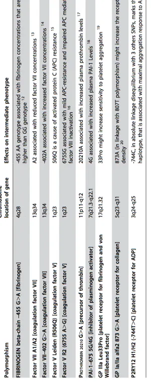
In this study we evaluated the combined effect of ten common genetic variants, with known modest effects on the hemostatic balance (listed in Table 2.1) [6,12-21], in modulating the risk of development of MI. Because of the relatively late occurrence of MI in the natural history of CAD, we focused on a selected population of high risk patients with angiographically documented, advanced CAD. A thrombin generation assay was also used in a subset of patients to explore the propensity to form blood clot as a function of the number of hemostatic polymorphisms.
2.3 Materials & Methods
2.3.1 Study population
This study was performed within the framework of the Verona Heart Project, a regional survey aimed to search for new risk factors for CAD and MI in subjects with objective angiographic documentation of their coronary ves-sels. Details about enrolment criteria have been described in detail elsewhere [35,36]. A total of 804 subjects, for whom complete analyses of 10
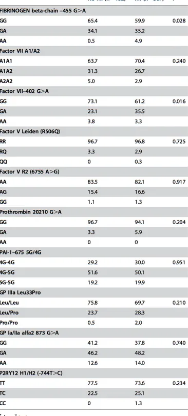
polymor-Table 2.1: Description of the haemostatic gene polymorphisms, analysed in this study, and their associated intermediate phenotype.
phisms of genes involved in hemostatic pathways were available, were in-cluded in the present study. Three-hundred fifteen subjects had completely normal coronary arteries, being submitted to coronary angiography for rea-sons other than CAD, mainly valvular heart disease (CAD-free group). These controls were also required to have neither history nor clinical or instrumen-tal evidence of atherosclerosis in vascular districts beyond the coronary bed. Fourhundred eighty nine subjects had angiographically proven CAD (the ma-jority of them being candidates for coronary artery bypass grafting) with ob-jective documentation of presence/absence of MI. The disease severity was determined by counting the number of major epicardial coronary arteries (left anterior descending, circumflex, and right) affected with ≥1 significant stenosis (≥50%). According to the hypothesis to be tested, subjects with non-advanced CAD (i.e. coronary stenosis <50%) were not included in the study. Cardiologists unaware that the patients were to be included in the study assessed the angiograms. Patients were classified into MI (n= 307) and non-MI (n= 182) subgroups on the basis of a thorough review of medi-cal records including history, electrocardiogram, enzyme changes, and/or the typical sequelae of MI on ventricular angiography.
All participants came from the same geographical area (Northern Italy), with a similar socio-economic background. At enrolment, a complete clinical history was collected, including the assessment of cardiovascular risk factors such as obesity, smoking, hypertension and diabetes. The study was approved by the Ethic Committee of our Institution (Azienda Ospedaliera, Verona). A written informed consent was obtained from all the participants after a full explanation of the study.
2.3.2 Biochemical analysis
Samples of venous blood were drawn from each subject at enrolment, before coronary angiography and after an overnight fast. Serum lipids, as well as other CAD risk factors, including high-sensitivity C-reactive protein (hs-CRP) were determined as previously described [26].
2.3.3 Genetic analysis and nomenclature
Genomic DNA was extracted from whole blood samples by a phenol-chloroform procedure using the Puregene kit (Gentra Systems) according to the manu-facturers protocol. The 10 genetic polymorphisms, selected on the basis of prior evidence of potential functionality in modulating the hemostatic path-way, are listed in Table 2.1. Seven out of ten polymorphisms (fibrinogen beta-chain - 455G>A, Factor VII A1/A2, Factor V Leiden, Prothrombin
20210 G>A, PAI-1 -675 5G/4G, GP IIIa Leu33Pro, GP Ia/IIa alfa2 873 G>A) were examined by a previously described and validated linear-array assays for candidate markers [37]. The accuracy of the linear-array genotyp-ing system as compared with standard genotypgenotyp-ing approaches reported else-where [35,36] was evaluated and the findings provide reassurance regarding the validity of the system used, as previously described [38]. The remaining three (Factor VII - 402 G>A, Factor V R2, P2RY12 H1/H2) were analyzed by previously described, standard genotyping approaches [36,39,40]. Geno-type interpretation for each polymorphism was performed independently by two investigators and very few samples (<1%) with unclear result were re-genotyped.
2.3.4 Measurement of thrombin generation activity
This assay was performed in a subset of CAD patients on samples drawn at enrolment, in order to evaluate a possible functional counterpart of an increasing number of prothrombotic alleles in terms of propensity to form blood clot. Plasma sample were centrifuged at 23,000 g at 4◦C for 1 hourbefore testing. Calibrated automated thrombin activity measurement was conducted according to Hemker et al. [41,42] in a microtiter plate fluo-rometer (Fluoroskan Ascent, ThermoLabsystems, Helsinki, Finland) using the Thrombinoscope software (Synapse BV, Maastricht, The Netherlands). The assay was carried out at 37◦C essentially as previously reported [43].
Coagulation was triggered in platelet poor plasma by recalcification in the presence of 1 pM recombinant human tissue factor and 4 µM phospholipids. Thrombin generation was then evaluated overtime by exploiting a specific fluorogenic substrate (Z-Gly-Gly-Arg-AMC). Thrombin generation measure-ment was conducted in parallel in plasma samples after the addition of a thrombin calibrator provided by the manufacturer (Synapse BV). The soft-ware enables the estimate of the following parameters: a) the Lag Time of thrombin generation, b) the time to reach the maximum concentration of thrombin (time to Peak), c) the maximum concentration of thrombin (Peak), d) the total duration of thrombin generation activity (Start Tail), and e) the total amount of thrombin activity assessed as the area under the curve, i.e. the endogenous thrombin potential (ETP). All experiments were carried out in duplicate.
2.3.5 Statistics
Calculations were performed mainly with SPSS 13.0 statistical package (SPSS Inc., Chicago, IL). Distributions of continuous variables in groups were
ex-pressed as means±standard deviation. Logarithmic transformation was per-formed on skewed variables, for whom geometric mean with 95% confidence interval (CI) are given. Quantitative data were assessed using the Students t-test or by ANOVA with Tukeys post-hoc comparison of the means. Correla-tions between quantitative variables were assessed using Pearsons correlation test. Qualitative data were analyzed with the χ2-test or the Fisher exact-test
when indicated. Hardy-Weinberg equilibrium was tested for each genotype within each group by means of χ2-test. A value of P<0.05 was considered
statistically significant.
Within each group examined, the frequencies of the genotypes associated with each of the polymorphisms were compared by the χ2- test, with the
values predicted on the basis of the Hardy-Weinberg equilibrium. To assess the extent to which gene polymorphisms were associated with MI, odds ra-tios with 95% CIs were estimated by univariate logistic regression analysis. Adjustment for other variables (i.e. number of affected vessels, age, sex, smoking, BMI, LDL- and HDL-cholesterol) was performed by adding those covariates in a set of multiple logistic-regression models.
The existence of gene-gene interactions was first explored by a data min-ing technique similar to the Adaboost algorithm, and based on classifica-tion and regression trees (CART): the gradient boosting machine [44]. The statistical significance of the interactions found with this method was then estimated by the likelihood ratio test applied on two logistic models (with and without the interaction terms). After observing that no significant in-teraction was present, we focused on an additive model. On this basis, we attributed to each patient a prothrombotic score (PS), reflecting the sum of 10 concomitant unfavourable prothrombotic alleles, theoretically ranging from 0 (no prothrombotic allele present) to 20 (all the prothrombotic alleles present). The association between the prothrombotic score and MI was eval-uated by χ2 for linear trend analysis. The prothrombotic score was analysed
by logistic regression both as continuous variable and as categorised variable. Odds ratios with 95% CIs were estimated by univariate logistic regression analysis and then by multiple logistic regression with adjustment for num-ber of affected vessels, age, sex, smoking, BMI, LDL- and HDLcholesterol. The predictivity of our models was then evaluated by the receiver-operating-characteristics (ROC) curve, estimating the area under the curve (AUC).
2.4 Results
2.4.1 Haemostatic polymorphisms in the CAD group
as a whole versus CAD-free subjects
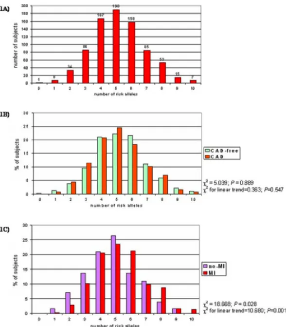
Supplemental Table 1 (Table S1) shows the genotype frequencies for each of the 10 polymorphisms in CAD-free (n=315; males 66.0%; mean age 59.26±11.9 years) and in CAD subjects (n=489; males 83.6%; mean age 60.36±9.3 years). All alleles were in Hardy- Weinberg equilibrium. For each polymor-phism there was no significant difference in genotype distribution between CAD and CAD-free groups. The distribution of the prothrombotic score (PS) in the whole study population (n=804) is shown in figure 2.1A. The score ranged from 0 (1 subject) to 10 prothrombotic alleles (7 subjects), with a median level of 5. Figure 2.1B shows the distribution of the PS in CAD-free and in CAD subjects. No association was found between the PS and CAD (P=0.889 by χ2-test).
2.4.2 Individual haemostatic polymorphisms and MI
risk in subjects with advanced CAD
Supplemental Table 2 (Table S2) shows the general characteristics of the CAD population divided in two groups on the basis of presence/absence of MI. As compared to CAD patients without MI, MI patients were sig-nificantly younger, more frequently males, had a higher degree of CAD in terms of number of diseased vessels, and lower HDL-cholesterol levels. No significant difference was found for the other variables. Table 2.2 shows the genotype frequencies of the 10 genetic variants in CAD patients with or with-out MI. Two polymorphisms, factor VII -402 G>A and fibrinogen β-chain -455 G>A, showed nominal association with MI at the univariate analysis. However, these associations were no longer significant after multiple logistic regression adjusted for sex, age, disease severity, smoking status, BMI, LDL-and HDLcholesterol (P =0.155 for factor VII -402 G/A LDL-and P= 0.998 for fibrinogen β-chain -455 G/A).
2.4.3 Combined effect of haemostatic polymorphisms
and MI risk
No significant interaction was found by CART among polymorphisms in de-termining MI risk (all P for interaction >0.05). As shown in figure 2.1C, the proportion of CAD patients with MI increased progressively with increasing
number of unfavourable alleles (χ2 for linear trend= 10.68; P= 0.001). In
a multiple logistic regression model the prothrombotic score remained sig-nificantly associated with MI after adjustment for sex, age, degree of CAD, smoke, BMI, LDL- and HDL-cholesterol (OR for 1-point increase in pro-thrombotic score= 1.22 with 95%CI 1.06-1.39, P =0.004). Using the median of PS as cut-off, CAD patients with >5 alleles had a significantly increased risk of MI as compared to subjects with ≤5 alleles (OR 2.02 with 95%CI 1.27-3.21, P= 0.003, by multiple logistic regression). Using approximately the 5th and the 95thpercentiles of PS distribution (i.e. 2 and 8, respectively),
the study population could be classified into in 3 subgroups: a low-risk group with less than 3 unfavourable alleles (n= 26), an intermediate-risk group with 3 to 7 unfavourable alleles (n= 417), and a high-risk group with more than 7 unfavourable alleles (n= 46). The prevalence of MI among these groups increased progressively (38.5% in low-risk; 62.6% in intermediate- risk; and 78.3% in high-risk; P= 0.001 by χ2 for linear trend), while they were similar
for the other clinical and laboratory variables (data not shown). Considering the intermediate-risk group as the reference group, carriers of <3 alleles had a lower risk of MI, while carriers of >7 alleles had an increased risk (Fig-ure 2.2). Comparing the two extreme groups, the subjects with >7 alleles had a remarkably higher MI risk (OR 7.28 with 95%CI 2.01-26.36, P= 0.002 adjusted by multiple logistic regression). The ROC curve for information provided by our polygenic approach for MI prediction in CAD patients is plotted in Supplemental Figure 1 (Figure S1). The AUC was 0.581 with a 95% CI from 0.530 to 0.632.
2.4.4 Combined effect of haemostatic polymorphisms
and thrombin generation activity
To get insights on the pathophysiological effect of combined hemostatic al-leles, we assessed the characteristics of thrombin generation activity curves as a function of the number of procoagulant alleles (i.e. fibrinogen β-chain -455 A, Prothrombin 20210 A, Factor V Leiden, Factor V R2, Factor VII A1, Factor VII -402 A and PAI-1 -675 4G). Since this assay pertains only to the coagulation pathway, the three platelet-related polymorphisms were not considered for this analysis. This assay was performed in a subset of 29 CAD patients (26 males and 3 females, 22 with and 7 without MI), selected among those without possible confounders (i.e. concomitant anticoagulant therapy or overt signs of inflammation, documented by hs-CRP,5 mg/l), in order to form three groups matched for age and sex representing the previously de-fined risk groups (low-risk: n= 9, 8 males and 1 female, mean age 53.7±8.5;
Figure 2.1: Study population (n = 804) stratified on the basis of number of risk alleles (1A). The distribution of number of risk alleles in CAD-free (n = 315) and in CAD patients (n = 489) (1B) and in CAD patients with (n = 307) or without a history of MI (n = 182) (1C).
intermediate-risk: n =10, 9 males and 1 female, mean age 57.8±7.4; high-risk: n =10, 9 males and 1 female, mean age 56.0±8.6). The number of procoagulant alleles was significantly associated with ETP and with Start Tail, but not with Lag Time, Peak or Time to Peak (Table 2.3). Similarly, subjects with a high number of procoagulant alleles (≥5) had significantly higher ETP values as compared to subjects with fewer alleles (Table 2.4). These two groups were similar not only for age and sex, but also for smok-ing, hypertension and diabetes (data not shown). Their median thrombin generation activity curves are showed in Figure 2.3.
2.5 Discussion
Evidence that a hypercoagulable state is associated with increased mortality has been provided by some recent studies [11,22]. To our knowledge, this is the first study that attempts to look at the impact of the combined effect of several common prothrombotic polymorphisms in the identification of CAD patients at different risk of developing MI. To put our results into perspective, we propose the following considerations.
2.5.1 Single haemostatic polymorphisms and MI risk
This study focused on relatively few genetic variants associated with de-fined biochemical alterations. While some of them (i.e. Factor V Leiden and prothrombin 20210 G>A) are established risk factors for venous throm-boembolism, their association with arterial thrombosis is much less convinc-ing [6,9]. Here too, despite some nominal significant P values, we found no consistent association when each polymorphism was considered individually. Indeed, CAD and MI are paradigms of complex disease, in which the effect of individual genes on the risk is anticipated to be weak [23,24]. Moreover, em-phasizing the principle that “the highest the allele effect, the lowest the allele frequency”[25], it is plausible that genetic variants such those investigated in the present study, relatively frequent in the general population, could have at best only a mild effect on a potentially lethal phenotype like MI. Indeed, until now only a recent large meta-analysis including tens of thousands of patients has been able to detect a moderate but significant increase in the risk of coronary disease associated with either the Factor V Leiden mutation or the prothrombin 20210A variant [8].Figure 2.2: OR for MI in groups stratified on the basis of number of unfavourable alleles. The intermediate group (from 3 to 7 unfavourable prothrombotic alleles), representing the 85.3% of the whole population, is considered as reference group.
Table 2.3: Correlations between number of procoagulant alleles and different character-istics of thrombin generation activity. The analysis was performed in a subgroup of CAD patients (n = 29) without anticoagulant therapies and without signs of overt inflammation. Procoagulant alleles were fibrinogen beta-chain -455 A, Prothrombin 20210 A, Factor V Leiden, Factor V R2, Factor VII A1, Factor VII -402 A, PAI-1 -675 4G.
Table 2.4: Characteristics of thrombin activity generation curves in groups stratified on the basis of number of procoagulant alleles, with a threshold level at 5 alleles. The analysis was performed in a subgroup of CAD patients (n = 29) without anticoagulant therapies and without signs of overt inflammation. Procoagulant alleles were fibrinogen beta-chain -455 A, Prothrombin 20210 A, Factor V Leiden, Factor V R2, Factor VII A1, Factor VII -402 A, PAI-1 -675 4G. †: by t-test.
Figure 2.3: Comparison between the median thrombin generation activity curves in groups stratified on the basis of number of procoagulant alleles, with a threshold level at 5 alleles.
2.5.2 Combined effect of haemostatic polymorphisms
and MI risk
Recently, a polygenic approach has been demonstrated to be a valid tool to identify subjects at risk for another complex trait such as type 2 diabetes [26]. A similar strategy was used in the present study, suggesting that in subjects with advanced CAD, an increasing number of prothrombotic alle-les may confer a significant risk of developing MI. It is biologically plausible that the simultaneous presence of several genetic variations with modest but defined effects on the hemostatic process could influence the risk of the ma-jor thrombotic complication in a given CAD patient. Under certain stimuli, such as plaque erosion or rupture, this condition may predispose to sustained thrombin generation leading to the acute thrombotic event [2]. Accordingly, our in vitro functional studies showed an association between the number of procoagulant alleles and thrombin generation. The latter is known to be a highly variable and complex phenomenon modulated by the interplay of sev-eral factors, none of them with predominant influence, many of them under genetic control [27]. It is noteworthy that our clinical model focused on a homogeneous group of patients with angiographically proven advanced CAD. Elegant studies in animal models, i.e. Factor V Leiden mice crossbred with apolipoprotein E-deficient mice, indicates that unregulated thrombin gener-ation is particularly harmful during the later stages of atherosclerosis. [3,4]. Conversely, a mild hypercoagulable state may be less meaningful in absence of underlying vulnerable atherosclerotic plaques. Our results may thus apply only to the specific clinical model of this study, and not to all CAD patients. While it is reasonable that genetically-induced excessive thrombin genera-tion may be clinically relevant in subjects with extensive coronary plaques, this excess might be less influential in the atherogenetic process, where other genetic factors (i.e. those involved in modulation of lipid metabolism, an-tioxidant balance, and so on) may be prominent. This could explain why we found no association between the hemostatic polymorphisms and the CAD phenotype.
2.5.3 Study limitations and strengths
One strength of our study is the clear definition of phenotypes, allowing comparison of patients with angiographically proven, advanced CAD, with or without MI. The CAD population had a substantial burden of traditional risk factors and thus represented a typical patient population seen in clinical practice.