UNIVERSITY OF CATANIA
DEPARTMENT OF BIOLOGICAL, GEOLOGICAL AND
ENVIRONMENTAL SCIENCES
Ph.D. PROGRAM COURSE IN GEOLOGICAL, BIOLOGICAL
AND ENVIRONMENTAL SCIENCES
XXIX CYCLE
LOREDANA LEGGIO
Functional analysis of two alternative transcripts from porin1
gene of Drosophila melanogaster and involvement of
corresponding 5'UTR sequences in the translation control
Analisi funzionale dei due trascritti alternativi del gene porin1 di
Drosophila melanogaster e coinvolgimento delle rispettive sequenze
5’UTR nella regolazione della traduzione
I
RIASSUNTO
Il VDAC (Voltage Dependent Anion-selective Channel) è un canale anionico dipendente dal voltaggio presente nella membrana mitocondriale esterna (OMM) di tutti gli organismi eucarioti. Tale canale permette il passaggio di nucleotidi, ioni e piccoli metaboliti da e verso il citosol grazie ad una struttura a barilotto β, con l’estremità N-terminale strutturata a formare un’α-elica rivolta verso lo spazio intermembrana e coinvolta nel processo di apertura e chiusura del poro. Esso assume la conformazione di massima apertura a voltaggi applicati di poco diversi da 0 mV; mentre a voltaggi superiori a 20 mV, sia in positivo che in negativo, il VDAC passa alla conformazione ―chiusa‖. L’importanza di tale poro è dettata dalla sua localizzazione cellulare, che gli permette di interagire con moltissimi enzimi e proteine direttamente o indirettamente partecipanti a numerosi pathways cellulari e, di conseguenza, ne giustifica il coinvolgimento in diverse patologie umane. Molte informazioni disponibili sul VDAC derivano da studi condotti sul ceppo Δpor1 del lievito Saccharomyces cerevisiae, un ceppo deleto del gene vdac (por1) endogeno. L’assenza del VDAC provoca nel lievito l’impossibilità di utilizzare i mitocondri per effettuare la respirazione cellulare. Questo difetto rende il ceppo Δpor1 incapace di crescere su fonti di carbonio non fermentabili, come il glicerolo, sia in condizioni normali e soprattutto alla temperatura restrittiva di 37°C. Tale difetto viene complementato in seguito a trasfezione con la sequenza codificante il VDAC umano che, una volta espresso, si inserisce in OMM permettendo così al lievito di recuperare il fenotipo indotto dalla delezione del VDAC endogeno. La complementazione del difetto di crescita del ceppo Δpor1 si ottiene anche con la sequenza codificante il VDAC (Porin1) di Drosophila melanogaster, sotto il controllo del promotore per il gene omologo di lievito. In D. melanogaster sono espresse due isoforme di VDAC, VDAC1 e VDAC2. Dm VDAC1 è dal punto di vista strutturale e funzionale equivalente all’isoforma 1 dei VDAC dei mammiferi o a POR1 del lievito. In seguito al completamento del progetto genoma della Drosophila è stata definita l’organizzazione genomica (numero di esoni, di introni, regioni UTR e siti di poliadenilazione) della famiglia dei geni VDAC: 4 geni (porin1, porin2, cg17139 e
gc17140) disposti a breve distanza l’uno dopo l’altro a formare un cluster sul
cromosoma 2L. Il gene porin1 codifica per la principale isoforma VDAC, sempre espressa in tutti i tessuti e stadi di sviluppo della mosca. Il gene porin1 è formato da 5 esoni, di cui gli esoni 1A e 1B, totalmente costituiti da sequenza 5’UTR, sono tra loro
II
alternativi. Infatti, in seguito a splicing alternativo vengono prodotti due trascritti contenti all’estremità 5’ l’esone 1A o l’esone 1B seguiti dalla medesima sequenza codificante per VDAC. I trascritti alternativi 1A-VDAC e 1B-VDAC vengono prodotti in tutti gli stadi di sviluppo dalla mosca e in tutti i tessuti ma in proporzioni diverse, con 1A-VDAC dieci volte più abbondante di 1B-VDAC. Sorprendentemente, dati di letteratura suggeriscono che il trascritto 1B-VDAC sia improduttivo, ovvero non sia tradotto, facendo così ipotizzare che possa svolgere una diversa funzione cellulare.
In base alle informazioni conosciute, i principali obiettivi di questa tesi sono stati: 1) comprendere i meccanismi molecolari responsabili della mancata traduzione dell’mRNA 1B-VDAC; 2) indagare sul significato in Drosophila melanogaster della produzione del trascritto alternativo 1B-VDAC improduttivo. A tal fine, in una fase iniziale del lavoro è stato studiato, nel lievito S. cerevisiae, il comportamento delle due varianti di splicing di porin1 di D. melanogaster. Si è così determinato che, in seguito a trasfezione con 1B-VDAC, il lievito Δpor1 non recupera il fenotipo difettivo, mentre la trasfezione con 1A-VDAC permette al lievito mutante di complementare il difetto di crescita su fonti di carbonio non fermentabili. Questo risultato dimostrava che il trascritto 1B-VDAC nel lievito come nella mosca non è tradotto, suggerendo l’esistenza di un meccanismo di controllo comune in entrambi gli organismi. Considerando che l’unica differenza fra i trascritti alternativi per VDAC riguarda la differente sequenza delle 1A- e 1B-5’UTR, abbiamo concentrato la nostra attenzione su di esse. In particolare, abbiamo innanzitutto verificato che le 5’UTR-1A e 1B svolgessero la stessa funzione anche in associazione a geni reporter come la Green Fluorescent Protein (GFP) e la Luciferase (Luc). Tali studi, condotti sia in lievito che in cellule HeLa, hanno evidenziato che con entrambi i reporter, e in entrambi i tipi cellulari, la sequenza 1A-5’UTR promuoveva la traduzione della coding a cui essa era stata fusa mentre la sequenza 1B-5’UTR inibiva la traduzione delle medesime ORF, in entrambi i tipi cellulari. Questi risultati ci hanno indotto a focalizzare la nostra attenzione sulle differenze esistenti fra le due sequenza 5’UTR alternative allo scopo di individuare soprattutto caratteristiche distintive proprie della 1B-5’UTR in grado di poter influire negativamente sul processo di traduzione. Pertanto, mediante esperimenti di mutagenesi, abbiamo individuato una regione interna alla 1B-5’UTR in grado di influire negativamente sulla traduzione della sequenza codificante la proteina VDAC o la proteina reporter. In seguito poi ad una analisi proteomica effettuata esclusivamente sulle proteine in grado di legare la sequenza interna ―regolatoria‖ della 1B-5’UTR sono
III
state individuate alcune putative RNA-binding protein (RBP) in grado di svolgere il supposto controllo regolatorio.
Infine si è voluto verificare che i risultati ottenuti in lievito e in cellule di mammifero fossero riproducibili in cellule di D. melanogaster. Gli esperimenti di trascrizione e traduzione in vitro del gene luciferase fuso alle due 5’UTR alternative in esame hanno dimostrato che 1A-Luc è molto più espresso rispetto a 1B-Luc in presenza di estratti cellulari di Dm. Tali risultati sono stati poi confermati dagli esperimenti condotti direttamente nelle cellule embrionali S2L di D. melanogaster.
Complessivamente, i dati ottenuti consentono di formulare le seguenti ipotesi:
- l’azione inibitoria sulla traduzione operata nel lievito dalla sequenza 1B-5’UTR è probabilmente associata all’azione di specifiche RBP in grado di legare la sequenza interna 16-31. Nel lievito tale meccanismo è di per sé sufficiente a garantire la repressione della traduzione della coding di un gene reporter come dell’mRNA full-length per VDAC, dimostrando così che la 1B-5’UTR contiene tutte le informazioni necessarie a produrre l’effetto inibitorio sulla sintesi proteica nel lievito;
- in Drosophila la sequenza 3’UTR del trascritto 1B-porin è indispensabile ai fini della messa in atto del meccanismo di repressione della traduzione dello stesso trascritto. Nella mosca infatti la sequenza 1B esplica il suo effetto inibitorio su coding diverse da quella del gene porin1 (es. per un gene reporter) e prive della 5’UTR originale. Viceversa, la sequenza 1B non spegne la traduzione della sequenza codificante porin1 mancante della 5’UTR originaria.
- la 1A-5’UTR rappresenta in generale una sequenza in grado di amplificare la traduzione della sequenza codificante a cui viene fusa. Infatti fondendo la 1A-5’UTR alla coding di geni reporter si ottiene, sia in Drosophila come in cellule HeLa, un notevole incremento dell’espressione della relativa proteina. Invece, trasfettando le cellule di mosca con il trascritto eterologo 1A-porin non si ottiene un aumento dei livelli di porina. Ciò induce a pensare che nella mosca le potenzialità della sequenza 1A-5’UTR sono in qualche modo tenute sotto controllo.
-
In Drosophila la sequenza 3’UTR del trascritto 1A-porin svolge probabilmente un ruolo ai fini del controllo dei livelli di porina endogena. Infatti transfettando le cellule di mosca con il costrutto 1A-VDAC-HA, quindi privo della sequenza 3’UTR, la corrispondente porina è tradotta a livelli di molto inferiori a quelli ottenuti con il costrutto 1B-VDAC-HA.IV
ABSTRACT
VDAC (Voltage Dependent Anion-selective channel) is a voltage-dependent anion selective channel, a pore forming protein located in the outer mitochondrial membrane (OMM) of all eukaryotic organisms. This protein allows the passage of nucleotides, ions and small metabolites between cytosol and mitochondria. VDAC has a β-barrel structure with its N-terminal forming an α-helix inserted into the pore and involved in the gating process. VDAC takes a maximum open state at voltage around 0mV; while at voltages greater than 20mV, for positive and negative values, VDAC switches to a ―closed‖ state. The crucial role of this channel is dictated by its strategic position, able to interact with many enzymes, proteins or metabolites directly or indirectly involved in several cellular pathways, explaining thus its involvement in many diseases. Most of the available information about VDAC derive from studies conducted on the Δpor1 strain of Saccharomyces cerevisiae, which is devoid of the endogenous VDAC gene (por1). Consequently, Δpor1 yeast strain is unable to use mitochondria to perform cellular respiration. This defect results in the inability for Δpor1 strain to grow on unfermentable carbon sources, such as glycerol, under usual grow conditions or at restrictive temperature of 37°C. This phenotype is complemented by transfection with sequences coding for mammalian VDACs, showing that heterologous VDACs work as well as yeast VDAC. Interestingly, the growth defect of Δpor1 yeast strain is recovered also by transfection with the sequence coding for porin1, the principal isoform of VDAC in Drosophila melanogaster (Dm). In Dm two isoforms of VDAC are expressed, VDAC1 and VDAC2. Dm VDAC1 corresponds, considering its structure and function, to isoform 1 of mammalian VDAC, VDAC1, or to POR1 of yeast. The Drosophila
melanogaster genome sequencing project was completed in March 2000. Thanks to it
has been possible to define in fly the genomic organization (number of exons, introns, UTR regions and polyadenilation sites) of VDAC gene family. Exactly, in Dm there are four VDAC genes (porin1, porin2, cg17139 and cg17140), tightly closed and forming a cluster on the chromosome 2L. porin1 gene encodes for the principal VDAC isoform, always expressed in any tissue and in all fly developmental stages. porin1 gene is made up by five exons, of which exon 1A and exon 1B, being 5’UTR sequences, are alternative between them. In fact, by means an alternative splicing process two transcripts are produced containing at the 5’-end or the exon 1A or the exon 1B, followed by the same coding sequence. The alternative transcripts 1A-VDAC and
1B-V
VDAC are produced in all developmental stage of fly and in any tissue. Thanks to a previous work from our team we know that 1B-VDAC transcript is unproductive because it is not translated. This result allowed us to speculate about a different cellular function for this 1B-VDAC transcript, respect the canonical 1A-VDAC mRNA.
Considering all data known, the main objectives of my thesis work were: 1) understanding the molecular mechanisms responsible of the failing of 1B-VDAC mRNA translation; 2) investigate about the meaning in D. melanogaster of the alternative unproductive 1B-VDAC mRNA. For this purposes, in the first part of the work, the behaviour of the two alternative splicing variants of D. melanogaster porin1 has been studied in S. cerevisiae. In particular, we determined that 1B-VDAC sequence is not able to recover the defective phenotype of Δpor1 yeast strain, while the transfection of Δpor1 strain with 1A-VDAC sequence complemented on glycerol the growth defect of mutant yeast. This result demonstrated that 1B-VDAC transcript is untranslated in yeast, like in fly, suggesting that in either organisms must be a common mechanism acting on the mRNA translation control.
In consideration that the only difference between the two porin1 alternative transcripts regards the sequence of 1A- or 1B-5’UTR regions, first of all we focused our work on the analysis of them. In particular we wanted to verify if 1A- or 1B-5’UTRs work in the same way of original porin1 mRNAs also when fused to reporter genes, as Green
Fluorescent Protein (GFP) or Firefly Luciferase (Luc). Therefore, we firstly studied in S. cerevisiae and in mammalian cells the effect of 1A and 1B sequences fused to
reporter genes: the obtained results showed that, in yeast as in HeLa cells, 1A-5’UTR always promotes the translation of any coding sequence fused to it, while 1B-5’UTR sequence inhibits the translation of the same tested ORFs. These results have thus induced us to focus our attention on the sequence differences present in the two alternative 5’UTRs, in order to identify the distinct features owned in each one 5’UTR. A proteomic analysis was performed exclusively on the proteins able to bind the regulatory sequence inside the 1B-5’UTR; thus we identified some RNA-binding proteins able to play the hypothesized regulative control.
Finally, we wanted to verify that results obtained in yeast and in mammalian cells were even reproducible in Drosophila melanogaster cells. Experiments of in vitro transcription and translation of luciferase gene fused to the 1A- and 1B-5’UTR revealed that 1A-Luc is much more expressed than 1B-Luc, in the presence of Dm cell extracts. Interestingly, by in vivo experiments performed by transfection of intact Dm SL2 cells
VI
we highlighted that the 1B-5’UTR sequence was able to inhibit the translation only when fused to ORF from reporter gene while was ineffective when fused to porin1 coding sequence. Our results also demonstrated that 1A-5’UTR sequence is able to produce, in any cell system tested, a remarkable expression increase of each gene reporter fused to it; while, the expression of VDAC protein was significantly reduced after transfection in SL2 cells of heterologous 1A-VDAC-HA mRNA.
Taken together, these data allow to formulate the following hypothesis:
- The inhibitor role played by the 5’UTR 1B sequence on translation in yeast is probably associated to the action of specific RBPs able to bind the inner sequence 16-31. In yeast this mechanism is itself sufficient to guarantee the translational repression of the coding sequence of a gene reporter as well as the full-length mRNA of porin1 gene, demonstrating in this way that the 5’UTR 1B contains all necessary information for inducing inhibition of protein synthesis in yeast;
- In Drosophila the 3’UTR sequence of 1B-VDAC transcript is indispensable for carry out the translational repression mechanism of the same transcript. In fly indeed, the 1B-Luc construct is never expressed while the same 5’UTR-1B fused to the porin1 coding sequence does not influence translation of the same porin;
- The 5’UTR 1A represents in general a sequence able to amplify translation of any coding sequence fused to it. Indeed, fusing the 5’UTR 1A with coding sequences of gene reporters we obtained always a noticeable increase in the expression of the relative protein. This effect is not detectable in fly cells where, after transfection with the heterologous transcript 1A-porin, an increase of the endogenous amount of VDAC protein is not obtained.
- In Drosophila the 3’UTR sequence of 1A-VDAC transcript plays probably a role in controlling endogenous levels of VDAC. Indeed, by transfecting fly cells with the 1A-VDAC transcript which does not contain the 3’UTR sequence, the VDAC protein is only weakly translated.
VII
INDEX
RIASSUNTO ... I ABSTRACT ... IV
1 INTRODUCTION ... 1
1.1 Bacterial and mitochondrial porins ... 1
1.1.1 The ancestors of VDAC ... 1
1.1.2 Porins of the outer mitochondrial membrane ... 2
1.1.2.1 VDAC, the principal porin of the outer mitochondrial membrane ... 4
1.2 VDAC structure and functions ... 5
1.2.1 VDAC and neurodegenerative diseases ... 6
1.2.2 Involvement of VDAC in Amyotrophic Lateral Sclerosis (ALS) ... 7
1.2.3 Human VDAC: three different isoforms ... 7
1.3 Saccharomyces cerevisiae life cycle ... 8
1.3.1 Yeast VDACs: por1 and por2 ... 9
1.4 Drosophila melanogaster: an important model organism for genetic and molecular studies ... 10
1.4.1 Drosophila life cycle ... 10
1.4.2 A powerful model system for studying human diseases ... 11
1.4.3 Studies of VDAC in Drosophila melanogaster ... 12
1.4.4 Genomic organization of the porin gene: three porin-like genes are located near the porin gene locus. ... 13
1.4.5 Characterization of the second isoform of porin in Drosophila melanogaster ... 14
1.4.6 Identification of alternative 5’UTRs in porin transcripts ... 15
1.4.7 Analysis of developmental specificity of VDAC transcript containing the exon 1B 17 1.5 The roles of RNA-binding proteins in post-transcriptional functions ... 18
1.6 Example of splicing regulation of Drosophila melanogaster genes ... 19
1.7 The function of RNA structure at 5’UTR in gene regulation mediated by microRNA ... 21
2 AIM OF WORK ... 22
3 METHODS ... 24
3.1 Generation of DNA recombinant constructs of porin1 gene and 5’UTRs of Drosophila melanogaster ... 24
3.1.1 Cloning of 1B-5’UTR, of porin1 gene, wild-type and mutant sequences in pYX212 vector. ... 24
VIII
3.1.3 Cloning of 1A-, 1B-, and 1B(∆16-31)-5’UTR, of porin1, in pEGFP-N1 vector ... 25
3.1.4 Cloning of 1A-, 1B-, and 1B(∆16-31)-5’UTR, of porin1, in pMK26-Firefly luciferase vector. ... 26
3.1.4 Cloning of 1A-, 1B-, and 1B(∆16-31)-5’UTR, of porin1, linked to Firefly luciferase in pBSK-A vector. ... 27
3.1.5 Cloning of 5’UTRs-VDAC-HA constructs in pAc5.1/v5-His vector. ... 28
3.2 Mutagenesis of 1B-5’UTR and 1A-5’UTR of porin1 gene ... 30
3.3 Transfection of yeast cells ... 32
3.4 Drop serial dilutions and complementation assay of yeast cells ... 33
3.5 Yeast mitochondria and ribosomes preparation ... 33
3.6 Production of anti-D.m. porin1 antibody ... 34
3.7 Immunoblotting assay of VDAC proteins extracted from yeast mitochondria ... 35
3.8 Immunoblotting assay of proteins extracted from yeast transfected with 5’UTRs-GFP constructs ... 35
3.9 RNA Electrophoresis Mobility Shift Assay (REMSA) ... 36
3.10 RNA pull down assay ... 37
3.11 Mass spectrometry analysis... 37
3.12 Bioinformatics analysis ... 38
3.13 Transfection of HeLa cells ... 39
3.13.1 Immunoblotting assay of proteins extracted from transfected HeLa cells with 5’UTRs-EGFP constructs ... 39
3.13.2 Flow cytometry assay ... 40
3.14 In vitro transcription assay ... 40
3.15 In vitro translation assay ... 41
3.16 Dual Luciferase Reporter Assay System ... 42
3.17 SL2 cells culture and transfection protocol ... 43
3.18 Measurements of luciferase activity in total SL2 lysates ... 43
3.19 Immunoblotting assay of protein extracted from transfected SL2 cells ... 44
3.20 Semi-quantitative PCR ... 44
3.20.1 RNA extraction from yeast cells ... 45
3.20.2 RNA extraction from transfected SL2 cells and qPCR analysis ... 45
4 RESULTS ... 47
4.1 Semi-quantitative PCR: Two splice variants of the mRNA coding for VDAC are present in D. melanogaster cells in different amounts. ... 47
4.2 Expression of D.m. VDAC in yeast lacking of por1 gene ... 48
IX
4.2.2 Characterization of 1B-VDAC mutants in yeast ... 51
4.2.3 Western blotting on yeast mitochondrial lysates... 54
4.2.4 Analysis of bound 1A-VDAC and 1B-VDAC mRNAs to ribosomes of VDAC-deleted yeast strain ... 55
4.2.5 RNA Electrophoresis Mobility Shift Assay (REMSA) ... 55
4.2.6 RNA pull down assay and mass spectrometry ... 57
4.2.7 5’UTR of 1B-VDAC mRNA strongly interacts with the helix 34 of yeast 18S rRNA ... 59
4.3 Expression of reporter genes fused to D. melanogaster 1A- or 1B-5’UTR sequences ... 60
4.3.1 Expression in yeast of GFP fused to wild type or mutant 5’UTRs from D. melanogaster porin1 gene ... 61
4.3.2 Expression in HeLa cells of GFP fused to wild-type or mutant 5’UTRs from D. melanogaster porin1 gene ... 62
4.4 In vitro transcription and translation experiments in D. melanogaster embryonic extracts ... 64
4.4.1 Measurements of Luciferase activity after in vitro translation of 5’UTRs-Luc constructs ... 64
4.5 Analysis of in vivo expression in D. melanogaster embryonic SL2 cells ... 65
4.5.1 Expression in SL2 cells of constructs carrying wild type or mutant 5’UTRs linked to Luciferase reporter genes ... 65
4.5.2 Expression in SL2 cells of constructs carrying the D. melanogaster porin1 coding sequence fused to the wild type or mutant 5’UTRs ... 66
4.6 Over-expression of 1A-VDAC enhances the transcription of 1B-VDAC mRNA in SL2 cells ... 69
4.7 1A- and 1B- 5’UTRs in species of Drosophila genus ... 71
4.8 RNA-binding sites on1A- and 1B- 5’UTRs ... 72
4.9 Bioinformatics analysis of D. melanogaster 1A- and 1B-VDAC mRNA ... 74
5 DISCUSSION ... 76
1
1
INTRODUCTION
1.1
Bacterial and mitochondrial porins
1.1.1 The ancestors of VDAC
Porin represents a specific class of proteins characterized by the ability of inserting in a lipid membrane and forming pores that allow the communication between both side of the membrane. Porins were discovered by Nikado and Varaa in 1985 in the outer membrane of Gram-negative bacteria. In order to protect themselves from a hostile environment, bacteria developed a protective complex cell envelope that allows selective passage of nutrients from the outside, and waste products from the inside. In bacteria three principal layers can be distinguished: the outer membrane (OM), the peptidoglycan cell wall, and the inner membrane (IM). The two membranes delimit two aqueous compartments, the cytoplasm and the periplasm, and are characterized by the presence of different types of proteins. The OM is a specific feature of Gram-negative bacteria because it lacks in Gram-positive bacteria (Galdiero et al.,2012). Proteins intercalated in the OM can be divided into two classes: lipoproteins and proteins that traverse the membrane. Lipoproteins have lipid moieties that fit them in the inner part of the OM and are thus not supposed to be trans-membrane proteins. Proteins that cross the OM can be also divided into three more specific classes: (I) non-specific or general porins, (II) specific channels, (III) high affinity, energy-dependent transport systems. (I) General porins are water-filled channels whose
permeation is based on the concentration of solutes at the two sides of the membrane, and no specific binding-sites are visible in the inner side of the porins (Galdiero et al., 2007; Pages et al., 2008).
(II) Specific channels produce water-filled channels, but, differently from the general porins, they have specific binding-sites that allow the passage of a unique class of solutes and the diffusion rate increases when the amount of solute is low, while it decreases when its concentration is high, following the same kinetic of the Michaelis - Menten enzyme
2
porins that for this reason are called channels to distinguish them from the general porins. (III) High affinity, energy-dependent transport systems are protein complexes that work together to allow the passage of a specific solute that binds the site in the channel with higher affinity (Nikaido, 1992). The bacterial general porins, called OMPs (outer membrane proteins) (Schirmer et al.,1995. Forst et al., 1998) have been well characterized and many of them assume a β-barrel conformation serving as molecular filters for hydrophilic substances or mediating the transport of nutrients and ions across the membrane into the periplasm. In some cases some small β-barrels act as anchor and promote bacterial adhesion to mammalian cells. Porins are the most abundant type of proteins in the OM and are essentially trimeric β-barrels forming channels with various grades of selectivity. The first high-resolution X-Ray structure of bacterial porins was published in 1990/1991 and since then many additional porin structures have been determined with a high similarity in architecture. The trimeric structure of porin shows the archetypical fold of 16 β-strands connected by extraplasmic loops and periplasmic turns and these types of porins were classified as general or non-specific porins (e.g. Omp32, OMPrp and Phoe); while 18 strands porins were classified as substrate-specific porins (e.g. Scry and Lamb), both of which are trimeric. Porins are typically of oval shape and the monomer has a dimension of laterally 30-35Ȧ and -50Ȧ in height. The principal feature of this structure is to be a closed barrel, obtained by pairing the first and the last β-strand in an antiparallel way. All strands are connected by eight or nine loops, facing to the extracellular environment and seven or eight small turns to the periplasmic side. Bacterial porins present a constriction at the barrel centre that is formed by the insertion of the long loop L3 that influences the permeability of the pore. OMPs are synthesized in the cytosol with the presence of N-terminal cleavable signal peptides that address them to the periplasm and probably are transported into the periplasm thanks to their hydrophobicity; once arrived in the periplasm, they are refolded into their stable β-barrel conformations and are ready to insert in the outer membrane.
1.1.2 Porins of the outer mitochondrial membrane
Given the similarity between bacteria and mitochondria, it was hypothesized that porin structures could be conserved during the evolution. In fact, the endo-symbiotic hypothesis of the origin of mitochondria and chloroplasts states that they are descendent
3
from specialized bacteria that were able to survive to endocytosis by another prokaryote, possibly an archeabacterium, to become stably incorporated into the cytoplasm. The symbiont bacteria retained the capacity to conduct cellular respiration using glycolysis and fermentation thus conferring a considerable evolutionary advantage to the host. As it is known, the present mitochondria contain only
the genes needed to code for mitochondria-specific proteins, but probably they lost much of their independence, since the protein-coding genes are not enough to organize a whole cell. The endosymbiotic theory proposes that former mitochondrial DNA constantly escaped from the organelle, becoming integrated into nuclear DNA. Nowadays the vast majority of mitochondrial proteins are encoded by nuclear genes, and many of these are endosymbiotic acquisitions from the mitochondrial ancestor. Mitochondria are organelles whose size, shape, structural organization and number per cell can significantly vary depending on the cell type, the specific tissue and, obviously, the organism; for example unicellular eukaryotes contain only one larger mitochondrion. In general, inside the cell, mitochondria usually are concentrated in metabolically active areas of the cell where significant ATP utilization is occurring, and their distribution can change with time. Most mitochondria are ovoid bodies with a diameter between 0.5 and 1.0 µm and a length up to 7 µm; they are enclosed by two distinct membranes, the outer membrane (OM) and the inner membrane. The inner membrane divides the organelle’s volume into two places: the matrix, consisting of a gel-like fluid, and the inner membrane space. The matrix contains some enzymes of the Krebs cycle, and suspended in the matrix are various copies of circular DNA and ribosomes. There are also other inclusions like filaments and tubules, as well as granules. The inter membrane space contains some enzymes and generally is devoid of specific inclusions. The inner membrane appears to be thicker (6.0-8.0 nm) than the outer membrane (about 6.0 nm) and has a greater surface area because it has folds that extend into the matrix. These projections are called cristae and noticeable vary in number and shape. The cristae of mitochondria in higher animal cells may almost bridge the matrix. Usually the cristae extend parallel to one another across the long axis of the mitochondrion but in some cells they lie longitudinally or form a network.
Figure 2: Various arrangements of cristae in the mitochondrion
4
Chemically, the IM and the OM are qualitatively and quantitatively distinct and also from other intracellular membranes. The IM is much richer in proteins than the OM and the proteins are more deeply anchored in the membrane. The OM contains much more phospholipids than the IM and contains most of the membrane cholesterol; the inner membrane indeed is rich in cardiolipin. Proteins of the IM are extrinsic and attached to the matrix side with spherical diameters of 8.0-9.0 nm which reflect the enzymatic function. In addition to their chemical and structural differences, the two membrane differ in permeability. The OM allows passage of substances of a molecular weight up to 4000 Da while, in contrast, the inner membrane has a reduced permeability and in particular it is only selectively permeable to substances with a molecular weight above 100-150 Da. This difference in permeability is necessary to have different environments where different processes can occur. The permeability of the outer mitochondrial membrane is due to the Voltage dependent anion selective channels, or VDAC, that represents the most abundant protein in this compartment.
1.1.2.1 VDAC, the principal porin of the outer mitochondrial membrane VDAC is the acronym of Voltage-Dependent Anion Channel for its ability to change its conformation in relation to voltage variations. In particular, it is able to sense the inner membrane potential at level of the junction between the two membranes. The channel properties of VDAC have been studied thanks to reconstitution experiments of the purified protein into a planar lipid bilayer. When inserted in artificial membranes, it shows symmetrical bell-shaped voltage dependent conductance with the highest conductance of 4 nS in 1M KCl at low potentials between -20 and +20 mV (Colombini, 1989). At low potentials, VDAC is in the fully open state and selectively conducts small ions with higher preference for anions, such as phosphate, chloride, adenine nucleotides, glutamate and others. At higher positive or negative potentials, > 30-60 mV, the VDAC conductance is reduced and the selectivity shifts to small cations becoming impermeable to ATP and ADP (Benz, 1994; Shoshan-Barmatz
Figure 3: the 3D structure overlaid on EM (left panel) and AFM images (right panel)
5
et al., 2010). The voltage sensor that responds to changes in transmembrane voltage is believed to be localized in the N-terminal α-helical segment of the channel, but the gating mechanism has not yet been resolved. Its relationship between measured conductance and applied potential can be seen as a bell-shaped curve:
Figure 4: Channel properties of bilayer-reconstituted purified VDAC1. (A) a typical activity recording of VDAC incorporated into a PLB is presented as current traces obtained in response to voltage steps form 0 mV to either -10 or -60 mV. In symmetric solution (1M NaCl), when the voltage was changed from -0 to 10-V, the channel opens and remains stable in this conformation for up to 2h. However, when the voltage was changed from 0 to -60 mV, the current first increased, due to a greater driving force. However, within less than 1 s, channel conductance decreased and VDAC assumed multiple conductance states. The dashed line indicates the zero current level, while the sub-states of the channel are indicated by arrowheads. (B) Multi-channel recordings of the average steady-state conductance of VDAC are presented as a function of voltage. The conductance (G0) at a given voltage was normalized to the conductance at -10 mV (Gmax). this voltage-dependent behaviour is well known for VDAC.
1.2
VDAC structure and functions
VDAC is a polypeptide of 283 amino acids and since the mid-1970s various hypothesis have been formulated to understand its transmembrane organization using numerous techniques, including circular dichroism (CD), atomic force microscopy (AFM), electron microscopy, NMR, crystallography and others. The 3D structure of recombinant VDAC1 was finally resolved in 2008 using a combination of the X-Ray crystallography and NMR. Such studies showed a barrel composed of 19 antiparellel and amphipathic β-strands with β1 and β19 being in parallel conformation; (Fig. 5) the β-barrel structure forms a pore of 2 nm in diameter.
6
The N-terminal domain of VDAC1 consists of 25 amino acids and has a strong tendency to fold as an α-helix located inside the pore (Ujwal et al., 2008). Because of its localization in the outer mitochondrial membrane, VDAC interacts with various cytosolic proteins mediating metabolic and signaling cross-talk between mitochondria and cytosol (Shoshan-Barmatz et al., 2006). The most important interactions of VDAC1 are with Glycerol kinase (Adams et al., 1991), Creatine kinase (Schlattner et al., 2001), Hexokinase (Azoulay-Zohar et al., 2004; Pastorino et al., 2005; Abu-Hamad et al., 2008), C-Raf kinase (Le Mallay et al., 2002), Tubulin (Rostovtseva et al., 2008b), ANT (Vyssokikh et al., 2003), mtHSP70, as well as Bcl-2 family members (Adams et al., 2007; Llambi et al., 2011).
1.2.1 VDAC and neurodegenerative diseases
Since few years it has emerged that VDAC is involved in various neurodegenerative diseases like ALS (amyotrophic lateral sclerosis), Alzheimer’s and Parkinson’s diseases and others. For example, Alzheimer’s disease, that involves multiple molecular and cellular events, is characterized by synaptic damage and mitochondrial dysfunction. These two events are related to an age-dependent accumulation of Aβ and phosphorylated tau in neurons. Several studies reported also mitochondrial abnormalities like changes in mitochondrial DNA, decreased mitochondrial enzyme activity, increased mitochondrial fragmentation and decreased mitochondrial fusion (Reddy, 2009). In particular Aβ and phosphorylated tau associated with mitochondrial membranes directly interact with VDAC1, resulting in the blockage of mitochondrial permeability transition pores and a disruption in the transport of proteins and metabolites (Manczak et al., 2012; Reddy, 2013).
Figure 5: Proposed three-dimensional structure of VDAc1 by Bayrhuber et al.,2008; Hiller et al., 2008; Ujiwal et al., 2008. (A) Side-view of the X-ray crystal structure of mouse VDAC1(Ujiwal et al., 2008) in a ribbon representation. The β-barrel is formed by 19 β-strands and the N-terminal helix is folded into the pore interior. Β-strands 1 and 9 are parallel and coloured blue. Loops and unstructured regions are coloured green. (B) Top-view of VDAC1 with the N-terminal inside the pore. (C) VDAC1 in a proposed conformation with the N-terminal outside the channel where it can interact with other
7
1.2.2 Involvement of VDAC in Amyotrophic Lateral Sclerosis (ALS)
Amyotrophic lateral sclerosis is a neurodegenerative disease characterized by the progressive degeneration of motor neurons. About 20-25% of familial ALS cases are associated to the presence of mutations in the superoxide dismutase 1( SOD1) and the mutant SOD1 is able to produce aggregates which associate with the cytoplasmic side of mitochondria, causing the mitochondrial failure. One of the most known mutations of SOD1 is the G93A. It has been demonstrated that mutant SODG93A, but not the wild type SOD1, is able to interact with VDAC reducing the VDAC cannel activity and producing thus mitochondrial dysfunction and cell death. VDAC represents the physiological receptor of Hexokinases, which catalyze the glucose phosphorylation and, by binding to VDAC1, they can preferentially access to ATP. When HKs bind to VDAC1, VDAC loses its propensity to interact with pro-apoptotic protein Bax, resulting in the protection of cell from apoptosis. Since in spinal cord of ALS rat have been detected reduced levels of HKs, degeneration of motor neurons is correlated to high levels of mutant SOD1G93A which interact with VDAC1 (Magrì et al., 2016).
1.2.3 Human VDAC: three different isoforms
Three isoforms of VDAC protein, VDAC1, VDAC2 and VDAC3 have been identified in mammals (Blachly-Dyson et al., 2001). These VDAC isoforms share approximately 70% identity with functional and structural homology. The three proteins have similar molecular weight (30-35 kDa) and are expressed in all tissue types, with VDAC1 higher by one or two orders of magnitude than the other two isoforms (Messina et al., 2012), and play different roles inside the cell. Recombinant VDAC1 and VDAC2 showed channel activity upon reconstitution into an artificial planar lipid bilayer (PLB) and recently hVDAC3 was also reconstituted into PLB showing slight voltage-dependent conductance (Checchetto et al., 2014) . VDAC1 was firstly thought to be localized exclusively in the OMM, but afterwards it was found in cell compartments other than mitochondria, including the plasma membrane (De Pinto et al., 2010), the sarcoplasmatic reticulum of skeletal muscles (Shoshan-Barmatz et al., 1996), the endoplasmic reticulum (ER) and in caveolae and caveolae-like domains. This kind of channel is thought to contain an N-terminal signal peptide responsible for targeting to the cell membrane (Buettner et al., 2000), but this form of the protein remains a matter
8
of dispute. The three-dimensional structure of the principal isoform of VDAC, VDAC1, was determined at atomic resolution by three independent groups in 2008 (Bayrhuber et al., 2008; Hiller et al., 2008; Ujwal et al., 2008). Mouse VDAC1 differs from human VDAC1 by just four amino acid substitutions that are, threonine 55 to asparagine, methionine 129 to valine, alanine 160 to serine and isoleucine 227 to valine. In contrast to VDAC1, structural characterization of VDAC2 and 3 is rather limited. Recently, the structure of Zebrafish VDAC2 was resolved at 2.8 Å resolution, revealing to be dimeric (Schredelseker et al., 2014). VDAC3 differs from the other two isoforms for the presence of six cysteines exposed to the oxidizing environment of the mitochondrial intermembrane space, with the possibility to form different types of intramolecular disulfide bonds, modulating its size pore and current (Guardiani et al., 2016).
1.3
Saccharomyces cerevisiae life cycle
S. cerevisiae is one of the most widely used
eukaryotic model organism to study aging, regulation of gene expression, signal transduction, cell cycle, metabolism, apoptosis, neurodegenerative disorders, and many others biological processes. At least 30% of genes implicated in human diseases may have orthologous genes in the yeast proteome. S.
cerevisiae is an unicellular organism which have
different phases in its life cycle. In the presence of sufficient nutrients, yeast cells duplicate every 100 minutes or so. The haploid cell has 17 chromosomes
that are duplicated during the mitotic cell cycle and then distributed to each cell. The ―mother‖ cell gives rise to an ellipsoidal daughter cell constituted of new cell surface material. Under particular environmental conditions yeast cells abandon the proliferation mode.
Figure 6: S. cerevisiae mitotic cell cycle. The phases of the cell cycle are in proportion of their length. The mother cell is drawn with a solid line; the daughter bud and cell are drawn with a dotted line. The shaded material represent the cell nucleus
9
For example, when nutrients are not enough, they stop as unbudded cells in the G1 phase of the cell cycle, where they survive well, whether nutrients are available. Another environmental influence that arrests the proliferation mode is the proximity of another yeast cell with which it can mate. In fact, the partner cell, of different mating type, transiently arrests other’s cell cycle in the G1 phase and then undergoes cell fusion. S. cerevisiae can exist in three specialized cell types, a, α and a/α, which play different roles in the life cycle. All cell types undergo mitotic cell divisions. The mating type a and α can mate efficiently each other, producing a diploid cell thanks to cell and nuclear fusion. The product of mating is the zygote that has a distinctive shape and gives rise to daughter diploid cells by budding. The a/α diploid cell is the third specialized cell type and it is unable to mate with either a or α cells. However it undergoes meiosis forming four haploid meiotic progeny, each of which is contained in a spore coat. All four products from a single meiosis are enclosed together in a sac, the ascus (Herskowitz, 1988).
1.3.1 Yeast VDACs: por1 and por2
For its characteristics, S. cerevisiae is one of the most important model organisms used in molecular biology, pharmacology and medical research. Being an unicellular organism it is easy to study metabolic pathways that resulted to be largely conserved. Considering the importance of VDAC, it has been thought, for a long time, that in S. cerevisiae there is only a single VDAC gene. This gene, named por1, encodes a 283-aminoacids protein which forms pores well characterized in artificial phospholipid bilayer. The presence of another VDAC isoform emerged when a ∆por1 yeast strain was produced. As VDAC is responsible for the primary pathway for the movement of metabolites across the OMM, it was expected that cells without VDAC Figure 7: Transition of the life cycle of S.
cerevisiae. All three cell types a, α and a/α, are capable of undergoing mitotic cell division. In this diagram is explained the transition that occur in the life cycle: mating of haploids yields a diploid and meiosis of a diploid yields haploid cells. a/α cells produce an ascus, which contains the four haploid spores that result from the meiosis of the diploid cell. The germinating spores have a specific mating type.
10
were not able to perform respiration. Surprisingly, ∆por1 cells were able to grow on glycerol-based media at elevated temperature (37°C) (Blachly-Dyson et al., 1990). In this way a second yeast VDAC gene (por2) was identified. YVDAC2 has 49% amino acid sequence identity to the previously identified VDAC protein,YVDAC1. The ability of YVDAC2 of forming pores is actually under investigation.
1.4
Drosophila melanogaster: an important model organism for
genetic and molecular studies
1.4.1 Drosophila life cycleDrosophila is a holometabolous insect,
with larval and pupal stage prior to the adult stage. The adult Drosophila live for more than 10 weeks and during this time, mating takes place. Fertilization is internal and, inside the female’s body, the sperms are stored in a seminal receptacle. The higher egg production occurs between the fourth and seventh day after females emerge and eggs hatch in 22-24 hours at 25°C. The larva that emerges is called the first instar
larva. After 25 hours, it molts into a larger wormlike form, the second instar larva, that
molts again into the third instar larva, after another 24 hours. This is the largest form of larva and it starts to climb upward out of its food, in order to undergo pupation after 30 hours. The pupa is a stationary stage where larva is metamorphosing into the adult fly, also called the imago. During this period, pupa lyses most of the larval structures, except the Malpighian tubules, fat bodies and gonads that are kept as well. After 3-4 days of pupal stage, the adult, or imago, emerges from the pupal eclosion. Adult males are sexually active within hours of emerging, while females don’t have ripe eggs until two days after eclosion.
Figure 8: The life cycle of Drosophila
11
1.4.2 A powerful model system for studying human diseases
Drosophila melanogaster is considered an important and powerful model system
for understanding the basic biological etiology of human disease and development, thanks to the higher degree of conservation of fundamental biological processes coupled with the broad repertoire of genetic approaches (Bier, 2005; Pandey et al., 2011; Ugur et al., 2016). In fact, 75% of human disease-related genes have a functional homologous in
Drosophila (Chien et al., 2002; Reiter et al., 2001), and homologous human and fly
proteins share about 40% sequence identity and this increases to 80-90% or higher in conserved functional domains (Rubin et al., 2000). Drosophila has about 14,000 genes organized in four chromosomes and three of these represent 96% of the animal’s genome (Adams et al., 2000). Compared to humans, the fly has 1/20 as much DNA, 1/8 as many chromosomes and 1/2 as many genes (Lander et al., 2001; Venter et al., 2001). The fly is an accessible model to work with thanks to the rapid generation time (8.5 days at 25°C); large family size (with hundreds of genetically identical progeny within days), and small size (Ashburner et al., 1987). At the organismal level, the fly has many structures equivalent to the human such as heart, lung, liver, kidney, gut, reproductive tract and brain (Behr, 2010; Jeibmann et al., 2009; Lesch et al., 2012; Roeder et al., 2012; Wolf et al., 2008; Ugur et al., 2016). In the fly brain there are more than 100,000 neurons, which govern insect locomotion, circadian rhythms, mating, aggression and feeding (Simpson, 2009). For example, thanks to the visual system of the adult fly, we have key information about vision as well as development (Baker et al., 2014; Borst et al., 2015; Paulk et al., 2013; Wernet et al., 2014). Two methods have proven successful in generating Drosophila models for human congenital disorders: the forward and reverse genetic approaches.
Forward genetic screen consists in a random genome-wide mutagenesis to generate progeny with an aberrant phenotype(s). Identification of individual mutated genes permits to discover genes involved in any given process. Identification of different genes with shared loss-of function phenotypes leads to the discovery of genetic pathways. Traditionally, X-ray, chemical and transposon mutagenesis were used for forward genetic screens in Drosophila, which have uncovered numerous genetic pathways involved in development and conserved in humans. Reverse genetic screen instead starts from the targeted mutagenesis of any given gene designed to understand the gene’s biological function. Mutagenesis can be obtained via numerous mechanisms,
12
such as RNAi and CRISPR/cas9. With the RNA interference method cells are depleted of a specific target mRNA. A cytoplasmic double-stranded RNA is expressed in the cell and processed into a single-stranded RNA molecules that are used then as templates to target and degrade complementary mRNA in the cell. The CRISPR/Cas9 method induces double-stranded breaks in the genome. Cas9 binds the guide RNA that pairs with genomic DNA and induces the double breaks. Improper repair at these breakpoints in the germline cells lead to mutations that can be isolated in the next generation. The double-stranded breaks can induce also the incorporation of foreign DNA containing homology arms surrounding the break point.
1.4.3 Studies of VDAC in Drosophila melanogaster
One of the first analysis of Drosophila melanogaster VDAC was done by Messina and coworkers., 1996. They purified VDAC from adult flies and used it as antigen to raise antibodies in mice. The antibodies were highly reactive against
Drosophila VDAC and thus were used for screening a λgt11 D. melanogaster cDNA
library. A positive clone was subcloned into the pUC 19 vector and the whole inserted sequence was sequenced. The subclone was used as a probe to isolate larger cDNA clones and several messages were collected. The coding sequence contained 840 nucleotides, including the starting ATG. By Edman degradation of the N-terminal sequence of the purified polypeptide, was confirmed the identity of this protein as D.
melanogaster mitochondrial porin VDAC. The open reading frame corresponded to a
protein of 279 amino acids and its deduced molecular weight was 30189 Da. Figure 9 : Forward and reverse genetic approaches in Drosophila. Forward genetics uncovers the genetic basis of phenotype. Mutagenesis by any means is used to generate mutant flies with aberrant phenotypes (indicated by the red fly), which are used as a starting point for gene discovery. Reverse genetics refers to the discovery of gene function through the targeted disruption of genes (here indicated by an *) and the analysis is of the resulting phenotype(s).
13
Comparison with other knows Porins showed the 55,7 % identity with HVDAC2, the 52% with bovine VDAC, the 30,3% with N. crassa VDAC and others.
Figure 10: multiple alignment of VDACs from different species
1.4.4 Genomic organization of the porin gene: three porin-like genes are located near the porin gene locus.
In all studied organisms more than one VDAC isoforms are present in the genome. In Drosophila melanogaster the 31-kDa VDAC protein encoded by the porin1 gene shares significant homology with the predicted proteins encoded by three genes that flank the porin1 gene at the chromosomal position 32B3-4. The genomic organization was deduced in silico by alignment of the genomic sequences with the EST sequences in the BDGP collection. The four transcription units are all on the same strand, and the coding sequences are all interrupted by two introns of similar length, and in particular the first exon occurs at the same position in all four genes. On the other hand, the intron in the 5’UTR is only present in the porin gene. The distances separating the four porin-like genes are very short and this suggests that they may be transcribed as a polycistronic transcript. When VDAC protein was aligned with the predicted proteins derived from the three flanking genes, it was observed that VDAC and CG17137
14
differed from CG17139 and CG17140 sequences because the latter pair possessed an additional sequence preceding the standard N-terminus. The CG17137 is also characterized by the insertion of a 10-amino acid stretch at position 290, that encodes for an acid cluster that is only found in this protein; this porin-like gene was named
porin2. The highest sequence conservation was observed between VDAC and CG17137
(42% identity and 65% similarity), and between CG17139 and CG17140 (47% identity and 67% similarity). A lower homology was observed between VDAC and CG17139/CG17140 (23% and 26% identity and 42% and 44% similarity, respectively) (Oliva et al., 2002).
Figure 9: Comparison of VDAC with the predicted proteins from the three porin-like genes
1.4.5 Characterization of the second isoform of porin in Drosophila
melanogaster
Given the elevated homology between porin1 gene and CG17137, recombinant proteins were produced using pQE30 expression vector, expressed into BL21 bacterial cells. Since the electrophysiological properties of the purified and refolded DmVDAC1
15
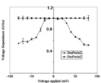
in black lipid bilayer membranes were identical to those measured with the native mitochondrial VDAC of D. melanogaster, the features of recombinant DmVDAC2 were studied. Recombinant DmVDAC2 was purified from inclusion bodies and refolded, showing a pore forming activity in lipid bilayer membranes with a long lifetime at low voltage. DmVDAC2 preferentially formed channels with a single channel conductivity of 4,5 nS in 1M KCl. The channel forming activity however was lower than that of DmVDAC1, since at least a 10 times highest concentration of DmVDAC2 was needed to obtain the same number of reconstituted channels as compared with DmVDAC1. The voltage dependence of DmVDAC2 was also investigated, and most surprising, DmVDAC2 did not display any voltage dependence. Furthermore, DmVDAC2 showed preferential movements of cations through the channel, indicating that the two VDAC isoforms exhibit different ion selectivity (Aiello et al., 2004).
1.4.6 Identification of alternative 5’UTRs in porin transcripts
As reported in Oliva et al. 1998, to study the genomic organization of porin1 gene in Drosophila melanogaster, a published cDNA clone containing the whole transcript sequence for VDAC (1370 bp), named 7T21 (acc. No. X92408) (Messina et al., 1996) was used as a probe to screen a genomic library in EMBL3 derived from Canton-S strain. The clone EM1T was chosen for further analysis of sequencing. The
Figure 12: Voltage dependence G/G0 comparison of DmVDAC1 and DmVDAC2.Voltage dependence G/G0 comparison of DmVDAC1 and DmVDAC2. Voltage dependence G/G0 was a function of the applied voltage. DmPorin2 is not voltage-dependent.
16
exons of porin were identified by comparison of the genomic sequences with the cDNA. The consensus dinucleotides for splice sites, GT and AG, were respectively found next to the 5’ end 3’ boundaries of the introns, showing that the D. melanogaster
porin gene contains four exons separated by three introns. The beginning of the first
exon was deducted by 5’RACE-PCR experiments performed on D. melanogaster Oregon-R third instar larvae poly(a)+ RNA. After reverse transcription, nested primers were designed in the DNA protein-coding region and the amplified PCR products were sub-cloned into a T-vector plasmid and some positive clones were sequenced. Sequence analysis showed the expression of two different 5’-untranslated extensions fused to the coding sequence. The ratio between the two 5’UTRs was 10:1 for the 5’UTR corresponding to the cDNA 7T21 sequence. The second sequence instead corresponded to another genomic region enclosed between exons I and II. This sequence is followed by the –GT splice-site canonical sequence. The exon corresponding to the cDNA was indicated as exon 1A, while the new one, corresponding to an alternative mRNA was named exon 1B. Exon II (320 bp) and exon III (228 bp) exclusively contain coding sequences. The first three bases of the exon II correspond to the ATG starting codon, while the TAA stop codon is contained in the remaining 298 coding bp in the exon IV, together with the 3’UTR sequence. Two putative poly-adenylation sites were detected in the 3’UTR at the positions 3607 and 3904.
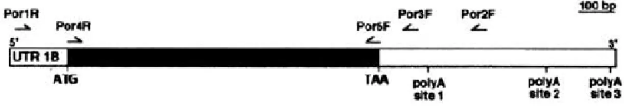
Figure 13: Structural organization of D. melanogaster porin gene (clone EM1T)
Exons are shown as boxes above the line and are numbered IA (486^600), IB (1014^1137), II (2485^2805), III (2881^3108), IV (3178 to the end). The positions of the ATG starting codon and of the TAA stop codon are evidenced. Exon II precisely starts with the ATG. Coding regions are clearer, while non-coding regions are shadowed. ER sites are indicated (A: AccI; B: BamHI; Bc: BclI; C: ClaI; E: EcoRI; P: PstI; S: SalI; X: XbaI; Xh: XhoI). IA and IB are two alternative leader exons. Under the gene structure the clones sequenced are reported. A SalI/SalI clone of about 7 kbp from an EMBL3 D. melanogaster genomic library was analyzed.
17
1.4.7 Analysis of developmental specificity of VDAC transcript containing the exon 1B
In D. melanogaster there is a low over-representation of specific sequences of
Drosophila promoters. Concerning exon IA, a TATA sequence is placed at 441, but its
position is -45 with respect to the transcription starting site. Also a CAAT sequence was found on the reverse strand at 405 (-80), and a CCAAT, also on the reverse strand, was found at 920 (-84) with respect to the transcription starting site of exon IB; two regions rich in GC, but without canonical sequences and no putative TATA-box were observed between this location and the IB transcription starting site. To verify the in vivo production of the VDAC transcript containing the exon IB, and to study its possible developmental specificity, RT-PCR experiments were performed on poly(A)+ RNAs from D. melanogaster embryos, larvae and adults of both sexes. The primers used were Por1R, a primer derived from the exon 1B, and two primers localized upstream the two putative poly-adenilation sites, por3F and Por2F.
Figure 10: A VDAC mRNA with an alternative 5'UTR is constitutively expressed in Drosophila melanogaster.
RT-PCR analysis was performed on poly(A)+ RNA from Oregon-R. Schematic representation of the transcript encompassing sequences complementary to exon 1B. possible polyadenylation sites are marked. The translated region is indicated by the filled box.
A single amplification product of the expected length (1065 bp) was observed at all developmental stages with the Por3F primer, but no product was observed using the Por2F primer. A nested PCR experiment using two primers on the coding sequence (Por4R and Por5F) verified that the amplification products of the expected length (849 bp), were obtained. Sequence analysis of the 1065-bp Figure 11: Agarose gel showing amplified RT-PCR products. In
the lanes 1, 3 and 5 the Por1R primer which is specific for exon 1B, was used together with Por3F primer, derived from the 3’UTR. The resulting products were re-amplified using Por4R and Por5F primers, inside the coding sequence (lanes 2,4,6). Lane 1, poly(A)+ RNA template isolated from 0-3 h embryos; lane 3, poly(A)+RNa template isolated from third-instar larvae; lane 5, poly(A)+RNA template isolated from adult flies; lane 7, control obtained by amplification on the porin cDNA with internal primers.
18
amplification product confirmed that the transcript without the exon IA and which uses the first polyadenilation site is present at all developmental stages tested. Mobilization of a P-Element (PlacW) inserted in the porin gene at the position 32B3-4 region of 2L chromosome, produced different kinds of mutants that have specific features. In particular, the PlacW element causes the lethal phenotype that can be reverted by precise or almost precise excision of the transposon in the porin gene. This experiments showed that exon 1B is not necessary for VDAC synthesis or for fly viability, since the total content of VDAC did not vary after 1B excision; conversely, exon 1A is indispensable because its excision caused the decrease of the total amount of VDAC protein (Aiello et al., 2004).
1.5
The roles of RNA-binding proteins in post-transcriptional
functions
In Drosophila melanogaster and in many others eukaryotes, gene expression needs a dynamic interplay between proteins and RNAs. The synthesis of RNAs involve the formation of ribonucleoprotein particles (RNPs) (Draper, 1995) ranging from small to large complexes. The interactions between proteins and RNA occur through one or more RNA-binding domains or via another protein that is itself directly bound to RNA (Glisovic et al., 2008). NOVA2, PTBP1, U2AF2, and RBFOX2, are examples of proteins that interact with RNA in a largely sequence-specific manner (Lewis et al., 2000; Jin et al., 2003; Kielkopf et al., 2004; Hall et al., 2013). In contrast, the protein SMN, which is involved in snRNP biogenesis, does not have any known RNA-binding domain and interacts with the snRNAs indirectly. RNA-binding proteins (RBPs) play a crucial role in cellular biology of higher eukaryotic organisms. RBPs indeed, participate in several essential post-transcriptional events, including pre mRNA splicing, 3’ end formation, RNA localization, turnover and translation (Glisovic et al., 2008). The Fragile X Mental Retardation Protein (FMR1) is one example of a pleiotropic RBP, encoded in D. melanogaster by Fmr1. FMR1 interacts with components of the RNAi machinery, forming a complex which includes Argonaute 2 (AGO2) of the RISC (RNA-induced silencing complex) complex (Ishizuka et al., 2002). FMR1 also binds ribosomes to block translation by inhibiting tRNA association (Chen et al., 2014a). RBPs recognize their RNA target through RNA-binding domains. In D. melanogaster there are common classes of RNA-binding domains, including the RNA-recognition
19
motif (RRM), the K homology domain (KH), both zinc finger motifs and the double-stranded RNA-binding motif (dsRBM). The difficult to identify functions of RBPs with their RNA-binding domains is due to the fact that there is no one-to-one mapping between domains and functional roles, and many RBPs with characterized functions appear pleiotropic. Many genes involved in post-transcriptional regulation tend to be often regulated post-transcriptionally. Feedback loops are considered the principal idea at the level of a regulatory process. Analysis of RBP and mRNA interaction profiles show ubiquitous interaction with mRNA and proteins products of the same gene; furthermore, RBPs of the same protein complex tend to reciprocally bind the mRNAs of their interaction partners. Post-transcriptional regulation, mediated by protein-RNA interaction, is a common process and constitutes a general mechanism of feedback in the hierarchy of gene regulation (Marcus et al., 2015).
1.6
Example of splicing regulation of Drosophila melanogaster
genes
Alternative splicing is a mechanism by which multiple mRNA from a single gene can be created, connecting different parts of a pre-mRNA. Alternative splicing can affect 5’- and 3’- untranslated regions of mRNAs modulating translation, stability or localization of mRNA. Also, by modulating insertion and exclusion of coding exons, the percentage of protein diversity dramatically increases. Moreover, using alternative splicing cells can produce functionally different proteins from a single gene in a temporal- or tissue-specific manner. In human, about 95% of gene undergo to an alternative splicing process, but the effects of these events are mostly unknown. Pioneer studies in Drosophila have shown the presence of regulatory sequences in the pre-mRNA required for alternative splicing regulation as well as some regulators that bind to them. Drosophila is known to use every alternative splicing strategy imaginable and there are a lot of examples as demonstration. The first example is the sex-specific splicing, since in Drosophila sex determination is controlled by splicing factors that lead to the sex-specific expression of two different variants of the Doublesex (Dsx) transcription factor. Splicing factors are themselves alternatively spliced, and the Dsx variants are differentially expressed in male and female bodies to control different set of genes. Moreover, the key splicing difference between male and female is the expression in females of the Sex-lethal (Sxl) protein that is expressed in embryos in response to X
20
chromosomes number, determining female development. In female a full length Sxl protein is expressed, while in males a truncated form of the protein is produced. Sxl is responsible for multiple aspects of female differentiation, governing several sets of genes.
Another important example of alternative splicing is given by the expression of the myosin heavy-chain gene (Mhc), which has in-frame alternative exons in five different regions (exons 3, 7, 9, 11 and 15) and a cassette exon (number 18) encoding the C-terminus. In this case, by multiplying these independent choices, 480 different myosin isoforms could be potentially produced in Drosophila, and therefore, different muscle types express specific Mhc isoforms through specific combinations of exons, in order to finely control the contractile properties of each kind of muscle.
Control of the alternative splicing. Usually, it represents an independent recruitment to the pre-mRNA of RNA-binding proteins. Most of them are direct or indirect controllers of one or more alternative splicing events, but not only alternative splicing factors, but also proteins previously considered as constitutive. Many and big differences are found in gene structure between D. melanogaster and mammals. For example, in humans, the dimensions of introns are usually much bigger than exons; in
Drosophila the reverse is true, and also multiple variants of mutually exclusive exons
and recursive splicing are not present in mammals. Moreover, the trans-splicing, which is very common in Caenorhabditis elegans and in D. melanogaster, is not found in mammals. On the other hand, between mammals and insects there is an extensive conservation of alternative splicing patterns. The spliceosome dynamics and composition are very similar between them and most known mammalian splicing factors have orthologous in flies. These include members of SR protein family (Gabut et al., 2007; Blanchette et al., 2005) , the hnRNP/hrp family and other like Tra2, TIAR/Rox, PUF60/pUf68. This means that studies in flies are highly relevant to the mechanisms of alternative splicing in humans and to understand the way that evolution adapts it to its needs.
21
1.7
The function of RNA structure at 5’UTR in gene regulation
mediated by microRNA
The 5’ untranslated region (5’UTR) of mRNA is the sequence preceding the start codon of translation, which plays important functions in regulating post-transcriptional events (Davuluri et al., 2000). In the 5’UTR has been found several regulatory elements, such as RNA binding sites for RNA binding proteins (RBPs), upstream open reading frames (uORF) and upstream start codons (uAUGs) that, together with the ability of the 5’UTR to assume a mRNA secondary structure, all these elements have been recognized as a major feature that regulates gene translation (Pickering et al., 2005). For example, in human about 60% of 5’UTRs present a structured RNA near the 5’cap site, which alone is able to block translational initiation. Recently, has been highlighted the role of 5’UTR in mRNA repression mediated by miRNA (Djuranovic et al., 2012), which is a class of non coding RNAs, 20-24 nt in length, that regulate gene expression by base-pairing with complementary sequences in mRNA transcripts. Usually miRNA acts silencing or degrading RNA (Bartel, 2009). The mRNA-miRNA interaction affects expression of the gene target at early stages of translation, i.e., translation initiation. For example, in Drosophila S2 cells, gene silencing mediated by miRNA occurs through translational inhibition, by mRNA deadenylation and subsequent decay. In HeLa cells, the function of the eIF4F initiation complex is impaired by mRNA-miRNA interaction confirming that the secondary structure of the 5’UTR is necessary to miRNA-mediated gene silencing. (Meijer et al., 2013). Moreover, Wanjun gu et al., 2015 found that mRNA secondary structure near the 5’cap, rather than the full length 5’UTR is increased to facilitate miRNA-mediated gene regulation, showing a universal trend of increased mRNA stability near the 5’cap in genes with miRNA targets (Wanjun et al., 2015).