_______________________________________________________________________________________________________________________________ 490 http://www.mjms.mk/ http://www.id-press.eu/mjms/ ID Design 2012/DOOEL Skopje, Republic of Macedonia
Open Access Macedonian Journal of Medical Sciences. 2017 Jul 25; 5(4):490-492.
Special Issue: Global Dermatology
https://doi.org/10.3889/oamjms.2017.137 eISSN: 1857-9655
Case Report
Erythema Ab Igne Caused By Laptop Computer
Claudio Guarneri1*, Georgi Tchernev2, Uwe Wollina3, Torello Lotti4, Mario Vaccaro51
Universita degli Studi di Messina, Clinical and Experimental Medicine, Section of Dermatology, Messina 98122, Italy; 2
Medical Institute of the Ministry of Interior, Dermatology, Venereology and Dermatologic Surgery; Onkoderma, Private Clinic for Dermatologic Surgery, Dermatology and Surgery, Sofia 1407, Bulgaria; 3Krankenhaus Dresden-Friedrichstadt, Department of Dermatology and Venereology, Dresden, Sachsen, Germany; 4Universitario di Ruolo, Dipartimento di Scienze Dermatologiche, Università degli Studi di Firenze, Facoltà di Medicina e Chirurgia, Dermatology, Via Vittoria Colonna 11, Rome 00186, Italy; 5Universita degli Studi di Messina, Policlinico Universitario, Via Consolare Valeria, Messina, Sicilia 98125, Italy
Citation: Guarneri C, Tchernev G, Wollina W, Lotti T,
Vaccaro M. Erythema Ab Igne Caused By Laptop Computer. Open Access Maced J Med Sci. 2017 Jul 25; 5(4):490-492. https://doi.org/10.3889/oamjms.2017.137
Keywords: Erythema ab igne; modern technologies;
panniculitis; cancer; differential diagnosis.
*Correspondence: : Claudio Guarneri. Universita degli
Studi di Messina, Clinical and Experimental Medicine, Section of Dermatology, Messina 98122, Italy. E-mail: [email protected]
Received: 02-Apr-2017; Revised: 02-May-2017; Accepted: 03-May-2017; Online first: 22-Jul-2017
Copyright: © 2017 Claudio Guarneri, Georgi Tchernev, Uwe Wollina, Torello Lotti, Mario Vaccaro. This is an open-access article distributed under the terms of the Creative Commons Attribution-NonCommercial 4.0 International License (CC BY-NC 4.0).
Funding: This research did not receive any financial
support.
Competing Interests: The authors have declared that no
competing interests exist.
Abstract
Erythema ab igne (EAI) represents the stereotype of a modern technology induced disease. Originally produced by repeated exposure of the skin to a heat source, more often because of habits related to the job or personal activities, this condition now tends to occur more frequently, being associated with a variety of modern instruments. The aim of our report is to discuss this strange medical condition with a focus on clinical features, possible confounding differential diagnoses and recommendations for prevention.
Introduction
Erythema ab igne (EAI) represents the stereotype of a modern technology induced disease.
Originally produced by repeated exposure of the skin to temperatures close to the burn threshold (43-47°C) [1], and being unchanged in its pathogenesis – based on the superficial injury on the dermal vascular plexus – the occurrence of EAI has been associated with a wide variety of ‘new’ warming sources, thus also involving a wider range of age of affected patients [2, 3]. In facts, due to its infrequency, EAI still constitutes a clinical conundrum, diagnostically misinterpreted and hard to be solved in the absence of suspected anamnestic data.
The aim of our report is to discuss this strange medical condition with a focus on clinical features, possible confounding differential diagnoses and recommendations for prevention.
Case report
A 32-year-old Caucasian woman, who was a secretary in a legal office, presented with skin changes of the left thigh. The lesion had appeared at some undetermined moment and got worse progressively in the last three months, always being only mildly pruritic.
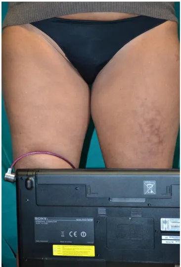
Physical examination disclosed a well-defined erythematous-violaceous, reticulated macular rash limited to the anterior part of the medium third of the left thigh (Fig. 1).
The patient was referred to our department by a rheumatologist with the suspect of vasculitis. Thus she had just received a diagnostic laboratory work-up, including tests for ANA and ANCA, all resulting within normal limits.
Guarneri et al. Erythema Ab Igne Caused By Laptop Computer ______________________________________________________________________________________________________________________________
_______________________________________________________________________________________________________________________________ Open Access Maced J Med Sci. 2017 Jul 25; 5(4):490-492. 491
Figure 1: Erythematous, violaceous, reticulated rash on the left thigh
On repeated anamnesis, the patient reported that she had been assigned to its actual job exactly four months before; in particular, due to her role of barrister assistant, she was used to having laptop on thighs during most of her work time. Checking patient’s laptop, we noted that the lesional area was exactly in contact with the ventilation fan (fig.2).
Figure 2: Lesional area was localised in correspondence of the ventilation fan of patient’s laptop
Since the history of persistent skin exposure
to the localised heat source, a diagnosis of erythema ab igne was made and proposed the use of a protective lap pad to create an efficient barrier between the user and the personal computer together with a favourable work surface.
Given the absence of significant symptoms, no pharmacological therapy was prescribed.
Discussion
Erythema ab igne is an initially transient, and then chronic cutaneous condition, caused by direct exposure of the skin to a heat source, more often because of habits related to the job or personal activities [4].
Originally also known as “hot water bottle rash”, typical of the pretibial area in subjects used to expose to space heaters [1], this condition now tends to occur more frequently, being associated with a variety of modern instruments including heating pillows [5], space heaters [6], electric blankets [7], heated car seats and backs [8], portable personal computers [9] and smartphones [2, 10].
Pathogenesis has not been fully understood. Long term and/or repeated heat exposure can determine, in general, superficial damage to the vascular structures, with vasodilatation and deposition of hemosiderin [1, 2]; other changes include changes in dermal elastic fibres, epidermal atrophy and melanin accumulation in the dermis [2, 11].
Depending on the frequency, temperature and time of exposure, we expect different degrees and onset of the condition [12].
Typical manifestations consist of transient-to-persistent, reticulated, initially erythematous or, later
hyper-/hypopigmented patches, sometimes
characterised by superficial changes (atrophy, xerosis, telangiectasia, bullae). The eruption is mainly asymptomatic, although warming in the acute phase and itch chronically have been anecdotally reported [1, 10, 13].
Diagnosis is based on the supportive history and the clinical picture, whereas histologic changes are non-specific [14], thus skin biopsy has to be considered only in limited cases. Differential diagnosis of EAI should include livedo vasculitis (the idiopathic as well as the symptomatic form in collagenosis) [4], panniculitis [15, 16], and side effects of some drugs (amantadine and memantine) [4]. As suggested by some authors, EAI may also mimic infectious diseases when anamnesis is not clear as well as original culture practices and conditions of the exotic patients [17].
Case Report
_______________________________________________________________________________________________________________________________
_______________________________________________________________________________________________________________________________ 492 http://www.mjms.mk/ http://www.id-press.eu/mjms/ preventive measures [18] usually lead to a favourable
prognosis. Chronic cases have been treated with topical retinoids and 5-fluorouracil, Nd-YAG, ruby and alexandrite lasers [2, 5, 19] plus antihistamines and/or FANS symptomatically. Monitoring of long-standing EAI is mandatory, whereas hyperkeratosis and ulceration have to be considered as a sign of premalignant changes at the epithelial level [20].
Finally, chronic pain and persistent systemic symptoms in EAI have to be carefully checked as a possible sign of occult internal malignancies. A total of 11 cases have been reviewed in a recent paper by Bunick and Ibrahim [21], with gastrointestinal (colorectal, pancreatic, gastric) cancer being the most represented tumour; lung, renal breast and hematologic malignancies have also been reported thus underlines the importance of complete assessment of this otherwise benign condition [21, 22].
References
1. Hernandez AC, Arbedol SB, Montalvo SM, Marugàn LT. Erythema ab igne. Reumatol Clin. 2016;12:233-4.
https://doi.org/10.1016/j.reuma.2015.08.009 PMid:26654294
2. Salgado F, Handler MZ, Schwartz RA. Erythema ab igne: new technology rebounding upon its users? Int J Dermatol. 2017; Mar
30. https://doi.org/10.1111/ijd.13609
3. Guarneri F, Guarneri C, Cannavò SP. An unusual case of cell phone dermatitis. Contact Dermatitis. 2010;62(2):117.
https://doi.org/10.1111/j.1600-0536.2009.01674.x PMid:20136896
4. Elsner P. Erythema ab igne as an occupational skin disease. J Dtsch Dermatol Ges. 2014; 12(7):621-2. PMid:24684647 5. Fareedy SB, Rettew A, Karmacharya P, Jehangir A, Shaikh B, Pathak R. erythema ab igne secondary to repeated heating pad use: an image case. J Community Hosp Intern Med Perspect. 2015;5(4):28335. https://doi.org/10.3402/jchimp.v5.28335
PMid:26333862 PMCid:PMC4558291
6. Morrison M, Cotton J, LaFond A. Reticulated erythematous patch on a teenager foot. J Fam Pract. 2014;63:537-9. PMid:25353026
7. Leal-lobato MM, Blasco-Morente G. electric blanket induced erythema ab igne. Semergen. 2015;41:456-7.
https://doi.org/10.1016/j.semerg.2014.12.008 PMid:25662480
8. Brodell D, Mostow EN. Automobile seat heater-induced erythema ab igne. Arch Dermatol. 2012;148:264-5.
https://doi.org/10.1001/archdermatol.2011.1158 PMid:22351837
9. Brazzelli V, Grassi S, Barruscotti S, Croci G, Borroni G. Erythema ab igne induced by laptop computer: an emerging disease among adolescents? G Ital Dermatol Venereol. 2015[epub ahead of print].
10. Kesty K, Feldman SR. Erythema ab igne: evolving technology, evolving presentation. Dermatol Online J. 2014;20:19.
11. Soholm Secher LL, Vind-Kezunovic D, Zachariae CO. Side-effects to the use of laptop computers: erythema ab igne. Dermatol reports. 2010;2:e11. https://doi.org/10.4081/dr.2010.e11
PMid:25386248 PMCid:PMC4211471
12. Turan E, Yesilova Y, Ucmak D, Celik OI. Thermal pillow: an unusual causative agent of erythema ab igne. Turk J Pediatr. 2013;55:648-50. PMid:24577987
13. Milchak M, Smucker J, Chung CG, Seiverling EV. Erythema ab igne due to heating pad use: a case report and review of clinical presentation, prevention and complications. Case Rep Med. 2016;2016:1862480. https://doi.org/10.1155/2016/1862480
PMid:26880929 PMCid:PMC4735926
14. Miller K, Hunt R, Chu J, Meehan S, Stein J. Erythema ab igne. Dermatol Online J. 2014;20:11.
15. Borgia F, De Pasquale L, Cacace C, Meo P, Guarneri C, Cannavò SP. Subcutaneous fat necrosis of the newborn: be aware of hypercalcemia. J Paediatr Child Health. 2006;42(5):316-8.
https://doi.org/10.1111/j.1440-1754.2006.00862.x PMid:16712567
16. Cannavò SP, Borgia F, Vaccaro M, Guarneri F, magliolo E, Guarneri B. Pretibial myxoedema associated with Hashimoto's thyroiditis. J Eur Acad Dermatol Venereol. 2002;16(6):625-7.
https://doi.org/10.1046/j.1468-3083.2002.00532.x PMid:12482050
17. Treister-Goltzman Y, Peleg R. Images in clinical tropical medicine. Erythema ab igne. A J Trop Med Hyg. 2015;92(3):476.
https://doi.org/10.4269/ajtmh.14-0474 PMid:25740953
PMCid:PMC4350531
18. Riahi RR, Cohen PR. Practical solutions to prevent laptop computer-induced erythema ab igne. Int j Dermatol. 2014;53:e389-e409. https://doi.org/10.1111/ijd.12407 PMid:24961574
19. Kim HW, Kim EJ, Park HC, Ko JY, Ro YS, Kim JE. Erythema ab igne successfully treated with low fluenced 1064-nm Q-switched neodymium-doped yttrium aluminium garnet laser. J Cosmet Laser ther. 2014;16:147-8.
https://doi.org/10.3109/14764172.2013.854623 PMid:24131068
20. Sigmon JR, Cantrell J, Teague D, Sangueza O, Sheehan DJ. Poorly differentiated carcinoma arising in the setting of erythema ab igne. Am J Dermatopathol. 2013;35:676-8.
https://doi.org/10.1097/DAD.0b013e3182871648 PMid:23872874
21. Bunick CG, King BA, Ibrahim O. When erythema ab igne warrants an evaluation for internal malignancy. Int J Dermatol. 2014;53:e347-e366. https://doi.org/10.1111/ijd.12329
PMid:24601874
22. Ashby M. Erythema ab igne in cancer patients. J R Soc Med. 1985;78:925-7. https://doi.org/10.1177/014107688507801110